Copyright © 2004, American Society for Microbiology. All Rights Reserved.
Enhanced Expression of Cell Cycle Regulatory Genes in Virus-Specific
Memory CD8
⫹
T Cells
Donald R. Latner, Susan M. Kaech,† and Rafi Ahmed*
Emory Vaccine Center and Department of Microbiology and Immunology, Emory University School of Medicine, Atlanta, Georgia
Received 4 April 2004/Accepted 15 June 2004
Unlike naive CD8ⴙT cells, antigen-experienced memory CD8ⴙT cells persist over time due to their unique
ability to homeostatically proliferate. It was hypothesized that memory cells might differentially regulate the expression of genes that control the cell cycle to facilitate homeostatic proliferation. To test this, the expression levels of 96 different cell cycle regulatory genes were compared between transgenic naive and memory CD8ⴙT
cells that specifically recognize the GP33-41 epitope of lymphocytic choriomeningitis virus (LCMV). It was discovered that relative to naive cells, memory cells overexpress several important genes that control the transition between G1and S phase. Some of these genes include those encoding cyclins D3, D2, B1, C, and H,
cyclin-dependent kinases (cdk’s) 4 and 6, the cdk inhibitors p16, p15, and p18, and other genes involved in protein degradation and DNA replication. Importantly, these differences were observed both in total popula-tions of LCMV-specific naive and memory CD8ⴙcells and in LCMV-specific CD8ⴙT-cell populations that were
in the G1phase of the cell cycle only. In addition, the expression differences between naive and memory cells
were exaggerated following antigenic stimulation. The fact that memory cells are precharged with several of the major factors that are necessary for the G1- to-S-phase transition suggests they may require a lower threshold
of stimulation to enter the cell cycle.
Historically, lymphocytic choriomeningitis virus (LCMV) in-fection has been proven to be an excellent model for studying CD8⫹-T-cell activation and development, because infection with LCMV provides a robust CD8⫹-T-cell response and viral clearance is primarily dependent on CD8⫹T cells (6). Naive, antigen-specific CD8⫹T cells respond to an LCMV infection by eliminating the target cell, which is facilitated by increased expression of several antiviral effector molecules, such as per-forin, granzyme B, gamma interferon, and tumor necrosis fac-tor alpha. In addition, the activated cells upregulate the inter-leukin 2 (IL-2) receptor and begin to synthesize IL-2, leading to rapid proliferation. The newly activated or “effector” T cells continue to proliferate until shortly after the virus is cleared, with the number of antigen-specific CD8⫹cells peaking at day 8 postinfection. After antigen clearance, the pool of effector cells contracts until it ultimately constitutes approximately⬃5 to 10% of the total T-cell pool. Current evidence indicates that the antigen-specific “memory” cells remaining after the con-traction phase progressively developed from the pool of effec-tor cells by slowly altering their gene expression pattern to provide them with unique characteristics. For example, mem-ory cells have the ability to more rapidly secrete antiviral cy-tokines and to kill infected cells than their naive precursors. In addition, one of the hallmark features of memory cells is their ability to homeostatically proliferate independently of contact with antigen. After antigen-specific memory cells are initially generated, their numbers are maintained by a gradual turnover of the population. Antigen-specific memory cells will divide
approximately once per 1 to 2 months (⬃1 to 5% of memory cells are in cycle at any given point in time), yet the total number of those cells does not change, suggesting there are equal rates of cell death and division in a given specific mem-ory-cell population (8, 12, 14, 26).
Although many details about the molecular mechanisms that control memory-cell homeostasis are not yet clear, several crit-ical components that govern this process are beginning to emerge. First, it has become apparent that the cytokine IL-15 is required for memory-cell homeostatic proliferation (2, 9, 17, 24, 25, 27). In mice lacking IL-15 or the IL-15 receptor alpha chain, memory CD8⫹T cells fail to undergo homeostatic pro-liferation and self-renewal, thus leading to a gradual reduction in their number over time (2). Interestingly, some evidence suggests that IL-7 can substitute for IL-15 to support ho-meostasis in immunodeficient mice, as long as it is not made limiting by competition from other cell types (9, 17, 23, 24). Second, it has been shown that memory cells can survive in the absence of major histocompatibility complex (MHC) class I contact, unlike their naive counterparts (22). These two dis-tinct properties of memory cells, the ability to homeostatically proliferate in an IL-15-dependent manner and the ability to survive without MHC class I contact, suggest that they have altered their gene expression pattern relative to their naive progenitors and are thus equipped to interact with and respond to their environment in different ways. In a comprehensive paper by Kaech et al. (13), memory cells were shown to have increased expression of genes encoding T-cell-effector mole-cules, such as gamma interferon, perforin, and granzyme B, in addition to a variety of genes involved in signal transduction, cell migration, apoptosis, and cell division pathways. It is rea-sonable to assume that some of these gene expression changes are fundamentally responsible for providing memory cells with
* Corresponding author. Mailing address: Emory Vaccine Center, 1510 Clifton Rd., G211, Atlanta, GA 30322. Phone: (404) 727-9301. Fax: (404) 727-3722. E-mail: ra@microbio.emory.edu.
† Present address: Section of Immunology, Yale Medical School, New Haven, CT 06520.
10953
on November 8, 2019 by guest
http://jvi.asm.org/
meostatic proliferation, the analysis was performed with simi-lar results on sorted populations of cells that were only in the G1phase of the cell cycle, based on DNA content. In addition,
it was discovered that the differences in expression of these genes between naive and memory cells was exaggerated fol-lowing antigenic stimulation.
MATERIALS AND METHODS
Mice.C57BL/6 mice were purchased from the National Cancer Institute (Frederick, Md.). Thy1.1⫹P14 mice bearing the Db-GP33-specific T-cell recep-tor were backcrossed to C57BL/6 and have been previously described (5). LCMV Armstrong virus was propagated, titers were determined, and virus was used as previously described (1). P14 chimeric immune mice were generated by adop-tively transferring⬃106total splenocytes from a naive P14 transgenic animal into naive B6 mice by intravenous injection. The chimeric animals were then infected with 2⫻105PFU of LCMV Armstrong by intraperitoneal injection. All chimeric immune animals were sacrificed at ⬎30 days postinfection. All experiments involving the use of mice were performed in compliance with all federal and institutional guidelines and policies.
Isolation of T-cell subsets.Single-cell suspensions were prepared in RPMI containing 10% fetal calf serum by mechanical disruption of spleens. Red blood cells were lysed in 0.83% ammonium chloride. CD8⫹cells were initially purified
by magnetic bead selection using anti-CD8 beads (Miltenyi Biotec, Auburn, Calif.). Enriched CD8⫹cells were stained with CD8 and Thy1.1
anti-bodies and further purified by fluorescence-activated cell sorting (FACS) to select antigen-specific, Thy1.1 CD8 double-positive cells. The purity for each population averagedⱖ94% across several experiments.
Flow cytometry, Hoechst staining, and cell sorting.Single-cell suspensions of spleen were prepared, and 106cells were stained in phosphate-buffered saline (PBS) containing 1% bovine serum albumin (FACS buffer) for 30 min at 4°C followed by three washes in FACS buffer. Cells were then fixed in 2% parafor-maldehyde diluted in PBS. Samples were acquired on a FACSCalibur flow cytometer (Becton Dickinson, San Jose, Calif.), and the data were analyzed by using CELLQuest software (Becton Dickinson Immunocytometry Systems). The anti-CD8 (clone 53-6.7) and anti-CD44 (clone IM7) antibodies were purchased from BD PharMingen. LCMV GP33-41 MHC class I/peptide tetramers were prepared and used as described previously (21).
Cell culture and peptide stimulation.Total splenocytes were cultured for 24 h with or without 1g of GP33-41 peptide/ml in RPMI plus 10% fetal calf serum, 40⫻106in each well of 6-well plate. Following stimulation, the cells were subjected to magnetic bead separation and cell sorting as described above.
RNA isolation and amplification.RNA was extracted from sorted cells using TRIZOL reagent (Invitrogen, Carlsbad, Calif.) according to the manufacturer’s instructions. The entire amount of mRNA isolated from approximately 5⫻105 to 1⫻106cells per sample was linearly amplified two times as previously described (13).
Gene expression analysis.Five micrograms of 2⫻amplified RNA was reverse transcribed into single-stranded cDNA and was then used as the substrate for probe synthesis according to the SuperArray (Frederick, Md.) [32P]dCTP radio-active labeling protocol, using the Mouse Q-series cell cycle array kit. Probe hybridization and blot washes were performed according to the manufacturer’s instructions. Following hybridization, the arrays were subjected to
phosphorim-analyzed using the Mouse Q-series cell cycle gene arrays pro-duced by SuperArray, Inc. Ninety-six different genes that con-trol the cell cycle are represented on each array, including cyclin-dependent kinases (cdk’s), cyclins, cdk inhibitors, cdk phosphatases, and cdk kinases. In addition, genes essential for DNA damage and mitotic spindle checkpoints and genes in some ubiquitin-conjugation pathways are represented.
Naive CD8⫹T cells were isolated directly from the spleens of uninfected P14 transgenic mice, which express T cell recep-tors that specifically recognize the GP33-41 epitope of LCMV in the context of MHC class I (5). Transgenic P14 memory CD8⫹T cells were harvested⬎30 days postinfection from the spleens of mice that initially received 106total P14 transgenic
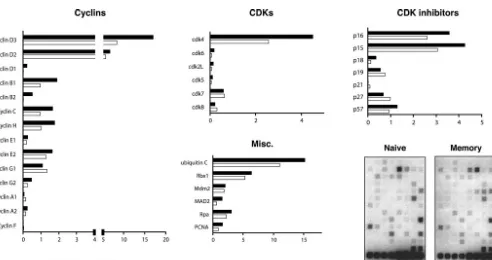
splenocytes by intravenous injection, followed by an acute in-fection with LCMV. Figure 1A shows a typical example of FACS analysis of cells harvested from naive and memory mice. The P14 transgenic CD8⫹cells in both naive and memory mice stain positive for GP33-41 MHC class I tetramer, the congenic marker Thy1.1, and the surface marker CD62L. CD44 is ex-pressed at low to intermediate levels on naı¨ve cells but is present at high levels on memory cells. In multiple indepen-dent experiments, both naive and memory cells were initially purified using anti-CD8 magnetic beads and then were stained with anti-CD8 and anti-Thy1.1 antibodies and sorted to⬎94% purity. A typical example of the FACS analysis from one such stain and sort is shown in Fig. 1B. The mRNA from sorted cells of each type was then isolated, linearly amplified, radioactively labeled, and hybridized to the gene arrays. The data were analyzed and quantified by using a phosphorimager. To deter-mine the relative amount of expression for each gene, the hybridization signal for each gene was calculated as a percent-age of the combined averpercent-age expression of two control genes, encoding cyclophilin A and rpl13A, following background cor-rection. Gene expression data from three independent exper-iments were averaged and are summarized in Fig. 2. The re-sults indicate that memory cells express higher levels of several types of genes that regulate the cell cycle than do naive cells. These genes include some cyclins, cdk’s, cdk inhibitors, and factors involved in DNA replication, mitosis, and protein deg-radation.
The comparison revealed that memory cells produce ele-vated levels of cyclins D3, D2, B1, C, and H. Notably, cyclin D3 was expressed in approximately twofold excess in memory cells. Cyclins D3 and D2 appeared to be the most abundantly expressed cyclins in both naive and memory cells, and they
on November 8, 2019 by guest
were among the most abundantly expressed of all genes exam-ined, assuming that the hybridization efficiencies for each gene were similar. In addition, it was revealed that cdk4 and cdk6, the major G1-phase cyclin-dependent kinases, are
overex-pressed by approximately twofold in memory cells. The cdk
inhibitors p16, p15, and p18 were also overexpressed in mem-ory cells, while the inhibitor p27 was slightly elevated in naive cells. The levels of the remaining cdk inhibitors appeared sim-ilar.
The ubiquitin-mediated protein degradation pathway is
[image:3.603.56.535.73.257.2]im-FIG. 1. Characterization of the naive and memory P14 transgenic CD8 T cells used for subsequent gene expression analysis. (A) Splenocytes from naive P14 transgenic or LCMV-immune P14 chimeric mice were isolated and stained with MHC class I tetramer specific for the LCMV GP33-41 epitope, anti-CD8, anti-Thy1.1, anti-CD44, and anti-CD62L antibodies and then analyzed by flow cytometry. Thy1.1, CD44, and CD62L expression is shown for tetramer and CD8 double-positive cells. (B) Thy 1.1 and CD8 double-positive transgenic T cells were sorted to⬎94% purity prior to RNA isolation. RNA was isolated from cells sorted as indicated here and was analyzed as described in the legend to Fig. 2.
FIG. 2. Relative expression of cell cycle control genes in naive and memory CD8⫹T cells. P14 transgenic naive and memory CD8⫹T cells were
sorted as described in the legend to Fig. 1. Total RNA from these cells was harvested and subjected to two rounds of linear amplification, labeling with [32P]dCTP, and array hybridization. Two representative arrays are shown. The relative expression level of each gene was quantified by
phosphorimager analysis, normalized to the signal of the cyclophilin A and rpl13a control genes on the same membrane, and expressed as arbitrary units. The formula used to calculate these units is described in Materials and Methods. The relative expression of cyclins, cyclin-dependent kinases, cdk inhibitors, and miscellaneous proteins involved in DNA replication and mitosis is indicated as an average for three independent experiments.
on November 8, 2019 by guest
http://jvi.asm.org/
[image:3.603.51.543.408.668.2]portant for controlling the concentration of cell cycle regula-tory proteins. Ubiquitin C and Rbx1 are encoded by two genes involved in ubiquitination that were both slightly upregulated in memory cells. Ubiquitin C is one of several genes that encode a polyubiquitin protein that is thought to be posttrans-lationally processed into ubiquitin monomers (3). Kamura et al. (15) found that yeast Rbx1 is a subunit and activator of the SCF-Cdc4 complex that is required for ubiquitination of the cyclin-dependent kinase inhibitor Sic1 and for the G1-to-S cell
cycle transition.
Other factors that were upregulated in memory cells include genes involved in DNA replication, such as proliferating cell nuclear antigen (PCNA), a subunit for DNA polymerase delta (16). Replication protein A (Rpa) (14 kDa), a single-stranded DNA binding protein that is involved in DNA damage repair (28), and mitotic arrest deficient protein 2 (Mad2) (7), which is required for execution of the mitotic checkpoint, were both also overexpressed in memory cells.
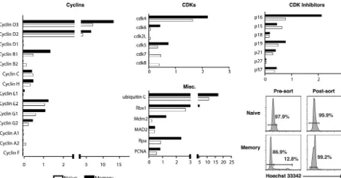
A primary concern was that these observed differences in the expression of cell cycle regulatory genes between naive and memory cells might be due to the fact that a small percentage of memory cells (1 to 3%) are in cycle at any given point in time, whereas naive cells essentially do not proliferate unless they are stimulated by antigen. To address this issue, naive and memory cells were stained with Hoechst 33342 prior to sorting (Fig. 3). The amount of fluorescence attributed to the Hoechst stain indicates DNA content, with the majority of cells having a diploid genome (i.e., residing in the G1peak). Those cells that
have progressed into S, G2, and M phases have a corresponding
increase in fluorescence intensity. Only the cells that were clearly
in the G1peak in each sample were harvested by sorting and used
for the array analysis. The data from two independent experi-ments were averaged and are shown in Fig. 3.
The gene expression profiles observed in Hoechst-stained, G1sorted cells were very similar to those observed in cells that
were not stained with Hoechst, with a few notable differences (Fig. 3). Specifically, the differences in the expression of cyclins cyclins C and H were diminished. Interestingly, two previously unobserved differences appeared in the G1sorted comparison.
First, the cdk inhibitor p16 appeared to be overexpressed in the G1 sorted memory cells by two- to threefold. Second,
mouse double minute 2 homolog (Mdm2), an inhibitor of the p53 oncogene, also appeared to be upregulated in G1sorted
memory cells. Finally, Mad2, which at first appeared to be overexpressed in memory cells, seemed to have equivalent expression between G1sorted naive and memory samples.
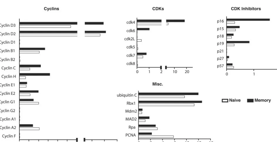
[image:4.603.47.542.67.326.2]Given the observed differences in expression of these genes in resting naive and memory cells and a recent report that memory cells enter the cell cycle slightly sooner than naive cells following antigen stimulation (29), it was hypothesized that additional differences in cell cycle gene expression might be observed after antigen stimulation. To test this, splenocytes from transgenic naive and immune chimeric mice were isolated and stimulated for 24 h with 1g of GP33-41 peptide/ml. As shown in Fig. 4, the cells responded to stimulation by upregu-lating the surface expression of CD69. In addition, the stimu-lated cells displayed an increase in forward scatter and Hoechst 33342 staining, which indicates that they were enlarg-ing and synthesizenlarg-ing DNA in preparation for division. How-ever, the cells had not yet divided at 24 h poststimulation, as
FIG. 3. Expression of cell cycle control genes in transgenic, G1sorted naive, and memory CD8⫹T cells. Naive and memory P14 transgenic
CD8⫹T cells were stained with Hoechst 33342, and the cells with 2N DNA content (G
1) were purified. RNA was isolated from the sorted cells
and analyzed as described in the legend to Fig. 2 and in the Methods section. The relative expression of cyclins, cyclin-dependent kinases, cdk inhibitors, and miscellaneous proteins involved in DNA replication and mitosis is indicated as an average for two independent experiments.
on November 8, 2019 by guest
indicated by staining with CFSE (carboxy-fluorescein diac-etate, succinimidyl ester). The antigen-specific cells were sorted and their RNA was harvested and analyzed as described above. The data from two independent experiments were av-eraged and are shown in Fig. 5. The results indicate that most of the genes that were examined were induced to a higher degree in memory cells than in naive cells. Following stimula-tion, memory cells expressed higher levels of every cyclin mRNA that was detectable with the exceptions of cyclins E1 and A2. Moreover, the differences between memory and naive cells were exaggerated compared to the unstimulated samples. Peptide stimulation dramatically induced cdk4 in memory cells
at nearly six-fold higher levels than in naive cells. cdk6 expres-sion remained higher in memory cells following stimulation but was not substantially increased relative to levels for unstimu-lated samples. Interestingly, antigen also induced the expres-sion of most cdk inhibitors to a higher degree in memory cells than in than naive cells. Notably, p16 and p19 were upregu-lated by eight- and twofold, respectively. The expression of other factors, such as ubiquitin c, Rbx1, Mdm2, and Mad2, was elevated in both samples following stimulation, but the relative levels between memory and naive cells remained unchanged. In contrast, PCNA was elevated to higher levels in naive cells following antigen stimulation.
[image:5.603.58.529.444.686.2]FIG. 4. Activation state of transgenic cells following peptide stimulation. P14 transgenic T cells were labeled with CFSE, stimulated for 24 h with 1g of GP33-41 peptide/ml, stained with Hoechst 33342 and anti-CD8, anti-Thy1.1, and anti-CD69 antibodies, and then analyzed by flow cytometry. CD8 Thy1.1 double-positive cells are shown in each histogram (gates not shown).
FIG. 5. Relative expression of cell cycle control genes in naive and memory CD8⫹T cells following antigen stimulation. Transgenic splenocytes
from naive and immune chimeric mice were harvested and stimulated for 24 h in vitro with 1g of GP33-41 peptide/ml. Following stimulation, the cells were stained with anti-CD8 and anti-Thy1.1 antibodies and sorted, and mRNA from each population was harvested and analyzed as described in the legend to Fig. 2. The data represent an average for two independent experiments.
on November 8, 2019 by guest
http://jvi.asm.org/
at higher levels in memory cells than in naive cells. This ob-servation was made from a comparison of whole populations of antigen-specific naive and memory cells in addition to naive and memory cells that were sorted based on DNA content to isolate only those cells that had not progressed beyond the G1
phase of the cell cycle.
The experiments described here indicate that cyclins D3, D2, B1, C, and H are all expressed at higher levels in resting CD8⫹ memory cells than in naive cells. Of these, cyclins D3 and D2 appeared to be the most abundantly expressed in both naive and memory cells. Cyclin D1 was essentially not detectable in either population. This result is supported by previously pub-lished reports. First, a recent paper by Veiga-Fernandes et al. demonstrated that cyclins D3 and D2 are overexpressed in antigen-specific memory cells (29). Second, a recent report of global changes in the gene expression of purified antigen-specific memory cells has indicated that cyclin B1 is overex-pressed relative to levels in naive cells (13). The work reported here confirms and extends both of these observations.
Cyclins D3 and D2 play important roles in the transition from the G phase (1) to the S phase of the cycle. In combina-tion with cdk4 or cdk6, the D-type cyclins induce expression of cyclins E1 and E2, which are active at the peak of the G1
-to-S-phase transition (18). Overexpression of either cyclin E1 or cyclin E2 in mammalian cells accelerates the G1phase,
indi-cating that these cyclins may be rate limiting for G1
progres-sion (11). Interestingly, recent work has shown that ligation of the costimulatory molecule 4-1BB enhances cell cycle progres-sion in CD8⫹T cells via both IL-2-dependent and -indepen-dent pathways (18). It does this in part by inducing the expres-sion of cyclins D2 and D3. It is plausible that increased expression of cyclins D3 and D2 in memory cells is the result of prior or increased signaling through 4-1BB during the process of antigen-induced activation or by continued signaling after antigen clearance. Corresponding to increased levels of the D-type cyclins, it was also observed that there were increases in the amounts of their binding partners, the major G1-phase
cyclin-dependent kinases cdk4 and cdk6. cdk4 and cdk6 are the main G1 kinases that cause progression into S phase when
bound to D-type cyclins. Cyclin B1, also upregulated in mem-ory cells, is predominantly expressed during the G2-M-phase
transition and complexes with cdk1 (cdc2) to form mitosis-promoting factor, which is responsible for causing the cell to enter mitosis following DNA replication (4). Interestingly, B1 can shuttle between the nucleus and the cytoplasm. A recent
important for maintaining the fidelity of DNA replication. Coincidentally, Rpa (replication protein A; 14 kDa) is a single-stranded DNA binding protein that is involved in DNA dam-age repair and was found to be overexpressed as well.
Additional support for the hypothesis that memory cells might have an altered threshold for entering S phase comes from the observation that Mdm2, a major negative regulator of the p53 oncogene, was also upregulated in G1sorted memory
cells. The well-characterized p53 oncogene induces the expres-sion of factors that inhibit cell diviexpres-sion. Mdm2 binds p53 and targets it for ubiquitination and subsequent degradation, thus promoting survival and cell cycle progression (10). It has been shown that DNA damage induces phosphorylation of p53, leading to decreased interaction with Mdm2. In light of the fact that the concentrations of many proteins that control the cell cycle, including p53, are regulated by ubiquitination, it is not that surprising to see a corresponding upregulation in memory cells of some of the factors, such as Mdm2, ubiquitin c, and Rbx1, that are important in the ubiquitination pathway.
In summary, it appears that memory cells express elevated levels of several factors that control the G1-S-phase transition,
including the major G1cyclins and cyclin-dependent kinases.
The reason that the levels of these transcripts are elevated is not yet known, but it could be due to a number of things, such as the following: (i) changes in chromatin structure at these loci, (ii) increased levels or activation of a common transcrip-tion factor that regulates these genes, (iii) an accumulatranscrip-tion of transcript from previous cycles, (iv) enhanced or prior signal-ing through 4-1BB, (v) stimulation by cytokines, such as IL-15 or IL-7, or (vi) increased sensitivity to cytokine signaling. Re-gardless, this preloading of memory cells with factors that are required for entry into S phase could have the effect of lower-ing the stimulus threshold that is required to induce prolifer-ation and may contribute to the ability of memory cells to homeostatically divide.
ACKNOWLEDGMENTS
This work was generously supported by grant RO1-AI30048 from the National Institutes of Health and by a postdoctoral fellowship from The Cancer Research Institute.
REFERENCES
1. Ahmed, R., A. Salmi, L. D. Butler, J. M. Chiller, and M. B. Oldstone.1984. Selection of genetic variants of lymphocytic choriomeningitis virus in spleens of persistently infected mice. Role in suppression of cytotoxic T lymphocyte response and viral persistence. J. Exp. Med.160:521–540.
on November 8, 2019 by guest
2. Becker, T. C., E. J. Wherry, D. Boone, K. Murali-Krishna, R. Antia, A. Ma, and R. Ahmed.2002. Interleukin 15 is required for proliferative renewal of virus-specific memory CD8 T cells. J. Exp. Med.195:1541–1548. 3. Board, P. G., M. Coggan, R. T. Baker, J. Vuust, and G. C. Webb.1992.
Localization of the human UBC polyubiquitin gene to chromosome band 12q24.3. Genomics12:639–642.
4. Brandeis, M., I. Rosewell, M. Carrington, T. Crompton, M. A. Jacobs, J. Kirk, J. Gannon, and T. Hunt.1998. Cyclin B2-null mice develop normally and are fertile whereas cyclin B1-null mice die in utero. Proc. Natl. Acad. Sci. USA95:4344–4349.
5. Brandle, D., K. Brduscha-Riem, A. C. Hayday, M. J. Owen, H. Hengartner, and H. Pircher.1995. T cell development and repertoire of mice expressing a single T cell receptor alpha chain. Eur. J. Immunol.25:2650–2655. 6. Buchmeier, M., and A. Zajac.1999. Lymphocytic choriomeningitis virus, p.
575–605.InR. Ahmed and I. Chen (ed.), Persistent viral infections. John Wiley & Sons, Ltd., London, United Kingdom.
7. Dobles, M., V. Liberal, M. L. Scott, R. Benezra, and P. K. Sorger.2000. Chromosome missegregation and apoptosis in mice lacking the mitotic checkpoint protein Mad2. Cell101:635–645.
8. Dutton, R. W., L. M. Bradley, and S. L. Swain.1998. T cell memory. Annu. Rev. Immunol.16:201–223.
9. Goldrath, A. W., P. V. Sivakumar, M. Glaccum, M. K. Kennedy, M. J. Bevan, C. Benoist, D. Mathis, and E. A. Butz.2002. Cytokine requirements for acute and basal homeostatic proliferation of naive and memory CD8⫹T cells. J. Exp. Med.195:1515–1522.
10. Grossman, S. R., M. E. Deato, C. Brignone, H. M. Chan, A. L. Kung, H. Tagami, Y. Nakatani, and D. M. Livingston.2003. Polyubiquitination of p53 by a ubiquitin ligase activity of p300. Science300:342–344.
11. Gudas, J. M., M. Payton, S. Thukral, E. Chen, M. Bass, M. O. Robinson, and S. Coats.1999. Cyclin E2, a novel G1cyclin that binds Cdk2 and is aberrantly expressed in human cancers. Mol. Cell. Biol.19:612–622.
12. Jameson, S. C.2002. Maintaining the norm: T-cell homeostasis. Nat. Rev. Immunol.2:547–556.
13. Kaech, S. M., S. Hemby, E. Kersh, and R. Ahmed.2002. Molecular and functional profiling of memory CD8 T cell differentiation. Cell111:837–851. 14. Kaech, S. M., E. J. Wherry, and R. Ahmed.2002. Effector and memory T-cell differentiation: implications for vaccine development. Nat. Rev. Immunol.
2:251–262.
15. Kamura, T., D. M. Koepp, M. N. Conrad, D. Skowyra, R. J. Moreland, O. Iliopoulos, W. S. Lane, W. G. Kaelin, Jr., S. J. Elledge, R. C. Conaway, J. W. Harper, and J. W. Conaway.1999. Rbx1, a component of the VHL tumor suppressor complex and SCF ubiquitin ligase. Science284:657–661.
16. Kelman, Z.1997. PCNA: structure, functions and interactions. Oncogene
14:629–640.
17. Kieper, W. C., J. T. Tan, B. Bondi-Boyd, L. Gapin, J. Sprent, R. Ceredig, and C. D. Surh.2002. Overexpression of interleukin (IL)-7 leads to IL-15-inde-pendent generation of memory phenotype CD8⫹T cells. J. Exp. Med.
195:1533–1539.
18. Lee, H. W., K. O. Nam, S. J. Park, and B. S. Kwon.2003. 4–1BB enhances CD8⫹T cell expansion by regulating cell cycle progression through changes in expression of cyclins D and E and cyclin-dependent kinase inhibitor p27kip1. Eur. J. Immunol.33:2133–2141.
19. Lee, M. Y., and N. L. Toomey.1987. Human placental DNA polymerase delta: identification of a 170-kilodalton polypeptide by activity staining and immunoblotting. Biochemistry26:1076–1085.
20. Moore, J. D., J. A. Kirk, and T. Hunt.2003. Unmasking the S-phase-pro-moting potential of cyclin B1. Science300:987–990.
21. Murali-Krishna, K., J. D. Altman, M. Suresh, D. J. Sourdive, A. J. Zajac, J. D. Miller, J. Slansky, and R. Ahmed.1998. Counting antigen-specific CD8 T cells: a reevaluation of bystander activation during viral infection. Immu-nity8:177–187.
22. Murali-Krishna, K., L. L. Lau, S. Sambhara, F. Lemonnier, J. Altman, and R. Ahmed.1999. Persistence of memory CD8 T cells in MHC class I-deficient mice. Science286:1377–1381.
23. Prlic, M., and S. C. Jameson.2002. Homeostatic expansion versus antigen-driven proliferation: common ends by different means? Microbes Infect.
4:531–537.
24. Prlic, M., L. Lefrancois, and S. C. Jameson.2002. Multiple choices: regula-tion of memory CD8 T cell generaregula-tion and homeostasis by interleukin (IL)-7 and IL-15. J. Exp. Med.195:F49–F52.
25. Schluns, K. S., K. Williams, A. Ma, X. X. Zheng, and L. Lefrancois.2002. Cutting edge: requirement for IL-15 in the generation of primary and mem-ory antigen-specific CD8 T cells. J. Immunol.168:4827–4831.
26. Sprent, J., and C. D. Surh.2001. Generation and maintenance of memory T cells. Curr. Opin. Immunol.13:248–254.
27. Tan, J. T., B. Ernst, W. C. Kieper, E. LeRoy, J. Sprent, and C. D. Surh.2002. Interleukin (IL)-15 and IL-7 jointly regulate homeostatic proliferation of memory phenotype CD8⫹cells but are not required for memory phenotype CD4⫹cells. J. Exp. Med.195:1523–1532.
28. Umbricht, C. B., L. F. Erdile, E. W. Jabs, and T. J. Kelly.1993. Cloning, overexpression, and genomic mapping of the 14-kDa subunit of human replication protein A. J. Biol. Chem.268:6131–6138.
29. Veiga-Fernandes, H., and B. Rocha.2004. High expression of active CDK6 in the cytoplasm of CD8 memory cells favors rapid division. Nat. Immunol.
5:31–37.