IL-23, A NOVEL MARKER IN THE DIAGNOSIS OF PSORIASIS
Dissertation submitted to
The Tamilnadu Dr.MGR Medical University
In partial fulfillment of the regulations for
the award of the degree of
M.D.BIOCHEMISTRY
Branch XIII
DEPARTMENT OF BIOCHEMISTRY
KILPAUK MEDICAL COLLEGE
CHENNAI - 600010.
THE TAMILNADU DR.MGR MEDICAL UNIVERSITY
CHENNAI-600032
CERTIFICATE
This to certify that the dissertation entitled “IL-23, A NOVEL
MARKER IN THE DIAGNOSIS OF PSORIASIS”- A CASE
CONTROL STUDY is the bonafide original work done by
DR.G.EZHIL,
Post graduate in Biochemistry
under overall supervision
and guidance in the Department of Biochemistry, Kilpauk Medical
College, Chennai, in partial fulfillment of the regulations of The
Tamilnadu Dr. M.G.R . Medical University for the award of M.D. Degree
in Biochemistry (Branch XIII)
Dr. NARAYANA BABU., M.D.DCH Dr . V. MEERA,M.D
DEAN, PROFESSOR & HEAD,
Kilpauk Medical College,
Department of Biochemistry,
Chennai – 600010.
Kilpauk Medical College,
Chennai – 600010
Date :
Date:
Station: Station:
DECLARATION
I solemnly declare that this dissertation entitled “IL-23, A
NOVEL MARKER IN THE DIAGNOSIS OF PSORIASIS”-
A CASE CONTROL STUDY was written by me in the Department of
Biochemistry, Kilpauk Medical College, Chennai, under the guidance and
supervision of
Prof. DR.V.MEERA,M.D.,
Professor & HOD,
Department of
Biochemistry & Kilpauk Medical College,
Chennai – 600010.
This dissertation is submitted to
THE TAMILNADU Dr.M.G.R
MEDICAL UNIVERSITY
Chennai, in partial fulfillment of the
university regulations for the award of
DEGREE OF
M.DBIOCHEMISTRY(BRANCH - XIII)
examinations to be held in
APRIL – 2017.
Date :
ACKNOWLEDGEMENT
“Gratitude is the humble gift, I can give to my beloved
Teachers”.
I express my profound gratitude to the
Dean Dr.N. NARAYANA BABU M.D.DCH,
Kilpauk Medical College
and Hospital, Chennai for granting me permission to utilize the facilities
and conduct the study at the Department of Biochemistry Kilpauk
Medical College and Hospital.
The author wishes to express her sincere thanks and special
gratitude to her beloved Prof.V.MeeraM.D,
Professor and Head of the
Department, Department of Biochemistry, Govt. Kilpauk Medical
College,Chennai
for her valuable guidance, suggestion and full support
throughout my study.
The author expresses her heartful and respectful gratitude to
Prof. Dr.R.Lalitha,M.D., Department of Biochemistry, Kilpauk Medical
College& Hospital , for her invaluable help and constant encouragement
during the course of the study.
of Biochemistry for their immense help, constructive ideas and
continuous support throughout the study.
The author is extremely thankful to Professor And Head Of
Department Dr.M. Vijayanand M.D.(DVL), And Assistant Professors
Dr.K. Rajkumar DDVL,M.D (DVL), Dr.B.T.Priya M.D(DVL) And
Dr.A.Krishnaveni M.D(DVL), Department of Dermatology, Govt
Kilpauk Medical College, Chennai for their great help in selecting cases,
guiding with useful ideas, and support throughout the study.
The author gratefully acknowledges the help rendered by
Mr.R. Ravanan M.Sc.,M.Phil.,Ph.D, Head of department of Statistics,
Presidency College (autonomous), Chennai-05
for guiding me in the
biostatistics.
The author expresses her special thanks to her colleagues and other
staffs of Biochemistry department for their immense help, constant
encouragement and unconditional support throughout the study.
The author is indebted to those patients and the persons from
whom blood samples were collected for conducting the study.
CONTENTS
SL.NO TITLE PAGE NO
1. INTRODUCTION 1-4
2. AIM AND OBJECTIVES 5
3. REVIEW OF LITERATURE 6-60
4. MATERIALS AND METHODS 61-73
5. RESULTS 74-85
6. DISCUSSION 86-91
7. CONCLUSION 92
8. LIMITATIONS OF THE STUDY 93
9. BIBILIOGRAPHY
10. ANNEXURES
1. PROFORMA 2. CONSENT FORM
3. ETHICS COMMITTEE APPROVAL CERTIFICATE
ABBREVIATIONS
IL :Interleukin
ICAM :Inter Cellular Adhesion Molecule
VCAM :Vascular Cell Adhesion Molecule
TNF :Tumor Necrosis Factor
INF :Interferon
TGF :Transforming Growth Factor
CBC :Complete Blood Count
VEGF :Vascular Endothelial Growth Factor
FGF :Fibroblast Growth Factor
KCF :Keratinocyte Growth Factor
NGF :Nerve growth factor
GM-CSF :Granulocyte macrophage colony stimulating factor
G-CSF :Granulocyte colony stimulating factor
CNTF :Ciliary neurotrophic factor
JAK :Janus kinases
STAT :Signal transducers and activators of transcription
MIP :Macrophage Inflammatory Protein
MCP :Monocytes Chemoattractant Protein
DC :Dentritic Cells
KC :Keratino Cytes
HLA :Human Leukocyte Antigen
IP-10 :IFN Inducible Protein -10
TCR :T Cell Receptor
TLR :Toll Like Receptor
YLD :Years Lived With Disability
YLL :Years Of Life Lost
RUNX 1 :Runt Related Transcription Factor
ROS :Reactive Oxygen Species
NOS :Nitric Oxide Synthase
NO :Nitric Oxide
ROR :Retinoic Acid Receptor Related Orphan Nuclear Receptor
APC :Antigen Presenting Cells
LFA-1 :LYMPHOCTE Function Associated Antigen-1
CAD :Coronary Artery Disease
PSA :Psoriatic Arthritis
MS :Multiple Sclerosis
IBD :Irritable Bowel Syndrome
SLE :Systemic lupus erythematosus
DM :Diabetes Mellitus
HT :Hypertension
INTRODUCTION
One of the most common dermatological problems is inflammatory skin diseases. They occur in various forms, from acute infrequent rashes in unison
with skin itchiness and redness, to chronic forms such as psoriasis, seborrheic
dermatitis, dermatitis (eczema), and rosacea. In these inflammatory
diseases, psoriasis is a common, chronic, mutilating, inflammatory, and
proliferative condition of the skin. In psoriasis both genetic and environmental
influences have a crucial role38
To understand the pattern of psoriasis, many studies are done recently.
This recent progression shows that the local and systemic cytokines regulation
contributes an important role in pathogenesis .
3
Psoriasis occurs in global. It affects almost the entire age irrespective of women and men, in all countries, despite the consequences of racial origin
.
78.
In 1979 to 2008, the scrutinisation of the global trends in prevalence shows that
existing prevalence has greater than before from 4.8% to 11.4% 78, but it is
middling about 4.6% in developing countries as per Parisi.R et al studies21. But
in majority of contribute, prevalence ranges 1.5 and 5% in developed countries.
There are more evidences to put forward that the psoriasis prevalence is in a
rising condition 78. The prevalence of psoriasis in India is 0.44 to 2.8%. The
point prevalence is 8%.
[20]
significant difference present in the age of onset. It appears as bimodal
distribution of the age of with two peaks in the occurrence – the first peak is
from 16 to 22 and the second one is from 57 to 60 years of age. It also occurs in
children76
In some families, psoriasis runs more frequently. 41% risk of child
developing psoriasis if both parents are affected, if one sibling is affected it is
6% and if one parent is affected the risk is 14% .
21
Most common representation of the disease is reddened
erythematous, scaly, sharply demarcated, indurated skin plaques, frequently
with itching, stinging and irritation
.
4,22,76. Psoriasis can occur anywhere on the
body, but the scalp, face, palms, elbows, knees, legs, lower back, and soles of
the feet are chiefly affected sites23. It is present in different types of
morphological appearances such as seborrheic, geographic, exfoliative,
eczematous, pustular, rupoid and guttate 38
The etiology is local hyper proliferation and increased turnover of
KCs due to climatic change, particularly the winter season and the
angiogenesis, induced by proangiogenic cytokines VEGF, IL-8 was first
thought to be the reason.
.
Later then some other studies suggested that the pathogenesis is due
to the vital participation of keratinocytes, natural killer cells, macrophages, T
cellsand antigen presenting cells and natural killer cells3,58. Lastly it was
systemic over expression of a number of proinflammatory cytokines; most
predominantly type-1 cytokines for example IL-2, IL-6, IL-8, IL-12,
IFN-gamma and TNF-alpha. After the detection of IL-23, this conclusion was
challenged and then experimental and clinical facts put the center of attention
on the IL-23/Th17 axis in psoriasis 5
Now the conclusion is, for the initiation, maintenance and recurrence
of skin lesions is attributable to over expression of these proinflammatory
cytokines especially the IL-23/Th 17 axis. As well the keratinocytes
hyperproliferation and the composition of inflammatory cells inside the plaques
are also to be headed by cytokines .
3,4,75.
As per Fotiadou et al study, IL-23, IL-17A, and IL-22 plays a crucial
role in the activity of psoriasis and the early stages of the disease. These
cytokines levels are when compare with the stable disease shows a significant
increase in active disease15. IL-23 highly significantly negatively correlated
with disease duration1.
Like other noncommunicable diseases, Psoriasis affects the quality
of life to a particular degree21. As compared with other skin inflammatory
diseases, they have greater percentage of comorbidity for example coronary
artery disease, arthritis, metabolic syndrome consist of diabetes mellitus,
dyslipidemia, and hypertension,.
Patient may experience considerable physical discomfort and
Itching and pain can disturb the day today life activities like, self-care and
sleep. Certain occupations and participating in some of the sports and mingling
and engaging with family members and others can be prevented by these skin
lesions. Some individuals may feel poor self esteem leads to depression and
anxiety 16,21. Epidemiological studies also have shown that an increased
standardized disease mortality in patients with psoriasis, particularly related to
cancer and heart16.
The diagnosis of psoriasis is usually based on the presence of typical
skin lesions. And rarely the biopsy is needed.21 Serum IL-10 and IL-23 levels
were notably raised in active psoriasis patients, indicates their role in disease
pathogenesis and IL-23 negatively associated with extent of the disorder.
Right now there is no cure for psoriasis.
1
So many researches are
underway for better treatment and possible cure.Major thing to improve lives
of patients with psoriasis is knowledge and wakefulness about the disease and
treatment107
In this way, the aim of the study is to assess the IL-23 levels in early
cases. By the IL-23 level, in future can cure or prevent the comorbidities and
mortality in the patients or the person with a family history of psoriasis.
. In these some works currently leads to molecular therapeutic
aspect.
AIM AND OBJECTIVES
AIM:
To assess the level of IL-23 in psoriatic patients.
OBJECTIVES:
• To assess the level of IL-23 in psoriatic patients and healthy individuals.
REVIEW OF LITERATURE
DEFNITION:
Psoriasis is a chronic, common, noncontagious,
The degree, periodicity of flares and duration is variable in each
patient. Frequent morphological variants are also seen.
inflammatory,
proliferative, disfiguring condition of the skin. It is stigmatizing skin disease
together with profound impaired quality of life, having genetic and
environmental impact in main play. The mainly distinctive lesions consist of
sharply demarcated, indurated, scaly, red plaques. Over the scalp and extensor
surfaces these plaques present predominantly.
76
HISTORY OF PSORIASIS:
. Psoriasis is not
spreaded by physical or sexual contact. Lifestyle, diet, or bad hygiene is also
not the cause for psoriasis.
Psoriasis was a known word only from the second century, before that
no such term was used by the people. By 19th century there was clear
relationship between psoriasis and articular symptoms were recognized36.The
terms psora and lepra was used for diseases that can be recognized as psoriasis
by Hippocrates. Later Willan named it, discoid lepra Graecorum and psora
leprosa in thought of two distinct form of psoriasis. But these two variants were
pointed out the one disease, i.e. psoriasis was implied by Ferdinand Von Hebra
the Viennese Dermatologist 57. The psoriatic arthritis was first described in
INCIDENCE AND PREVALENCE:
For the incidence of psoriasis, only few studies are there. Evocative
reliable datas are difficult to find because of there is no compulsion for
registration of cases of psoriasis in hospitals. In 3 countries Morocco, Algeria
and Tunisia, a 14 days psoriasis screening study was performed parallel in
2012 to know the incidence. The projected psoriasis incidence are
correspondingly15.04, 10.36 and 13.26 78,45
Worldwide psoriasis is a common disease. In most of the countries
apart from of ethnic origin, men and women of all ages are affected. The
psoriasis prevalence from a variety of numbers of studies it is evidence for a
discrepancy between 0.09% and 11.4% among all countries. Prevalence is
around 1.5 and 5% in developed countries generally. There is a steady state
increase on the prevalence of psoriasis is suggested by some studies .
78
.
Population prevalence is of 1.5-3%4. The prevalence in south India was only
2.8% 20
In clinical category, the frequently occur one is chronic plaque
psoriasis (50%) . In descending order, the quotient of involved sites were the
trunk, limbs, scalp, face, palms-soles and flexures.30% shows worsened
clinical picture in winter season. .
AGE AND SEX DIFFERENCE:
The onset of psoriasis stands for a constant lifelong risk. Any age
can develop the disease64. It has a diagnostic problem as well as has
insurmountable clinical representation even in neonates but incidence among
neonates is rare.66 1.1: 1 is the male female ratio in the psoriasis patients.
Highest prevalence was noted in the age group of 21-30 and 41-50 years,
comprising 25% each.
SEASONAL INFLUENCE/CLIMATIC GOVERN: 39
Cool temperature, dusk, and low moisture of wintry weather trigger the blazing of psoriasis. This type of climatic oversee can augment skin
permeability, epidermal thickening, and arouse inflammatory mediator
assembly8. The deterioration of severity in summer may be attributed the
immunomodulatory and bactericidal effects of the sunlight.
FAMILY HISTORY:
8, 96
Lower numbers of family history was account by the Indian studies.
14% of positive family history was reported by Bedi in his study. While only
2% of family history was accounted by Kaur et al. 84% affected in First
degree relatives while 12% of cases in second degree family relatives. In
psoriasis family history, barely minimal studies are done, so the specific
MODE OF INHERITANCE:
Probability of a positive family history and earlier onset is greater 104.
Various researchers agree that genetic basis take part as an etiologic role, for
the prevalence of psoriasis in family’s leads to the guess that there is no
conformity on the mode of inheritance. The elucidation of the observations
ranges from simple dominance to digene recessivity.
The elemental theories of the form of inheritance of psoriasis are:
94,83
* Autosomal dominant with incomplete penetrance;
* Double autosomal recessive;
* Multifactorial94
Genomic imprinting proposes an epigenetic effect that is, based on the
sex of the transmitting parent, the degree of difference in expression of gene
occurs. A non-mendelian mode of transmission has also been proposed for
psoriasis and PsA by genomic imprinting. .
In genetics of psoriasis this is evidenced most convincingly by the
report that, 63% concordance among identical twins versus 15% concordance
among dizygotic twins. The risk for offspring is 14-15% if one parent is having
psoriasis, and it is 41-75% if both parents are involved. The risk of subsequent
children is 6-20% if neither parent has the disease but the child affected
84, 83
89
psoriasis weigh 270 g more than offspring of females with psoriasis. The same
authors reresearched the same population in the Faroe Island and documented
that a higher penetrance of psoriasis if the male parent was presumed gene
carries or was affected 84
GENETICS:
.
IL 23A gene codes for the IL-23p19 subunit has recently been found
as proof for the genetic association with psoriasis.A familiar risk haplotype was
recognized in IL-23R receptor gene, at 310 aminoacid proline, and at 381
aminoacid arginine . In IL-23R, at 381 position if change the glutamine for
arginine then it is establish, it would be defensive against psoriasis. IL-12Rβ1,
IL-12Rβ2, p35 and p19sites polymorphism was not complexed with psoriasis
vulnerability in these studies35
In sibling pair analysis, it is confirm that chromosome 6p has the
liability gene for psoriasis. They ascertained out that there is a strong linkage to
replication loci on chromosomes 17 and 4, once the allele was of paternal
origin, and was most considerable one in those families without psoriatic
arthritis83
GENETIC STUDIES:
.
Within the past decade, on the basis of genome-wide linkage studies,
They found out one locus in the major histocompatibility-complex (MHC)
region on chromosome 6 has been replicated in several populations91
It is most commonly stated that HLA-Cw6, HLA-B13, HLA-Bw57 and HLA-DR7 are the HLA types associated with psoriasis. 50% percent of
psoriasis cases coupled with PSORS1 gene
.
91, 110
. Homozygotes for
HLA-Cw0602 have 2.5 fold advanced risk of having psoriasis in compare with
[image:19.595.101.504.340.547.2]heterozygotes.
TABLE 1: Genetic susceptibility loci in psoriasis 64
Location
66, 91,76
Position concerned Genes
PSORS1 6p21.3 Corneodesmosin, HCR
PSORS2 17q25
PSORS3 4q
PSORS4 1q21 Epidermal differentiation complex gene cluster
PSORS5 3q SLC12A8
PSORS6 19p
PSORS7 1p
Most recently it has been revealed that an extra gene locus for
psoriasis liability was on chromosome 17q25. This locus is a runt-related
transcription factor 1 (RUNX1) binding-site variant, encodes for a gene
GLOBAL BURDEN OF PSORIASIS:
In 2010, to evaluate the degree of dissuade or dearth of healthiness
owing to diverse diseases was assessed by ‘The Global Burden of Disease
Study’. The Disability -Adjusted Life Year (DALY) in one of the metrics
frequently used for this assessment. The whole total of and years of life lost
(YLLs) and years lived with disability (YLDs) is equaled to DALY. One lost
year of a healthy life is meant to be one DALY. .
DALY = YLD + YLL 1 DALY = 1 lost year of healthy life
In psoriasis the scrutiny of the Global Burden of Disease Study
suggested that the burden is high-pitched. For psoriasis at 2010, the global
average of DALY was estimated 1050660, which is twice in so far as for acute
hepatitis C 12, 78
HISTORY OF PATIENTS:
.
The foremost symptom is generally pruritus and disturbed sleep is
encountered by the majority of the patients. In unstable pustular or
erythrodermic psoriasis, the common features are skin tightness and burning of
the skin. In palmoplantar or flexural disease pain may be encountered in areas
of fissure formation. In scalp psoriasis, peeling of scale from the lesion is a
major symptom.
At any age first sign of psoriasis may appear and in common those with
have a family history of psoriasis, sooner the onset disease are more likely to.
relapses and remissions varies to a particular extent. The degree of involvement
of specific sites by psoriasis that may not be expressed by the patient, for
[image:21.595.166.504.217.318.2]example the ano-genital region it is essential to ask about 67
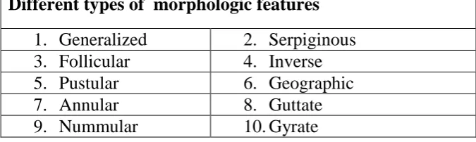
Table 2: Different morphology of psoriasis
.
57
Different types of morphologic features :
1. Generalized 2. Serpiginous
3. Follicular 4. Inverse
5. Pustular 6. Geographic
7. Annular 8. Guttate
9. Nummular 10.Gyrate
PRESENTATION:
The typical lesion is a well-demarcated from normal skin, sharply
declined edges, itchy, pink to salmon-colored plaque covered by slackly
adherent scales that is typically silvery white in color21,34,65,67. There is silvery
or yellow white scaly lesion similar to mica 66. Some lesions are presenting as
annular, linear, gyrate, and serpiginous like different configurations 65. A clear
peripheral zone encircles the plaques called, the halo ring of Woronoff
NUMBER: Number of lesions may be several to single one. From one to several centimeters, the diameter differs. On the legs and sacral region it is
commonly seen that the formation of coalescence of smaller plaques into large
plaques. Involuting lesions often clear from the center initially, producing
annular or arcuate shapes.
HISTOLOGICAL APPEARANCE:
In early budding lesions vasodilatation, papillary edema and leukocytes infiltration pave the way to the epidermal changes. Following this compact
hyperkeratosis, disappearance of granular layer and slight epidermal
hyperplasia will be continued76
Acanthosis (augmented epidermal cell turnover leads to distinct
thickening in epidermis) with uniform rete ridges descending extension and
appearance of mitotic figures in keratinocytes .
64,76.
Characteristic plaque, the majority portion of epidermis is shrunken
particularly overlying the dermal papillae (suprapapillary plates). Inside these
papillae, circuitous, dilated blood vessels can be seen. This assemblage of
changes costs in unusual closeness of vessels within the dermal papillae to the
overlying parakeratotic scale, and accounts for the typical clinical occurrence
of multiple, minute, bleeding points when the scale is moved up from the
plaque(Auspitz sign)
The stratum granulosum is
frailed or not present and widespread parakeratotic scale is visualized.
64
Neutrophils form small compilation within the parakeratotic stratum
corneum which is called as munro microabscesses and inside the spongiotic
foci of the superficial epidermis is known as spongiform pustules
.
64
Histologically there is mixed dermal infiltrate, including CD4 .
+ T cells,
dentritic cells, macrophages and mast cells present along with distinct
In a plaque lesion, the bulged endothelial cells are activated with having
intracellular Weibel-Palade bodies and prominent Golgi apparatus. Dermal
blood vessel dilatation and widening of intracellular spaces occurs because of
this. For easy migration of inflammatory cells like macrophages etc into the
skin, these lesional capillary loops express E- selection, present inside the
lesion having venous structural similarity, bridged fenestrations, put up it
easier58,81
LATENT PSORIASIS:
.
It is a state which appears before the manifestation of the signs of clinical psoriasis. During this period may be a few aberration present but there
is absence of clinical manifestation or the extinction of some hidden defect.
This latent stage may possibly occur at birth or later. No consideration of the
age at which psoriasis appears, the clinical phase can come into view from the
latent stage at any time. The changeover to clinical psoriasis from latent one
mostly presents as stable form which waxes and wanes in severity, but
infrequently transfer back to the latent period.These switchovers are "natural"
in that no treatment brings them about. It is evident that the progression of
clinical psoriasis often depends upon one of the "triggering stimuli" or whether
PATHOGENESIS:
It is seem to be multifactorial, whether symbolize the immune system or
the principal disease of skin, or the immune system, or contributions from
genetic and environmental factors is being discussed for some years.
ROLE OF OXIDATIVE STRESS:
1,58,64
Recently some theories have been proposed the participation of oxidative stress in the pathogenesis of psoriasis. Oxidative stress is unevenness
among oxidants and antioxidants in favor of the oxidants. This imbalance leads
to interference in the redox signaling and control and/or molecular damage60.
First it has been suggested that there is a compromised function of antioxidant
system and increased reactive oxygen species (ROS) production 34
As it is constantly exposed to UV and other environmental stresses, skin
is a vulnerable target for oxidative damage and producing ROS .
34
. The nitric
oxide synthase (NOS) expression is seen in keratinocytes the major
composition of epidermis and fibroblasts in the dermis. Following the UV
exposure these above mentioned cells liberated NO which plays a considerable
role in immunosupression and erythema and 61. Increased NO• end products has
important role in etiopathogenesis of psoriasis34. ROS mediated oxidative
damage causes a notable raise in arachidonic acid, DNA modification, and
secretion of inflammatory cytokines. Lipid peroxidation of plasma membrane
IMMUNOPATHOLOGY57
Most important hints of participation of immune system in pathogenesis:
:
1. The occurrence and emergence of abundant activated T cells in active
psoriatic lesions.
2. Immune mediated emergence of adhesion molecules on the surface psoriatic
keratinocytes.
3. The relative absence of TH
4. Lymphokine profiles signifying a T
2 related skin disorders, for example utricaria
and atopic dermatitis.
H
INFLAMMATORY MARKERS:
1 disarray.
The picture of psoriasis is thought that extremely increased rate of
epidermal production, along with set off mononuclear infiltrate in the dermis
beneath the affected epidermal layer3.Then subsequently following cells and
cytokines have been thought to take part as vital role in the pathogenesis, that
includes; Keratinocytes, Natural killer cells, Langerhans' cell,
Antigen-presenting cells, macrophages and T cells. The assembly of Th1 type cytokines,
growth factors like keratinocyte growth factor (KGF) and vascular endothelial
growth factor (VEGF) and etc 58
There is unusual expression of antigens such as heterodimers keratin 6–
α, along with
cytokine significant to disease processing, present in psoriatic epidermis, was
evidenced by in vitro and in vivo researches. In adding up, there are
intercellular adhesion molecule 1 (ICAM-1) and provoked expression of MHC
class II antigens91,57
Primary source for angiogenetic activity in the growing plaque is
epidermal keratinocyte proangiogenetic cytokines like VEGF and IL-8. Blood
vessels that are present psoriatic lesions have features like dilated and knotty,
entering directly in the dermal papillary vicinity, beneath the epidermis.
ICAM-1 (CD54), E-selectin (CD62E), vascular-cell adhesion Molecule ICAM-1 (CDICAM-106),
and MHC class II antigens, all are uttered by the lesional vascular cells shows
the activation
.
91
Recent progress in understanding the important role of T cells in
pathogenesis of psoriasis is going on. Along with INF-γ and other above
mentioned cytokines, the T helper-1 cells dominated cytokine milieu also
accounts for the skin lesions.
.
Current evidences suggest that IL-23 might be a key cytokine in
psoriatic lesion. INF-γ production and proliferation of memory Th1 cells are
stimulated by IL-23. IL-23 would maintain a Th1-committed memory response
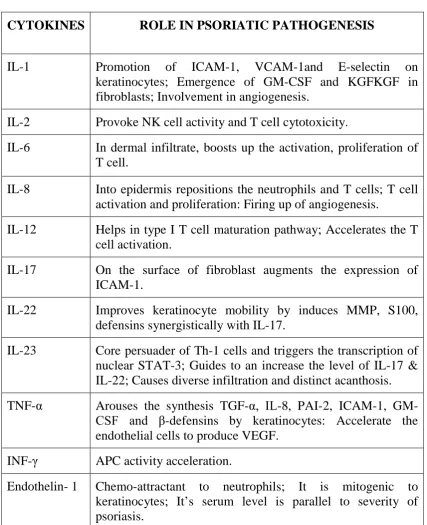
Table 3: Cytokines in the pathogenesis of psoriasis
CYTOKINES ROLE IN PSORIATIC PATHOGENESIS
IL-1 Promotion of ICAM-1, VCAM-1and E-selectin on
keratinocytes; Emergence of GM-CSF and KGFKGF in fibroblasts; Involvement in angiogenesis.
IL-2 Provoke NK cell activity and T cell cytotoxicity.
IL-6 In dermal infiltrate, boosts up the activation, proliferation of
T cell.
IL-8 Into epidermis repositions the neutrophils and T cells; T cell
activation and proliferation: Firing up of angiogenesis.
IL-12 Helps in type I T cell maturation pathway; Accelerates the T
cell activation.
IL-17 On the surface of fibroblast augments the expression of
ICAM-1.
IL-22 Improves keratinocyte mobility by induces MMP, S100,
defensins synergistically with IL-17.
IL-23 Core persuader of Th-1 cells and triggers the transcription of
nuclear STAT-3; Guides to an increase the level of IL-17 & IL-22; Causes diverse infiltration and distinct acanthosis.
TNF-α Arouses the synthesis TGF-α, IL-8, PAI-2, ICAM-1,
GM-CSF and β-defensins by keratinocytes: Accelerate the
endothelial cells to produce VEGF.
INF-γ APC activity acceleration.
Endothelin- 1 Chemo-attractant to neutrophils; It is mitogenic to
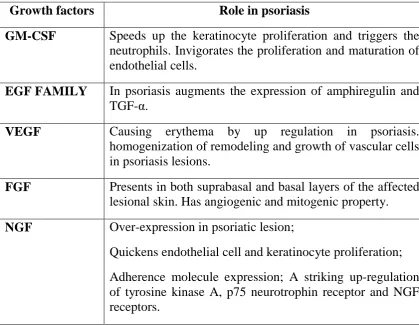
Table 4:Growth Factors
Growth factors Role in psoriasis
GM-CSF Speeds up the keratinocyte proliferation and triggers the neutrophils. Invigorates the proliferation and maturation of endothelial cells.
EGF FAMILY In psoriasis augments the expression of amphiregulin and TGF-α.
VEGF Causing erythema by up regulation in psoriasis. homogenization of remodeling and growth of vascular cells in psoriasis lesions.
FGF Presents in both suprabasal and basal layers of the affected
lesional skin. Has angiogenic and mitogenic property.
NGF Over-expression in psoriatic lesion;
Quickens endothelial cell and keratinocyte proliferation;
Adherence molecule expression; A striking up-regulation of tyrosine kinase A, p75 neurotrophin receptor and NGF receptors.
ROLE OF ICAM IN PSORIASIS:
Intercellular Adhesion Molecules (ICAMs) found in psoriatic plaques in a
significant quantity are otherwise known as adhesion molecules. It includes
ICAM-1, ICAM-2 which are along with VCAM-1, make availability of
costimulatory signals obligatory for T-cell activation by assists the binding of T
cells to antigen-presenting cells and keratinocytes. The interactions among
cellular adhesion molecule facilitates the continuous recirculation of T
LEUKOCYTE FUNCTION–ASSOCIATED ANTIGEN 1 (LFA-1):
LFA-1was recognized in humans in 1982. Tthis is the element of the
leukocyte β2-integrin family of adhesion molecules. Heterodimer shares a
common β chain (CD18) and a unique α chain (CD11a for LFA-1).Expression
of LFA-1 is constrained to leukocytes.
LFA-1 is an integral molecule in T cell activation and leukocyte
trafficking. LFA- 1 interrelates chiefly with ICAM-1, and too intermingles with
ICAM-2, ICAM-3 as well as JCAM-1. Memory T cells expresses LFA-1 in
higher concentration compared to naive T cells. The b2 subunit of LFA-1 has
been drawn in as important for signaling actions thought to be associated with
this LFA-1-ICAM-1 engagement. Early studies recommended that great
increase in the functional gluttony of T cell:APC interactions is by the primary
role of LFA-113
IL-23:
.
In 2000 during, searching for the members of IL-6 family, IL-23 was
discovered by Oppmann and colleagues40,19
This is discovered in 2000, more or less 15 years back, IL-23 has
promptly shifted as a key player, and a probable therapeutic object in psoriasis
than just a pro-inflammatory cytokine. .
5
IL-23, IL-12, IL-27 and IL-35 are
belongs to the IL-12 family which is wholly are heterodimeric cytokines. Even
though are having several structural similarities in the cytokines, downstream
differ in their biological activities. In the development of Th1 and Th17 cells,
these two IL-12 and IL-23 play a role of predominant proinflammatory/
prostimulatory cytokines which contribute to the above respectively14. IL-23
is a heterodimeric cytokine, consisting of a unique IL-23p19 subunit 5,40.
Belonging to the same family, IL-23 and IL-12 share a common p40 subunit6
Synthesis: It is mainly synthesized by activated myeloid cells, epithelial and endothelial cells
.
5. In monocytes, monocyte derived dentritic cells(DCs)
,and in mature DCs IL-23p19 in strongly expressed and mainly localized in
papillary dermis19. In psoriasis, dentritic cells and keratinocytes mainly
produced IL-235,41,42
IL-23, through TNF-α and IL-20R2 arbitrates epidermal hyperplasisa,
hyperkeratosis, acanthosis and orthohyperkeratosis .
47
Structural relationship between IL-23 and IL-12:
.
IL-12 and IL-23 are unique because of the way of secretion as binary
complexes82. IL-23 comprises unique IL-23p19 and IL-12/23p40 2 and
IL-23p19 is most closely related to the IL-12 p35 subunit5
IL-12 and IL-23 are heterodimeric typical four-helix cytokines. They are
secreted as a complex with a common binding protein termed p40 and a
disulfide-linkage linking the helical cytokines p19 and p35. Because of
together p35 and p19 have sequence homology to G-CSF and IL-6, both the
two are belongs to the members of the glycoprotein (gp) 130-class of
receptors such as the non-signaling alpha receptors for IL-6 and CNTF. In
essence, soluble α-receptor subunit which represented by IL-23 and IL-12 is
constitutively associated with the class I cytokine receptors. 82
In compared to nonlesional skin, the psoriatic skin lesion has increased
level of both p19 and p40 mRNA shows a clear cut indication that elevation of
IL-23 and its involvement in pathogenesis of psoriasis
.
35
Based on their common subunit use of p40, these ILs are important for
cell-mediated antimicrobial and cytotoxic activities that is T helper (Th) 1-type
responses by this it seems that these two cytokines would have superfluous
roles in immune homeostasis. Later,it was hastily exposed that the functions
are non-redundant
.
82
The relative restricted expression of p40 subunits limits potential IL-23
producing cells to monocytes, macrophages and dentritic cells. The receptor
complex for IL-23/12 is expressed or up regulated on T and NK cell and
myelomonocytic lineage including DC. The immune response of both IL-23
and IL-12 are comparable, but distinct.IL-12 mainly stimulates the IFN-γ
production in naïve T cells. Production of memory Th1 cells and IFN-γ
synthesis is better .
stimulated by IL-236
Functional difference between IL-23 and IL-12 in psoriasis:
.
• IL-23 signals through IL-23R and IL-12Rβ1, while IL-12 signals through
12Rβ1 and IL 12Rβ2 subunits.
• IL-23 accelerates the JAK-STAT pathway activity but acts largely on
STAT3 and IL-12 of JAK2 and TYK2 pathway leads to phosphorylation of
STAT4 and other STAT molecules.
• . IL-23 brings on IL-17A, IL-17F activity and/or IL-22, and stabilizes Th-17
cell but IL-12 promotes the synthesis of IFN-γ, which is needed for the
progression of Th1 immune responses 2,9
IL-23/ Th-17 AXIS:
.
In past years major advances in understanding of psoriasis from genetic,
immunological and clinical findings, all unambiguously converge on the
pivotal role of the IL-23/Th17 axis5,19
The pathophysiology of psoriasis can be divided into two discrete
immune-mediated phases:
.
Initial Phase
Amplification Phases.
Initial phase:
11,59
Resident dentritic cells and keratinocytes perturbation by trauma and/or
the following stimulation of pattern recognition receptors (e.g. dectin-1,
TLR-2, and TLR-4) in a genetically prone skin lead to stimulation of the innate
immune system. This cataclysm of macrophages, dentritic cells and diverse
Amplification phase:
By the adaptive immune response, these two cytokines make the bridge
from initiation to the amplification phase11,59. But this leads to persistence and
proliferation of Th17 cells inside the lesion. Following this Th17 cells may
enter into the skin, and expression of chemokine receptors, was recently
explained 35
Th17/IL-23pathway promotes chronic inflammation .
11. And this pathway
adds to the complexity of psoriasis pathogenesis and provides targets for newer
drug development 11. IL-23 attaches and signals by means of its heterodimeric
complex receptor compiled of subunits IL- 23R and IL-12Rβ1. IL-23R is an
exclusive in IL-23Rcomplex. IL-23R expresses at memory T cells, DCs,
natural killer cells and monocytes.43,44,45
IL-23 is implicated in propagation of memory T cells, when compare to
naïve T cells which does not responds to IL-23 due to minimal expression or no
subunit of IL-23R. IL-23 is responsible for differentiation and expression of
Th17 cell population which is exemplified by the synthesis of the IL-17A and
related proinflammatory cytokines
.
19,17,40,46
IL-23R is acquaintance with Jak2. Stimulation of IL-23 is directed to
ligand induced transphosphorylation and autophosphorylation of Jak2.
Activated Jak2 in sequence phosphorylates the tyrosine molecules situated in
tyrosine residues. After tying up, phosphorylation of these molecules occur.
Now in IL-23 signaling pathway, these above mentioned molecules particularly
STAT3 acts as the main participant19,43
For Th17 cell development, activation of STAT3 and involvement of the
orphan nuclear receptor RORα, aryl hydrocarbon receptor and the
Transforming growth factor-β1(TGF-β1) are needed .
19,35
. Phosphorylated
STAT-3 particles dimerizes from two identical monomers and transfer into the
nucleus, invoking cytokine transcription, like IL-17A, IL-17F, IL-22 and
INF-γ. Aminoacid switching, in the IL-23R subunit that is, arginine to glutamine
and leucine to proline, confer protection against psoriasis.IL-23 thus promotes
the IFN-γ production and type-1 immunity9
The term “type-1immunity” relates to a environment enhanced natural
killer (NK) activities and distorted towards cytotoxic functions of TH1, CD8+
T cell and. The major function of type 1 immunity is to execute intracellular
pathogens or cancer cells. Many tissue destructive inflammations for which
firstly TH1 cells were blamed, but in reality it is mediated by TH17 cells.
Immunopathology, tissue damage and disease onset is due to uncontrolled
type-1 cellular immune responses
.
9
TH-17 CELLS DERIVATION:
.
T cells:
of TCR is recognition of antigen. It can act in response to only an antigen
which is processed and presented by the macrophages like antigen presenting
cells.
TCR: (T cell receptor)
CD4+ T cells migrate into skin causing aggravation of disease and presenting as a new lesions. As compared with T cells of peripheral blood or
normal skin, CD4+ T cells on psoriatic lesional skin showed significant greater
presentation of Vβ2, Vβ5.1 and Vβ6 T cell receptor. The CD4+ T cells are the
majority of T cells localized in the affected dermis, whereas those migrating
into the epidermis are predominantly CD8 killer cells. The lesional CD8+ cells
have constant oligoclonal expression of Vβ3 and Vβ13.1 T cell receptors.
These T cells are activated and expressed in high levels of MHC class II
molecules and CD25, IL-2. 57
Most TCR comprise 2 chains- α and β.TCR does not have α/β and γ/delta chains. TCR is active only when both the chains (α and β), complex with CD3
molecules.
Factors promote the T cell migration into lesional plaque:
The above mentioned receptors in the papillary and dermal endothelial
cells receptors guide the T cell to ramble into the lesional skin. This relocation
into the psoriatic skin is accelerated by lipid mediators such as
MCP-2, MCP-3 and IL-8, MIP-1α and MIP-1β, IP10 and as yet other uncharacterized
peptides57
T cell development:
.
The majority of important events of T cell development take place in thymus. The progenitor T cells are originated from the bone marrow and then
drifted to thymus via blood stream. The chief maturation events take place in
the cortex, under the control of thymic hormones and lymphopoietic growth
factor IL-17 which are secreted by thymic stromal cells.
The antigen presenting cells differentiate the naïve T cells into Th1,
Th2, Th17 or T-regulatory cells by stimulation of the T cell receptor and the
fastidious release of cytokines 35
TH CELLS:
.
A novel TH subset, has been recently recognized as a distinct TH
lineage named as THIL-17, TH17 or inflammatory TH (THi). Because of
secretion of the following proinflammatory cytokines, such as TNF-α, IL-6,
from IL-17A to IL-17F, IL-21 and IL-22, it is defined as Th 17 cell 35
DEVELOPMENT AND DIFFERENTIATION:
.
In 2 ways:
1) IL-23 dependent:
On developing Th17 cells, the generation of IL-23R is by, intracellular
signaling through STAT3, RORγT, and extracellular TGF-β. IL-23R upholds
the sensitivity of IL-23 as the master cytokine by inducing the continued
existence and multiplication of Th17 cells. The IL-23R receptor is a
heterodimer made up of IL-12Rβ1, IL23R subunits1535
2) IL-23 independent:
.
The switch from naive CD4 cells to Th-17 cells happens in the presence
of IL-6 and exposure to extracellular transforming growth factor (TGFβ) as
well as Toll-like receptor-activated monocytes 19. IL-1β and Tumor necrosis
factor (TNF)-α, both these cytokines causes amplification of the Th-17 cell
differentiation, which are mediated by IL-6 and TGF-β. The differentiation of
the Th 17 cells from naive T cell precursors is also dependent on the
intracellular transcription factors RORγ and STAT335
IL-1β was enough for production of both IL-17A and IFN-γ and to
create the expression of RORC. In IL-17A-producing cells, a new marker in
detected and named as CD161and was recommended as a novel marker for
Th17 cells
.
The Th17 differentiation was initiated by TGFβ and IL-6 and mediated
by STAT3 via regulating the chromatin remodeling of the IL-17-IL-17-F locus,
which is further reinforced by IL-23
19,24
Apart from the cytokine induced Th lineage formation, the
differentiation is shown to be directly endorsed by the prostaglandin E2. PGE2
synergizes with IL-1β and IL-23 to persuade the up regulation of IL-23R, and
IL-1R to promote Th 17-associated profile of transcription factor, cytokine and
chemokine receptor expression 50
Transcription factors comprised in Th cell development:
.
• IFN-regulatory factor 4(Irf-4)
• Signal transducer and activator of transcription 3 (STAT 3),
• T cell-specific splice isoform of retinoic acid receptor-related orphan
receptor (Ror) known as Ror-γt
• Splice isoform of Ror- αd
• Aryl hydrocarbon receptor (AhR)
Cytokines produced by Th-17 cells:
• IL-17A to IL-17F
• Tumor necrosis factor (TNF)-α
• IL-6, IL-22, and IL-26.
IL-17:
TH17 cells produce IL-17A, IL-17F, IL-6 and IL-22, all of which
regulate inflammatory responses by tissue cells49
It is undetectable in normal skin. It is a member of a newly identified cytokine family comprising 17A, 17B, 17C, 17D, 17E and
chromosome, as well as co-ordinate expression pattern IL-17F shares a
maximum homology with IL-17A50,35
IL-17RA is uttered on B and T cells, epithelial cells, fibroblasts,
monocytic cells and bone marrow stroma. IL-17RA signaling activates both the
nuclear factor-kB, and mitogen activated protein kinase intracellular
pathways
.
35
IL-17 role in psoriasis:
.
Proinflammatory activity induces the neutrophils, monocytes/macrophages
T cells and epithelial cells to express proinflammatory cytokines, colony
stimulating factors and chemokine 16
Acts on macrophages to promote their recruitment and survival. .
Conscription and activation of neutrophils via effects on granulopoiesis. 35
In inflamed tissue, inhibit the neutrophils apoptosis directly
.
62
It’s marked role in sub corneal accumulation of neutrophils and stimulates
the production of matrix metalloproteases and aniogenic factors to cause
tissue remodeling and angiogenesis.
.
CXC chemokine induction.
Stimulates the mixture of immune and non-immune cells for the synthesis
of proinflammatory cytokines and anti-microbial peptides.
Also enhance the IL-2 production capacity of human CD4+ T cells.
Mechanism of action of IL-17:
In psoriasis, the mechanism of IL-17 activity in stems from its
co-operative gene regulation IL-22 and other stimuli. Together with IL-17, IL-22
synergistically enhances expression of skin antimicrobial peptides including
S100A7 (psoriasin), b-defensin-2(BD-2), and S100A8/9 (calprotectin)103.
Supporting this, S100A7–9 is elevated in psoriasis, correlating with disease
onset. Interestingly as the result of elevated antimicrobial peptide production
psoriasis, patients are more challenging to skin infections than non psoriatic
people. Another antimicrobial peptide, cathelicidin (LL37), is synergistically
increased by treatment with IL-17 in combination with 1,25-dihydroxyvitamin
D3. LL37-bound self-DNA fragments trigger TLR9 in DC, which induces a
potent adaptive immune response, possibly one of the mechanisms by which
self-tolerance is broken102
COMORBIDITIES:
.
Manifestations of psoriasis are not only restricted to the skin. In
moderate to severe psoriasis, several other multiple comorbidities also
complicate the other systems. Ischemic heart disease, stroke, hypertension,
dyslipidemia, obesity/metabolic syndrome, diabetes mellitus, autoimmune
diseases, sleep apnea and crohn’s disease are some diseases arise in psoriatic
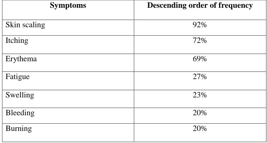
Table 5: The symptoms most often stated by the psoriasis patient 86
Symptoms
:
Descending order of frequency
Skin scaling 92%
Itching 72%
Erythema 69%
Fatigue 27%
Swelling 23%
Bleeding 20%
Burning 20%
ASSOCIATIONS OF PSORIASIS:
Psoriatic arthritis, immunobullous disorder, vitiligo, metabolic syndrome,
acne, pustulosis, synovitis and hyperostosis are some clinical entities associated
with psoriasis51
1. Psoriatic Arthritis: .
Arthritis is the most common association with psoriasis was established
out by Jean Louis Alibert in 1818. The incidence of arthritis in psoriatic
patients is from 1.3% to34.7%. On sex predilection of PsA, there is no proper
reliable data. Psoriatic arthritis (PsA) falls under the type of seronegative
spondyloarthropathies.
on the national psoriasis foundation, up to 30% of the patients have psoriatic
arthritis65. Usually within 10 years of the onset of psoriasis the PSA affects the
joints and occurring PSA after 10 years of onset is low53. 27.5% of psoriatic
arthritis patients have family history of psoriasis54
5 types of psoriatic arthritis;
.
Symmetric,
Asymmetric,
Distal Interphalangeal Predominant (DIP),
Spondylitis And
Arthritis Mutilans.
Symmetric arthritis looks a lot like rheumatoid arthritis, but more often
than not is milder. Asymmetric arthritis be capable of involve several joints
with the presentation as sausage digits.DIP is classic form, but arises only in
relation to 5% of the patients with PSA. Arthritis mutilans is dangerous and
deforming. The small joints of hands and feet are affected primarily by this
type. Less than 5% of PsA will suffer by this form65
2. Obesity/Metabolic syndrome:
.
The other different names are insulin resistance syndrome and syndrome
X. This syndrome includes metabolic abnormalities with high risk of coronary
artery diseases and diabetes mellitus. The primary pathophysiology linking the
metabolic syndrome and psoriasis engages overlie of genetic propensity and
factor-α dysregulation leads to chronic inflammation interceded by Th-1 and Th-17
cells. . These Th-1 and Th-17 cellsand the cytokines released by these cells
persuades epidermal hyperplasia in psoriasis, and alienates signaling function
of insulin, intervene the resistance of insulin resistance and change the
expression of adipokine, and obesity. Susceptibility to psoriasis or seriousness
of disease is due to long term inflammation and angiogenesis and these are
promoted by hyperinsulinemia in metabolic syndrome. And moreover, the
subsistence of genetic loci, e.g., PSORS2-4, CDKAL1 and ApoE4 and, is too
been drawn into the joint of genetic vulnerability of metabolic syndrome and
psoriasis. The alliance relating metabolic syndrome and psoriasis is due to the
above mentioned shared measures 48
Different types of surveys has accounted that increased levels of serum
immunological markers, like IL-6, IL-2, ICAM-1 and TNF-α , in order to prove
that it is a systemic immunological disorder. Inflammation is mainly controlled
by hormones and cytokines derived from adipose tissue and liver by IL-1, IL-6
and TNF-α. Common cytokine pathways are responsible for both psoriasis and
obesity but it is yet to be answered which pathology comes first when
psoriasis-associated obesity and metabolic syndrome is considered .
79
There are so many evidences that prove the relationship between
psoriasis and a number of lifestyle factors for example alcohol intake and
smoking and other diseases with psoriasis
.
3. Cardiovascular diseases:
Gelfand et al used the General Practice Research Database (GPRD) to decide the psoriasis is an independent risk factor for myocardial infarction.
CAD risk factors prevalence is greater in psoriatic patients. The psoriatic
patients with raised TNF-α level has the risk of increased frequency of CAD,
pulmonary emboli and cerebrovascular diseases occur 79.When put side by
side to healthy population psoriatic patients had a 1.6 fold increased risk for
venous occlusion, 2.6 fold increased risk for other occlusive vascular diseases.
Arteriothrombotic markers like fibrinogen and plasminogen activator
inhibitor-1 (PAI-1) levels are also seem to be increased in psoriasis79.
4. Bullous pemphigoid and vitiligo:
Along with a systemic inflammation, antipsoriatic prescriptions are
believed (acitretin, cyclosporine) for the adverse CAD risk factors like
elevation of blood pressure, elevation of serum levels of lipids are other
possible optional mechanisms for CAD comorbidity of psoriasis .
Co localization of vitiligo and psoriasis may be possible because of structural abnormalities between anti stratum corneum antibodies and anti
melanocyte antibodies. In addition, a common neuropeptide might be also
accountable for co-habitation of psoriasis and vitiligo51
5. Psychiatric co morbidity:
.
Psoriasis causes important adverse effects on the psychological and
and treatment for disease were most affected physical and psychosocial factors.
Sufferers mostly tends to avoid the social contacts because of disturbed feel,
low self conscious, live in a constant fear of relapse or bothered by the peeling
of the skin.
Patients with psoriasis describe feeling of annoying or defenselessness.
They reveal a higher rate of suicidal ideations Compare with other patients,
thinking of suicide attempts is in a greater proportion. Among 127 psoriatic
patients a study was conducted where about active suicidal thought was
reported in 5.5% and “want to die” was 9.7% during the study period78
ASSOCIATION WITH PREGNANCY:
.
Many reports have recommended that physiological changes during
pregnancy habitually lessening of systemic and cutaneous inflammatory
diseases88, 89
In pregnancy, there is a improvement in psoriasis due to hormone
mediated down regulation of the immune system. The greatest role in the
improvement of psoriasis is played by progesterone. The metabolizing capacity
of keratinocyte cells on steroid hormones like estrogen and progesterone also
believed to be altered by the hormonal changes of pregnancy. High levels of
IL-10 in pregnancy have a favorable effect on psoriasis. In addition, some
theories include roles of human chorionic gonadotrophin and human placental
Up-regulation of proinflammatory Th-1 cytokines also plays a key role
in the inflammatory streams of psoriasis. It is likely that, anti-inflammatory and
antagonizing effects of Th-2 cytokine-mediated down-regulation on the Th-1
cytokines improves psoriasis during pregnancy88
ASSOCIATION OF PSORIASIS AND AUTOIMMUNE DISEASES: .
We reviewed studies published in the MEDLINE database from January
1, 1980, to June 1, 2011, and recapitulated the associations between psoriasis
and several key autoimmune diseases, including celiac disease (CD),
inflammatory bowel disease (IBD), multiple sclerosis (MS), systemic lupus
erythematosus (SLE), and autoimmune thyroid disease. This review shows that
the association among psoriasis and CD and IBD appears to be well
described98
ANEMIA IN PSORIASIS:
.
Psoriasis is known to be one of the skin diseases which can cause folate deficiency. Touraine et al101 reported reduced serum and red blood cell
folate levels in 22 out of 50 patients with psoriasis. Similar observations have
also been reported by Shuster and Marks and Fry et al. They attributed the
folate deficiency mainly to the increased utilization of folate by the rapid
turnover of epidermal cells in psoriasis. The malabsorption of folate first
proposed to occur in psoriasis by Shuster and Marks has since been noted only
in rare cases by Touraine et al. Folate deficiency due to excessive loss in
contrast to the reduced folate levels, serum vitamin B12 levels are reported to
be normal in most psoriasis patients and no evidence of impaired vitamin B12
absorption has been detected despite abnormal Schilling test results in some
cases. In addition, there is no increased incidence of pernicious anemia in
psoriasis patients to our knowledge. These findings suggest that vitamin B12
deficiency is unlikely to be a contributory factor in the megaloblastic anemia in
psoriasis99
C-REACTIVE PROTEIN:
.
In 1939, Tillet and Francis described material in the sera of acutely ill patients. It was attached to the cell wall C-Polysaccharide of Streptococcus
pneumonia and agglutinated the organisms. In 1941 that material was revealed
to be a protein and called as C - reactive protein.
Evolution of CRP and pentraxin:
It belongs to pentraxin family of calcium dependent ligand binding
plasma protein. The pentraxin family, is vastly conserved in evolution. It is
called for due to its electron microscopic appearance and from the Greek
penta(five) ragos(berries) with homologous proteins throughout the vertebrates
and even in phylogenetically distant arachnid, limulus polyphemus, a
horseshoe crab71
The disparities are based on with regard to
• Presence and character of glycosylation,
• capability to precipitate and aggregate ligands
• Fine ligand-binding specificity,
• Protomer assembly
• Behavior as acute-phase proteins
• Base line circulating concentrations
• Capacity to activate autologous complement.
Indeed, only human CRP has been meticulously known to activate
complement in isologues serum. These differences dominate in extrapolating
from animal models to humans71
Synthesis:
.
In the hepatocytes it is synthesized, nearly exclusively a large amount under the control of cytokines. But extra hepatic sites of CRP production have
also been reported 69
IL-1, IL-6 and TNF-α mostly controls the synthesis of CRP. These
cytokines has power to alter the CRP levels as well and increase of CRP in
blood and body fluids by a steady release of these proinflammatory cytokines .
79
Biochemistry of CRP:
.
or disk configuration by way of radial symmetry. The total mass of CRP is ~
115kDa. Every subunit having 206 aminoacids.
The family name pentraxin for CRP has come because of its pentameric
structure. Other related proteins to CRP, belongs to pentraxin family are
proteins such as serum amyloid-P and pentraxin-3.
CRP has circulating half life of 18-20 hours70,71. Each protomer has
the characteristic “lectin fold”, composed of 2 layered β-sheets with flattened
jellyroll topology. The ligand-binding site, composed of loops with two
calcium ions bound 4 Å apart by protein-side chains, is located on the concave
face. The other face carries a single α helix71
Function of CRP:
.
Against break down products of cells and infectious organisms it has
non specific host defense.
Stimulate the classical complement pathway by starts at C1q, resulting
in paghocytosis via C3b receptors.
Has positive feedback via alternative pathway by make a complex with
factor H, a complementary inhibitory factor and to a great extent reduces
the activation of late components(C5- C9).
No genetic abnormalities have been reported for circulating CRP. It is
catabolized when complexes are engulfed by phagocytes.
Clinical significance of CRP:
The synthesis rate is the only determinant of circulating levels of CRP
because the plasma half-life of CRP is stable in all conditions of health and
disease. Thus the rate of synthesis is directly proportional to the intensity of
pathological process inducing the CRP production
72
It is one among the strongest acute phase reactants .
69
Moreover, CRP may serve interchangeably with Psoriasis Area and
Severity Index (PASI) as a measure of disease severity in the case of untreated
psoriatic patients who do not have disease related arthritis
. The plasma levels
can rise up to 1000 fold after stress, trauma, myocardial infarction, infection or
neoplastic proliferation. In the infection and inflammatory conditions it may
go up to more than 5 to 10 mg/L. Only in moderate and severe forms of disease
might have the high CRP level inferred from the literature and there is no
enough data signifying a similar connection for mild disease.
56
Role of CRP in psoriasis:
.
It is recognized as the most sensitive indicator of inflammation, even
though it is a nonspecific cytokine. Depends amount of tissue injury and
inflammation severity, the magnitude of CRP level will be increased.72
When compare the Psoriatic patients with severity of the disease the
severe forms (PASI > 10) had appreciably elevated levels of CRP than with
mild disease (PASI < 10) (44% vs 25%) (
.
show the characterization of CRP level in psoriasis. That is as an inflammatory
response that worsens with increasing disease severity. Several other studies
have also reported a correlation between PASI and increased levels of CRP.
Thus, CRP can be considered as a useful marker of disease severity. And can
be used to observe the disease course and severity and can able to decide the
treatment68
CRP estimation is widely available, inexpensive, and can be simply
carried out in an outpatient clinical setting. To evaluate psoriasis disease
severity, CRP along with PASI can be used as a powerful and sensitive marker,
when it is difficult to evaluate, based on visual evaluation of the lesions. It can
be used for screening of the disease course and treatment. Elevation of CRP
may be thought as a risk factor for CVD in psoriatic patients because there is
some research supporting the association between inflammation and CVD in
psoriatic cases .
68
CLASSIFICATION OF PSORIASIS:
.
I. Depends on natural history or morphology. (clinical appearance) II. Depends on the precipitants or age.
I. Depends on natural history or morphology: (clinical appearance)
a. Plaque psoriasis (psoriasis vulgaris)
b. Acute guttate psoriasis
c. Unstable
d. Erythrodermic
e. Pustular
f. Inverse
II. Depends on the environmental factors:
a. Photo aggravated
b. Drug induced or exacerbated
c. HIV-induced or exacerbated
d. Alcohol misuse
e. Cigarette smoking.
f. Trauma
g. Metabolic factors
III. Depends on the involved specific sites:
a. Scalp
b. Flexural (inverse)
c. Genital
d. Nonpustular palmoplantar
e. Nail
f. Mucosal
g. Ocular
I. Depends on natural history or morphology: (clinical appearance)
a. Plaque psoriasis: (psoriasis vulgaris)
This is the most common type of psoriasis. In this type, there is a stable;
red (salmon pink) scaly lesion with gradual broadening of plaques lesions
persists for months to years. They expand very steadily and progress as
indolent course and rarely occurs abruptly. The scaling extension may differ
and orange-brown or waxy yellow. The most commonly involved areas are the
elbows, knees, gluteal cleft and the scalp. There is a symmetrical involvement
in the affected areas65.
b. Guttate psoriasis: (eruptive psoriasis)
The clear marginal zone, called the halo ring of
Woronoff may enclose the lesion.
Most frequent in children and young adults. It arises in persons with no
psoriasis or in people with chronic plaque psoriasis. Patients present with
shower of tiny 2 to 3 mm erythematous, scaling papules, mostly follow the
upper respiratory tract infection due to β-hemolytic streptococci. Pityriasis
rosea and secondary syphilis are the differential diagnosis for the guttate
type65,76. Mainly affected sites are the trunk, face, and proximal portions of
limbs. Guttate psoriasis also detected after withdrawal of corticosteroid therapy
c. Unstable psoriasis:
This term usually explain the stages of disease, which is highly active and with unpredictable outcome76. Patients have frequent complaints of more itchiness, irritation and even pain. They are more intense inflammation with angry looking lesions. These seem to be ill demarcated, mild scaling and more
red in color with intermittent exudation and crust. Further spontaneous
conversion to pustular or erythrodermic psoriasis can happen. Inappropriate use
of corticosteroids, excessive irritation, sunburn are some of the reasons usually
associated with unstable psoriasis
d. Erythrodermic psoriasis: 25,57
When affects around 90% of the body surface as a generalized form, it is described as erythrodermic psoriasis. Although the affected patient is having
all the symptoms of the disease, but the generalized erythema is the most
outstanding feature with less scaling. The face is rarely involved. It may
present as different stages as sudden appearance of generalized erythema or
gradual evolving from chronic plaque psoriasis. Triggering causes are
occasionally identified25,57
e. Pustular psoriasis:
.
Numerous tiny pustules develop an erythematous skin with pustular
plaques and inconsistent scale. This type of psoriasis is either mild and
localized in soles and palms, or extensive and life threatening [64]. Localized
fever for a number of days, , diffuse cutaneous and mucosal pustules with a
background of severe erythema, leukocytosis, arthralgia, secondary infection
and electrolyte disturbances64,65 episodes of fever and pustules are
recurrent.[65]
f. Inverse psoriasis:
A severe, acute form (the von Zumbusch variant) can cause
life-threatening complications.
Affect the intertriginous areas including the axilla, groin,
submammary region and naval. It also affects scalp, palms and soles. The
individual lesions are finely demarcated plaques, with absence of scales
because of their presenting areas and they may be moist65
II. Depends on the environmental factors:
.
a. Photoaggravated:
Usually the sunlight is favorable, but in little insignificant number of patients, psoriasis is aggravated by strong sunlight and it is the reason for
summer exacerbations in exposed skin. Recent work has indicated that severely
photosensitive psoriasis is predominantly female, strongly associated with
onset age, HLA-Cw6 and family history, and different from polymorphic light
eruption (PLE). Photochemotherapy (PUVA) may be useful in these patients.
b. Drug induced or exacerbated:
76
In current clinical practice, the generally common drugs which may be
Non-steroidal anti-inflammatory agents, Synthetic antimalarials, Angiotensin
converting enzyme inhibitors, and withdrawal of Corticosteroids76,67
Action of some medications is partially described. Such as, lithium salts
raise proinflammatory cytokines, and rousing the cutaneous leukocyte
recruitment; beta-adrenergic blockers may