PHARMACOLOGICAL EVALUATION OF GOLD
NANOPARTICLES
A Dissertation Submitted to
THE TAMIL NADU Dr. M. G. R. MEDICAL UNIVERSITY CHENNAI-600 032
In partial fulfillment of the requirement for the award of the Degree of
MASTER OF PHARMACY
IN
PHARMACOLOGY
OCTOBER-2017
DEPARTMENT OF PHARMACOLOGY
KMCH COLLEGE OF PHARMACY,
KOVAI ESTATE, KALAPATTI ROAD,
PHARMACOLOGICAL EVALUATION OF GOLD
NANOPARTICLES
A Dissertation Submitted to
THE TAMIL NADU DR. M. G. R. MEDICAL UNIVERSITY CHENNAI-600 032
In partial fulfillment of the requirement for the award of the Degree of
MASTER OF PHARMACY
IN
PHARMACOLOGY
OCTOBER-2017
Submitted by
Anusree. E
(Reg. No. 261525803)
Under the Guidance of
Dr. K. T. Mani Senthil Kumar, M Pharm, Ph. D,
Head, Department of Pharmacology
DEPARTMENT OF PHARMACOLOGY
KMCH COLLEGE OF PHARMACY,
KOVAI ESTATE, KALAPATTI ROAD,
Prof. Dr. A. Rajasekaran, M. Pharm., Ph.D.,
Principal,
KMCH College of Pharmacy, Kovai Estate, Kalapatti Road, Coimbatore - 641 048.
Tamil Nadu
CERTIFICATE
This is to certify that the dissertation work entitled
“PHARMACOLOGICAL EVALUATION OF GOLD NANOPARTICLES” was
carried out by Ms. Anusree. E (Reg. No. 261525803). The work mentioned in the dissertation was carried out at the Department of Pharmacology, KMCH College of Pharmacy, Coimbatore, Tamil Nadu, under the guidance of Dr. K. T. Mani Senthil Kumar, M Pharm, Ph. D for the partial fulfillment for the degree of Master of Pharmacy during the academic year 2016-2017 and is forwarded to the Tamil nadu Dr. M. G. R. Medical University, Chennai.
Date: Prof. Dr. A. RAJASEKARAN, M. Pharm., Ph.D.
Place: Coimbatore Principal
Dr. K. T. Mani Senthil Kumar, M Pharm, Ph. D,
Professor and Head,Department of Pharmacology, KMCH College of Pharmacy, Kovai Estate, Kalapatti Road, Coimbatore -641 048.
Tamil Nadu
CERTIFICATE
This is to certify that the dissertation work entitled “PHARMACOLOGICAL
EVALUATION OF GOLD NANOPARTICLES” is a bonafide work carried out by
Ms. Anusree. E (Reg. No. 261525803). The work mentioned in the dissertation was carried out at the Department of pharmacology, KMCH College of Pharmacy, Coimbatore, Tamil Nadu, under my supervision and guidance during the academic year 2016-2017.
This research work either in part or full does not constitute any of any thesis / dissertation.
Date:
Place: Coimbatore Dr. K. T. Mani Senthil Kumar, M Pharm, Ph. D,
DECLARATION
I do here by declare that to the best of my knowledge and belief ,the dissertation work entitled “PHARMACOLOGICAL EVALUATION OF GOLD
NANOPARTICLES’’ submitted to the Tamil Nadu Dr. M. G. R. Medical university,
Chennai, in the partial fulfillment for the Degree of Master of Pharmacy in
Pharmacology, was carried out at Department of Pharmacology, KMCH College of Pharmacy, Coimbatore under the guidance of Dr. K. T. Mani Senthil Kumar, M. Pharm, Ph. D, during the academic year 2016-2017.
Date:
Place: Coimbatore Anusree. E (Reg. No.261525803)
EVALUATION CERTIFICATE
This is to certify that the work embodied in the thesis entitled
“PHARMACOLOGICAL EVALUATION OF GOLD NANO PARTICLES”
submitted by Ms. Anusree. E (Reg. No:261525803) to the Tamil Nadu Dr. M.G.R. Medical university, Chennai, in the partial fulfillment for the Degree of Master of Pharmacy in Pharmacology, is a bonafide research work carried out by the candidate during the academic year 2016-2017 at KMCH College of Pharmacy, Coimbatore, Tamil Nadu and the same was evaluated by us.
Examination Center: KMCH College of Pharmacy, Coimbatore
Date:
Internal Examiner External Examiner
PHARMACOLOGICAL EVALUATION OF
GOLD NANOPARTICLES
A Dissertation Submitted to
THE TAMIL NADU Dr. M. G. R. MEDICAL UNIVERSITY CHENNAI-600 032
In partial fulfillment of the requirement for the award of the Degree of
MASTER OF PHARMACY
IN
PHARMACOLOGY
OCTOBER-2017
DEPARTMENT OF PHARMACOLOGY
KMCH COLLEGE OF PHARMACY,
KOVAI ESTATE, KALAPATTI ROAD,
PHARMACOLOGICAL EVALUATION OF GOLD
NANOPARTICLES
A Dissertation Submitted to
THE TAMIL NADU Dr. M. G. R. MEDICAL UNIVERSITY CHENNAI-600032
In partial fulfillment of the requirement for the award of the Degree of
MASTER OF PHARMACY
IN
PHARMACOLOGY
OCTOBER-2017
Submitted by
Reg. No. 261525803
DEPARTMENT OF PHARMACOLOGY
KMCH COLLEGE OF PHARMACY,
KOVAI ESTATE, KALAPATTI ROAD,
Prof. Dr. A. Rajasekaran, M. Pharm., Ph.D.,
Principal,
KMCH College of Pharmacy, Kovai Estate, Kalapatti Road, Coimbatore - 641 048.
Tamil Nadu
CERTIFICATE
This is to certify that the dissertation work entitled
“PHARMACOLOGICAL EVALUATION OF GOLD NANOPARTICLES” was
carried out by Reg. No. 261525803. The work mentioned in the dissertation was carried out at the Department of Pharmacology, KMCH College of Pharmacy, Coimbatore, Tamil Nadu, for the partial fulfillment for the degree of Master of Pharmacy during the academic year 2016-2017 and is forwarded to the Tamil Nadu Dr. M. G. R. Medical University, Chennai.
Date: Prof. Dr. A. RAJASEKARAN, M. Pharm., Ph.D.
Place: Coimbatore Principal
GUIDE,
Dept. of Pharmacology, KMCH College of Pharmacy, Kovai Estate, Kalapatti Road, Coimbatore -641 048.
Tamil Nadu
CERTIFICATE
This is to certify that the dissertation work entitled
“PHARMACOLOGICAL EVALUATION OF GOLD NANOPARTICLES” is a
bonafide work carried out by Reg. No. 261525803. The work mentioned in the dissertation was carried out at the Department of Pharmacology, KMCH College of Pharmacy, Coimbatore, Tamil Nadu, under my supervision and guidance during the academic year 2016-2017.
This research work either in part or full does not constitute any of any thesis / dissertation.
Date:
DECLARATION
I do here by declare that to the best of my knowledge and belief, the dissertation work entitled “PHARMACOLOGICAL EVALUATION OF GOLD
NANOPARTICLES” submitted to the Tamil Nadu Dr. M. G. R. Medical university,
Chennai, in the partial fulfillment for the Degree of Master of Pharmacy in
Pharmacology, was carried out at Department of Pharmacology KMCH College of Pharmacy, Coimbatore during the academic year 2016-2017.
Date:
EVALUATION CERTIFICATE
This is to certify that the work embodied in the thesis entitled
“PHARMACOLOGICAL EVALUATION OF GOLD NANOPARTICLES”
submitted by Reg. No:261525803 to the Tamil Nadu Dr. M.G.R. Medical university, Chennai, in the partial fulfillment for the Degree of Master of Pharmacy in
Pharmacology, is a bonafide research work carried out by the candidate during the academic year 2016-2017 at KMCH College of Pharmacy, Coimbatore, Tamil Nadu and the same was evaluated by us.
Examination Center: KMCH College of Pharmacy, Coimbatore
Date:
Place: Coimbatore
Internal Examiner External Examiner
Plan of Work
Bibliography
DEDICATED TO ALMIGHTY,
MY BELOVED PARENTS,
BROTHERS, SISTERS
ACKNOWLEDGEMENT
“Through gratitude comes deep from the heart, if left unexpressed loses its memory, charm and above all, the biggest asset, its beauty”
My dissertation entitled “PHARMACOLOGICAL EVALUATION OF GOLD
NANOPARTICLES” would not have beena feasible one without the grace of God almighty who gave me moral till the completion of my project.
First and foremost I am extremely beholden to my esteemed research guide, Dr.
K. T. Manisenthil Kumar, M. pharm, Ph.D. HOD and Professor, Department of Pharmacology, for his constant insight, personal advice, countless serenity and pain taking efforts in all stages of my study.
With great pleasure I wish to place my indebtedness to Dr. A. Rajasekaran, Principal, KMCH College of Pharmacy for his support and for giving me an opportunity to do my project work.
I submit my sincere thanks and respectful regards to our beloved Managing
Trustee, Dr. Nalla G. Palaniswami and respected trustee madam Dr. Thavamani D.
Palaniswami, Kovai Medical Centre Research and Education Trust, Coimbatore for all the facilities that were provided to me at the institution.
I owe my debt of gratitude to our esteemed and staffs Dr. Ariharasivakumar, M.pharm., Ph. D., Professor and Mr. Saravanan J. M. pharm., Ms. Sanju. K M.pharm., for their immense support, timely help and valuable suggestions.
I express my sincere thanks to Biostatistics mam Mrs. J. Vennila for the helps
offered during my work.
This project would not be a resplendent one without the timely help and
continuous support by my ever-loving buddies Anna, Anu, Boopathi, Neethu and Parthipan.
I take this opportunity to extend my indebtedness to Manimaran, Jopson, Revathy and Swathy for helping me every aspects.
I express my sincere thanks to all my juniors from Dept. of pharmacology for their memorable company and involvement.
It gives me immense pleasure to express thanks to my dearest friends, Abdul Kaleel, Nikhil. M, Ayesha, Rincy, and my roomies Anjali, Ansu, Aparna and Athira who were always there whenever I needed.
I take great opportunity to extend my indebtedness to Kokila, Sangeetha, Alga,
Treeza.
I am greatful to Lab technicians Mr. Tamilarasan (Dept. of Pharmacology), Mrs. Anandhi, Mrs. Muneeswari, librarian, for their timely support and help during the course of the entire work.
I express my special thanks to Mrs. Dhanalakshmi for helping in animal maintenance during the study.
Above all I dedicate myself before the unfailing presence, constant love,
immense support and encouragement given to me by my beloved Achan, Amma, Chechi, Evamol and my brothers who deserves the credit of success in whatever work i did.
Last but not least, I would like to thank everyone who was important to the
successful realization of this thesis.
Thank you all for the support and motivation
LIST OF ABBREVIATIONS
SL
NO ABBREVIATIONS FULL FORM
1 ALP Alkaline Phosphate
2 ANOVA Analysis of Variance
3 DEN Diethyl Nitrosamine
4 DMSO Dimethyl sulfoxide 5 DNA Deoxyribonucleic acid
6 EDTA Ethylenediaminetetraacetic acid 7 EGFR Epidermal growth factor receptors
8 ER Endoplasmic reticulum
9 FBS Fetal bovine serum
10 GCS Gold colloid solution.
11 gm Gram
12 GNP Gold Nanoparticles 13 HBV Hepatitis B Virus
14 HCC Hepatocellular Carcinoma
15 HCL Hydrochloric Acid
16 HCV Hepatitis C Virus 17 i.p Intra peritoneal
18 IL Interleukin
19 kg Kilo gram
20 LDH Lactate dehydrogenase 21 mg/dl Milligram deci Litre
22 ml Milli Litre
23 MTT Microculture tetrazolium Assay 24 NADH Nicotinamide adenine nucleotide
25 NADPH Nicotinamide adenine dinucleotide phosphate 26 NCCS National Centre for Cell Science
28 NPs Nanoparticles
29 p.o Per Oral
30 PG Prostaglandin
31 PI Percentage inhibition 32 ROS Reactive Oxygen Species
33 SD Standard Deviation
34 SEM Standard Error Mean
35 SGOT Serum Glutamate Oxaloacetate Transaminase 36 SGPT Serum Glutamate Pyruvate Transaminase 37 TCA Trichloro Acetic Acid
38 TNF-α Tumor necrosis factor alpha
39 μg Micro gram
LIST OF TABLES
TABLE NO. TITLE PAGE NO.
1. Pharmacological Model for DEN induced HCC 36
2.
Experimental design for carrageenan induced paw
edema 43
3. Experimental design for Histamine induced paw edema 44 4. Experimental design for Serotonin induced paw edema 45 5. Elemental composition of gold nanoparticles 49 6. Elemental composition of colloidal gold 49 7. Cytotoxic activity of GNP and GCS in HeLa cell line 50 8. Effect of GNP& GCS on the RBC& WBC counts 55
9.
Effect of the GNP& GCS on the Body weight of the
Experimental rats 57
10.
Effect of the GNP& GCS on the Liver weight of the
Experimental rats 58
11.
Effect of the GNP& GCS on the Serum biochemical
parameter 59
12.
Effect of GNP and GCS on Carrageenan induced paw
oedema in rats 63
13.
Effect of GNP and GCS on Histamine induced paw
oedema in rats 64
14.
Effect of GNP and GCS on Serotonin induced paw
LIST OF FIGURES
FIGURE
NO. TITLE PAGE NO.
1. Pathophysiology of Cancer 2
2. Mechanism of inflammation in cancer development 13 3. SEM image and XRD value of GOLD NANOPARTICLES 47
4. SEM images and XRD values of GOLD COLLOID
SOLUTION 48
5. Graphical representation of Cytotoxic activity of GNP in
HeLa Cell line 51
6. Graphical representation of Cytotoxic activity of GCS in
HeLa cell line 51
7. Pictoral view of Cytotoxic activity of GNP in HeLa Cell line 52 8. Pictoral view of Cytotoxic activity of GCS in HeLa Cell line 53 9. Development of Tumor in experimental rats 54
10. Estimation of RBC 56
11. Estimation of WBC 56
12. Estimation of Body weight analysis 57
13. Effect of the GNP& GCS on the Liver weight of the
Experimental rats 58
14. Effect of GNP and GCS in serum SGOT level 60 15. Effect of GNP and GCS in serum SGPT level 60
16. Estimation of GNP and GCS in Serum ALP level 61
17. Estimation of GNP and GCS on Serum Total protein level 61
CONTENTS
SL.NO CONTENTS PAGE NO
LIST OF ABBREVIATIONS LIST OF TABLES LIST OF FIGURES
1 INTRODUCTION 1
2 REVIEW OF LITERATURE 14
3 AIM AND OBJECTIVES 27
4 PLAN OF WORK 28
5 MATERIALS AND METHODS 30
6 RESULTS 47
7 DISCUSSION 66
8 CONCLUSION 71
INTRODUCTION
Department of Pharmacology Page 1
1.
INTRODUCTION
CANCER
Among the entire diseases, cancer ranks high as a major killer worldwide. Cancers are a large family of diseases that includes the abnormal cell growth with the potential to invade or spread to other parts of the body. The term cancer derives from the Greek word (Karkinoma) for crab, by Hippocrates used to describe the appendage-like projections extending from tumours. A tumor is any abnormal proliferation of cells, which may be either benign or malignant. Both benign and malignant tumor attacks surrounding normal tissue and spread throughout the body via the circulatory or lymphatic systems. Cancer refers to a disease of cells that show unlimited proliferation, dedifferentiation, invasiveness and the ability to metastasis .The branch of science dealing with the study of tumours or neoplasms is known as oncology. [1, 2]
The features are common to all types of cancer:
Abnormal cell growth
Capacity to invade other tissues
Capacity to spread to distant organs via blood vessels or lymphatic channels (metastasis)
Cancers can be grouped into 5 main categories based on the type of cell they start in.
Carcinoma – cancer starts in the skin or in tissues that cover internal organs. There are a number of subtypes, including adenocarcinoma, basal cell carcinoma, squamous cell carcinoma, and transitional cell carcinoma
Sarcoma – cancer starts in the connective or supportive tissues such as bone,
cartilage, fat, muscle, or blood vessels
Leukaemia – cancer that starts in blood forming tissue such as the bone marrow and causes large numbers of abnormal blood cells to be produced and go into the blood
Lymphoma and myeloma – cancers that begin in the cells of the immune system
INTRODUCTION
Department of Pharmacology Page 2
PATHOPHYSIOLOGY [4]
Cancers, occurs by a sequence of mutations, which causes the variation of the behavior of the cells. Normal cell mutated into cancer cells, reason for the failure of regulation of the cell growth and the carcinogenisis
Figure: 1 Pathophysiology of Cancer
SIGNS AND SYMPTOMS
A symptom is a signal that is felt or noticed by the person who has. It may not be easily seen. A sign is a signal that can be seen by the person.
Fatigue, fever
Weight changes( loss or gain)
Yellowing, darkening or redness of the skin, sores that won't heal, or changes to existing moles
INTRODUCTION
Department of Pharmacology Page 3
Bowels to anemia or rectal bleeding
Ulceration can cause bleeding
A persistent cough or trouble breathing
Esophageal cancer can cause narrowing of the esophagus difficulty swallowing
Hoarseness
Persistent indigestion or discomfort after eating
Persistent, unexplained muscle or joint pain
Unexplained bleeding or bruising [5]
LIVER CANCER
Liver cancer, also known as hepatic cancer or primary hepatic cancer. Cancer which has spread from another place to the liver is known as liver metastasis, is more common than that which starts in the liver. The major site of metabolism of ingested materials in liver, it is more susceptible to carcinogenic insult. Moreover, due to the high acceptance of liver, hepatocellular carcinoma is seldom detected at the early stage and once detected treatment has a poor prognosis in most cases. The liver can be affected by primary liver cancer that originates in the liver or by cancer which forms in other parts of the body and then spreads to the liver. The most common cause of liver cancer is cirrhosis which occurs due to hepatitis B, hepatitis C, or alcohol. The viruses induce malignant changes in the cells by affecting gene expression, altering gene methylation and repressing or promoting various cellular signal transduction pathways. Secondary liver cancer occurs as a result of metastasis of cancer from different parts of the body (intestine or pancreas) that drain into the liver via the portal vein or from other cancers. Liver cancer can also form from various other structures within the liver such as the blood vessels, bile duct and the immune cells.
Liver cancers include:
Hepatocellular carcinoma (HCC)
Cholangiocarcinoma
INTRODUCTION
Department of Pharmacology Page 4
Hepatocellular carcinoma is a cancer formed by the liver cells, called as hepatocytes that become malignant.
The Hepatocellular carcinoma (HCC) is most common primary malignancy in liver and the third leading cause of cancer deaths worldwide, with few effective therapeutic options for this severe disease. HCC is associated with abdominal pain, abdominal mass, emesis, anaemia, jaundice and fever Most of the HCC appears in cirrhotic livers after years of chronic liver inflammation caused by hepatitis viral infection, alcoholic and non-alcoholic steatohepatitis. [6]
LIVER CANCER RISK FACTORS: Hepatitis B virus (HBV)
Hepatitis C virus (HCV)
Cirrhosis
Aflatoxin exposure
Heavy alcohol drinking
Tobacco
Smoking, Monogenic syndromes such as hereditary hemochromatosis and α-1 antitrypsin deficiency. [7]
SYMPTOMS
Pain in the upper right part of your belly
A lump or feeling of heaviness in your upper belly
Bloating or swelling in your belly
Loss of appetite and feelings of fullness
Weight loss
Weakness or deep fatigue
Nausea and vomiting
Yellow skin and eyes
Pale, chalky bowel movements and dark urine
INTRODUCTION
Department of Pharmacology Page 5
DIAGNOSIS
Abdominal CT scan
Abdominal ultrasound
Liver biopsy
Liver enzymes (liver function tests)
Liver MRI
Serum alpha fetoprotein [9]
TYPES OF TREATMENT
Primary liver cancer is rarely detectable early, when it is most treatable. Secondary or metastatic liver cancer is hard to treat because it has already spread. The liver's complex network of blood vessels and bile ducts makes surgery difficult. Most treatment concentrates on making patients feel better and perhaps live longer.
Patients with early-stage tumors that can be removed surgically have the best chance of long-term survival.
In some patients, chemotherapy is given directly into the liver (chemoembolization) to reduce tumors to a size that may make surgery possible.
Cryotherapy, or freezing the tumor, and radiofrequency ablation (RFA), using radio waves to destroy the tumor, may be used to treat some cases of liver cancer
A liver transplant may be an option for those with both liver cancer and cirrhosis. Although this procedure is risky, it offers some chance of long-term survival. [10]
PREVENTION
Preventing and treating viral hepatitis may help reduce your risk. Childhood vaccination against hepatitis B may reduce the risk of liver cancer in the future.
Do not drink excessive amounts of alcohol.
People with certain types of hemochromatosis (iron overload) may need to be screened for liver cancer.
INTRODUCTION
Department of Pharmacology Page 6
EXPERIMENTAL MODELS FOR HCC
Several rodent models have been used in defining the pathogenesis of HCC and have contributed to the current knowledge of HCC. Because of the physiologic and genetic similarities between rodents and humans, the short lifespan and the breeding capacity, rodents are often used for cancer research. Many chemically induced experiments have been conducted on rats (Rattus norvegicus) but Mice (Mus musculus) are also a favourite model for cancer because of the availability of gene targeting methods and the possibility of xenograft implantation. A Broad range of models are available which mainly includes
i. Chemically induced models ii. Transgenic models
iii. Xenograft models
CHEMICALLY INDUCED MODELS
A few of the chemical compounds has been found to be carcinogens when administered in sufficient dose and given particular time span. They either belong to any of the two classes by the way which it induces tumor formation.
i. Genotoxic compounds - which are capable of inducing structural DNA changes, and
ii. Promoting compounds - which lack direct genotoxic capability, but enhance tumour formation after initiation by a hepatotoxic compound.
Chemical models of hepatocarcinogenesis often involve initiation by a carcinogen followed by a growth stimulus promoter to induce clonal expansion of initiated cells or by repeated administration of the carcinogen for a prlonged period of time.
DIETHYLNITROSAMINE INDUCED HCC
INTRODUCTION
Department of Pharmacology Page 7
is oxygen-and NADPH-dependent and is mediated by cytochrome P450, an enzyme which has its highest activity in the centrilobular hepatocytes. After cleavage of acetaldehyde, an electrophilic ethyldiazonium ion is formed. This ethyldiazonium ion causes DNA damage by reacting with nucleophiles such as DNA-bases. Furthermore, oxidative stress caused by DEN can contribute to hepatocarcinogenesis. DEN works in a dose dependent manner, a single low initiation dose does not lead to the formation of neoplasm; administration of a high dose induces HCC after a period of latency. The development of HCC after administration of DEN not only depends on the dose but also on several other factors such as age, sex and strain of the animal. The yonger animals are found to develop HCC faster due to its high hepatocyte proliferation rate. Male rats are found to show 100% development while female rats show only 30% of tumor development. This gender related difference is due to the inhibitory effect of estrogen and stimulating effect of androgens on hepatocarcinogenesis. The tumor sensitive rats are found to develop HCC fater than tumor resistant rat strains. [12]
INFLAMMATION [13]
Inflammation is defined as the local response of living mammalian tissues to injury due to any foreign agent, in order to eliminate or limit the spread of injurious agent, followed by removal of the necrosed cells and tissues.
The agents causing inflammation are:
a) Infective agents like bacteria, viruses and their toxins, parasites, fungi. b) Immunological agents like cell-mediated and antigen antibody reactions. c) Physical agents like heat, cold, radiation, mechanical trauma.
d) Chemical agents like organic and inorganic poisons. e) Inert materials such as foreign bodies
AGENT CAUSING INFLAMMATION [14, 15]
Infectious agent : Bacteria, viruses and their toxins, fungi. Immunological agent : Cell mediated and antigen anti body reaction Chemical agents : Organic and inorganic poisons
INTRODUCTION
Department of Pharmacology Page 8
SIGNS OF INFLAMMATION
The inflammation was first described by Celsus who identified cardinal signs of inflammation as:
Rubor (Redness) Tumor(Swelling) Calor (Heat) Dolor (Pain)
Function Laesa(Loss Of Function)
Redness of inflammation is due to dilation of vascular bloods in injured area and heat is due to increased blood flow. Swelling occurs due to edema formation caused by fluid accumulation and plasma protein in the extra vascular spaces. Pain inflammation due to increased pressure in the tissue which leads to increased firing of pain afferents in affected area.
MEDIATORS OF INFLAMMATION
Histamine Prostaglandins Leukotrienes Serotonin Lysosome
Platelet activation factors Nitric oxide
Cytokines Bradykinins
PHASES OF INFLAMMATION [16]
Vasodilatation: Vasodilatation is the first phase of inflammation, caused by increase in vascular permeability result in exudation of fluid from blood into interstitial space
INTRODUCTION
Department of Pharmacology Page 9
Emigration of cells: It is the third phase of inflammation; it involves granuloma migration and tissue repair.
TYPES OF INFLAMMATION
Based on the defense capacity of the host and duration of response, inflammation can be classified as acute and chronic
A. Acute inflammation is of short duration, enduring less than 2 weeks and represents the early body reaction, resolves quickly and is usually followed by healing.
The main features of acute inflammation are:
I. Accumulation of fluid and plasma at the affected site. II. Intravascular activation of platelets.
III. Polymorphonuclear neutrophils as inflammatory cells
B. Chronic inflammation is of longer duration and occurs either after the causative agent of acute inflammation persists for a long time, or the stimulus is such that it induces chronic inflammation from the beginning. The characteristic feature of chronic inflammation is presence of chronic inflammatory cells such as lymphocytes, plasma cells and macrophages, granulation tissue formation, and in specific situations as granulomatous inflammation. [12]
CAUSES OF INFLAMMATION
Microbial infections: Microbes include viruses, bacteria, protozoa, fungi and various parasites.
Hypersensitivity reactions
Physical agents, irritant and corrosive chemicals: Physical trauma, ultraviolet or
other ionizing radiation, burns or excessive cooling ('frostbite') may cause tissue damage leading to inflammation. Corrosive chemicals such as acids, alkalis, oxidizing agents are also inflammatory stimulus that can cause direct tissue damage.
Tissue necrosis: Lack of oxygen or nutrients results into inadequate blood flow
INTRODUCTION
Department of Pharmacology Page 10
ROLE OF INFLAMMATION
1) Physiological role
- Eliminate cause of inflammation and to minimalize tissue damage - To stop spreading of the cause of inflammation
- To activate processes of regeneration and repair
- Without inflammation, the tissues are not capable of healing 2) Pathological role
- Excessive or long-lasting reaction leading to tissue damage - Role in pathogenesis of many diseases [17]
GOLDNANOPARTICLES /GOLD COLLOID SOLUTION
Nanotechnology is used for controlling multiple medical processes with effective influence on medicine .Nanotechnology is the engineering of functional systems at the molecular scale which involves several interdisciplinary fields, such as medicine, electronics, and biomaterials. Nanoparticles offer alternative option in cancer therapy both as drug delivery carriers and as direct therapeutic agents for cancer cell inactivation.
INTRODUCTION
Department of Pharmacology Page 11
Bioimaging, agents for medical therapy, drug delivery, medical applications and electronic instrumentalists. They are used in therapeutic agent carriage due to its large surface area/volume ratio, allowing their surface to be coated with numerous types of molecules including therapeutics and targeting agents. [18]
Among the currently investigated metal-based chemotherapeutic agents, gold nanoparticles hold a promising future as these compound exhibit different oxidation states and lower toxicity compared to other metal-based drugs. Generally, Gold nanoparticles with oxidation states II and III have potential applications in the field of medicine, particularly as chemotherapeutic agents for various cancers.
CHARACTERISTICS OF GOLD NANOPARTICLES
Gold nanoparticles are chemically inert These have greater biological compatibility
Optical properties like plasmon resonance are fluorescence and chemiluminescence having better exhibited by gold nanoparticles
Gold nanoparticles provide microscopic probes for the study of the cancer cell Gold nanoparticles accumulate in the cancerous cell and show the cytotoxic
effect i.e. apoptosis or necrosis of the specific cell and cell specific receptor These have high stability due to the gold-sulphur bonds
BIOLOGICAL PROPERTIES OF GOLD NANOPARTICLE
Most of the biological applications of gold nanoparticles use it as a passive agent, either as a probe for electron microscopy or as a vehicle to deliver biomolecules into cells
gold nanoparticles can be used as active agents to interfere directly with the cellular processes and possess anti-angiogenic and anti-tumor properties
Gold nanoparticles can also be fabricated as a multifunctional nanoplatform for various biomedical applications such as detection and diagnosis
INTRODUCTION
Department of Pharmacology Page 12
GOLD NANOPARTICLES AGAINST CANCER
Metal complexes have dominated the modern-day use of drugs in cancer. Gold nanoparticles have been used widely in the treatment of various malignant tumors as chemotherapeutic agents. [20] Gold compounds are potential antitumor agents their use has been limited due to their mandatory side effects in the modern-day treatment of cancer. Development of resistance to drugs has also minimized the use of gold compounds in the current treatment of cancer. These unresolved disadvantages potentiate the research on cancer in the quest to find alternatives for the treatment of cancer .Gold complexes in cancer were highlighted in the early 1970s and 1980s. Gold phosphine complexes were the first to investigate for their antitumor activity in gold complexes. Auranofin a gold I phosphine complex showed excellent antitumor property in mice bearing in P388 leukemia. [21]
GOLD NANOPARTICLES AGAINST ANTI INFLAMMATORY
INTRODUCTION
Department of Pharmacology Page 13
Figure: 2. Summary of mechanisms for the involvement of inflammation in cancer development. Tumor promotion indicates the process during which initiated cells develop into benign lesions. Tumor progression defines the process during which benign tumors progress to malignant carcinomas. [24]
APPLICATIONS OF GOLD NANOPARTICLES
A list of some of the applications of nano materials to biology or medicine is given below:
Fluorescent biological labels
Drug and gene delivery
Bio detection of pathogens
Detection of proteins
Probing of DNA structure
Tissue engineering
Tumour destruction via heating (hyperthermia)
Separation and purification of biological molecules and cells
MRI contrast enhancement
[image:47.595.97.519.97.356.2] [image:47.595.95.517.549.765.2]REVIEW OF LITRATURE
Department of Pharmacology page 14
2. REVIEW OF LITERATURE
Nikolaos m. dimitriou et al., (2017) studied gold nanoparticles, radiations and the immune system: Current insights into the physical mechanisms and the biological interactions of this new alliance towards cancer therapy. Considering both cancer's serious impact on public health and the side effects of cancer treatments, strategies towards targeted cancer therapy have lately gained considerable interest. Employment of gold nanoparticles (GNPs), in combination with ionizing and non-ionizing radiations, has been shown to improve the effect of radiation treatment significantly. GNPs, as high-Z particles, possess the ability to absorb ionizing radiation and enhance the deposited dose within the targeted tumors. [26]
Sejal Patel et al., (2016) revealed that annonaceous acetogenins are the major constituents of A. muricata. More than 100 annonaceous acetogenins have been isolated from leaves, barks, seeds, roots and fruits of A. muricata. It has wide potent anticancerous agents and it plays a key role towards many varieties of cancer, Acetogenins are potent inhibitors of NADH oxidase of the plasma membranes of cancer cells. These activities include anticancer, anticonvulsant, anti-arthritic, antiparasitic, antimalarial, hepatoprotective and antidiabetic, analgesic, hypotensive, antiinflammatory, and immune enhancing effects. The most promising activities are found to be its anticancer. [27]
REVIEW OF LITRATURE
Department of Pharmacology page 15
Showed to possess anxiolytic, anti-stress, anti-inflammatory, contraceptive, antitumoral, antiulceric, wound healing, hepato-protective, anti-icteric and hypoglycemic activities.[28]
Liu N et al., (2016) showed the extracts of Annona muricata used to cause apoptosis of various cancer cells in vitro, and inhibit tumor growth in vivo in animal models. Investigated the molecular mechanisms underlying liver cancer cell apoptosis triggered by the ethanol extract of leaves of Annona muricata .Liver cancer HepG2 cells were used as experimental model. MTT assay has employed and evaluated the cell viability. Showed that the extract was able to reduce viability and trigger apoptosis of the cancer cells. Proteomic analysis identified 14 proteins associated with the extract-elicited apoptosis, which included the increased expression levels of HSP70, GRP94 and DPI-related protein 5. Western blot analysis confirmed that the extract did up-regulated the protein levels of HSP70 and GRP94. Results from bio informatic annotation pulled out two molecular pathways for the extract notably, included endoplasmic reticulum (ER) stress. Results indicated that the ethanol extract of leaves of Annona muricata. Apoptosis of liver cancer cells through ER stress pathway, which supports the ethnomedicinal use of this herb as an alternative or complementary therapy for cancer. [29]
L.A. Dykman et al., (2016) studied Biomedical Applications of Multifunctional Gold-Based Nanocomposites andActive application of gold nanoparticles for various diagnostic and therapeutic purposes started in recent decades due to the emergence of new data on their unique optical and physicochemical properties. In addition to colloidal gold conjugates,growth in the number of publications devoted to the synthesis and application of multifunctional nanocomposites has occurred in recent years. This review considers the application in biomedicine of multifunctional nanoparticles that can be produced in three different ways. The first method involves design of composite nanostructures with various components intended for either diagnostic or therapeutic functions. [30]
REVIEW OF LITRATURE
Department of Pharmacology page 16
Metabolites such as alkaloids, flavonoids, phenols, terpenoids, alcohols, sugars and proteins which act as reducing agents to produce nanoparticles. They also act as capping agent and stabilizer for them. They are used in medicine, agriculture and many other technologies. The attention is therefore focussed on all plant species which have either aroma or colour in their leaves, flowers or roots for the synthesis of nanoparticles because they all contain such chemicals which reduce the metal ions to metal nanoparticles. [31]
Ferdousi Akter et al., (2016) evaluated pharmacological and toxicological studies of an ayurvedic medicine Rasaraj Ras on biological system of the male Sprague-Dawley rats. In this study, the pharmacological and toxicological effects alongwith possible side effects of the classical ayurvedic formulation Rasaraj Ras (RR) which is used as a traditional medicine in the treatment of hemiplegia in the rural population were evaluated. All throughout the experimental period the RR treated animals were always maintaining negligible changes in body weight, but all throughout the experimental period no statistically significant increase or decrease was noted. There is a statistically significant decrease in the relative percent weight of the male rat heart. There is a statistically highly significant decrease in the absolute weight of the male rat liver. [32]
Micah D. K. Glasgow et al., (2016) studied Recent Developments in Active Tumor Targeted Multifunctional Nanoparticles for Combination Chemotherapy in Cancer Treatment and Imaging. Nanotechnology and combination therapy are two major fields that show great promise in the treatment of cancer. The delivery of drugs via nanoparticles helps to improve drug’s therapeutic effectiveness while reducing adverse side effects associated with high dosage by improving their pharmacokinetics. Taking advantage of molecular markers over-expressing on tumor tissues compared to normal cells, an “active” molecular marker targeted approach would be beneficial for cancer therapy. These actively targeted nanoparticles would increase drug concentration at the tumor site, improving efficacy while further reducing chemo-resistance. [33]
REVIEW OF LITRATURE
Department of Pharmacology page 17
lines of breast, prostate and cervix; as well as the antioxidant and antimicrobial properties of those extracts. The methanolic extract of soursop seed was obtained by two methods: Soxhlet apparatus and maceration. Human tumor cell lines from breast (MCF-7 and SKBr3), prostate (PC3) and cervix (HeLa), and fibroblasts (as control) were determined by the cytotoxic activity by the MTT (3-(4,5-dimethylthiazol-2-yl)-2,5-diphenyl-2H-tetrazolium bromide) assay.Antioxidant and antimicrobial activity were determined by the DPPH (1,1-diphenyl-2-picrylhydrazyl) and disc diffusion method respectively. This research suggested that consumption of soursop fruit will be a good alternative to prevent illness such as cancer of prostate and cervix. [34]
Shamshad Alam et al., (2015) suggested that the plant-based dietary supplements can reduce the risk of liver cancer. Nexrutine(NX), an herbal extract from Phellodendronamurense, has been shown the inflammatory, microbial and anti-tumor activities. The study has shown an anti-anti-tumor potential of NX against Solt-Farber model with elimination of PH, rat liver tumor induced by diethylnitrosoamine (DEN) as carcinogen and 2-acetylaminofluorene (2-AAF) as co-carcinogen. The elucidation of mechanistic path-ways has explored in human liver cancer cells. Dietary intake has significantly decreased the cell proliferation and inflammation, as well as increased apoptosis in the liver sections of DEN/2-AAF-treated rats. Moreover, NX significantly decreased the viability of liver cancer cells and modulated the levels of Bax and Bcl-2 proteins levels. NX treatment resulted there is an increase in cytochrome-c release and cleavage of caspases 3 and 9. In addition, NX decreased the expression of CDK2, CDK4 and associated cyclins E1 and D1, while up-regulated the expression of p21, p27 and p53 expression. NX also enhanced phos-phorylation of the mitogen-activated protein kinases (MAPKs) ERK1/2, p38 and JNK1/2.Collectively, that these study suggested that NX- mediated protection against DEN/2-AAF-induced liver tumorogenesis involves decrease in cell proliferation and enhancement in apoptotic cell death of liver cancer cells. [35]
REVIEW OF LITRATURE
Department of Pharmacology page 18
are extracted by these solvents. The extract showes broad spectrum antimicrobial effects which compared favorably with those of reference drugs – streptomycin and amphotericin B. Moreover, the n-butanol could be a better solvent for the extraction of the antifungal compounds from A. muricata leaf. This study has demonstrated appreciable biocidal activities of A. muricata leaf extracts which justify the ethnomedicinal use of the plant. [36]
Jyothy k bhaskaran et al., (2015) evaluate the effect of swarna bhasma on memory and learning in swiss albino mice. The study was conducted to evaluate the effect of swarna bhasma on memory and learning against hyoscine/ scopolamine induced amnesia in albino mice. Administration of gold in children is a popular practice in ayurveda. it is stated that pure gold if administered along with honey and ghee for a period of 6 months will enable the infant to remember things which are just heard. The trial drug showed significant decrease in transfer latency (p<0.05) both on 2nd and 3rd day of the study when compared with initial values and the control group. Swarna bhasma exhibited significant therapeutic effects on memory and learning in albino mice. [37]
Neha Mohan. P. V et al., (2015) evaluated anti-inflammatory activity in ethanolic extract of Coriandrum sativum L. using carrageenan induced paw oedema in albino rats. The present study investigates the anti-inflammatory activity in ethanolic extract of Coriandrum sativum. L using carrageenan induced paw edema in albino rats.The medicinal values of the Coriandrum sativum. L has been mentioned ancient literature as useful in disorders of inflammation.Dried leaves of Coriandrum sativum,powdered and extracted with ethanol using shaker. The anti-inflammatory was done by carrageenan induced hind paw edema method using plethysmometer. Indomethacin used as a standard drug. [38]
REVIEW OF LITRATURE
Department of Pharmacology page 19
Carvacrol for its promising use in clinical applications. The present study is an attempt to reveal the protective role of Carvacrol against N-Nitrosodiethylamine (DEN) induced hepatic injury in male wistar albino rats. DEN is an egregious toxin, present in numerous environmental factors, which enhances chemical driven liver damage by inducing oxidative stress and cellular injury. [39]
Sherien k hassan et al., (2014) therapeutic and chemopreventive effects of nano curcumin against diethylnitrosamine induced hepatocellular carcinoma in rats. The present study is designed to investigate the preventive and therapeutic effects of nano curcumin (Nano Cur) against diethylnitrosamine (DEN) induced hepatocellular carcinoma (HCC) in rats. Administration of DEN to rats in group II significantly increased relative liver weight, serum liver function enzymes, serum sialic acid, vascular endothelial growth factor and hepatic thiobarbituric acid reactive substances this was accompanied by significant decrease in serum albumin and tissue antioxidants (GPx, GST, SOD, CAT and GSH). [40]
B. Arirudran et al., (2014) alteration in levels of minerals in den induced hepatocellular carcinoma in wistar albino rats. The present attempt has been made to evaluate, and examine the levels of minerals in serum and liver in DEN induced hepatocellular carcinoma in wistar albino rats for possible chemopreventive effect. In hepatocellular carcinogenesis complications such as hepatic fibrosis and cirrhosis may lead to several abnormalities in mineral metabolism, hence attempt is made to evaluate on the level of minerals. Hepatic cancer was induced by single dose of intraperitoneal injections of DEN (200mg/kg body weight) followed by phenobarbital of 0.05% mixed with drinking water for 20 weeks. Concentration of calcium, magnesium, sodium and potassium were assessed in the serum and liver at the end of experimental period. [41]
REVIEW OF LITRATURE
Department of Pharmacology page 20
resonance in near-infrared light, their interaction with radiation to generate secondary electrons, and their ability to be conjugated with drugs or other agents. [42]
Rajesh kumar soni et al., (2014) Observed anti-inflammatory activity of kirganelia reticulata (poir). baill. root by carrageenan-induced rat paw oedema model. to evaluate anti-inflammatory activity of ethanolic roots extract of kirganelia reticulata (poir). baill. by carrageenan- induced rat paw oedema . ethanolic roots extract of k. reticulata was investigated for anti-inflammatory activity by carrageenan induced right hind rat paw oedema in wistar rats at the dose of 200 and 300mg/kg, p. o. (per orally). The ethanolic extract of k. reticulata root shows significant anti-inflammatory activity (p<0.05 and p<0.01) at the dose of 300mg/kg, p.o. p < 0.001 .when compared to control. The results obtained demonstrated that ethanolic roots extract of k. reticulata (poir) baill. has potential health benefits as it showed dose dependant anti-inflammatory activity. [43]
Khetbadei Lysinia Hynniewta Hadem et al., (2014) Observed Inhibitory potential of methanolic extracts of Aristolochia tagala and Curcuma caesia on hepatocellular carcinoma induced by diethylnitrosamine in BALB/c mice.This study was to evaluate the anti-carcinogenic properties of the crude methanolic extracts of roots of AT and rhizomes of CC in BALB/c mice.Exposed to a hepatocarcinogen, diethylnitrosamine (DEN). To evaluate the effects of these two HPE either alone or following DEN exposure, serum transaminases (aspartate aminotransferase [AST], alanine aminotransferase [ALT]), alkaline phosphatase (ALP), and cancer marker enzyme acetylcholine esterase (AChE) were assayed in mice. [44]
REVIEW OF LITRATURE
Department of Pharmacology page 21
extract of Annona muricata against Escherichia coli. So it could be a reliable source of potent pharmacophore for treatment of disease like cancer. [45]
Mitsuru Futakuchi et al., (2013) observed that N-nitrosomorpholine (NMOR) given after a multi-carcinogenic treatment induced liver carcinomas with 56% lung metastasis. An additional treatment with diethylnitrosamine (DEN) with NMOR further enhanced the incidence of hepatocellular carcinoma (HCC) with lung metastasis. It is further revised the duration of NMOR treatment to establish an animal model with a simple experimental protocol and mechanisms of HCC metastasis and development of anti-metastatic therapeutics. Observed that the DEN exposure followed by 16-week treatment with NMOR to be a most efficient protocol for the induction of hepatocellular metastasizing to the lung. [46]
Attalla f. el-kott et al., (2013) studied the histopathological, immunohisto chemical and ultrastructural alterations following administration of nigella sativa in rats hepatocellular carcinoma. Hepatocellular carcinoma (HCC) is the 3rd greatest cause of carcinoma-related deaths. In the present study, the Nigella sativa was used as a pharmaceutical agent in the hepatocarcinogenesis of rats which induced by Diethylnitrosamine (DEN). Four groups of animals were used and fed ad libitum. The 1st group was a control; the 2nd was fed ad libitum and given 0.2 g/rat/day orally in a watery suspension till the time of sacrifice. The 3rd group was intraperitoneally injected with a single dose of DEN 150 mg k-1 b.wt. The 4th group was intraperitoneally injected with DEN and after one week, the each rat was given Nigella sativa as in group 2 at dose 0.2 g/rat/day. [47]
REVIEW OF LITRATURE
Department of Pharmacology page 22
Magda Ismail Youssef et al., (2012) studied Expression of Ki 67 in hepatocellular carcinoma induced by diethylnitrosamine in mice and its correlation with histopathological alterations. Hepatocellular carcinoma (HCC) is one of the most common malignant tumors worldwide and the prognosis still remains dismal, so the present work was planned to assess the prognostic value of Ki67 in mice model of HCC induced by diethylnitrosamine (DEN), in addition to its correlation to the histopathological changes. histopathological study revealed spotty necrosis with enlarged nuclei and cholestasis 6 weeks after DEN injection. [49]
Anil Kumar et al., (2012) observed the Gold nanoparticles functionalized with therapeutic and targeted peptides for cancer treatment. Functionalization of nanostructures such as gold nanoparticles (AuNPs) with different biological molecules has many applications in biomedical imaging, clinical diagnosis and therapy. Researchers mostly employed AuNPs larger than 10 nm for different biological and medicinal applications in previous studies. Herein, we synthesized a novel small (2 nm) AuNPs, which were functionalized with the therapeutic peptide, PMI (p12), and a targeted peptide, CRGDK for selective binding to neuropilin-1(Nrp-1) receptors which overexpressed on the cancer cells and regulated the process of membrane receptor mediated internalization. [50]
REVIEW OF LITRATURE
Department of Pharmacology page 23
Erik C Dreaden et al., (2012) Studied size matters: gold nanoparticles in targeted cancer drug delivery. Cancer is the current leading cause of death worldwide, responsible for approximately one quarter of all deaths in the USA and UK. Nanotechnologies provide tremendous opportunities for multimodal, site-specific drug delivery to these disease sites and Au nanoparticles further offer a particularly unique set of physical, chemical and photonic properties with which to do so. This review will highlight some recent advances, by our laboratory and others, in the use of Au nanoparticles for systemic drug delivery to these malignancies and will also provide insights into their rational design, synthesis, physiological properties and clinical/preclinical applications, as well as strategies and challenges toward the clinical implementation of these constructs moving forward. [52]
K. Balamurugan et al., (2012) evaluation of Luteolin in the Prevention of N-nitrosodiethylamine-induced Hepatocellular Carcinoma Using Animal Model System. Hepatocellular carcinoma (HCC) is one of the commonest tumors worldwide. The treatment of HCC is vital for disease diagnosis and prognosis, as the liver is the most important organ controlling metabolic functions. Now-adays, western folklore medicines are largely dependent on the phyto compounds which are highly effective in therapy and with low side effects. Luteolin is a flavonoid (3,4,5,7-Tetrahydro flavones) possess anti-inflammatory, anticancer and anti-allergic property. [53]
REVIEW OF LITRATURE
Department of Pharmacology page 24
Zhao-Zhin Joanna LIM et al., (2011) studied the Gold nanoparticles in cancer therapy. The rapid advancement of nanotechnology in recent years has fuelled a burgeoning interest in the field of nanoparticle research, in particular, its application in the medical arena. A constantly expanding knowledge based on a better understanding of the properties of gold nanoparticles (AuNPs) coupled with relentless experimentation means that the frontiers of nanotechnology are constantly being challenged. At present, there seems to be heightened interest in the application of AuNPs to the management of cancer, encompassing diagnosis, monitoring and treatment of the disease. [55]
Naina Mohamed Pakkir Maideen et al., (2011) evaluated the chemopreventive effect of methanol extract of Phyllanthus polyphyllus (MPP) against N-nitrosodiethylamine (DEN) induced liver Phenobarbital promoted tumor. These agents significantly reduces tumor incidence, delay tumor onset and also have minimal long-term toxicity, The present study was undertaken to establish the cancer chemopreventive efficacy of MPP against DEN induced malignancy of liver.The extract phyllanthus polyphyllus shows result for hepatocarcinoma. [56]
C. Lasagna-Reeves et al., (2010) performed the Bioaccumulation and toxicity of gold nanoparticles after repeated administration in mice. Gold nanoparticles (GNPs) offer a great promise in biomedicine. Currently, there is no data available regarding the accumulation of nanoparticles in vivo after repeated administration. The purpose of the present study was to evaluate the bioaccumulation and toxic effects of different doses (40, 200, and 400 μg/kg/day) of 12.5 nm GNPs upon intraperitoneal administration in mice every day for 8 days. The gold levels in blood did not increase with the dose administered, whereas in all the organs examined there was a proportional increase on gold, indicating efficient tissue uptake. [57]
REVIEW OF LITRATURE
Department of Pharmacology page 25
injected into fibrotic livers of mice pretreated with TAA and EtOH, 2.Transgenic method: inducible transgenic: Tet-inducible Met expression under albumin promoter: 60% HCC at 12 months; tumors regressed when transgene (Tg) has been inactivated 3.endogenous GEM: conditional gene targeting: Cre-mediated liver specific PTEN-/- knockout: 66% HCC at 8 months. They had described both traditional models of carcinogenesis had an expression of oncogenes and tumor suppressor genes in genetically altered to produce HCC, and in other models tumor formation occurs dependent on inflammation, hence these (mus musculus) mice may show good response for animal while hepatocellular carcinoma screening is done. [58]
Nermin A. H. Sadik et al., (2008) designed to study the efficacy of dietary supplementation with blueberries (BB) on diethylnitrosamine (DEN)-initiated hepatocarcinogenesis in male wistar rats. The results showed that BB caused significant decrease in the elevated serum levels of alpha-fetoprotein (AFP), homocysteine (Hcy) along with levels of glutathione (GSH), deoxyribonucleic acid (DNA), ribonucleic acid (RNA)and activity of Glutathione reductase (GR) in liver. Normalization of elevated 2-macroglobulin (2M) and total antioxidant capacity (TAC) levels in serum, hepatic glutathione-S-transferase (GST), glutathione peroxidase (GPx) activities and liver weight was achieved whereas body weight was significantly decreased. Moreover, no significant change was observed in elevated relative liver weight, hepatic glucose-6-P-dehydrogenase (G6PD), lactate glucose-6-P-dehydrogenase (LDH) along with serum amino transferases, alkaline phosphatase (ALP) and glutamyltransferase (GT) activities. Significant increase in reduced hepatic activity of xanthine oxidase (XO) was achieved and histopathological damage was minimized in BB-treated group. It is suggested that BB suppress DEN- induced hepatocarcinogenesis and could be developed as a promising chemopreventive natural supplement for liver cancer. [59]
REVIEW OF LITRATURE
Department of Pharmacology page 26
in modern fast-paced life with a multitude of stressful conditions While gold is used only for the treatment of rheumatoid arthritis in modern medicine 7-9 it is attributed with varied medicinal properties in Ayurveda and Unani-Tibb including nervine tonic effects and utility in neuropsychiatric. [60]
AIM & OBJECTIVE
Department of Pharmacology Page 27
3. AIM & OBJECTIVE
AIM
The aim of the present study was to evaluate the pharmacological action of Gold nanoparticles.
OBJECTIVE
The objectives of the present study include:
Evaluation of in vitro anti-cancer activity
Evaluation of in vivo anti-inflammatory activity
PLAN OF WORK
Department of Pharmacology Page 28
4. PLAN OF WORK
Review of literature
Qualitative analysis (Particle size, Surface property, Elemental composition)
Acute toxicity studies
Evaluation of anti-cancer study
1. DEN induced liver cancer (in vivo) 2. MTT assay (in vitro)
Evaluation of anti-inflammatory activity 1. Carrageenan induced paw oedema 2. Histamine induced paw oedema 3. Serotonin induced paw oedema
Parameters to be evaluated
Estimation of serum biochemical parameters 1. SGOT
2. SGPT
3. Alkaline phosphate (ALP) 4. Total protein
Estimation of Haematological parameters 1. Red blood cells (RBC) 2. White blood cells (WBC) Physical parameters
1. Body weight 2. Liver weight
Histopathological studies
MATERIALS AND METHODS
Department of Pharmacology Page 30
5. MATERIALS AND METHODS
5.1. Collection of the Formulation
Thanga parpam was procured from Jaya indian medicine pharmaceutical PVT.LTD (Chennai) [62]
5.1.1. Uses
Neuromuscular disorders Respiratory disorder Tremors(shake)
Supportive therapy in cancer Arthritis
Low sperm count
5.2. QUALITATIVE ANALYSIS (PARTICLE SIZE, SURFACE PROPERTY, ELEMENTAL COMPOSITION)
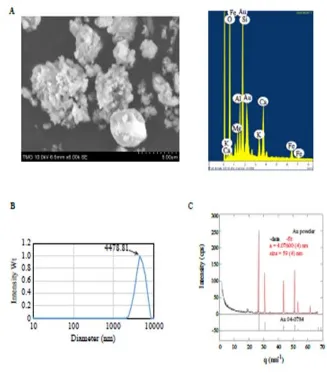
5.2.1. Synthesis and Characterization.
Gold colloids were synthesized by reduction of chloroauric acid with sodium citrate using the Turkevich method. [63]
The elemental composition of gold colloids was measured by ICP-MS (inductively Coupled Plasma Mass Spectroscopy) 7700x Agilent and Energy Dispersive Spectroscopy (EDS attached to SEM Hitachi S 3400N. The gold nanoparticle (Jaya) was suspended in DI water and deposited on pre-cleaned glass slides for SEM measurements. Dynamic Light Scattering (DLS) measurements were done by using a Nicomb 380 instrument. Elemental composition was measured by EDS-SEM and ICP-MS after digestion with aqua regia.
5.2.2. Turkevich method
MATERIALS AND METHODS
Department of Pharmacology Page 31
around 10–20 nm in diameter. Larger particles can be produced, but this comes at the cost of monodispersity and shape. It involves the reaction of small amounts of hot chloroauric acid with small amounts of sodium citrate solution. The colloidal gold will form because the citrate ions act as both a reducing agent and a capping agent.
Recently, the evolution of the spherical gold nanoparticles in the Turkevich reaction has been elucidated. It is interesting to note that extensive networks of gold nanowires are formed as a transient intermediate. These gold nanowires are responsible for the dark appearance of the reaction solution before it turns ruby-red.
To produce larger particles, less sodium citrate should be added (possibly down to 0.05%, after which there simply would not be enough to reduce all the gold). The reduction in the amount of sodium citrate will reduce the amount of the citrate ions available for stabilizing the particles, and this will cause the small particles to aggregate into bigger ones (until the total surface area of all particles becomes small enough to be covered by the existing citrate ions).
5.2.3. Dynamic light scattering (DLS)
Dynamic light scattering (DLS) is a technique in physics that can be used to determine the size distribution profile of small particles in suspension or polymers in solution.
In the scope of DLS, temporal fluctuations are usually analyzed by means of the intensity or photon auto-correlation function (also known as photon correlation spectroscopy or quasi-elastic light scattering).
In the time domain analysis, the autocorrelation function (ACF) usually decays starting from zero delay time, and faster dynamics due to smaller particles lead to faster decorrelation of scattered intensity trace. It has been shown that the intensity ACF is the Fourier transformation of the power Spectrum, and therefore the DLS measurements can be equally well performed in the spectral domain.
MATERIALS AND METHODS
Department of Pharmacology Page 32
DLS is used to characterize size of various particles including proteins, polymers, micelles, carbohydrates, and nanoparticles. If the system is not dispersing in size, the mean effective diameter of the particles can be determined.
Dynamic light scattering provides insight into the dynamic properties of soft materials by measuring single scattering events, meaning that each detected photon has been scattered by the sample exactly once.
5.2.4. Field Emission Scanning Electron Microscopy (FESEM)
To study the surface structure of the Gold nanoparticles and Gold colloid solution FESEM analysis was carried out on FEI Nova NanoSEM 450. Resolution: 1.0 nm at 15kV, 1.4 nm at 1kV and 1.8 nm at 3kV and 30Pa. Software used was xT microscope control.
5.2.5. X-Ray Diffraction (XRD)
XRD measurements were carried out in symmetric reflection mode with a custom-built diffract meter equipped with pyrolytic graphite monochromator and analyzer crystals.
The elemental composition of the Gold nanoparticle was confirmed used XRD analysis.
The XRD pattern of Gold nanoparticle and Gold colloid solution sample reflects gold metal as the major phase along with certain impurities
Cu K-alpha radiation = 0.15418 nm was used for the measurements
5.2.6. Energy Dispersive X-ray Spectrometry (EDS)
MATERIALS AND METHODS
Department of Pharmacology Page 33
5.3. IN VITRO ANTICANCER ACTIVITY
5.3.1. MTT assay
3-[4,5-dimethylthiazol-2-yl]2,5-diphenyltetrazolium bromide (MTT) is a yellow water soluble tetrazolium salt. A mitochondrial enzyme in living cells, succinate-dehydrogenase, cleaves the tetrazolium ring, converting the MTT to an insoluble purple formazan. Therefore, the amount of formazan produced is directly proportional to the number of viable cells.
After 48 h of incubation, 15µl of MTT (5mg/ml) in phosphate buffered saline (PBS) was added to each well and incubated at 370C for 4h. The medium with MTT was then discarded and the formed formazan crystals were solubilized in 100µl of DMSO and then measured the absorbance at 570 nm using micro plate reader. The percentage cell viability was then calculated with respect to control as follows
% Cell viability = [A] Test / [A]control x 100
5.3.2. Cell line
The human breast adenocarcinoma cell line (MCF7) was obtained from National Centre for Cell Science (NCCS), Pune and grown in Eagles Minimum Essential Medium containing 10% fetal bovine serum (FBS). The cells were maintained at 370C, 5% CO2, 95% air and 100% relative humidity. Maintenance cultures were passaged weekly, and the culture medium was changed twice a week.
5.3.3. Cell treatment procedure
MATERIALS AND METHODS
Department of Pharmacology Page 34
final maximum test concentration with serum free medium. Additional four serial dilutions were made to provide a total of five sample concentrations. Aliquots of 100 µl of these different sample dilutions were added to the appropriate wells already containing 100 µl of medium, resulting in the required final sample concentrations. Following sample addition, the plates were incubated for an additional 48 h at 370C, 5% CO2, 95% air and 100% relative humidity. The medium con