Intelligent Classification Technique for Lung
Cancer
R. Saritha, C. Parthasarathy
Abstract: Lung Cancer is the due to irregular cells that begin off in one or the two lungs, ordinarily in the cells that line the air entries. Metastasis alludes to malignancy spreading past its site of starting point to different pieces of the body. At the point when malignancy spreads, it is a lot harder to treat effectively. Essential lung malignancy begins in the lungs, while auxiliary lung disease begins elsewhere in the body, metastasizes, and achieves the lungs. In ebb and flow therapeutic conclusion, treatment, and medical procedure, restorative imaging plays a standout amongst the most critical jobs, since imaging gadgets, for example, Computerised Tomography (CT), Magnetic Resonance Imaging (MRI), and ultrasound diagnostics yield a lot of data about sicknesses and organs. In any case, radiologists need to break down and assess some restorative pictures thoroughly in a brief timeframe, which is a gigantic weight. To help the weight, PC innovation research has been utilized all the more regularly to examine medicinal pictures lately. The proposed strategy which is observed to be exact for tumor discovery, utilizes Gray Level Co-event Matrix (GLCM). The Support Vector Machine (SVM) classifier characterizes the given info stage was ordinary or unhealthy and in the event that it is ailing, further it arranges the tumor pictures into considerate (non-destructive) or dangerous (malignant).
Keywords: GLCM, SVM, ROI, Wavelet transform, GLSDM
I. INTRODUCTION
Normal breath starts by breathing air through the oral pit and nasal. These wind currents down your trachea, that partitions into the left and right bronchi, that convey air to each respiratory organ. Once inside the respiratory organ, the bronchi isolate into littler cylinders alluded to as channels and each bronchiole closes with alveoli. The alveoli are at fault for oxygenating the blood for dissemination also as expelling nursery discharge from the blood. the epithelial cell is that the probable carcinoma to blessing as a Pancoast's neoplasm, that is high inside the respiratory organ zenith with expansion to the chest divider, dispensing shoulder torment that emanates down the nervusulnaris moreover called epidermoid threatening neoplastic malady. Most basic kind of lung most malignant growths makes up 40-half of all lung tumors a large portion of these diseases start at the fringe territories of the lung, similar to the bronchial mucosa can likewise start in scars provoked through fibrosis, easily considered on x-beams two can appear in non-smokers additional basic in ladies, firmly connected to smoking Slow metastasis can happen for the term of the lung or distinctive body organs.
Revised Manuscript Received on July 05, 2019.
R. Saritha, Research scholar, Dept. of CSE, SCSVMV University, Kanchipuram, India.
Dr. C. Parthasarathy, Assistant Professor, Dept. of IT, , SCSVMV University, Kanchipuram, India.
The most incessant indication is a constant and non gainful hack breathing issues: shortness of breath quickened shortness of breath all through substantial movement, wheezing because of the bronchus being somewhat discouraged , piercing respiratory sounds (stridor) changes in mucus (sputum): expanded sum lood in the mucus (hemoptysis) lung tainting (pneumonia): far-reaching lung diseases may fortify the lung pollution might be situated in a similar district as the tumour.Horner's disorder intention recessed eyeball, understudy narrowing, saggy higher eyelid and brought down sweat on the influenced part of the face. two This kingdom is presented on my loss of motion of the storage compartment of the cervical thoughtful nerve by such things as a lung tumor. A chest x beam is an easy, non-intrusive test that makes pictures of the developments inward your chest, for example, your heart, lungs, and veins. "Non-intrusive" expertise that no medicalprocedure is done and no gadgets are embedded into your body. They regularly appear gentle on a chest x beam. Your lungs, which are packed with air, typically appear dim. An infection in the chest that changes how radiation is ingested additionally will show up on a chest x beam. Chest x beams have few dangers. The amount of radiation utilized in a chest x beam is exceptionally little. A lead cover might be utilized to shield certain segments of your body from the radiation. CT outputs recommends the size, shape, and position of your lungs and various structures in your chest.Follow up on strange discoveries from elegant chest x beams, Find the reason for lung indications, for example, shortness of breath or chest torment, Find out whether you have a lung issue, for example, a tumor, overabundance liquid around the lungs, or a pneumonic embolism. X-ray resembles a CT exclusively it utilizes attraction as an option of xrays, dispense with every single metallic article, and round out a screening structure, mentioned to rests on a basically cushioned work area that tenderly skims you into the scanner. Most MRI tests take between 15 to forty five minutes to whole contingent upon the body area imaged and what numbers of pictures are required.
II. LITERATURE REVIEW
In perspective on bosom malignancy, the death toll rate is transforming into raised. As per WHO (World Health Organization) bosom undermining impacted over 1.5 million female always by and mammoth [1]. In 2015; 570,000 young ladies outperformed out because of the reality of chest perilous risk that used to be around 15% of entire passing rate. In 2017 cycle 252,710 patients of bosom perilous hazard are recognized and cycle 40, 610 female died in America [2]. India is in danger expense with 90,000 sufferers and extending every yr and passing ladies is around 40,000 reliably [4].
Support Vector Machine (SVM) [5], is proposed to consider mammography pictures. During the procedure, the region of distraction is depicted sooner than applying the method.Biopsy is an evaluated logical way in recognizing chest threatening development which is an exorbitant, dull and troublesome system. Radiologist proposal is crucial in this stage, once there should be a competition of unsatisfactory confirmation; influenced need never again to do unfortunate biopsy [6]. Another hearty Global Kernel Fuzzy-C Means (NRGKFCM-F) grouping calculation [7], the spot F alludes to kernelized trademark space. It fathoms every transitional inconvenience utilizing Kernel-based Fuzzy C-Means-F (KFCM-F) as a close-by pursuit technique strategy. It uses the division of Genius MRI image,and then morphological sifting can be shaped definitely to maintain a strategic distance from the mis-bunched regions.Mini MIAS enlightening accumulations [8] is used for evaluating the structure. The people group usage is assessed as affectability, distinguishing proof and precision. Affectability and precision finished from the structure are 97% and 97.89% independently. A technique for division [9] of MRI is basically situated in a half breed procedure the utilization of volumes got with the guide of a strategy division dependent on Growth Regions as contribution to a calculation Parametric Deformable Models.CAD framework [10] for perceiving bosom disease in ROIs of advanced mammograms. The examination furthermore explores the general execution of the contraption with wave molecule drastically change and SVM technique.
III. PROPOSED SYSTEM
Hence, the gained pictures are first exposed to pre-handling steps that include: 1) Segmentation of ROI, 2) edge detection and 3) image enhancement to separate the ROI
[image:2.595.60.545.478.833.2]part of the lung picture. Every one of the procured lung pictures is first exposed to binarization, utilizing a fixed limit esteem, to coarsely confine the inside part (ROI) in the picture. A few parts of foundation still show up as associated with the splendid areas, transcendently because of uneven brightening. Thusly, the circle molded foundation in the subsequent pictures is disposed of from the thresholding, i.e., in view of the low force estimations of the foundation. The subsequent twofold veil is utilized to section the ROI from the first lung picture. The photos with low qualification and uneven light are exposed to picture improvement the utilization of CLAHE strategy. Balance is connected in my view for every one of the three RGB shading spaces. These leveled RGB elements are blended all things considered to result the hue evened out picture. CLAHE used to be toward the begin produced for effectively bettering the low refinement logical pictures. A dark dimension co-event grid (GLCM) incorporates records about the places of pixels having similar dim degree esteems .The factual estimates depicted so far are anything but difficult to figure, anyway don't allow any records about the rehashing idea of surface. GLCM incorporates realities about the places of pixels having similar dim dimension esteems. Bolster vector machines are managed picking up information of models with related learning calculations that examine data and perceive designs, utilized for grouping The essential SVM takes a lot of enter insights and for each given information, predicts, which of two exercises assortments the info, making it a non-probabilistic parallel direct classifier. From given arrangement of preparing precedents,each set apart as having a place with one of two classifications, a SVM instruction calculation assembles a mannequin that appoints new models into one class or the other. In the proposed procedure we are utilizing straight classifier. Best hyper airplane is the one that speaks to the greatest partition or edge between the two classes.
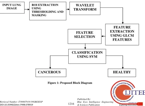
Figure 1: Proposed Block Diagram INPUT LUNG
IMAGE
ROI EXTRACTION USING
THRESHOLDING AND MASKING
WAVELET
TRANSFORM
FEATURE
EXTRACTION
USING GLCM
FEATURES
FEATURE
SELECTION
CLASSIFICATION
USING SVM
IV. GLCM BASED FEATURES EXTRACTION
Feature extrication plays a fundamental role in classifying pattern. Gray Level Co occasion Matrix (GLCM) feature is chosen in 0° for entire mammograms. Per users urged to scrutinize the basic minimizing of GLCM. The span of GLCM was chosen from the dark measurement value in an image. For each recipe gave in the conditions, n choose the amount of dim measurement used. The grid part Q (i, j) is the recurrence related with two pixels, secluded by separating the pixel occurred in the area with power i and j. Textual features achieved from GLCM were presented underneath
.
Contrast
It evaluates gray dimension extent between reference and the neighborhood pixels, variance in the mammogram is assessed from it.
Correlation
Connection delineates the direct needy of gray value. The correlation value is vast if the mammogram contains direct shape for extensive sum.
where
, Sum entropy
It is the whole of small scale (nearby) contrasts in the picture.
SE Difference entropy
It demonstrates the variations in small scale contrasts.
DE Where
[image:3.595.306.546.48.464.2] [image:3.595.47.269.370.502.2]
|i-j| = k, k=0,…n-1
Entropy
Entropy is a proportion of likelihood which illustrates the difference circulation in a region. It is evaluated from the condition appeared as follows.
N
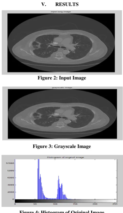
V. RESULTS
Figure 2: Input Image
Figure 3: Grayscale Image
Figure 4: Histogram of Original Image
Figure 6: Lung Cavities Image
Figure 7: Lung Image-Roi
[image:4.595.46.295.43.619.2]Figure 8: Lung Image-Shape
Figure 9: Tumour Extraction-Thresholding
[image:4.595.304.552.46.386.2]Figure 10: Tumour Image
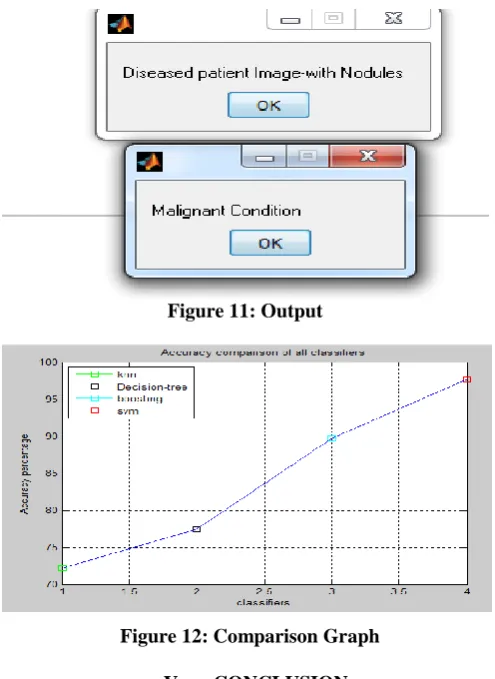
Figure 11: Output
Figure 12: Comparison Graph
V. CONCLUSION
We proposed a lung cancer classification system which classifies automatically by simultaneously utilizing the normal and cancerous images. We presented GLCM Algorithm and region properties measurement for the feature extraction, which can extract the shape features and SVM Algorithm for classification which achieves much higher accuracy than previously proposed approaches.
REFERENCES
1. 1.http://www.who.int/cancer/prevention/diagnosis-screening/breast-cancer/en/
2. Siegel RL, Miller KD, Jemal A. Cancer statistics, 2017. CA: A Cancer Journal forClinicians.Am Cancer Society 67: 7-30.
3. 3.http://htv.com.pk/health/breast-cancer-growing-at-alarming-rate-in-pakistan
4. https://www.dawn.com/news/1344915
5. 5.A.Rajesh,Dr.E.Mohan“Classification of Mammogram Using Wave Atom Transform and Support Vector Machine Classifier,”International Journal of Computer Science and; Information Technologies– Volume 7 ,issue 2, 467-470, Feb 2016
6. Jalalian A, Mashohor S, Mahmud R, Karasfi B, Saripan MIB, et al. (2017) Foundation and methodologies in computer-aided diagnosis systems for breast cancer detection. Excli 16: 113.
7. 7.R.Sugumar ,Dr.E.Mohan. “Magnetic Resonance Imaging Segmentation for Brain Tumor Detection Using New Robust Global Kernel Fuzzy C-Means Clustering Algorithm (NRGKFCM-F),”International Journal of Applied Engineering Research – Volume 9 ,issue 21, 10889-10908, 2014, (ISSN: 0973-4562).
8. Suckling J, Parker J, Dance D, Astley S, Hutt I, et al. (1994) The mammographic image analysis society digital mammogram database. InExerptaMedica. International Congress Series 1069: 375-378. 9. 9.Dr.E.Mohan,R.Sugumar and K.Venkatachalam “Automatic Brain
and Tumor Segmentation in MRI Using Fuzzy Classification with Integrated Bayesian,”International Journal of Applied Engineering Research – Volume 9 ,issue 24, 25859-25870, 2014, (ISSN: 25859-25870).
of computer science, 10 (9), 1543-1547, 2014.
AUTHORS PROFILE
Mrs. R. Saritha, received her B.E degree in CSE from Arulmigu Meenakshi Amman College of Engineering, Tiruvannamalai district in 2002.She received the Master’s degree in Computer Science and Engineering from Arulmigu Meenakshi Amman College of Engineering, Tiruvannamalai district in 2011, she is currently working towards the PhD degree in the department of Computer Science Engineering in Sri Chandrasekarandra Saraswathi Viswa Maha Vidyala University, Kancheepuram, India. Her Research interest is Image mining and Image Processing.
.
![Dimethyl (2Z) 2 [4 ((1Z) 1 {2 [(2Z,5Z) 5 (2 methoxy 2 oxoethylidene) 4 oxo 3 phenyl 1,3 thiazolidin 2 ylidene]hydrazin 1 ylidene}ethyl)anilino]but 2 enedioate](data:image/gif;base64,R0lGODlhAQABAIAAAP///wAAACH5BAEAAAAALAAAAAABAAEAAAICRAEAOw==)