Copyright 01975 AmericanSocietyforMicrobiology Printed inVol. 16,U.S.A.
Differential
Expression
of
Helper
Viral
Structural
Polypeptides
in
Cells Transformed
by
Clonal Isolates
of
Woolly
Monkey
Sarcoma Virus
STUART A. AARONSON,* JOHN R. STEPHENSON, SHIGEO HINO, AND STEVEN R. TRONICK ViralCarcinogenesis Branch, National Cancer Institute, Bethesda, Maryland 20014
Received forpublication9July 1975
Celllines transformed by woolly monkeysarcomavirus(WSV) in theabsence
of infectiousvirusproductionwereanalyzedfor theexpressionofwoolly monkey
helper viral p30, p12, and gp7O antigens. Several linesproduced high levels of
both p30 and p12, whereas gp7O was not detectable. One transformed clone
expressed only p12,and in another cellline,noneof thehelperviralantigenswere
detected. The properties of each sarcoma virus bred true upon transmission,
indicating that each variant represents a distinct genotype. The different cell
lineswereexamined with respecttoproperties characteristic ofthe transformed
state.The in vitrogrowth propertiesandoncogenicityof eachWSV-transformed
clone were indistinguishable, indicating that transformation by WSV occurs
independentlyoftheexpressionofatleast threehelperviralpolypeptides.
RNA-containing sarcoma viruses that have
beenisolated from anumberofspecies differin
their competence for replication. Many avian
sarcoma virus strains appear to replicate
inde-pendently (12, 14, 29), while infection by
mam-malian sarcoma viruses requires the presence of
a nontransforming typeC RNA helper virus (3,
5, 7, 15). Among known replication-defective
murine sarcoma viruses, there appears tobe a
range of abilities to express helper viral
anti-gens. Certain strains transform cells in the
absence ofdetectable
helper
viral antigenex-pression (3, 5, 15, 17). Another classof murine
sarcoma virus genome, termed S+L-
(6,
7),which codes for the mousehelper virus 30,000-mol wt major structural polypeptide (p30) (6,
30) and a 12,000-mol wtpolypeptide (p12) (30),
also produces low levels ofnoninfectious virus
(6).Finally, cells transformedbyathird murine
sarcoma virus variant have recently been
re-portedtoexpressa70,000-molwt mousehelper
virus glycoprotein (gp7O) in the absence of
detectable p30 orcomplete virus (10).
A sarcoma virus isolated from a New World
primate, the woolymonkey (16, 28, 32), has also
been shown to transform cells in the absence
ofinfectious virus production (2, 22). Radioim-munological techniques have been developed for
detectionofgp7O(15),p30(21, 26), andp12 (31)
of the woolly helper virus initially isolated in
associationwiththis sarcoma virus. In the
pres-ent studies, we have utilized competition
im-munoassays for these viral polypeptides to
in-vestigate the expression of woolly helper viral
antigens by cells transformed by individual
clones of woolly sarcoma virus. The results
provideevidence concerning the genetic
compo-sition of this sarcoma virus and preliminary
information concerning the ordering of some of
its genes.
MATERIALS AND METHODS
Media.Cellsweregrown inDulbecco modification ofEaglemediumsupplemented with 10% calf serum
(Colorado Serum Co.,Denver, Colo.) in 50-mm plas-ticpetridishes (Falcon Plastics, Los Angeles, Calif.).
Cells and viruses. Cells used included a line of normal ratkidney (NRK) cells (11). Marmoset cells,
chronically infected with woolly sarcoma virus
(WSV), were generously provided by F. Deinhardt,
RushPresbyterian St. Luke's Hospital, Chicago,Ill. Theviruswas passagedinastrain of normal human fibroblasts immediately prior to use in these studies. The isolation of the woolly type C helper virus associated with WSV has been reported (2, 32). A clone of Kirsten murineleukemiavirus(KiMuLV)(5) was also used.
Virus assays.Thefocus-forming assay forsarcoma virus was performed as previously described (5). An assayfordetection ofvirion-associated reverse tran-scriptaseactivityin tissueculture fluidutilizing the synthetic template poly(rA)-oligo(dT) has also been reported (27).
Isolation of WSV transformants.
WSV-trans-formed lines were isolated as previously described (2). Briefly, exponentially growing NRK cultures were 1117
on November 10, 2019 by guest
http://jvi.asm.org/
exposed to limiting dilutions of WSV (1 to 2 focus-forming units [FFUlplate) in 0.5ml of medium for4 hat 37C.Cultures werethen overlaid withmedium
containing 0.3% agar to minimize infectious virus spread.At around7days, theagar wasaspirated and
transformed fociwereselectedby the cloning cylinder
technique (3). Each focus-derived line was recloned
by the microtiter procedure (27) priortoanalysis. Analysis of biological propertiesof WSV-trans-formed cells. Cellswereinoculatedonto50-mmpetri dishes at a density of 5 x 10⁹ cells/cm2, and the
mediumwaschangedevery3days. Cells from
dupli-cate plates were counted with a hemocytometer at 2-day intervals. The doubling time was determined
during growth in exponential phase. Thesaturation densitywastakenasthe valuewherethreesuccessive
cell counts showed no increase in cellnumber. Cell
colony formation on confluent monolayers of NRK
cells and in soft agar was determined according to methods previously reported (4, 19).
Tumorigenicity studies. Athymic (nude) mice (6 to8weeksold)were inoculatedsubcutaneously in the
intrascapular region with washed cells suspended in 0.2mlofphosphate-buffered saline. The animalswere
examined at weekly intervals for the appearance of tumors.
Radioimmunoassays. Competition immunoassays for woolly viral gp7O (14a), p30 (21, 26), and p12 (31) have been described in detail. Reaction mix-tures contained 0.01 M Tris, 0.3 M NaCl, 0.15 M EDTA, 0.4% Triton X-100, and 1% bovine serUim aIbumin, pH 7.8,inavolume of 0.7 ml. Antiserawere
assayed atserialtwofold dilutions for theirabilityto precipitate approximately 10,000 counts/minof 20I-labeled antigen.Unlabeled viral antigensweretested
atserialtwofold dilutions for theirabilitytocompete with "25I-labeledgp7O,p30,orp12forbinding limiting
amounts of antiserum. Antiserum and unlabeled competing antigen were incubated at 37 C for 1 h;
1211-labeled antigenwasthenadded, and the
incuba-tionwascontinued for 3 hat37 C andafurther 18 hat
4C. After addition of 0.025 ml of undiluted swine anti-goat immunoglobulin G to eachtube, the mix-turewasincubated3hat4C and centrifugedat2,500
rpm for 15 min, and the resulting precipitate was
measured for radioactivity. The antisera used were
generously provided by R. Wilsnack through the Office of Resources and Logistics, National Cancer Institute. Protein concentrationsweredeterminedby the method ofLowryetal. (18).
RESULTS
Sarcoma virus-release by cells infected at
limiting dilution with WSV. Cells transformed
at limiting sarcoma virus dilution by WSV
associated with woolly helper viruswere grown
up asfocus-derived linesforanalysis of virus re-lease. Twenty of 27 tranformants tested
re-leased high levels of virion-associated reverse
transcriptase activity and focus-forming virus (Table 1). With each of these lines, it was
[image:2.498.267.462.75.192.2]possible to isolate woolly helper virus beyond
TABLE 1. Virological properties ofindividual WSV-transformed lines
Virusreleasea No. of Virion-associatedreverse
lines transcriptase activity WSV titer (pmol/ml of tissue (FFU/ml)
culturefluid)
20 103- 104 JOS.8_104-4
1 100.0 10°
6 <1O-l° <10-l °
aTissue culture fluids of exponentially growing
cultures of each transformed line were assayed for virion-associated reverse transcriptase activity and for focus-forming activity on NRK assay cells as described in the text.
the end point for focus formation. Thus, they werepresumed tohave arisen from co-infection by both WSV and helper virus. One
transform-ant (Table 1) released a very low level of
focus-forming virus (100°0 FFU/ml), and it was
possible to detect a low level of reverse
tran-scriptase activity in tissue culture fluids of this line. The remaining six transformants showed
noevidence of virus release, but contained WSV rescuable by superinfection with either woolly helper virusorKiMuLV.
Woolly helper viralantigen expression by
non-virus-releasing WSV transformants.
Each non-virus-releasing transformant was
tested inimmunoassaysforwoolly viral p12 and p30. Goat antiserum prepared against the
woolly helpervirus wasusedtoprecipiateeither
"2'I-labeled woolly viral p12 or p30 in the presence of increasing quantities of unlabeled WSV-transformed cell extracts. Immunoassays
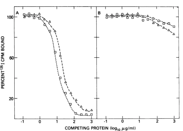
forwoolly viral p12 and p30detected high levels
ofboth viral polypeptides inNRK cells
chroni-callyinfected withwoollyhelper virus (Fig. 1A).
In contrast, uninfected NRK cells (Fig. 1B)
showed only slight reactivity at very high
pro-tein concentration. This was considered to be
nonspecific because at high concentrations the
same extractsalso showed similar lowdegreesof
reactivityinimmunoassays for unrelatedtypeC
viral polypeptides (data not shown).
Threedistinct patterns ofimmunological
re-activity were observed among the
non-virus-releasing WSV transformants examined. One
clone demonstrated no greater reactivity than
uninfected NRK cells in the immunoassay for
woollyviral p30and only slightly greater
reac-tivity in the assay for viral p12 (Fig. 2A). In
contrast, another WSV-transformed line (Fig.
2B) was strongly positive in the immunoassay
for woolly viral p12 but lacked detectable p30
on November 10, 2019 by guest
http://jvi.asm.org/
1119
-1 0 1 2 3 -1 0 1 2 3
COMPETING PROTEIN(log1o,LgImI)
FIG. 1. Analysis ofviralantigen expressionin cellextractsprepared from(A) NRKchronically infectedwith woolly monkey virus and(B) control NRK. Extracts were prepared by sonication and clarified by low-speed cen-trifugation aspreviously described (26). Protein determinationswere performed by the method of Lowry et al. (18). Assays included an immunoassay for woolly viral p12 utilizing antiserum to woolly monkey virus to precipitate '25I-labeled woolly monkey virus p12 (0), and an immunoassay forp30 utilizing antiserum to woolly monkey virus to precipitate '25I-labeledwoollymonkey virusp30 (A).
-o Ia- a A
-Ob
\4\b
B ~~~~~~~C
AA A.AAAA
A1A
-V
b0 'A
H--1 0 1 2 3 -1 0 1 2
COMPETINGPROTEIN(Iog1gopg/mlI
FIG. 2. Viralantigenexpressionincell extractsprepared from (A) WSVCl9,(B) WSVCl2,and(C) WSVCl 11.Extractswereassayed for woolly monkeyvirusp12 (0)andp30 (A)immunological reactivityasdescribedin thelegendtoFig.1.
expression. Finally, high levels of both woolly viral p12 and p30 weredetected in each of the
remaining non-virus-releasing WSV
transform-antsasrepresentedby results withonesuch line
showninFig.2C.ItcanbeseeninFigure2that, where reactivity of WSV-transformed lines was
detected in either assay, it closely
approx-imated the amount of viral polypeptide ob-served with cells chronically infected with
woolly helper virus.
Recently, immunoassays that specifically
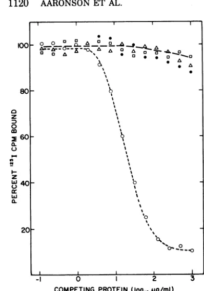
de-tect gp7O of the woolly helper virus have been developed (14a). Itwasofinteresttoinvestigate theexpression of thisviral antigen by
represen-tative non-virus-releasing WSV transformants. NRK cells productively infected with woolly helper viruswerehighly positive for woolly viral
gp7O(Fig. 3). Incontrast,the reactivities of the threephenotypic variants of WSV-transformed cells were no different than that of uninfected
NRK cells.
Evidence that the differences in woolly
100
60
a z
0
0~
co
m
0-
I-z C:
0
A
_=D__
_C'"
. AA
B A~~~~~~N
AA0
Z-s
20
100
z8
z 0
co
W4
20
on November 10, 2019 by guest
http://jvi.asm.org/
[image:3.498.94.388.62.273.2] [image:3.498.94.386.344.490.2]00
80
a
z
3
0
260
a. u 0.
*-z
w40
w
a.
201
_1 0 2 3
[image:4.498.57.256.52.336.2]COMPETING PROTEIN (Iogj,oug/ml)
FIG. 3. Analysis of gp70 expression in cellextracts prepared fromWSVCl9(A), WSVCl2(O), WSVCl 11 (0), and NRK cells chronically infected with woolly monkey virus (0). Extracts, preparedas
de-scribed in the legend to Fig. 1, were tested in the
woolly viralgp70immunoassay in whichantiserumto woolly monkey virus was used to precipitate 1251_ labeledwoolly monkey virusgp70.
helper viral antigen expression by WSV
transformants are virus coded. The
varia-tions in expression ofwoolly type C viral
anti-gens among differentnon-virus-releasing WSV
transformants could reflect virus-coded
differ-encesorcell-specificeffectsontheexpressionof
an identical viral genome. To examine this
question, WSV was rescued by KiMuLV from
cells of eachtransformedphenotype and
trans-mitted to new NRK cells. The expression of
woolly viral antigenswasthenanalyzedin these
cultures. As with the parental line, cells
in-fected with sarcoma virus rescued from the
WSV Cl 9 transformant failed to demonstrate detectable woolly viral p12 cross-reactive anti-gen(Table 2), despite the factthatthe cellswere
morphologically transformedand released high-titered WSV (102 FFU/ml). Similarly, like
their respective parental WSV transformants, NRK cultures infected with sarcoma virus
res-cued from WSV Cl 2 and Cl 11 transformants
expressed high levels of woolly viral p12 (410
and 320 ng/mg of cell protein, respectively).
Due to cross-reactivity of KiMuLV in the
im-munoassay forwoolly viralp30, it was difficult
to quantitate the expression of woolly viral p30
in these cells. These results argue that the
differences in expression of p12 cross-reactive
antigensby eachWSVvariant are viruscoded.
The genetic stability of viral p30 expression
by WSV present in awoolly viral
p30-contain-ing transformant, WSV Cl 11, was next
exam-ined. Cells transformed by progeny sarcoma
virus ofthis linein theabsence of helper virus
co-infectionwereisolated and analyzed for viral
polypeptide expression. Six separate clonal
transformants induced by thefirstcycleof virus
from WSV Cl 11-transformed cells each
con-tainedboth woolly viral p30 and p12
cross-reac-tive antigens at levels similar to those of the
parentaltransformant(Table3). A second cycle
ofsarcoma virus transmission to new cells was performed after its rescue by KiMuLV from a
first-generation transformant. Again, each of
fourclonal transformantsexpressed both woolly
viral polypeptidesatlevelscomparabletothose
detected in the original parental line. These
findings strongly indicate thatwoolly viralp30,
in addition to p12, is coded by this WSV
genome and provide further evidencethat each of theimmunologically distinguishable
replica-tion-defective WSV variants represents a
dis-tinct genotype.
TABLE 2. Woolly type C viralp12expressionby cells infected withKiMuLVpseudotypesof
differentWSVvariants
Level of viralantigen WSV associated with Cellsinfected (KiMuLV) infectedcells with KiMuLV release (ng/mgofcell pseudotypeof: (FFU/ml)a protein)'
Woolly virus MuLVc
p12 p12
WSVCl2 104.8 410 440
WSVCl9 105.2 <2 380
WSV Cl11 104.5 320 430
aNRKcultures were infected with WSV rescued by KiMuLV from each WSV-transformed clone and thenpassagedintissueculturefor 3weeks. Thetiter of WSV in tissue culture fluids of each infected culturewasassayedasdescribed inthetext.
Extracts were prepared and immunoassays were performed as described in the legend to Fig. 1. The results, expressed asnanograms ofp12permilligram ofcell protein, werecalculated from standard calibra-tion curves (30, 31) and represent mean values of three separate determinations.
IMuLV, Murine leukemia virus.
& ~~~* A
5 0
00
Li
I~~~~~~'
on November 10, 2019 by guest
http://jvi.asm.org/
[image:4.498.268.461.412.536.2]CLONAL ISOLATES OF WOOLLY MONKEY SARCOMA VIRUS 1121
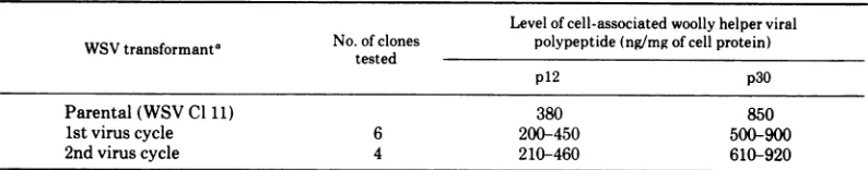
TABLE 3. Genetic stability of woolly viral p12 andp30expression by progeny of one WSV variant
Level ofcell-associated woolly helper viral WSVtransformanta No. ofclones polypeptide(ng/mgofcellprotein)
tested
p12 p30
Parental (WSVC1 1) 380 850
1stviruscycle 6 200-450 500-900
2nd viruscycle 4 210-460 610-920
aWSV was rescued from WSV-transformed NRK cells by superinfection with KiMuLV and transmitted at
limiting dilution to NRK assay cells. Transformed foci were isolated as described in the text and those transformants that failed to release high-titered WSV were tested immunologically. Transformed foci, the product of a second cycle of WSV release, were inducedby.WSVrescued from a transformant of the first cycle of virus transmission. Each line was assayed for reactivity in woolly viral p12 andp30immunoassays as described inthe text.
Characterization of a low level
virus-re-leasing WSV transformant. The properties
of individual transformed foci induced by the
low level virus-releasing WSV-transformed line
WSV Cl 4 were next investigated. Each of five
separate transformants isolated after infection
with tissue culture fluids of this line
demon-strated levels of infectious sarcoma virusrelease
and reverse transcriptase activity similar to
that of the parental line. In contrast, cells
infected beyond the end point for focus
forma-tion and even nontransformed cells isolated
from the same petri dishes were virus negative
by these assays. Analysis ofwoolly viral
anti-gens expressed bytheparental line and several
ofitsdaughtertransformants revealed that each
containedhighlevels ofwoollyviralp12andp30
in the absence of detectable gp7O (data not
shown).
Biological properties of different WSV
gen-otypes. The
biological
properties oftransform-ants containing representative WSV variants
were nextcompared. Themorphological
charac-teristics of cells transformed by each were
indistinguishable. The transformants all grew
to similar saturation densities (1.8 x 101to 2.3
x 10'
cells/cm'),
and the generation time ofeach line resembled that of uninfected NRK
cells (18 to 22 h). Properties of cells in tissue
culture that are known to correlate with their
malignant potential in vivo include the
ability
toformcoloniesonmonolayersof
contact-inhib-ited cells (4) and to grow in suspension in
semisolid medium (19). Each WSV
transform-anttested, incontrast touninfectedNRKcells,
possessed the capacity to form colonies under
both selective conditions (Table 4). The
tu-morigenicity ofeach cell line was also
investi-gated. Representative WSV-transformed lines
of each genotype produced solid tumors at a
dose of 5 x 10⁶cells inapproximately40to50%
of athymic mice at the site of subcutaneous
inoculation; control NRK cells, similarly tested,
were nontumorigenic. Tumors that formed
were histopathologically diagnosed as
fibrosar-comas. The lack of detectable differences in the transformed properties of the different WSV
variants both in vitro and in vivo argues that
the differences in their capacities to induce
helper viral antigens have little, if any,
re-lationship with those viral functions involving transformation.
DISCUSSION
Analysis of the immunological properties of
cells transformed by clonal sarcoma viruses
obtained from a natural isolate of WSV has
revealed the existence of several
immunologi-cally distinguishable variants. One isolate
ap-pearsanalogoustopreviouslydescribed
nonpro-ducer transforming sarcoma viruses of murine
origin (3, 5, 15, 17). Cell extracts of this
WSV variant lacked detectable reactivity in
immunoassays forwoolly helper viralp12, p30,
orgp7Oand didnotreleasefocus-formingvirus.
Asecond variantinduced theexpressionofhigh
levels of both woolly viral p12 and p30 in the
absenceofdetectablegp7Oorvirusrelease. This
WSV variant resembles the S+L- genome of
MSV, whichhasbeen shownto induce
produc-tion of mouse type C viral p30 and p12 in
infected cells (7, 30). However, unlike
S+L--transformed mouse or rat cells which also
pro-ducealow level of noninfectious virus (6), cells
transformed bythis WSV variantdid notshow
evidence of virus release. Cells transformed
by
athird WSV variant expressed woolly viral p12
cross-reactiveantigen inthe absenceof
detecta-blep30, gp7O, orinfectiousvirus.Findingsthat
the properties of each variant bred true upon
passageto newcellsargue that thevirusesdiffer
genetically.
In addition to several non-virus-releasing
WSV transformants, one clone, which released
VOL.16,1975
on November 10, 2019 by guest
http://jvi.asm.org/
TABLE 4. Invitro and in vivobiological properties of WSV-transformed cells
In vitroacolony-forming efficiency (%) Invivo'tumor-forming NRK cell line _________________________abilityinathymicmice
(no.oftumors/no.
On monolayers In agar inoculated)
Uninfected <0.001 <0.001 0/10
Transformedby
WSV Cl 2 15 1.0 4/10
WSV Cl9 10 1.5 5/10
WSV Cl11 12 0.8 4/10
a Cellswereinoculatedatserial 10-folddilutions eitherontoconfluent NRKmonolayersorinsuspensionin medium containing0.3% agar. Colonieswerecountedat 12to 15daysandthecolony-forming efficiencywas expressed asthe percentageofcellsinoculated. The results representmeanvalues from twoexperiments.
'Athymic mice (6 to 8 weeks old) were inoculated with 5 x 10⁶ cells suspended in 0.5 ml of phosphate-buffered saline. The site of subcutaneous inoculation was observed weekly for 12 weeks. Tumors reaching adiameter of 1 cm were scored as positive.
a verylowlevel of infectioussarcomavirus, was
isolated. This virus also appearedtobreedtrue
upon transmission to new cells, since each
daughter transformant possessed the parental
phenotype. These results could not be
ac-counted for by the presence of excess helper
virus,sinceattempts todetect suchaviruseven
at a titer comparable to that of the sarcoma
virus were unsuccessful. The transformed cells
werefoundtocontainhigh levels of woolly viral
p12and p30butnotdetectable gp7O, indicating
that if this WSV genome were competent for
replication it must be relatively restricted in
viral gp7O production. The present results are
also consistent with the possibility that this
sarcomaviruscomplements theendogenousrat type C virus present in NRK cells (1, 17),
causing activation and release of infectious
sarcoma virus in the envelope ofthis virus.
Murine and avian sarcoma viruses contain
genetic information both unrelated (8, 23, 25)
and related (8, 9)tothatofknowntypeChelper
viruses. Thus, one possibility is that these
transforming viruses have arisen as a result of
recombination between the helper virus and
some other cellular genetic information.
Analy-sis ofnucleic acid homologyof WSV expressed
in one WSV-transformed nonproducer clone
with woolly helper virus has indicated some
sequence relatedness between the two viruses (24). The presentfindings, byestablishing that
different amounts of helper viral information
are expressed by different WSV variants, and
that these properties breed true upon virus
passageto newcells, indicatethatwoollyhelper
virus genetic sequences canbestably associated
with the WSVgenome.
The present findings do not resolve whether
the WSV variants isolated here differ at levels
involving their transcription or translation or
result from specific deletions of helper viral
information. However, the pattern of woolly
viral antigens detected among the
transform-antstested in the presentstudies would suggest an ordering of genes coding for viral structural polypeptides relative to those coding for
trans-formation as follows: transformation
gene(s)-p12-p30-gp70. A recent report (10) of a murine sarcoma virus genome which codes for murine
leukemiavirus gp7O in the absence of p30 would
appear to be at variance with the above data.
However, when more information is available
concerning the origin of sarcoma virus and the mechanisms by which recombination with helper viruses occur, it may be possible to explain the apparentdifferencesbetween these twosystems.
The present evidence, as well as previous findings with murine and avian sarcoma virus, indicates that sarcoma viral genes coding for transformation may be expressed
independ-ently of those codingfor helper viralstructural components. Evidence is accumulating that
high-molecular-weight precursor polypeptides
are involved in thereplicationof typeCviruses (13, 20; J. R. Stephenson, S. R. Tronick, and S. A. Aaronson, Cell, in press). Ifthe sarcoma viralgenescoding fortransformationand helper viralpolypeptidesare present in the same mes-senger RNA, WSV genomes expressing woolly viral p12aloneor in combinationwithp30 may provide antigenic markers to monitor
purifica-tion ofthose proteins responsible for the
trans-formed phenotype. Studies of this nature are
currently in progress.
ACKNOWLEDGMENTS
WethankMae C.Wong, LindaK.Long,andVirginiaK.
Steiner for excellent technical assistance.
This work wassupportedbycontract no.NCI-E-73-3212of
the VirusCancerProgramofthe NationalCancerInstitute
on November 10, 2019 by guest
http://jvi.asm.org/
[image:6.498.75.466.85.181.2]CLONAL ISOLATES OF WOOLLY MONKEY SARCOMA VIRUS 1123
and by a Public Health Service International Research Fellowship(FO5TW-2207-01) toS. Hino.
LITERATURE CITED
1. Aaronson, S. A. 1971.Chemical inductionof
focus-form-ing virus from nonproducercellstransformed by
mu-rine sarcoma virus. Proc. Natl. Acad. Sci. U.S.A.
68:3069-3072.
2. Aaronson, S.A. 1973. Biologic characterization of
mam-malian cells transformed byaprimate sarcoma virus.
Virology52:562-567.
3. Aaronson, S. A., and W. P. Rowe. 1970. Nonproducer clonesofmurine sarcomavirustransformed BALB/3T3 cells.Virology42:9-19.
4. Aaronson, S. A., andG. J. Todaro. 1968. Basisfor the
acquisition of malignant potential by mouse cells
cultivated in vitro. Science 162:1024-1026.
5. Aaronson, S. A., andC. Weaver. 1971. Characterization
ofmurine sarcoma virus (Kirsten) transformation of
mouse andhuman cells.J. Gen.Virol. 13:245-252.
6. Bassin, R. H., L. A. Phillips, M. J. Kramer, D. K.
Haapala, P. T.Peebles, S. Nomura, and P. J. Fisch-inger. 1971. Transformation of mouse 3T3 cells by
murinesarcomavirus: release ofvirus-like particlesin
the absence of replicating murine leukemia helper
virus. Proc. Natl.Acad. Sci.U.S.A. 68:1520-1524.
7. Bassin, R. H., N. Tuttle, and P. J. Fischinger. 1970.
Isolationof murinesarcomavirustransformed mouse
cellswhicharenegative forleukemiavirusfromagar
suspensioncultures. Int.J.Cancer6:95-107. 8. Beeman, K.,P. Duesberg, and P.Vogt. 1974. Evidence
forcrossing-overbetweenaviantumorvirusesbasedon
analysisofviralRNAs. Proc. Natl.Acad. Sci.U.S.A.
71:4254-4258.
9. Benveniste, R. E., and E. M. Scolnick. 1973. RNA in
mammalian sarcoma virus transformed nonproducer cells homologous to murine leukemia virus RNA.
Virology51:370-382.
10. Bilello, .J.A.,M. Strand,and J. T.August. 1974. Murine
sarcoma virus gene expression: transformants which
expressviral envelope glycoprotein in the absenceof
the major internal protein and infectious particles. Proc. Natl. Acad. Sci.U.S.A. 71:3234-3238.
il. Duc-Nguyen, J., E. M. Rosenblum, and R. F. Zeigel.
1966.Persistentinfection of a ratkidney cellIiiie with
Rauscher murine leukemia virus. J. Bacteriol.
92:1133-1140.
12. Dougherty,R.M.,and R.Rasmussen.1964.Propertiesof
astrain ofRoussarcoma virus that infects mammals.
Natl.Cancer Inst. Monogr. 17:337-350.
13. Eisenmnan, R., V. M. Vogt, and H. Diggelman. 1975. Synthesis ofavian RNA tumorvirus structural
pro-teins. Cold Spring Harbor Symp. Quant. Biol.
39:1067-1075.
14. Hanafusa, H., and T. Hanafusa.1966.Determiningfactor inthecapacityofRoussarcomavirustoinducetumors inmammals. Proc.NatI.Acad.Sci.U.S.A.55:532-538.
14a. -lino, S., J. R. Stephenson, and S. A. Aaronson. 1975.
Antigenic determinants of the 70,000 molecular weight glycoprotein of woollv monkey type C RNA
virus. J. Immunol. 155:922-927.
15. Huebner,R.J.,J. W. Hartley,W.P.Rowe,W. T.Lane,
and W.I.Capps. 1966.Rescue of the defectivegenome
ofMoloneysarcomavirusfromanoninfectious hamster
tumor and the production of pseudotvpe sarcoma
viruses with various murine leukemia viruses. Proc.
Natl. Acad. Sci. U.S.A. 56:1164-1169.
16. Kawakami, T. G., S. E. Huff, P. M. Buckley, D. C.
Dungworth, S. P. Snyder, and R. V. Gilden. 1972. C-typevirus associated with gibbon lymphosarcoma.
Nature(London)New Biol. 235:170-171.
17. Klement, V., M. 0. Nicolson, and R.J.Huebner. 1971.
Rescueof the genome offocus-formingvirus from rat
non-productivelinesby5'-bromodeoxyuridine. Nature
(London)234:12-14.
18. Lowry, 0. H., N. J. Rosebrough, A. L. Farr, and R. J. Randall. 1951. Protein measurement with the Folin phenolreagent.J.Biol.Chem. 193:265-275.
19. MacPherson, I. A., and L. Montagnier. 1964. Agar suspension culture for the selective assay of cells
transformedby polyoma virus.Virology23:291-294.
20. Naso, R. B., L. J.Arcement,and R. B.Arlinghaus. 1975.
BiosynthesisofRauscher leukemia viralproteins.Cell
4:31-36.
21. Parks, W. P.,E.M. Scolnick, M.L.Noon,C.J. Watson,
and T. G. Kawakami. 1973. Radioimmunoassay of
mammalian type-C polypeptides. IV.Characterization
of woolly monkev and gibbon viral antigens. Int. J.
Cancer 12:129-137.
22. Scolnick, E. M., and W. P. Parks. 1973. Isolation and
characterization of a primate sarcoma virus:
mecha-nismof rescue.Int.J.Cancer 12:138-147.
23. Scolnick, E. M.,E.Rands,D.Williams,and W. P. Parks.
1973.Studiesonthe nucleic acid sequences of Kirsten
sarcoma virus: a model for formation of a mammalian
RNA-containingsarcomavirus.J.Virol. 12:458-463. 24. Scolnick, E. M., W. P. Parks, T. G. Kawakami, D.
Kohne, H. Okabe, R. V. Gilden, and M. Hatanaka.
1974. Primate and murine type-C viral nucleic acid
association kinetics: analysis of model systems and
natural tissues. J. Virol. 13:363-369.
25. Stephenson, J. R., and S. A. Aaronson. 1971. Murine
sarcoma andleukemiavirus:genetic differences
deter-mined byRNA-DNA hybridization. Virology
46:480-484.
26. Stephenson,J.R., and S.A. Aaronson. 1973.Expression
ofendogenous RNA C-typevirusgroupspecific
anti-gens in mammalian cells.J.Virol. 12:564-569.
27. Stephenson,J.R.,R. K.Reynolds,and S. A. Aaronson.
1972. Isolation of temperature sensitive mutants of
murineleukemiavirus.Virology48:749-756.
28. Theilen, G. H., D. Gould,M. Fowler, and D. L. Dung-worth. 1971. C-type virus in tumor tissue of awoolly
monkey (Lagothrix spp.) with fibrosarcoma. J. Natl.
CancerInst. 47:881-889.
29. Toyoshima, K., R.R. Friis, and P. K. Vogt. 1970. The
reproductiveandcell-transformingcapacitiesofavian
sarcoma virusB77; inactivation withUVlight.
Virol-ogy 42:163-170.
30 Tronick, S. R., J. R. Stephenson, and S. A. Aaronson.
1973.Immunologicalcharacterizationofalow
molecu-larweightpolypeptideof murine leukemia virus. Virol-ogy 54:199-206.
31. Tronick, S. R.. J. R. Stephenson, andS. A. Aaronson.
1974. Comparative immunological studies ofprimate
RNA C-type viruses. Radioimmunoassay for a low
molecular weight polypeptide ofwoolly monkey
leu-kemia virus.Virology57:347-356.
32. Wolfe, L. G., R. K. Smith, and F. Deinhardt. 1972.
Simian sarcoma virustype 1 (Lagothrix): focusassay
and demonstration ofnontransforming associated
vi-rus. J. Natl.CancerInst. 48:1905-1908.
VOL.16,1975
on November 10, 2019 by guest
http://jvi.asm.org/