Surface Modification of Acetaminophen Particles by
Atomic Layer Deposition
Tommi O. Kääriäinen a, b, d, *, Marianna Kemell a, Marko Vehkamäki a, Marja-Leena Kääriäinen b, d,, Alexandra Correia c, Hélder A. Santos c, Luis M. Bimbo c, Jouni Hirvonen c, Pekka Hoppu d,
Steven M. George b, David C. Cameron e, Mikko Ritala a, and Markku Leskelä a
a Laboratory of Inorganic Chemistry, University of Helsinki, P.O. Box 55 (A.I.Virtasen aukio 1), FI-00014 Helsinki, Finland. Email: [email protected]
b Department of Chemistry and Biochemistry and Department of Chemical and Biological Engineering, University of Colorado, Boulder, Colorado 80309
c Division of Pharmaceutical Chemistry and Technology, Faculty of Pharmacy, University of Helsinki, FI-00014 Helsinki, Finland
d NovaldMedical Ltd Oy, Telkäntie 5, 82500 Kitee, Finland
e R&D Centre for Low-Cost Plasma and Nanotechnology Surface Modification, Masaryk University, Kotlářská 267/2, 611 37 Brno, Czech Republic.
ABSTRACT
Active pharmaceutical ingredients (APIs) are predominantly organic solid powders. Due to their bulk properties many APIs require processing to improve pharmaceutical formulation and manufacturing in the preparation for various drug dosage forms. Improved powder flow and protection of the APIs are often anticipated characteristics in pharmaceutical manufacturing. In this work, we have modified acetaminophen particles with atomic layer deposition (ALD) by conformal nanometer scale coatings in a one-step coating process. According to the results, ALD, utilizing common chemistries for Al2O3, TiO2 and ZnO, is shown to be a promising coating method for solid pharmaceutical powders. Acetaminophen does not undergo degradation during the ALD coating process and maintains its stable polymorphic structure. Acetaminophen with nanometer scale ALD coatings shows slowed drug release. ALD TiO2 coated acetaminophen particles show cytocompatibility whereas those coated with thicker ZnO coatings exhibit the most cytotoxicity among the ALD materials under study when assessed in vitro by their effect on intestinal Caco-2 cells.
KEYWORDS
ABBREVIATIONS
1. INTRODUCTION
Primary drug particles in powder form which comprise active pharmaceutical ingredients (APIs) are typically solid organic particles. Drug powders are used to create enteral drug dosage forms (such as tablets, capsules, pellets and granules), inhalation powders, parenteral injectable preparations, topical transdermal patches and emulsions, as well as ophthalmic systems such as contact lenses and eye drops. Pharmaceutical manufacturing can benefit from improved surface characteristics of powders that optimize drug loading efficiency and improve powder processability (Ghoroi et al., 2013; Shi and Sun, 2011; Vanhoorne et al., 2014; Ehlers et al., 2009; Sauer et al., 2013; Jallo et al., 2015; Beach et al., 2010). Quite often there is a need to stabilize the desired solid state of the API. These can be amorphous or hydrate states, which make the pharmaceutical processing hard to perform unless the drug powders are coated (Airaksinen et al., 2005; Han and Suryanarayanan, 1999; Matsuo and Matsuoka, 2007; Wu et al., 2011). The coating may also provide chemical stabilization for the pharmaceutical (Wu et al., 2011) or control of the drug release (Chen et al., 2006). There are many biological interfaces, such as between nanoparticles and carbon nanotubes and APIs, where control of the surface characteristics of pharmaceutical powders can improve their therapeutic response and bioavailability (Terracciano et al., 2015; Taylor et al., 2014; Schäfer et al., 2013; Chow et al., 2007).
atomization is usually obtained with high pressure air, electrostatics or ultra sound. Coating of small particles can be difficult, because the size of the individual coating liquid droplets is usually over 30 µm (Ehlers et al., 2009; Werner et al., 2007). Spray-drying, a widely used operation in pharmaceutical manufacturing, is also used for particle encapsulation (Vanhoorne et al., 2014; Chow et al., 2007; Vehring, 2008; Dobry et al., 2009), in an attempt to overcome poor tabletability of pharmaceutical crystals (Shi and Sun, 2011; Vanhoorne et al., 2014), or to mask the undesired taste of the API (Shi and Sun, 2011). Spray drying is used to produce solid dispersions, which have already been investigated for some time for increasing the drug release rate and bioavailability of poorly water-soluble drugs, as well for controlling the drug release rate (Dobry et al., 2009; Giri et al., 2012; Huang and Dai, 2014).Dry-particle coating is an alternative method to spray drying where the addition of excipient particles is done by blending them with the API particles. Dry particle coating is favourable, for example, for moisture sensitive drugs since it does not involve the use of aqueous solvents. The removal of solvents from the final formulation is energy-consuming and complicates the process (Sauer et al., 2013; Jallo and Dave, 2015; Beach et al., 2010; Hoashi et al., 2013).
method, which can be used both for drug particle synthesis and particle coating by physical vapour deposition, has been developed and studied for synthesizing drug particles (Eerikäinen et al., 2003), for coating salbutamol sulfate particles with L-leucine (Raula et al., 2008), for encapsulating indomethacin nanocrystals in mannitol microparticles with an L-leucine coating (Laaksonen et al., 2011), and for combining budesonide and salbutamol in microparticles coated with L-leucine (Raula et al., 2013; Vartiainen et al., 2016). In this method, APIs, binders and coating material are dissolved in water to form a precursor solution which is used in an aerosol process. The atomized solution mixed with nitrogen gas forms an aerosol which is introduced to a heated laminar flow reactor. In the reactor the solvent will be evaporated forming particles from the solute which will undergo further deposition by the evaporated coating substance (L-leucine) in the gas phase.
State-of-the-art drug processing requires well defined and controlled surface modification techniques. Novel drug delivery systems (Terracciano et al., 2015) are needed for the stabilization of acetaminophen nanocrystals in aqueous medium (Das et al., 2013), targeted drug delivery based on metal cation attachment to active pharmaceutical ligand forming coordination compounds (Ledeti et al., 2013), and biomedical imaging and molecular diagnostics (Park et al., 2010).
Ommen, 2010). It is suggested that pharmaceutical manufacturing can benefit from capabilities of ALD. Potential applications can be e.g. particle surface functionalization, stabilization of APIs, and improved bulk properties such as better powder flowability.
In this work, we have studied ALD on solid pharmaceutical particles by depositing conformal nanometer scale metal oxide films on particles of the drug acetaminophen. Acetaminophen is a widely used analgesic and antipyretic drug agent. In order to show the proof-of-concept for the application of ALD on API we have investigated the physicochemical characteristics after ALD coating, the drug dissolution, and cytotoxicity of ALD oxide coated acetaminophen.
2. MATERIAL AND METHODS
2.1. Atomic layer deposition (ALD) on acetaminophen particles
temperature while TTIP was vaporized from the source at a temperature of 65 to 70 ºC. TTIP– water chemistry is a commonly used and studied process for ALD and allows the growth of TiO2 at low temperatures suitable for organic and temperature sensitive materials (King et al., 2008; Ritala et al., 1993; Aarik et al., 2000; Knez et al., 2006). TTIP–water chemistry is also favorable for avoiding HCl production, which is a byproduct of TiCl4–water ALD chemistry.
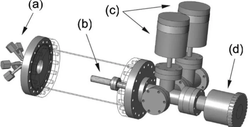
Figure 1. Schematic diagram of rotary ALD particle reactor with, a) multiple-input reactant dosing flange, b) small porous metal cylinder inside the main vacuum chamber, c) capacitance manometers to control the partial pressure, and d) magnetically coupled rotary manipulator to agitate particles inside metal cylinder during the deposition. Reprinted with permission from J. A. McCormick et al. 2007, J. Vac. Sci. Technol. A 25, 67 © 2007, American Vacuum Society.
The reactant exposure sequence for each ALD cycle was the following (McCormick et al., 2007): 1) exposure of metal precursor at a vapor pressure of 0.5 to 2 Torr, 2) hold for metal precursor reaction time, 3) pump out excess metal precursor and reaction products, 4) dose N2 at 10 to 20 Torr for purging the reactor, 5) pump out N2, 6) expose H2O at 1 to 2 Torr, 7) hold for H2O reaction time, 8) pump out excess H2O and reaction products, 9) dose N2 at 10 to 20 Torr for purging the reactor, and 10) pump out N2. The reaction times were between 60 to 240 s. The pump out times used for the precursors were between 60 to 360 s. The N2 exposure time was 60 s and pump out time for the N2 was 60 s. After each reactant exposure and pump out 5 N2 exposure – pump out purging steps were applied. Due to the low vapor pressure of TTIP it was exposed multiple times at 0.5 Torr separated by 360 s pump out during the TTIP exposure step to ensure enough reactant for the entire surface area to be coated. The needed amount of reactant can be estimated by using simple calculations based on the ideal gas law. The metal precursor partial pressure was varied based on the particle load, thus the surface area to be coated. The estimation for the specific surface area of acetaminophen was 1 m2/g.
2.2 Thermal analysis of the particles
loss. The residual mass of ALD coated acetaminophen at 390 °C was considered to be ALD oxide. This information was used to estimate the composition of ALD oxide material in coated acetaminophen.
Differential scanning calorimetry (DSC) Netzsch DSC 204 F1 was used to investigate the change in enthalpy within the temperature range of 25 °C to 180 °C during the heating-cooling cycles. Two heating-cooling cycles were performed for each sample. Samples of around 10 to 12 mg were heated and cooled in an aluminum pan, which was first sealed with an aluminum lid prior to piercing the lid to avoid the pan deforming and losing proper physical contact between the pan bottom and the instrument thermoelement. The heating and cooling rate used was 10 K min-1 in a dynamic inert N2 atmosphere with flow rates of 20 ml min-1 for purging and 70 ml min-1 for protective gas flow, respectively. The structure and the structural changes in acetaminophen and ALD coated acetaminophen prior to and after DSC analysis were studied with attenuated total reflectance Fourier transmittance infrared (ATRFTIR) spectroscopy using a Nicolet 4700 FTIR spectrometer with a Smart Orbit ATR accessory with a diamond crystal internal reflection element.
2.3. X-ray powder diffraction
2.4. Surface characterization of the particles
Untreated acetaminophen and ALD coated acetaminophen surface morphology, particle size and conformality of ALD coating were analyzed by using a scanning electron microscope with energy dispersive X-ray spectroscopy (SEM-EDS) (Hitachi S4800). The particle cross-section samples were prepared with a focused ion bean scanning electron microscope (FIB-SEM) (FEI Quanta 3D 200i). The samples were coated with Au-Pd alloy and carbon prior to loading them into the FIB-SEM to protect the interface against ion beam damage. The protection was completed with an ion beam deposited Pt layer just before the ion beam cutting. EDS elemental maps were measured across the cross-section surfaces with an Oxford INCA 350 EDS connected with the FEI Quanta microscope. Beam energies were 5 keV for the Al2O3-coated acetaminophen sample and 10 keV for the TiO2-coated acetaminophen sample.
2.5. Drug stability and release profile
medium was retained constant by replacing 200 µl of the fresh pre-warmed release medium after each sampling. The samples were then centrifuged at 21,382gfor 3 min and the released amount of acetaminophen was measured in the supernatant by the HPLC method described above. All experiments were performed at least in triplicate.
2.6. Cell culture and viability assays
The in vitro studies with the ALD powders were carried out on the human epithelial colorectal adenocarcinoma Caco-2 cell line (from American Type Culture Collection). The cells were cultured in 75 cm2 culture flasks (Corning Inc. Life Sciences) using Dulbecco’s modified Eagle’s medium (DMEM, HyClone). The medium was supplemented with 10% heat inactivated fetal bovine serum (HIFBS, Gibco, Invitrogen), 1% sodium pyruvate (HyClone), 1% nonessential aminoacids, 1% L-glutamine, penicillin (100 IU/ml), and streptomycin (100 mg/ml) (all from EuroCloneS.p.A). The cultures were maintained in a BB 16 model (Heraeus Instruments GmbH) gas incubator at 37 °C in an atmosphere of 5% CO2 and 95% relative humidity. The growth medium was changed every other day until the time of use. Caco-2 cells from passage numbers 35–40 were used in all the experiments.
Prior to each test, the cells were harvested using 0.25% (v/v) trypsin-ethylenediamine
added to the wells. After 3 h, 6 h and 24 h of incubation for TiO2 (TiCl4) and ZnO, and 24h of incubation for Al2O3 and TiO2 (TTIP), the wells were washed once with HBSS and 50 µl of fresh HBSS was then added to the wells along with 50 µl of the CellTiter-Glo® reagent assay (Promega Corporation), according to the manufacturer’s instructions. In this assay the number of viable cells in culture is quantified based on the amount of adenosine triphosphate (ATP) produced by metabolically active cells. Thus, the amount of ATP produced is directly proportional to the number of living cells presented in the culture. The plate was then measured for luminescence using a Varioskan Flash fluorometer (Thermo Fisher Scientific). The luminescence of the sample wells was then compared with their time-paired controls. The results are the averages of three independent experiments and error bars represent mean ± standard deviation (SD).
2.7. Reactive oxygen species (ROS) determination
emission wavelengths of 498 and 522 nm, respectively. All the assays were conducted at least in triplicate and error bars represent ± SD.
2.8. Tumor necrosis factor alpha (TNF-α) assay
Varioskan Flash (Thermo Fisher Scientific). The amount of TNF-α present in each sample was determined by subtracting the absorbance values at 550 from the absorbance values at 450 nm to correct from optical imperfection on the plate, and then calculating the total amount from the standard curve of known amounts of human TNF-α.
2.9. Statistical analysis
Results from the several tests are expressed as mean ± SD from of at least three independent experiments. A one-way analysis of variance (ANOVA), followed by a Dunnett’s multiple comparison test was used to analyze the cell viability, ROS levels and TNF-α induction. The level of significance was set at a probability of p < 0.05 for *, p < 0.01 for **, and p < 0.001 for ***. The analysis was carried out using GraphPad Prism v. 6.07 (GraphPad Software).
3. RESULTS AND DISCUSSION 3.1. DSC
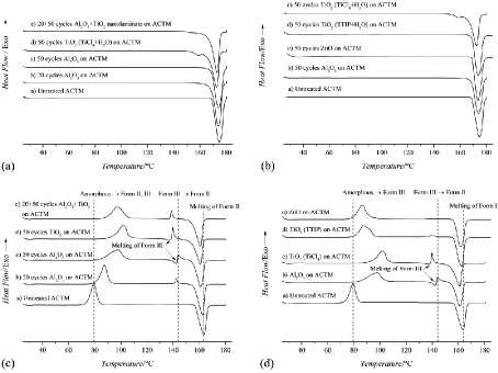
amorphous via melting point depression. It is possible that the endothermic reaction seen at 159 °C for the ALD TiO2 (TiCl4) sample is due to acetaminophen crystallization from amorphous to form II during the DSC measurement. This mechanism would mean that TiO2 coating prolongs higher energy amorphous acetaminophen crystallization. The thermograph of 20 ALD cycles Al2O3 and 50 cycles TiO2 (TiCl4) nanolaminate on acetaminophen in Figure 2a (thermograph e) shows that Al2O3 coating protects acetaminophen from the formation of a mixed system between acetaminophen and TiO2 (TiCl4).
peak broadening. In pure substances the melting curve give a symmetric peak while in impure substances the melting temperature decreases and the curve appears broader and asymmetric. The theory of thermodynamics of two-component systems allows the determination of the purity of eutectic mixtures (Höhne et al., 1996).
Figure 2. DSC thermographs during the first heating cycle of pure acetaminophen and ALD oxide coated acetaminophen (a and b) and during the second heating cycle of pure acetaminophen and ALD oxide coated acetaminophen (c and d) with the heating rate of 10 K min-1 in a dynamic inert N2 atmosphere. The scans have been offset vertically for clarity.
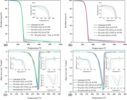
3.2. Thermogravimetric analysis (TGA)
Figure 3. Thermogravimetric curves of acetaminophen and ALD oxide coated acetaminophen obtained during the heating in N2 (a and b), and differential thermogravimetric (DTG) curves of the same samples, presenting the mass loss rate at certain temperature (c and d).
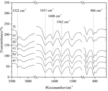
3.3. ATR-FTIR
[image:25.612.89.538.76.429.2]-2008; Zimmerman and Baranovic, 2011; Zhao et al., 2014). The IR spectra of ALD coated acetaminophen do not reveal any structural changes in acetaminophen when compared to uncoated acetaminophen. This does not rule out the possibility of chemical reactions between the acetaminophen and nanoscale ALD coating since the penetration depth of ATR-FTIR is typically higher than 0.5 µm. However, the sensitivity of ATR-FTIR is improved by higher surface area of the particle samples compared to a single planar sample. This benefit can be seen especially from the IR spectra of 50 cycles TiO2 (TTIP) and ZnO samples between 3000 and 3500 cm-1 (Figure 4e and 4f), where the increase in intensity of OH stretching vibration attributed to the OH in ALD oxide coating is observable.
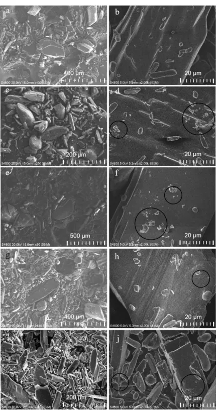
3.4. SEM/EDS analysis
Scanning electron microscopy images of uncoated and ALD coated acetaminophen particles in Figure 5 confirm the irregular shape and size distribution of the particles. Deposition on acetaminophen does not induce any apparent change in particle shape or size distribution or in surface properties, which is revealed in the higher magnification images in Figures 5b, d, f, h and j. To confirm the existence and uniformity of ALD oxide coating on acetaminophen, energy dispersive X-ray spectroscopy (EDS) elemental mapping and point spectrum collection were performed on the ALD coated acetaminophen samples.
[image:27.612.82.298.291.700.2]
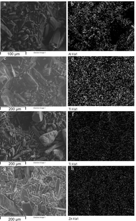
The elemental mapping from images shown in Figure 6 confirms the uniform distribution of metal elements (Al, Ti, Zn) on the acetaminophen particle samples, indicating uniform ALD metal oxide deposition over the particles. The presence of ALD oxide coating on individual particles was further demonstrated by the EDS point analysis on the particle surfaces and elemental mapping across the particle cross-section surface. The images in Figure 7 show points where EDS point analysis were done. The EDS point analysis confirmed the existence of the metal element from ALD oxide in each point measured except the point for spectrum 5 in Figure 7c indicating absence of ALD coating. The difference between the areas on the individual particle showed by spectra 5, 1 and 6 is evident.
Figure 7. SEM images of point spectrum collected from a) 50 cycles ALD Al2O3 coated acetaminophen sample, b) 50 cycles ALD TiO2 (TiCl4) coated acetaminophen sample, c) 50 cycles TiO2 (TTIP) coated acetaminophen sample, and d) 50 cycles ZnO coated acetaminophen sample by SEM with EDS.
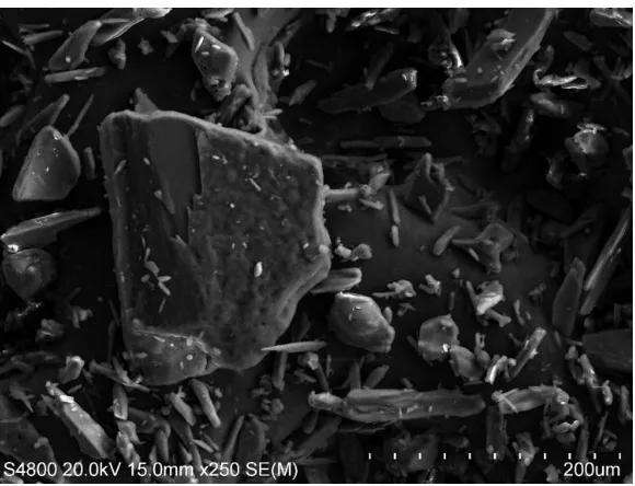
[image:29.612.83.550.101.465.2]In earlier work where acetaminophen was mixed with sodium caseinate and lecithin in distilled water and then spray dried at 130 °C (spray outlet temperature 75 °C), the attached crystals on the particle surface were attributed to the crystallization of the drug onto the surface (Thi et al., 2012). In our study, the crystallization of acetaminophen on the particle surface would require amorphization of crystalline drug during the deposition. Based on XRD, the acetaminophen remains crystalline during the deposition so the possible amorphization takes place on the surface of the crystalline particles, which is not detected by XRD (Figure S1). Change in acetaminophen particle surface roughness has also been observed after leucine dry powder coating (Ghoroi et al., 2013).
3.5. Stability after ALD Coating
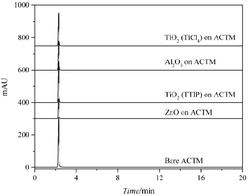
Figure 9. Chromatograms of acetaminophen and 50 cycles ALD oxide coated acetaminophen samples in working solution. Peak 1, acetaminophen.
3.6. Drug Release Studies
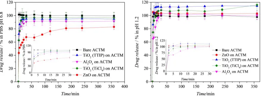
[image:32.612.176.429.77.277.2]Figure 10. Drug release profiles of uncoated acetaminophen and 50 ALD cycles oxide coated acetaminophen samples in PBS at pH 6.8 (a), and in buffer solution at pH 1.2 (b).
3.7. Cell viability and ROS production
ZnO. Metal oxides, especially in the form of nanoparticles, have been found to lead to the formation of ROS in cells with deleterious effects on cell viability. Usually the first marker of oxidative aggression, ROS can lead towards inflammation and ultimately cytotoxicity (Nel et al., 2006). As such, we subsequently investigated ROS generation in Caco-2 cells by TiO2 (TiCl4) and ZnO at 3, 6 and 24h and found that none of these ALD acetaminophen powders induced any statistically significant ROS across all concentrations and time points tested (Figure 10 c and d). It is worth mentioning that ROS generation induced by ZnO nanoparticles has been found to be surface- and cell-type dependent (Hanley et al., 2009; Jeong et al., 2013; Moos et al., 2010) and that ZnO-induced toxicity has also been suggested not to be dependent on either the classical extrinsic or mitochondrial signaling pathway, but rather, a caspase-independent alternative apoptosis signaling mechanism, in which ROS is not a major contributor (Buerki-Thurnherr et al., 2013).
[image:34.612.76.509.412.657.2]and 24 h. Statistical analysis was made by ANOVA, followed by a Dunnett’s multiple comparison test. The level of significance was set at a probability of p < 0.05 for *, p < 0.01 for **, and p < 0.001 for ***. Error bars represent SD (n ≥ 3).
Finally, the inflammatory response measured by TNF-α release from Caco-2 cells incubated with the ALD powder which gave the highest toxicity (ZnO) was also investigated. No statistically significant TNF-α induction was detected in the concentrations and incubation times tested (Figure 11), which is corroborated by several reports that have shown that cytotoxic concentrations of ZnO do not elicit a broad inflammatory response, measured by increased TNF- expression levels (Moos et al., 2011; Roselli et al., 2003).
[image:35.612.94.528.340.538.2]4. CONCLUSION
ASSOCIATED CONTENT
Supporting Information. Supporting information is available on the Elsevier website.
ACKNOWLEDGMENTS
REFERENCES
Aarik, J., Aidla, A., Uustare, T., Ritala, M., Leskelä, M., 2000. Titanium isopropoxide as a precursor for atomic layer deposition: characterization of titanium dioxide growth process. Appl. Surf. Sci. 161, 385-395.
Airaksinen, S., Karjalainen, M., Shevchenko, A., Westermarck, S., Leppänen, E., Rantanen, J., Yliruusi, J., 2005. Role of water in the physical stability of solid dosage formulations. J. Pharm. Sci. 94, 2147-2165.
Beach, L., Ropero, J., Mujumdar, A., Alcalà, M., Romanach, R. J., Davé, R. N., 2010. Near-Infrared Spectroscopy for the In-Line Characterization of Powder Voiding Part II: Quantification of Enhanced Flow Properties of Surface Modified Active Pharmaceutical Ingredients. J. Pharm. Innov. 5, 1-13.
Behzadi, S. S., Toegel, S., Viernstein, H., 2008. Innovations in coating technology. Recent Pat. Drug Deliv. Formul. 2, 209-230.
Bimbo, L. M., Mäkilä, E., Laaksonen, T., Lehto, V. –P., Salonen, J., Hirvonen, J., Santos, H.A., 2011. Drug permeation across intestinal epithelial cells using porous silicon nanoparticles. Biomaterials 32, 2625-2633.
Calinescu, O., Badea, I. A., Vladescu, L., Meltzer, V., Pincu, E., 2012. HPLC separation of acetaminophen and its impurities using a mixed-mode reversed-phase/cation exchange stationary
phase. J. Chromatograph. Sci. 50, 335-342.
Chen, R., Okamoto, H., Danjo, K., 2006. Particle design using a 4-fluid-nozzle spray-drying technique for sustained release of acetaminophen. Chem. Pharm. Bull. 54, 948-953.
Chow, A. H. L., Tong, H. H. Y., Chattopadhyay, P., Shekunov, B. Y., Particle Engineering for Pulmonary Drug Delivery. Pharm. Res. 24, 411-437.
Das, S., Paul, A., Chattopadhyay, A., 2013. Nanocrystalline p-hydroxyacetanilide (paracetamol) and gold core–shell structure as a model drug deliverable organic–inorganic hybrid nanostructure. Nanoscale 5, 9247-9254.
Dobry, D. E., Settell, D. M., Baumann, J. M., Ray, R. J., Graham, L. J., Beyerinck, R. A., 2009. A Model-Based Methodology for Spray-Drying Process Development. J. Pharm. Innov. 4, 133-142.
Eerikäinen, H., Watanabe, W., Kauppinen, E. I., Ahonen, P. P., 2003. Aerosol flow reactor method for synthesis of drug nanoparticles. Eur. J. Pharm. Biopharm. 55, 357-360.
Ehlers, H., Räikkönen, H., Antikainen, O., Heinimäki, J., Yliruusi, J., 2009. Improving flow properties of ibuprofen by fluidized bed particle thin-coating. Int. J. Pharm. 368, 165-170.
Elam, J. W., George, S. M., 2003. Growth of ZnO/Al2O3 Alloy Films Using Atomic Layer Deposition Techniques. Chem. Mater. 15, 1020-1028.
Ferguson, J. D., Weimer, A. W., George, S. M., 2004. Atomic layer deposition of Al2O3 films on polyethylene particles. Chem. Mater. 16, 5602-5609.
Ferguson, J. D., Yoder, A. R., Weimer, A. W., George, S. M., 2004. TiO2 atomic layer deposition on ZrO2 particles using alternating exposures of TiCl4 and H2O. Appl. Surf. Sci. 226, 393-404.
Ghoroi, C., Han, X., To, D., Jallo, L., Gurumurthy, L., Davé, R. N., 2013. Dispersion of fine and ultrafine powders through surface modification and rapid expansion. Chem. Eng. Sci. 85, 11-24.
Giri, T. K., Kumar, K.; Alexander, A.; Ajazuddin, Badwaik, H., Tripathi, D. K., 2012. A novel and alternative approach to controlled release drug delivery system based on solid dispersion technique. Bull. Fac. Pharm. Cairo Univeristy. 50, 147-159.
George, S. M., 2010. Atomic layer deposition: An overview. Chem. Rev. 110, 111-131.
Hakim, L. F., Blackson, J., George, S. M., Weimer, A. W., 2005. Nanocoating individual silica nanoparticles by atomic layer deposition in a fluidized bed reactor. Chem. Vap. Deposition 11, 420-425.
Han, J., Suryanarayanan, R., 1999. A method for the rapid evaluation of the physical stability of pharmaceutical hydrates. Thermochim. Acta 329, 163-170.
Hanley, C., Thurber, A., Hanna, C., Punnoose, A., Zhang, J., Wingett, D. G., 2009. The influences of cell type adn ZnO nanoparticle size on immune cell cytotoxicity and cytokine induction. Nanoscale Res. Lett. 4, 1409-1420.
Huang, Y., Dai, W-G., 2014. Fundamental aspects of solid dispersion technology for poorly soluble drugs. Acta Pharm. Sin. B. 4, 18-25.
Hyde, G. K., Park, K. J., Stewart, S. M., Hinestroza, J. P., Parsons, G. N., 2007. Atomic Layer Deposition of conformal inorganic nanoscale coatings on three-dimensional natural fiber systems: effect of surface topology on film growth characteristics. Langmuir 23, 9844-9849.
Höhne, G., Hemminger, W., Flammersheim, H.-J., 1996. Differential scanning calorimetry: An introduction for practioners, Springer-Verlag.
Jallo, L. J., Dave, R. N., 2015. Explaining electrostatic charging and flow of surface-modified acetaminophen powders as a function of relative humidity through surface energetics. Pharm. Drug Deliv. Pharm. Technol. 104, 2232-2232.
Jeong, S. H., Kim, H. J., Ryu, H. J., Ryu, W. I., Park, Y-H., Bae, H. C., Jang, Y. S., Son, S. W., 2013. ZnO nanoparticles induce TNF-α expression via ROS-ERK-Egr-1 pathway in human keratinocytes. J. Dermatol. Sci. 72, 263-273.
Johnson, R. W., Hultqvist, A., Bent, S. F., 2014. A brief review of atomic layer deposition: from fundamentals to applications. Materials Today 17, 236-246.
Kaialy, W., Larhrib, H., Chikwanda, B., Shojaee, S., Nokhodchi, A., 2014. An approach to engineer paracetamol crystals by antisolvent crystallization technique in presence of various additives for direct compression. Int. J. Pharm. 464, 53-64.
King, D. M., Liang, X., Zhou, Y., Carney, C. S., Hakim, L. F., Li, P., Weimer, A. W., 2008. Atomic layer deposition of TiO2 films on particles in a fluidized bed reactor. Powder Technol. 183, 356-363.
King, D. M., Liang, X., Weimer, A. W., 2012. Functionalization of fine particles using atomic and molecular layer deposition. Powder Technol. 221, 13-25.
Klímová, K., Leitner, J., 2012. DSC study and phase diagrams calculation of binary systems of paracetamol. Thermochimica Acta 550, 59-64.
Knez, M., Kadri, A., Wege, C., Gösele, U., Jeske, H., Nielsch, K., 2006. Atomic layer deposition on biological macromolecules: Metal oxide coating of tobacco mosaic virus and ferritin. Nano Lett. 6, 1172-1177.
Knez, M., Kornelius, N., Niinistö, L., 2006. Synthesis and surface engineering of complex nanostructures by atomic layer deposition. Adv. Mater. 19, 3425-3438.
Kääriäinen, T., Cameron, D. C., Kääriäinen, M-L., Sherman, A. 2013. Atomic layer deposition: Principles, characteristics and nanotechnology applications, 2nd edition. Wiley-Scrivener.
Laaksonen, T., Liu, P., Rahikkala, A., Peltonen, L., Kauppinen, E. I., Hirvonen, J., Järvinen, K., Raula, J., 2011. Intact nanoparticulate indomethacin in fast-dissolving carrier particles by combined wet milling and aerosol flow reactor methods. Pharm. Res. 28, 2403-2411.
Ledru, J., Imrie, C. T., Pulham, C. R., Céolin, R., Hutchinson, J. M., 2007. Pharmacokinetics, pharmacodynamics and drug metabolism: High pressure differential scanning calorimetry investigations on the pressure dependence of the melting of paracetamol polymorphs I and II. J. Pharm. Sci. 96, 2784-2794.
Longrie, D., Deduytsche, D., Detavernier, C., 2014. Reactor concepts for atomic layer deposition on agitated particles: A review. J. Vac. Sci. Technol. A. 32, 010802.
Matsuo, K., Matsuoka, M., 2007. Solid-state polymorphic transition of theophylline anhydrate and humidity effect. Cryst. Growth Des. 7, 411-415.
McCormick, J. A., Cloutier, B. L., Weimer, A. W., George, S. M., 2007. Rotary reactor for atomic layer deposition on large quantities of nanoparticles. J. Vac. Sci. Technol. A. 25, 67-74.
Miikkulainen, V., Leskelä, M., Ritala, M., Puurunen, R. L., 2013. Crystallinity of inorganic films grown by atomic layer deposition: Overview and general trends. J. Appl. Phys. 113, 021301(1-101).
Moos, P. J., Chung, K., Woessner, D., Honeggar, M., Cutler, N. S., Veranth, J. M., 2010. ZnO particulate matter requires cell contact for toxicity in human colon cancer cells. Chem. Res. Toxicol. 23, 733-739.
Moos, P. J., Olszewski, K., Honeggar, M., Cassidy, P., Leachman, S., Woessner, D., 2011. Responses of human cells to ZnO nanoparticles: a gene transcription study. Metallomics 3, 1199-1211.
Nevalainen, K., Suihkonen, R., Eteläaho, P., Vuorinen, J., Järvelä, P., Isomäki, N., Hintze, C., Leskelä, M., 2009. Mechanical and tribological property comparison of melt-compounded nanocomposites of atomic-layer-deposition-coated polyamide particles and commercial nanofillers. J. Vac. Sci. Technol. A. 27, 929-936.
Park, D-H., Kim, J-E., Oh, J-M., Shul, Y-G., Choy, J-H., 2010. DNA Core@Inorganic Shell. J. Am. Chem. Soc. 132, 16735-16736.
Peltonen, L., Hirvonen, J., 2014. Drug nanocrystals and nanosuspensions in medicine, in: Torchilin (ed.), Handbook of Nanobiomedical Research, World Scientific, 169.
Qi, S., Avalle, P., Saklatvala, R., Craig, D. Q. M., 2008. An investigation into the effects of thermal history on the crystallization behaviour of amorphous paracetamol. Eur. J. Pharm. Biopharm. 69, 364-371.
Rahtu, A., Ritala, M., 2002. Reaction mechanism studies on titanium isopropoxide-water atomic layer deposition process. Chem. Vap. Dep. 8, 21-28.
Raula, J., Lähde, A., Kauppinen, E. I., 2008. A novel gas phase method for the combined synthesis and coating of pharmaceutical particles. Pharm. Res. 25, 242-245.
Raula, J., Rahikkala, A., Halkola, T., Pessi, J., Peltonen, L., Hirvonen, J., Järvinen, K., Laaksonen, T., Kauppinen, E. I., 2013. Coated particle assemblies for the concominant pulmonary administration of budesonide and salbutamol suphate. Int. J. Pharm. 441, 248-254.
Roselli, M., Finamore, A., Garaguso, I., Britti, M. S., Mengheri, E., 2003. Zinc oxide protects cultured enterocytes from the damage induced by Escherichia coli. J. Nutr. 12, 4077-4087.
Rossi, A., Savioli, A., Bini, M., Capsoni, D., Massarotti, V., Bettini, R., Gazzaniga, A., Sangalli, M. E., Giordano, F., 2003. Solid-state characterization of paracetamol metastable polymorphs formed in binary mixtures with hydroxypropylmethylcellulose. Thermochimica Acta 406, 55-67.
Sacchetti, M., 2001. Thermodynamic analysis of DCS data for acetaminophen polymorphs. J. Thermal Anal. Calorimetry 63, 345-350.
Sauer, D., Cerea, M., DiNunzio, J., McGinity, J., 2013. Dry powder coating of pharmaceuticals: A review. Int. J. Pharm. 457, 488-502.
Sarnes, A., Østergaard, J., Jensen, S.S., Aaltonen, J., Rantanen, J., Hirvonen, J., Peltonen, L., 2013. Dissolution study of nanocrystal powders of a poorly soluble drug by UV imaging and channel flow methods. Eur. J. Pharm. Sci. 50, 511-519.
Schäfer, C., Schröder, K. R., Höglinger, O., Tollabimazraehno, S., Lornejad- Schäfer, M. R., 2013. Acetaminophen changes intestinal epithelial cell membrane properties, subsequently affecting absorption processes. Cell. Physiol. Biochem. 32, 431-447.
Shi, L., Sun, C. C., 2011. Overcoming poor tabletability of pharmaceutical crystals by surface modification. Pharm. Res. 28, 3248-3255.
Taylor, A. J., McClure, C. D., Shipkowski, K. A., Thompson, E. A., Hussain, S., Garantziotis, S., Parsons, G. N., Bonner, J. C., 2014. Atomic Layer Deposition Coating of Carbon Nanotubes with Aluminum Oxide Alters Pro-Fibrogenic Cytokine Expression by Human Mononuclear Phagocytes In Vitro and Reduces Lung Fibrosis in Mice In Vivo. PLOS ONE 9, e106870, 1-14.
Terracciano, M., Shahbazi, M-A., Correia A., Rea, I.; Lamberti, A., De Stefano, L., Santos, H. A., 2015. Surface bioengineering of diatomite based nanovectors for efficient intracellular uptake and drug delivery. Nanoscale 7, 20063-20074.
Thi, T. H. H., Morel, S., Ayouni, F., Flament, M-P., 2012. Development and evaluation of taste-masked drug for paediatric medicines – Application to acetaminophen. Int. J. Pharm. 434, 235-242.
Trasi, N. S., Taylor, L. S., 2012. Effect of polymers on nucleation and crystal growth of
amorphous acetaminophen. Cryst. Eng. Comm. 14, 5188-5197.
Van Ommen, J. R. 2010. Delft University of Technology, EP2403976.
Vanhoorne, V., Peeters, E., Van Snick, B., Remon, J. P., Vervaet, C., 2014. Crystal coating via spray drying to improve powder tabletability. Eur. J. Pharm. Biopharm. 88, 939-944.
Vartiainen, V., Bimbo, L. M., Hirvonen, J., Kauppinen, E. I., Raula, J., 2016. Drug permeation and cellular interaction of amino acid-coated drug combination powders for pulmonary delivery. Int. J. Pharm. 504, 89-97.
Werner, S. R. L., Jones, J. R., Paterson, A. H. J., Archer, R. H., Pearce, D. L., 2007. Air-suspension particle coating in the food industry: Part I – state of the art. Powder Technol. 171, 25-33.
Wu, Y., Levons, J., Narang, A. S., Raghavan, K., Rao, V. M., 2011. Reactive impurities in excipients: Profiling, identification and mitigation of drug–excipient incompatibility. AAPS Pharm. Sci. Tech. 12, 1248-1263.
Zhao, M., Barker, S. A., Belton, P. S., C. McGregor, C., Craig, D. Q. M., 2014. Development of fully amorphous dispersions of a low Tg drug via co-spray drying with hydrophilic polymers. Eur. J. Pharm. Biopharm. 82, 572-579.