“ PROSPECTIVE RANDOMISED CONTROL STUDY
COMPARING THE INCIDENCE OF POSTDURAL
PUNCTURE HEADACHE FOLLOWING SPINAL
ANAESTHESIA USING 25 GAUGE WHITACRE SPINAL
NEEDLE AND 25 GAUGE QUINCKE SPINAL NEEDLE
IN OBSTETRIC PATIENTS”
Dissertation submitted to
THE TAMILNADU DR. M.G.R.MEDICAL UNIVERSITY
in partial fulfilment for the award of the degree of
DOCTOR OF MEDICINE
IN
ANAESTHESIOLOGY
BRANCH X
INSTITUTE OF ANAESTHESIOLOGY & CRITICAL CARE
MADRAS MEDICAL COLLEGE
CHENNAI- 600 003.
CERTIFICATE
This is to certify that the dissertation entitled,“Prospective
randomised control study for comparing the incidence of postdural
puncture headache following spinal anaesthesia using 25 gauge
Whitacre and 25 gauge Quincke spinal needle in obstetric patients”
submitted by Dr.G.MALINI, in partial fulfilment for the Degree of
Doctor of Medicine in Anaesthesiology by the Tamilnadu Dr. M.G.R.
Medical University, Chennai., is a bonafide record of the work done by
her in the GOVT. KASTHURBA GANDHI HOSPITAL FOR
WOMEN AND CHILDREN, TRIPLICANE, CHENNAI, during the
academic year 2013 – 2014.
Prof. Dr. G.R. Rajashree, M.D. D.A.,
Professor of Anaesthesiology Institute of Social obstetrics Govt. Kasthurba Gandhi Hospital for Women and Children,
Triplicane, Chennai – 600 005.
Prof. Dr.B.KALA, M.D.,D.A,
Director and Professor, Institute of Anaesthesiology And Critical Care, Madras Medical College, Chennai-600 003
Prof. Dr.R.VIMALA, M.D.
DEAN,
Rajiv Gandhi Govt. General Hospital, Madras medical college,
DECLARATION
I, Dr. G.MALINI, solemnly declare that this dissertation entitled
“Prospective randomised control study for comparing the incidence
of Postdural puncture following spinal headache in using 25gauge
Whitacre and 25 gauge Quincke spinal needle in obstrtric patients
”
is a bonafide work done by me in the Institute of Social ObstetricsGovernment Kasthurba Gandhi Hospital for Women and Children,
Triplicane, Chennai, during the period 2013 to 2014 under the guidance
of Prof. Dr. G.R. Rajashree, M.D. D.A., Professor of Anaesthesiology
and Prof. Dr. B.KALA, M.D.,D.A, Director, Institute of
Anaesthesiology and Critical Care, Madras Medical College, Chennai – 3
and submitted to The TamilnaduDr. MGR Medical University,
Guindy, Chennai – 32, in the partial fulfillment of the requirements for
the award of the degree of MDAnaesthesiology (Branch X).
Place: Chennai,
Date:
ACKNOWLEDGEMENT
I am extremely thankful to Dr. R.VIMALA, M.D., Dean, Madras
Medical College & Rajiv Gandhi Govt General Hospital, for her
permission to carry out this study.
I am immensely grateful to Prof. Dr. B. KALA, M.D., D.A.,
Director and Professor, Institute of Anaesthesiology and Critical Care, for
her concern and support in conducting this study.
I am extremely grateful and indebted to my guide
Prof Dr. G.R. RAJASHREE, M.D., D.A.,Professor of Anaesthesiology,
Govt. Kasturba Gandhi Hospital For Woman And Children, for her
concern, inspiration, meticulous guidance, expert advice and constant
encouragement in preparing this dissertation.
I am especially thankful to our former guide, Prof. Dr. S.NELLAI
KUMAR, M.D.,D.A., for his invaluable help, guidance and constant
encouragement.
I am very grateful to express my sincere gratitude to the Professors,
Dr.ESTHER SUDHARSHINI RAJKUMAR, MD.,DA, Dr.LAKSHMI MD.,DA.,
Institute of Anaesthesiology and Critical Care, for their constant
motivation and valuable suggestions.
I am thankful to Prof. ARUN MURUGAN ,statistician who
helped me in statistical aspects of my study.
I am thankful to the Institutional Ethical Committee for their
guidance and approval for this study.
I am thankful to all my colleagues and friends for their help and
advice in carrying out this dissertation.
I am grateful to my family and friends for their moral support and
encouragement.
Last but not the least, I thank all the patients for willingly
CONTENTS
S.No
TOPICS Page
No.
1. INTRODUCTION 1
2. AIM OF THE STUDY 3
3. ANATOMY OF THE SUBARACHNOID SPACE 4
4. PHYSIOLOGY OF SPINAL ANAESTHESIA 14
5. PHARMACOLOGY OF THE DRUG 30
6. POSTDURAL PUNCTURE HEADACHE 35
7. REVIEW OF LITERATURE 53
8. MATERIALS AND METHODS 66
9. OBSERVATION AND RESULTS 71
10. DISCUSSION 89
11. SUMMARY 91
12. CONCLUSION 92
13. BIBLIOGRAPHY
14. PATIENT CONSENT FORM
15. PROFORMA
16. MASTER CHART
17. ETHICS COMMITTEE APPROVAL FORM
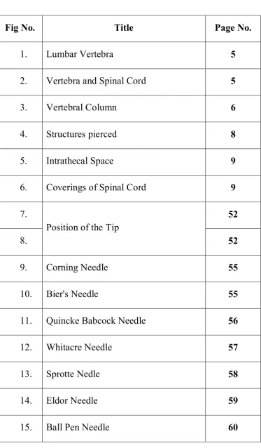
LIST OF FIGURES
Fig No. Title Page No.
1. Lumbar Vertebra 5
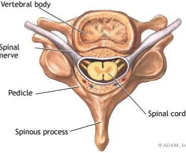
2. Vertebra and Spinal Cord 5
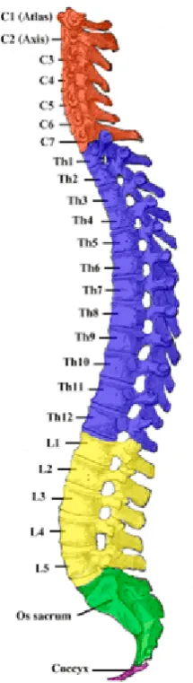
3. Vertebral Column 6
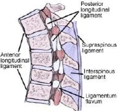
4. Structures pierced 8
5. Intrathecal Space 9
6. Coverings of Spinal Cord 9
7.
Position of the Tip
52
8. 52
9. Corning Needle 55
10. Bier's Needle 55
11. Quincke Babcock Needle 56
12. Whitacre Needle 57
13. Sprotte Nedle 58
14. Eldor Needle 59
LIST OF TABLES
S. No. Title Page No.
1. Incidence of PDPH 38
2. Estimated rate of Spontanous recovery 42
3. Age Distribution 74
4. Height Distribution 76
5. Weight Distribution 78
6. Gestational Age 80
7. PDPH Frequency 82
8. Severity of PDPH 83
9. Onset of PDPH 83
10. No. of attempts 85
11. Failure of SAB 57
LIST OF BAR CHARTS
S. No. Title Page No.
1. Age Distribution 74
2. Height Distribution 76
3. Weight Distribution 78
4. Gestational Age 80
5. PDPH Frequency 81
6. No. of attempts 85
7. Failure of SAB 87
LIST OF ABBREVIATION USED
CNS : Central nervous system
CVS : Cardiovascular system
CSF : Cerebrospinal fluid
EBP : Epidural blood patch
GIT : Gastrointestinal system
MAP : Mean arterial blood pressure
mg/dl : Milligrams per deciliter
PDPH : Postdural puncture headache
ABSTRACT
BACKGROUND AND OBJECTIVES
Spinal anaesthesia is one of the most commonly used regional
technique in anaesthesia. It is economical,safe and cost
effective.Preferred technique of choice in obstetric patients.
The objective of the present study is to compare the incidence of
Postdural puncture headache in obstetric patients following spinal
anaesthesia using 25 gauge Whitacre and 25 gauge Quincke spinal
needles.
METHODOLOGY
This one year randomized control study was conducted in the
Department of Anaesthesiology, Institute of Social Obstetrics,
Government Kasthurba Gandhi Hospital For Women And Child,
Triplicane, Chennai-5, during the year 2013-2014. Obstetric patients
posted for elective LSCS were included in the study. The Institutional
Ethical Committee clearance and written informed consent from the
patients obtained. Incidence of PDPH following spinal anaesthesia
,number of attempts for a successful blockade and failed blocks are
RESULTS
In this study, study groups contain subjects with same
demographic characters like age, height and weight. The occurance of
PDPH was meaningfully less(3.3%) when pencil point whitacre is used
compared to cutting quincke babcocks needle (10.8 %).
It was mild in 6%, moderate in 12%, severe in 6% of patients in
whitacre group. In quincke group it was mild in 29%, moderate in 35%,
severe in 5%
INTERPRETATION AND CONCLUSION
There is a real advantage of using pencil point whitacre needle in
comparison to cutting bevelled quincke needle.
KEYWORDS
Post dural puncture headache , failed spinal, subarachnoid
1
INTRODUCTION
Subarachnoid block is the widely used technique by the
anaesthesiologist worldwide. Spinal anaesthesia dates back to late 1800
with the work of Wynter,Quincke and Corning. However Dr.Karl August
Bier is given the credit for introducing spinal anaesthesia into clinical
practice in 1898.
The main advantage of SAB is due to its
· simplicity.
· ease of performance.
· requirement of minimal apparatus.
· minimal effect on blood biochemistry
· conscious patient maintaining airway
· good immediate postoperative pain relief
· blunts stress response to surgery
· decreased thromboembolic events
Though popular still,subarachnoid block is not without
2
headache. Bier gained first-hand experience of the disabling headache
related to dural puncture. He correctly summarized that the headache was
related to excessive loss of CSF.
PDPH causes considerable morbidity, with symptoms lasting for
several days, at times severe enough to immobilize the patients. It
increases the duration of hospital stay. In addition to this, it warrents a
battery of investigations to rule out different causes of headache. This
PDPH is more distressing to the mother who is supposed to take care of
the newborn baby.
There are lot of studies done regarding the measures for the
reduction of PDPH. It is said that with the help of fine gauge spinal
needle and needle tip modification, we can reduce the incidence to a
greater extend.
The 25 gauge Quincke babcock with medium cutting bevel is the
most popular and widely used spinal needle. A balance has to be done
between the incidence of PDPH and the technical difficulties with the use
of fine gauge spinal needles.
My study is to compare the incidence and the severity of PDPH in
obstetric patients using 25 gauge cutting tip Quincke spinal needle and 25
3
AIM OF THE STUDY
The aim of the study is to compare the incidence of PDPH,using 25
gauge Quincke spinal needle and 25 gauge Whitacre spinal needle in
obstetric patients.
PRIMARY AIM
Incidence and Severity of PDPH.
SECONDARY AIM
Number of attempts for successful blockade.
ANATOMY OF
SUBARACHNOID
4
ANATOMY OF THE SUBARACHNOID SPACE
The key to successful subarachnoid blockade is combining
appropriate technique with a three dimensional picture and appreciation
of vertebral anatomy.
VERTEBRAL COLUMN
It consists of 33 vertebra.
· 7 cervical
· 12 thoracic
· 5 lumbar
· 5 fused sacral
· 4 fused coccygeal bone.
VERTEBRAL CANAL
The bony canal is formed by the body of vertebra anteriorly,
posteriorly by two pedicles and the laminae connecting the pedicles. This
canal protects the spinal cord and contains spinal cord with surrounding
spinal meninges, spinal nerves and cerebrospinal fluid. The lamina gives
5
Figure 1 : Lumbar Vertebra
[image:20.595.142.512.436.739.2]6
Figure 3 : Vertebral column
CURVES OF SPINE
There are 4 curves. They are
i. Cervical curve-convex anteriorly.
ii. Dorsal curve-concex posteriorly.
iii. Lumbar curve-convex anteriorly.
7
These curves are important when the patient lies supine and these
can be modified by posture and the flexibility of the spine.
The highest point is at the level of L3 and the lowest point is at T5
level.
In Pregnancy, the lumbar curve is exaggerated
STRUCTURES PIERCED
The following structures are pierced when performing a SAB
· skin and subcutaneous tissue
· supraspinous ligaments
· interspinous ligament
· ligamentum flavum
· areolar tissue or epidural space
· duramater
ANATOMY OF LIGAMENTS
(i) SUPRASPINOUS LIGAMENT
It is a strong, thick, fibrous band extending from the spine of
8
(ii) INTERSPINOUS LIGAMENTS
It is a thin, fibrous structure connecting adjacent spines. These
fibres meet supraspinous ligament posteriorly and blend with ligamentum
flavum anteriorly.
(iii) LIGAMENTUM FLAVUM
It is a yellow elastic tissue, perpendicular fibres running from the
anterior inferior surface of upper lamina to the antero superior surface of
the lower lamina. It exist as right and left half in each intervertebral
space, with the halves fusing in midline.
TOPOGRAPHIC LINE OF TUFFIERS
A line joining the highest point of the iliac crest passes over
the spine of the fourth lumbar vertebra in the upright position or between
[image:23.595.207.402.564.746.2]L4-L5 interspace in the lateral position is called Tuffiers line.
9
Figure 5 : Intrathecal Space
[image:24.595.178.478.419.743.2]10
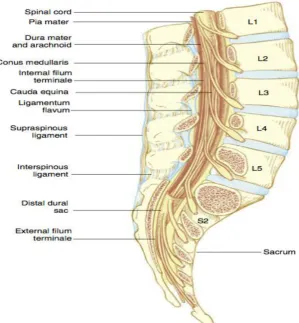
SPINAL CORD
The spinal cord is surrounded by three protective membranes
duramater, piamater, arachnoid mater, which are continuous with cranial
meninges. It is continuous cephalad with the brain stem through the
foramen magnam and terminates distally in the conus medullaris.
Differential growth rates between the bony vertebral canal and CNS
causes this distal termination to vary from L3 in infants to lower border
of L1 in adults.
SUBARACHNOID SPACE
It is the space between piamater and arachnoidmater. It has CSF,
Spinal nerves, trabecular network between the membranes, blood vessels
that supply the spinal cord, lateral extension of piamater, dentate
ligaments providing lateral support from the spinal cord to the duramater.
The subarachnoid space ends at the level of S2.
SPINAL MEMBRANES
Innermost layer is the piamater, which is highly vascular and
closely invest the spinal cord.
Middle layer is arachnoid mater which is non-vascular, delicate
11
Duramater is outermost, randomly organized fibroelastic
membrane, which extends from foramen magnum to S2,where the
filumterminale blends with the periostium in coccy
CEREBROSPINAL FLUID
It is a complex solution produced by plasma ultra filteration in
choroid plexus within cerebral ventricles.
Volume of CSF-100 to 160ml.
Rate of production-20 to 25ml/hr.
Absorbed in the arachnoid villi in the superior saggital sinus and/or
into lymphatics via perineural sheaths of cranial and spinal nerves.
CSF contains
Proteins : 15-45mg/dl
Glucose : 50-80mg/dl
Non protein : 20-30mg/dl
Chloride : 120-130meq/l
Sodium : 140-150meq/l
Bicarbonate : 25-30meq/l
12
ARTERIAL BLOOD SUPPLY
Anterior two third of spinal cord is supplied by anterior spinal
artery, formed at foramen magnum by a branch from terminal portion of
vertebral artery.
Posterior one third is supplied by two pairs of posterior spinal
artery derived from vertebral artery directly or from posterior inferior
cerebellar artery.
One of the anterior radicular branches arising from segmental
artery is considerably large and called Artery Of Adamkiewics. It
provides rich reinforcement to the flow of anterior spinal artery in its
lower distribution. It arises from the descending aorta at the lower
thoracic or upper lumbar level usually on the left side
VENOUS DRAINAGE
Veins are situated in the piamater. They are six in numbers and
form longitudinal plexiform channels after draining the parenchyma of
the cord. They emerge chiefly from the median fissure of the cord and are
larger in lumbar region. In this plexus, there are
i. two median longitudinal veins,one anterior in the anterior
13
ii. four lateral longitudinal veins,one pair runs dorsal to the
attachment of nerve roots and the other ventral to the nerve
root.
These veins communicate with the internal vertebral plexus, from
which blood drains into intervertebral veins. It then drains into segmental
veins and external vertebral plexus.
Near the base of the skull, the longitudinal channels surrounding
the cord units and form two or three small trunks, which communicate
with internal vertebral plexus, then intervertebral veins and the segmental
veins, then with cranial venous sinuses, ending in inferior petrosal
sinuses.
VEINS OF THE VERTEBRAL COLUMN:
The plexuses and the veins of the vertebral system are
· External plexus
· Internal plexus
· Basivertebral veins
· Intervertebral veins
PHYSIOLOGY OF
SPINAL
14
PHYSIOLOGY OF SPINAL ANAESTHESIA
The principle site of action of SAB is believed to be the nerve root.
Local anaesthetic agent injected into the subarachnoid space, baths the
nerve roots floating in CSF. They act by blocking the neural transmission
in the posterior nerve roor fibres and interrupts somatic and visceral
sensation. They block the anterior nerve root fibre also and prevent
efferent motor and autonomic outflow.
SOMATIC BLOCKADE
SAB provides excellent muscle relaxation by blocking afferent
transmission of painful stimuli and efferent impulse responsible for
skeletal muscle tone. Sensory blockade interrupts both somatic and
visceral painful stimuli. The size and character of the fibre type and the
fact that the concentration of local anaesthetic agent decreases with
increasing distance from the level of injection, explains the phenomenon
of Differential Blockade.
Differential Blockade results in sympathetic blocked(judged by
temperature sensitivity),that may be two segments or more cephalad than
the sensory block (pain, light touch),which, in turn, is usually several
15
AUTONOMIC BLOCKADE
Interruption of efferent autonomic transmission at the spinal nerve
roots during neuraxial block produces sympathetic blockade. The
physiological response of neuraxial blockade results from decreased
sympathetic tone and/or unopposed parasympathetic tone.
CARDIOVASCULAR MANIFESTATIONS:(CVS)
Neuraxial blockade produces decrease in blood pressure,
accompanied by decrease in heart rate.T5-L1 sympathetic fibres
determine the vasomotor tone. Blocking these nerves causes vasodilation
of venous capacitance vessels and pooling of blood in the viscera and
lower extremity, thereby decreasing effective circulating blood volume
and venous return to the heart.
Arterial vasodilation decreases systemic vascular resistance. The
effect of arterial vasodilation may be minimized by compensatory
vasoconstriction above the level of block. A high sympathetic block not
only prevents compensatory vasoconstriction, but also block the
sympathetic cardio acceleratory fibres that arise at T1-T4.Profound
hypotension can result from arterial vasodilation, venous pooling and
bradycaria. Unopposed vagal tone explains the sudden cardiac arrest seen
16
PULMONARY MANIFESTATIONS
Alteration in Pulmonary physiology is minimal with neuraxial
blockade because the Phrenic nerve is innervated by fibres from C3-C5.
Tidal volume is unchanged. There can be a mild decrease in Vital
capacity, which results from a loss of the abdominal muscle contribution
to forced expiration. Patients with severe respiratory disease depent on
accessory muscle of respiration to actively inspire and expire. High
neuraxial blockade in these patients may impair effective coughing and
clearing of secretions. Hence neuraxial blocks should be used with
caution in patients with respiratory reserve.
GASTROINTESTINAL MANIFESTATIONS
Sympathetic outflow originates at T5-S1. Neuraxial block-induced
sympathectomy allows vagal tone predominance and results in a small
contracted gut with active peristalsis. This improves operative conditions.
Postoperative epidural analgesia hastens the return of
gastrointestinal function after open abdominal procedures.
17
URINARY TRACT MANIFESTATIONS
There is little effect on renal function as the autoregulation
maintains renal blood flow. Neuraxial blockade at the lumbar and sacral
segments blocks both sympathetic and parasympathetic control of bladder
function. Loss of autonomic bladder control results in urinary retention
until the block wears off.
METABOLIC AND ENDOCRINE MANIFESTATIONS
In addition to local inflammatory response produced by surgical
trauma, systemic neuroendocrine response occurs via activation of
somatic and visceral afferent nerve fibres. This response includes
increased concentration of adrenocorticotropic harmone, cortisol,
epinephrine, norepinephrine and vasopressin levels. Activation of
Renin-Aldosterone-Angiotensin system also occurs. Neuraxial blockade can
partially suppress or totally block the neuroendocrine stress response. To
maximize this blunting effect, Neuraxial blockade should preceed
18
INDICATIONS
· Lower abdominal surgery
· Inguinal surgery
· Urogenital procedures
· Rectal surgery/Lower extremity surgery.
CONTRAINDICATIONS
ABSOLUTE
· Raised intracranial pressure
· Coagulopathy/blood dyscrasias
· Skin sepsis
· Severe mitral stenosis/aortic stenosis
· Patients refusal
· Hypovolemia
RELATIVE
· Preexisting neurological deficits
19
TECHNIQUE OF SPINAL ANAESTHESIA
To perform spinal anaesthesia, the technique is divided into four
steps.
· Preparation
· Position
· Projection
· Puncture
PREPARATION
Preparation of the equipment and drugs is essential for
subarachnoid injection. The drug chosed should match the duration of
surgery and the level of blockade needed for the procedure.
POSITION
The three primary position includes
i. The lateral decubitus position
ii. Sitting position
20
LATERAL DECUBITUS POSITION
It is the most commonly used position. It allows easier
administration of sedation and less dependent on a assistant for
positioning. Patients are placed with their back parallel to the edge of the
operating table nearest the anaesthesiologist, their thighs flexed on their
abdomen and their neck flexed to allow the forehead to be as close as
possible to the knees.
SITTING POSITION
This position is ideal in two situations.
a. Low lumbar and sacral levels of sensory anaesthesia are required
for the Perineal or Urological procedures.
b. Obesity or Scoliosis making identification of midline difficult in
the lateral position.
When placing the patient in this position, a stool can be provided as
a footrest and a pillow placed in the lap. The assistant then maintains the
patient in a vertical plane while flexing the patients neck and arm over the
pillow to open up the lumbar vertebral space. The assistant prevents the
21
PRONE POSITION
This position is appropriate for Rectal, Perineal and lumbar
procedures using hypobaric technique. After the patient in position,
Lumbar lardosis should be minimized and a paramedian approach used.
The anaesthesiologist may have to aspirate for CSF because CSF pressure
is minimized when inserting spinal needle in this position.
PROJECTION AND PUNCTURE
After proper preparation and patient positioning ,the midline or the
paramedian spinal puncture can be performed.
The midline approach relies on the ability of the assistant to
minimize lumbar lardosis and allow easy access to the subarachnoid
space between adjacent spinous process, usually L2-L3,L3-L4 or
L4-L5.It is the technique of choice because it requires anatomic projection in
only two planes and provides a relatively avascular plane.
The paramedian approach is choosed when there is a difficulty in
needle insertion with midline approach.The needle is inserted 1cm cauded
22
TAYLORS APPROACH
This is a variation of paramedian approach.It is carried out at
the L5-S1 space.The needle is inserted in a cephalomedial direction ,1cm
medial and cauded to the lowermost prominence of the posterior superior
iliac spine.
STOUT’S PRINCIPLE FOR SPREAD OF SOLUTIONS:
1. Height of anaesthesia varies directly with concentration
2. Extent of anaesthesia is inversely proportional to rapidity of
fixation
3. Extent of anaesthesia is directly proportional to the speed of
injection.
4. Extent of anaesthesia is directly proportional to the volume of fluid
5. Extent of anaesthesia is inversely proportional to the spinal fluid
pressure
6. Extent of anaesthesia is directly proportional to specific gravity for
hyperbaric solution
7. With hypobaric or hyperbaric solution extent depends upon on
23
INFLUENCE OF PREGNANCY ON SPINAL ANAESTHETIC
SPREAD
Pregnant women in second and third trimester require smaller dose
of anaesthetic agent. In 1953, the influence of pregnancy on spinal
anaesthetic dose requirement was reported in Pitkins Conduction
anaesthesia.
Two principle factors for the decreased anaesthetic dose
requirement in pregnant women for SAB are
i. Mechanical factor
ii. Hormonal factor
Compression of Inferior vena cava by gravid uterus causes
shunting of blood to the venous plexus in the vertebral column. This
decreses the vertebral canal space and CSF volume, Thus given spinal
anaesthetic mixture will be more diluted in a small amount of CSF than
in the non preganant women.
Hormonal factors for an increased sensitivity of the spinal nerves
to local anaesthetic agents in pregnancy are
24
ii. Increased endorphine level
iii. Increased alkalinity of CSF
FATE OF THE INJECTED DRUG:
There is a fall in the concentration immediately after injection of
the agent into the subarachnoid space due to four pharmacokinetic
processes
i. Dilution and mixing in CSF
ii. Diffusion and distribution to neural tissues
iii. Uptake and fixation by neural tissues
iv. Vascular absorption and elimination through arachnoid
villi and directly from capillary bed of parenchyma.
FACTORS AFFECTING SPREAD OF LOCAL ANAESTHETIC
AGENTS
1. Patient age
2. Height
3. Patient position
4. Configuration of spine
25 6. Site of injection
7. Speed of injection
8. Direction of needle
9. Local anaesthetic agent baricity
10. Local anaesthetic agent dose/volume.
FACTORS NOT AFFECTING CSF SPREAD
1. Patient weight.
2. Patient gender.
3. CSF composition /CSF circulation.
DISADVANTAGES AND COMPLICATIONS
1. Controllability- Continuous technique afford a greater margin of
control and flexibility.
2. Circulatory disturbances – hypotension occurs with high spinal
especially above T4
3. Not suitable for surgery above diaphragm
4. Respiratory impairment with high spinal level
26
6. Post lumbar puncture sequelae – head ache, cranial nerve
disturbances(ocular and auditory), infection(meningitis, epidural
abcess).
7. Neurological sequelae following injection
i. Meningismus
ii. Adhesive arachnoidits
iii. Cauda equine syndrome/myelitis.
ADDITION USES OF SPINAL ANAESTHESIA
(i) Paralytic ileus
Nonobstructive ileus has been satisfactorily treated with spinal
anaesthesia
(ii) Hyperthyroidism
Spinal anaesthesia to the level of fifth thoracic segment has a
beneficial effect upon patients in thyroid crisis and thyrotoxic patients
27
(iii) Pulmonary edema
The rationale of treatment with spinal anaesthesia is that this
diminishes venous return to the heart and relieves the cardiac load.
(iv) Diagnostic test
For evaluating hypertensive patients, which patients will have a
successful treatment from sympathectomy.
(v) Megacolon
In Hirschsprung disease, spinal anaesthesia has been used to block
the predominant action by the sympathetic system which causes
dilatation.
(vi) Pain evaluation
Differential spinal blockade may be used to evaluate and
differentiate functional chronic interactable pain from pain of somatic or
28
USE OF AN INTRODUCER
The concept of the introducer for performance of spinal puncture in
anaesthesia was that of Lincoln Sise. The advantages of the introducer are
i. prevents deflection of spinal needle by tough supra and
intraspinous ligament as well as resistant skin.
ii. Enables the introduction of fine gauge spinal needle into the
subarachnoid space.
iii. Safeguard against contamination of subarachnoid space and
carrying of skin bacteria to the thecal space.
iv. Avoid skin fragmentation entering subarachnoid space and
formation of Epithelioma.
The purpose of the introducer is to enable the entry of the spinal
needle into the depth of interspace without touching skin, subcutaneous
29
TESTING OF THE SPINAL NEEDLE QUALITY
The flexibility and the resiliency of the needle is to be tested. The
hub of the needle is grasped with one hand and the shaft of the needle
with the other hand.The ends should be depressed gently to make the
needle bent.On release of the shaft,the needle should spring back to a
straight position,demonstrating the above properties.
The needle with the stylet snugly in position,should be thrust
through a sterile gauze square.If there is a poor matching of the stylet
with the needle or if a burr is present at the point of the needle,then a
30
PHARMACOLOGY OF THE DRUG
Local anaesthetics are drugs that inhibit sensory,motor or
autonomic nerve function when injected or applied near the neural tissue.
These drugs consist of a lipophilic,unsaturated aromatic ring and a
hydrophilic,tertiary amine separated by a hydrocarbon chain that includes
an ester or amide linkage.These drugs are weak bases,carrying a positive
charge at the terminal amine group at physiological pH
The minimum concentration of local anaesthetic that will block the
nerve impulse is affected by
i. Fibre size, type, myelination
ii. pH
iii. frequency of nerve stimulation
iv. electrolyte concentration.
Onset of action depends on lipid solubility,relative concentration of
nonionized lipid soluble and ionized water soluble form expressed as
pKa.Local anaesthetic with pKa closer to physiological pH will have a
greater concentration of nonionized base that permeates the nerve cell
31
BUPIVACAINE
It is a synthetic drug,prepared by A.F.Ekenstam in 1957 and
marketed as Marcaine.
· Molecular weight of chloride salt is 325 and that of the base
is 288.
· Melting point-258 degree Celsius.
· pH of the solution with adrenaline is about 3.5
· The chemical name is 1-n-butyl-DL-piperidine-2-carboxylic
32
CHEMICAL PROPERTY OF THE DRUG
· The base is sparingly soluble, the hydrochloride is readily
soluble in water.
· The drug is stable and can withstand autoclaving.
· It is more potent and long acting than lidocaine.
· Anaesthetic index is 3-4.
PHARMACODYNAMICS
The onset of action is between 5-7minutes,maximum anaesthesia is
obtained between 15-25 minutes.
PLASMA BINDING
The drug binds avidly with protein to the extend of 70-90%.
METABOLISM
As its an amide,Liver is the primary site of metabolism mostly by
alkylation.10% drug is excreted unexchanged in the urine.The high
protein-binding capacity explains the less diffusion of the drug across the
33
MECHANISM OF ACTION
The drug inhibits passage of sodium ions through ion-selective
sodium channel in the nerve membrane.It slows the rate of depolarization
so that the threshold potential is not reached and action potential not
propagated.
Bupivacaine blocks the sodium chanel in inactivated closed
state,and prevent their change to the resting closed and activated open in
response to nerve impulse.
The drug also binds to activated open sodium channels to produce
frequency dependent blockade.
They also act on voltage dependent potassium channel.Evidence
suggest that they can act on G-protein coupled receptor also.
SIDE EFFECTS
The principal side effects are allergic reactions and systemic
toxicity due to excessive plasma and tissue concentration of the agent.
ALLERGIC REACTION
The Ester local anaesthetics producing metabolites of
34
anaesthetics.Allergic reaction also develop for methylparaben,a
preservative in commercial preparation. History of
rash,utricaria,laryngeal edema,with or without bronchospasm and
hypotension is suggestive of local anaesthetic induced allergic reaction.
SYSTEMIC TOXICITY
Excess plasma concentration due to accidental intravascular
injection or absorption from injection site accounts for systemic toxicity.
CARDIOVASCULAR SYSTEM (CVS)
The CVS toxicity is more resistant to treatment than CNS toxicity.
During diastole,highly lipid soluble bupivacaine dissociates slowly from
sodium ion channel,thus accounting for persistant cardiac
toxicity.Ropivacaine is a pure S-enantiomer that is less lipid soluble and
less cardiotoxic than bupivacaine.Drug toxicity presents as precipitous
hypotension,arrhythmias and atrioventricular blocks.
CENTRAL NERVOUS SYSTEM (CNS)
The CNS toxicity ranges from perioral numbness, seizures, CNS
depression, accompanied by hypotension and apnea. Transient
neurological symptoms,Cauda Equina syndrome and Anterior spinal
35
POSTDURAL PUNCTURE HEADACHE
Apart from intentional dural puncture in subarachnoid
block,epidural anaesthesia performed for postoperative and labour pain
relief can result in unintentional dural puncture.However dural puncture
is a commonly performed invasive procedure for diagnostic lumbar
puncture,myelography and intrathecal chemotherapy.
12% claims of malpractice filed against anaesthesiologist providing
obstetric anaesthesia care were because of postdelivery headache in
patients who received epidural anaesthesia and probable dural puncture.
CHARECTERISTICS OF PDPH
PDPH is
· dull aching or throbbing in nature.
· occurs within 7 days of dural puncture.
· It is self-limiting and disappears within 14 days.
· It worsens in upright position and eases when supine.
· Headache is often in occipital region.
· Pressure over the abdomen with the women in upright position
36
pressure secondary to the increase in intraabdominal
pressure.(GUTSCHE SIGN)
CAUSES OF POSTPARTUM HEADACHE
After delivery, 39% of Parturients report symptoms of headache
unrelated to dural puncture. There are many causes that mimics PDPH in
postoperative period that need to be diagnosed as the treatment modalities
varies.Causes include
(i) Infective
Meningitis and encephalitis
(ii) Vascular causes
Migraine
Cerebral vein thrombosis
Cerebral infarct
Subdural hematoma
Subarachnoid hemorrhage
(iii) Neoplastic
37 (iv) Pharmacological or metabolic
Dehydration / Caffeine withdrawal
(v) Others
Postdural puncture headache
Pre-eclampsia
Tension headache
Benign intracranial hypertension
Pneumocephalus
Lactational headache.
PATHOPHYSIOLOGY OF PDPH
With dural puncture, there is loss of CSF and lowering of CSF
pressure.Rate of CSF loss is greater than the rate of CSF production
particularly with spinal needle size greater than 25 gauge.
Possible explanation for headache includes
i. Lowering of CSF pressure causes traction on pain sensitive
intracranial structures like meninges,vessels and cranial
38
ii. According to Monro-Kellie Hypothesis,loss of CSF volume
is compensated by venodilatation resulting in increased
blood volume.
INCIDENCE:
In 1898,the incidence of PDPH was as high as 66% due to larger
gauge, medium bevel, cutting spinal needles.
Needle design Gauge Incidence
Quincke 22 36%
25 3-25%
26 0.3-20%
27 1.5-5.6%
29 0-2%
Whitacre 20 2-5%
22 0.6-4%
25 0-14.5%
27 0%
Sprotte 24 0-9.6 %
Atraucan 26 2.5-4 %
The incidence of PDPH is directly proportional to the gauge of
39
anaesthetist. The incidence is less if the bevel of the needle is oriented
parallel to the direction of dural fibres. The loss of resistance with air
increases the risk of PDPH than loss of resistance with saline during
epidural anaesthesia.
RISK FACTORS
The risk factors for the development of PDPH may be either needle
related or patient related.
The needle related factors are
i. size of the needle.
ii. shape of the needle tip.
Larger the gauge, more is the incidence of PDPH. Fine gauge
spinal needles have a low incidence of PDPH but are technically more
difficult to use with high failure rate. Pencil tipped non-cutting needle tips
40 Patient related factors are
· Age./
· Sex.
· Body mass index.
· Obstetric population.
PDPH is more common in young age group. In children,
incidence is less because of low reporting rate and low CSF pressure. In
old age, incidence is less because of the following reasons
· Decreased elasticity of dural fibres
· Decreased reactivity of cerebral vessels
· Decreased A delta and C fibres
· Attenuated central sensititation.
· Elevated pain threshold.
PDPH is more common in Women.In a series report by Vandane
and Dripps,women have twice the incidence as in male.It was found to be
12% in women and 7% in men. It is stated that the greater incidence is
41
PDPH is more common in low BMI patients.
Obstetric patients are at high risk because of their sex,young age
and the frequency of epidural anaesthesia as a part of labour analgesia.
SYMPTOMS
· Postural headache.
· Neckache
· Nausea.
· Vomiting.
· Tinnitus.
· Blurring of vision.
· Hearing loss.
· Vertigo.
· Dizziness
42
DIAGNOSIS
There is a positive history of accidental or deliberate dural
puncture.
Diagnosic LP demonstrates low CSF pressure or a dry tap,a slight
increase in CSF protein and increase in CSF lymphocyte count.
MRI shows diffuse dural enhancement with evidence of dural
sagging,descent of brain,optic chiasma and brain stem,obliteration of
basal cisterns and enlargement of pituitary.
CT myelogram, Retrograde Radionuclide Myelography,
Cisternography can locate the site of dural leak.
ESTIMATED RATE OF SPONTANEOUS RECOVERY
DURATION % RECOVERY
1-2 days 27
3-4days 39
5-7days 19
8-14 days 8
3-6weeks 5
3-6months 2
43
The largest follow-up of PDPH is still that of Vandam and Dripps
et al in 1956.They reported that 72% headache resolved within 7 days
and 87% within 6 months.
TREATMENT
1. Adequate hydration is to be maintained.Studies have been done to
know effect of preloading with PDPH and found that Preloading
cannot prevent PDPH.Its better to maintain adequate hydration
than dehydration.
2. Most of the patients respond to simple analgesics like
NSAIDS,Acetaminophen.
3. Supportive treatment like antiemetics can be given.
4. Prone posture increases intraabdominal pressure and epidural
pressure.A clinical trial of prone position following dural puncture
failed to demonstrate any decrease in PDPH.
5. Pharmacological treatment includes
i. Caffeine
ii. Cosyntropin(ACTH analogue)
44
(i) CAFFEINE
It is a CNS stimulant,produces cerebral vasoconstriction,blocks
peripheral adenosine receptors and other central mechanisms.It is given
as Caffeine sodium benzoate orally or intravenously.Dose is 500mg
orally or 500mg in one litre intravenous fluid iv over one hour.
The most frequently quoted work on treatment of PDPH with
caffeine is that of Sechzer. He concluded that iv caffeine is an effective
treatment for PDPH.
The side effects of caffeine are
· CNS Stimulation.
· Seizures.
· Gastric irritation.
· Cardiac arrhythmias.
Treatment with caffeine does not decrease CSF leak nor restores
normal CSF dynamics,therefore the recurrence rate is 60%.
(ii) COSYNTROPIN
It is an ACTH analogue, less antigenic than naturally occurring
harmone. It stimulates adrenal cortex to secret glucocorticoids,
45
resultant increase in intracellular cyclic AMP. Increased CSF production
through a sodium active transport mechanism, as well as possible
increase in beta endorphin in CNS, increased pain threshold are the
possible mechanism of action.
Dose is 0.25-0.75mg intravenous.
(iii) SUMATRIPTAN
It is a 5HT1D receptor agonist. Carp et al reported administration
of sumatripten 6mg to six patients with headache within six hours and
reported good results.
EPIDURAL BLOOD PATCH
Introduced by Gormley in 1960.usually performed for severe
headache not responding to conservative management after 24 hours.
Contraindication includes
· Fever
· Coagulopathy
· Infection at the site
46
Blood samples to be sent for microbiology for culture before
proceeding for the procedure. Patient is placed in lateral position,epidural
space identified with tuohy needle at the level of supposed dural puncture
or one space below. Two persons are required to perform the technique.
One person identifies Epidural space while the other draws 10-20 ml of
blood under aseptic precaution. Withdrawn blood is injected into the
epidural space at the rate of one ml every three seconds. The injected
blood clots and seals the dural rent and the compressed dural space
restores the subarachnoid pressure. The injection should stop if the
patient experiences lancinating pain or backache. The patient should lie
flat on the back for one or two hours, then allowed to walk.
Complication of this invasive technique is that of
· Backache
· Infection,arachnoiditis
· Failure of treatment.
EPIDURAL SALINE/DEXTRAN
Saline is relatively inert and sterile. It can be given as bolus or
47
· 1 to 1.5 litres of epidural hartmann solutions over
24hours,starting on day 1 after spinal anaesthesia.
· Upto 35ml/hr for 24 hours on development of headache.
· Single 30ml epidural bolus after development of headache.
· 10-120ml of saline injected as bolus via caudal epidural
space.
EPIDURAL,INTRATHECAL,PARENTERAL OPOIDS
Majority of the studies on use of morphine is either case reports or
inadequate controlled trials.
FIBRIN GLUE
Fibrin glue is a preparation of pooled human plasma obtained from
plasmapheresis. It is prepared by mixing two solutions. First one contains
fibrinogen, factor VIII, fibronectin ,aprotinin, plasminogen. The second
one contains thrombin and calcium. The mixture forms a gel with high
tensile strength. It is applied blindly or CT- guided percutaneous
48
INTRATHECAL CATHETERS
The placement of spinal catheters through the perforation may
provoke an inflammatory reaction that will seal the hole.Complication
include risk of Cauda equina syndrome and infections.
NUMBER OF ATTEMPTS
Lybecker et al didn’t support a association between frequency of
PDPH and the number of dural puncture.But recent prospective analysis
of SAB performed on 8034 patients showed increased incidence of PDPH
with repeated attempts.
FAILED SPINAL ANAESTHESIA
Though spinal anaesthesia is safe,cost effective and reliable
technique commonly used,failed spinal block is a possibility if the
technique is not performed meticulously.Inadequate/failed spinal
anaesthesia is very difficult to manage.
Two factors are very important determinant of a successful
blockade.They are
i. Dural puncture
49
Spinal anaesthesia is technically easy procedure with a clear cut
end point,the CSF.The appearance of CSF at the needle hub gives us an
indication that we are in the thecal space.At the same time,appearance of
CSF alone doesn’t guarantee successful blockade.
Failed block can be of three types, namely
i. Inability to get a flow of CSF,called as Dry tap.
ii. subarachnoid block attempted,no resultant block.
iii. Block ensues,but inadequate for the extend and qualityof
anaesthesia and duration of surgery.
Failure of spinal anaesthesia is avoidable provided the technique is
performed properly. With experienced hands,incidence of failed spinal is
less than 1%
There are five phases of spinal anaesthesia which can influence the
failure incidence. They are
i. Lumbar puncture
ii. Injection of the drug
iii. Spread of the drug
iv. Action of the drug
50
LUMBAR PUNCTURE
The problems encountered during lumbar puncture are due to two
factors,
· poor positioning of the patient
· incorrect needle insertion
Abnormalities of the spine like kyphoscoliosis and calcification of
the ligaments, obesity, patients anxiety plays a role in poor positioning of
the patient.
The spinal needle is inserted in midline, midway between the
posterior spines.The shaft of the needle is placed at right angle to the back
of the patient.When there is resistance to insertion,the needle is angulated
in cephalad direction.While advancing the needle,a mental picture of the
spinal anatomy helps to appreciate where the needle tip lies.
INJECTION OF THE DRUG
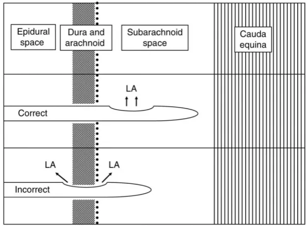
The drug should be injected into the thecal space for a successful
blockade.Anterior or posterior displacement of the spinal needle while
attaching the syringe to the needle or while injecting the drug can result
in an inadequate block or failed spinal.With pencil tip needles,the
51
loss of drug injected into the epidural space and a forward movement can
elicit parasthesia by the tip contacting the nerve fibres.
SPREAD OF THE DRUG
The CSF acts as a medium through which the drug acts.The spread
of the drug is dependent on the baricity of the drug injected,position of
the operating table and the anatomical abnormality of the vertebral canal.
PATIENT FACTOR
Surgical stimulus can stimulate the unblocked parasympathetic
and phrenic nerve ,making the patient anxious.The patients should be
explained the technique to be performed and what to expect before
52
[image:70.595.170.483.489.719.2]53
REVIEW OF LITERATURE
HISTORY OF SPINAL ANAESTHESIA
In 1885,Leonard Corning,a Newyork Physician,accidentally
injected cocaine intrathecally in a dog.He proposed that local anaesthesia
of the spinal cord with cocaine may have therapeutic property.He injected
cocaine in a man at the level of T11-T12 interspace to treat seminal
incontinence.He coined the term SPINAL ANAESTHESIA.
In 1891,Wynter and Quincke aspirated CSF from subarachnoid
space to treat increased ICT associated with TB meningitis.
In 1898,Karl August Bier,a german surgeon and his assistant
Hildebrandt underwent spinal anaesthesia with cocaine,the assistant first
injecting Bier and then Bier his assistant.While injecting the assistant
struggled to connect the syringe to the needle,resulting in significant loss
of CSF.He developed severe headache and he correctly noted that the
headache was related to the loss of CSF.
In 1901,a swiss obstetrician used intrathecal cocaine for pain relif
in second stage of labour.Though high incidence of both vomiting and
PDPH noted,a high mortality rate in LSCS performed under subarachnoid
54
referred to as Dark Age Of Obsteric Anaesthesia,when natural child birth
and psychoprophylaxis were encouraged.
Development of fine gauge spinal needles and needle tip
modification has enabled a significant reduction in the incidence of
PDPH.
HISTORY OF SPINAL NEEDLES
The development of spinal needles and needle tip modification
began with the understanding of anatomy and physiology of CNS that
pertained at the time of introduction of spinal anaesthesia.
In 1841,Zophar jayne of illinos designed a syringe attached to a
small sharp,hollow beak with an opening on the side near the tip.
In 1853,Alexander Wood of Edinburg is credited the first in
developing hollow hypodermic needle. He used Fergusons needle for
injecting into deeper structure.
In 1885,Leonard Corning developed his own needle made of gold
or platinum. cannula was flexible with a metal stop and a set of screw to
fix the needle at correct place once the subarachnoid space has been
entered. Tip is sharp,short cutting bevel.Introducer was short with a right
55
Bier took a historic step of using spinal anaesthesia to provide analgesia
for surgical procedure. He designed a large bore needle,15gauge or
17gauge that needed no introducer. It has a long cutting bevel and a sharp
tip. Biers needle was criticized for causing pain on injection and leaving a
large hole in dura.
Figure 10 : Bier's Needle
Bainbridge in 1900,described a needle with a small circular hub,a
short sharp cutting bevel and a stylet with a matching bevel,made of
flexible metal.
Barker appreciated the problem with long bevel and he designed a
hollowed out point to secure sharpness without lengthening the terminal
opening.Barkers needle is 18 gauge or 19 gauge with a medium length
[image:74.595.162.445.97.157.2]bevel and a stylet with a matching bevel.
56
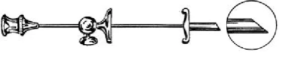
In 1914,Babcock described a sharp,medium length bevel with the
matching stylet,made of iridized pplatinum or gold.It was 20 gauge in
diameter and referred as Quincke babcock needle.It was a very successful
needle and became the standard needle for comparative studies.
Figure 11 : Quincke Babcock Needle
Gaston labot needle was a medium gauge cannula with a
short,sharp bevel and a matching stylet. The tip ground to match the bevel
of the cannula.
ATRAUMATIC SPINAL NEEDLE TIPS
In 1920,realization of the fact that CSF leak is associated with
cutting of duramater led to the development of needle tip modifications.
In 1923,Hebert Morton Greene used an ordinary 23 gauge cutting
needle that he shaped to a rounded tip by removing the cutting edge of the
bevel.The needle was sized between 20gauge and 26 gauge.The point was
a rounded,non-cutting bevel of medium length with a matching,bevelled
fitted stylet.It was very popular in obstetric practice till the invent of
57
MODIFICATION OF CUTTING BEVEL
Pitkin needle is 20gauge or 22 gauge,made of relatively flexible
rustproof steel with a collar to mark the depth of insertion.Tip has
short,sharp bevel ground off to a taper of 45 degrees,resulting in rounded
blunted bevel heel.He advocated the insertion of cutting edge of the bevel
parallel to the longitudinal fibres of dura.
PENCIL POINT NEEDLES
Barker was credited the first directional closed-end spinal
needle,which forms the basis of pencil-point tip used commonly
nowadays.
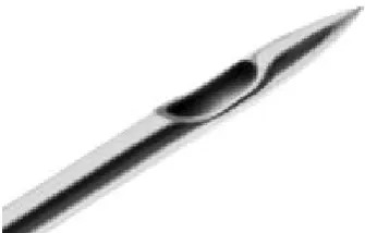
Hart and Whitacre designed a closed ended,lateral orifice,pencil
point tip needle in 1951.It is a 20 gauge needle with a solid end drawn to
a point similar in shape toa finely sharpened pencil.These needles have
decreased incidence and severity of PDPH and a more appreciable dural
click.The needle has a small hole making aspiration and injection
difficult.
58
CONTINOUS SPINAL NEEDLE
Various spinal needle were described in literature.
· Lemmon needle
· Hingson Fegurson needle
· Tuohy needle
· Cappe and Deutsch needle.
SPROTTE NEEDLE
It is a modification of Whitacre needle. It has increased size of
lateral orifice to combat the problem of slow CSF flow and resistance to
injection. The tip is also elongated in an attempt to allow more gradual
[image:77.595.224.392.533.640.2]separation of dura.
59
ATRAUCAN NEEDLE
In 1993,Atraucan needle has a cutting double bevel with a sharp
point making an initial incision. The second part then dilates the incision
rather than cutting leaving a small hole in the dura.
DOUBLE HOLE PENCIL-POINT NEEDLE
Eldor in 1996 was based on pencil point tip but with two lateral
[image:78.595.218.401.353.493.2]holes opposite to each other.
Figure 14 : Eldor Needle
BALLPEN NEEDLE
It’s a modification of Quincke needle designed in 2000.This needle
is similar to Levy needle of 1957.Advantages of the needle are
i. tip of the needle is always in the subarachnoid space on removal
60
ii. needle tip doesn’t project beyond the orifice to cause
neurological damage
iii. no mechanical weakning at the tip caused by the presence of
lateral opening.
Figure 15 : Ball Pen Needle
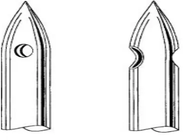
Shutt Le et al in 1992,compared the incidence of PDPH in 150
obstetric patients using 26gauge Quincke,22gauge Whitacre and 25 gauge
Whitacre needles.The problem with the study was that the power of study
was not disscussed.Power of study estimated by Altmans nomogram is
0.55 which is too low to expect a significant result.They stated difference
in the incidence of headache was statistically insignificant.The incidence
of PDPH was 4% with Quincke needle and 1% with Whitacre
needle.(Br.J.Anaesth;1992,69:589-94)
A study includes 300 patients posted for lower limb/ortho
surgery.They were allotted into 3 groups.Group 1 had SAB with 25 gauge
Quincke needle, with bevel oriented perpendicular to the fibres.Group 2
61
fibres.Group 3 had SAB with 24 gauge Sprotte.The incidence of PDPH
was 17.9%,4.5% and 2.4% respectively.The results shows that Quincke
needle should not be inserted perpendicular to the dural fibres and tha
Sprotte needle does not solve the problem of PDPH or
backache.(Reg.Anaesth 1992;17(5):283-7)
A study comparing 24 gauge Sprotte and 25 gauge Quincke spinal
needle showed lower incidence of severe PDPH with 24 gauge Sprotte
but was not significantly different when compared with 25 gauge
Quincke spinal needle.In this study,194 patients were allotted into group
1(n-96,Sprotte) and group 2(n-98,Quincke).Fentanyl was given
intrathecally in 47 patients in group 1 and 49 patients in group 2.All
patients followed for 4 days postoperatively for headache.Incidence of
PDPH was found to be 4.2% in Sprotte group and 7.1% in Quincke
group.(Reg Anaesth 1993;18:222-5)
Campbell et al in 1993,studied in 300 patients with 24 gauge
Sprotte and 25 gauge Whitacre needle.Each group includes 150
patients.The incidence of PDPH is 4% with sprotte needle(6/150) and
0.66% with whitacre needle(1/150).(Can.J.Anaesth 1993;40(12):1131-5)
A study of 400 patients, comparing 25 gauge Whitacre and 25
62
Whitacre and 8.5% with Quincke needle.No significant difference in age
or sex in both groups.The duration lasted for 1-3 days in Whitacre group
and 1-9 days in Quincke group.(Reg Anaes.1993;18:166-9)
The frequency and severity of headache after Lumbar myelography
using 25 gauge Whitacre in 63 patients aged 20-81 years showed an
incidence of 4.7%.3 patients had PDPH.(Neuro radiology 1995;37:553-6)
Incidence of PDPH was 4% with 26 gauge Atraucan and 4.3% with
25 gauge Whitacre spinal needle in a study.Use of 26 gauge helps in
better identification of subarachnoid space,faster CSF back flow and
fewer parasthesia.(Can J Anaesth 1995;42:706-10)
Lambert et al reported the rate of PDPH with 25gauge Whitacre as
1.2%, 2.7% with 27gauge Quincke cutting needle and 5.2% with 26
gauge Quincke needle. He included 4125 patients over 4 years
period.(Reg.Anesth 1997;22(1):66-72)
In Acta Anaesthesiology Scand 1997;41:779-84,200 patients
posted for day care procedure were studied for the severity and incidence
of PDPH in using 27 gauge Quincke and 27 gauge Whitacre.Incidence of
PDPH is 0% with 27 gauge Whitacre spinal needle and 5.6% with 27
gauge Quincke needle.All PDPH occurred with Quincke needle oriented
63
Hwang JJ,HOST et al in 1995,studied 90 patients with 26 gauge
Quincke and 25 gauge Whitacre needle.Incidence of PDPH was 6.66%
with Quincke and 1.52% with Whitacre.
Jost u et al in 2000,studied 600 patients using 26 gauge Quincke
and 27 gauge Whitacre. Incidence of PDPH was 6% with Quincke and
1% with Whitacre. (Anasthesiol Intensive Med;2000,35:381-7)
Vallejo et al studied the incidence of PDPH and Epidural Blood
Patch using 5 spinal needles in obstetrics.He used 2 cutting needle(26
gauge atraucan and 25 gauge Quincke) and 3 pencil point needles(24
gauge Gertie Marx,24 gauge Sprotte,25 gauge Whitacre).The incidence
of PDPH and EBP are 5%, 8.7%, 4%, 2.8%, 3.1% and 55%, 66%, 12.5%,
0%, 0% respectively. (Anaesthesia and Analgesia 2000;91-4:916-920)
Invitro study of the dural lesion evaluated by scanning electron
microscopy was done while using 25 gauge Quincke and 25 gauge
Whitacre.T11-L4 dural membrane from 5 fresh male patients declared
brain dead,aged 23,46,48,55 and 60 were excised by anterior
laminectomy.100 punctures (20 on each samples) at 90 degrees made.50
with 25 gauge Quincke and Whitacre each. It is shown that the dural hole
64
accounts for more inflammation seen with Whitacre needle and therefore
better healing.(Reg Anaesth Pain Med 2000;25:393-402)
Landau R et al in 2001,studied 400 patients with 25 gauge
Whitacre and 27 gauge Whitacre needle.The incidence of PDPH was
1.32% with 25gauge and 0.03% with 27gauge Whitacre
needle.(Int.J.Obstet Anaesth;2001,10:168-71)
75 female patients scheduled for LSCS were grouped into 3 of 25
each.Group 1,2,3 received SAB using 25 gauge Quincke,27 gauge
Quincke and 27 gauge Whitacre needles respectively.The patients were
assessed for the severity of PDPH and the technical difficulties with the
needle insertion.All the patient had mild form of PDPH.25 gauge needle
had 100% successful dural puncture and 27 gauge Whitacre had 12%
failure rate.(Indian J.Anaesth 2002;46(5):373-377)
Tabedar in 2006,studied 60 patients with 25 gauge Quincke and
26gauge Whitacre needle. Incidence was 8% with Quincke and 2.2% with
Whitacre. (Kathmandu Uni. Med.J;2003,1:263-6)
Bano F et al in 2004,studied 100 patients with 25 gauge Quincke
and 25gauge Whitacre.The incidence of PDPH was 4% with Quincke and
0.75% with whitacre.(Dow University of Health science
65
In a study 480 obstetric patients were included as group 1-(25
gauge Quincke,n-168),group 2(27 gauge Quincke,n-160)and group 3(27
gauge Whitacre,n-152). The study comparing 25 gauge Quincke,27 gauge
Quincke and 27 gauge Whitacre needle showed the incidence of PDPH as
8.3%(14/168) with 25 gauge Quincke,3.8%(6/160) with 27 gauge
Quincke,2%(3/152) with 27 gauge Whitacre needle.The severity of
PDPH was mild in 5,moderate in 7 and severe in 2 patients in group 1.In
group 2,it was mild in 2,moderate in 3 and severe in 1 patient.In group
3,2 patients had mild headache and 1 patient had moderate
66
MATERIALS AND METHODS
The study is to compare the incidence and severity of
PDPH,number of attempts for successful block and number of failed
spinal in using 25 gauge Quincke and 25 gauge Whitacre spinal needles
in 240 obstetric patients after getting approval by the Institutional Ethical
Committee.It is a double blind study where the patient is unaware of the
needle type used and the postoperative assessment of headache is done by
a person unaware of the needle type used.
The study was done in the Department Of
Anaesthesiology,Institute of Social Obstetrics,Government Kasthurba
Gandhi Hospital for Child and Women,Triplicane,Chennai-5.
240 obstetric patients posted for elective LSCS under
Subarachnoid block were randomly allocated into group A and group B
by using a computer generated randomization table .
SELECTION CRITERIA
INCLUSION CRITERIA
· Age 20 to 36 years.
· Weight 58 to 87 kilograms.
· Height 148 to 170 cms.
67
· Elective surgery
· Valid informed consent.
EXCLUSION CRITERIA
· Not satisfying inclusion criteria
· Lack of Written informed consent.
· Contraindication for neuraxial anaesthesia.
· H/O Recurrent headache in the past.
· H/O Occipital neuralgia/Migraine.
PROCEDURE
PREOPERATIVE EVALUATION
A thorough history taking and clinical evaluation done to include
patients who satisfy inclusion and exclusion criteria.Preoperative
investigations done as all cases posted for elective surgery.All patients
were briefed about the nature of the study,procedure to be performed and
a valid written informed consent obtained.
The patients were instructed fasting from midnight, atleast for 8
hours.
The patients transported to the operation theatre in left lateral