Dissertation on
MORPHOLOGICAL ANALYSIS AND MORPHOMETRIC
STUDY OF THE FORAMEN MAGNUM
Submitted in partial fulfillment for M.D. DEGREE EXAMINATION
BRANCH- XXIII, ANATOMY
Upgraded Institute of Anatomy
Madras Medical College & Rajiv Gandhi Government General Hospital,
Chennai- 600 003
THE TAMILNADU Dr.M.G.R. MEDICAL UNIVERSITY CHENNAI – 600 032
TAMILNADU
CERTIFICATE
This is to certify that this dissertation entitled
“MORPHOLOGICAL ANALYSIS AND MORPHOMETRIC STUDY OF THE FORAMEN MAGNUM”
is a bonafide record of the research work done by Dr.M.ANURADHA, Post graduate in the Institute of Anatomy, Madras Medical College and Research Institute, Rajiv Gandhi Government General Hospital, Chennai-03, in partial fulfillment of the regulations laid down by The Tamil Nadu Dr.M.G.R. Medical University for the award of M.D. Degree Branch XXIII- Anatomy, under my guidance and supervision during the academic year from 2012-2015.
Dean
Madras Medical College and
Rajiv Gandhi Government General Hospital
Chennai-600 003
Dr.Sudha Seshayyan, M.B.B.S.,M.S.,
Director and Professor, Institute of Anatomy, Madras Medical College, Chennai 600003.
ACKNOWLEDGEMENT
I wish to express exquisite thankfulness and gratitude to my most respected teacher and guide Dr. Mrs. SudhaSeshayyan, M.S., Director and Professor, Institute ofAnatomy, Madras Medical College, Chennai – 3, for their invaluable guidance, persistent support and quest for perfection which has made this dissertation take its present shape.
I’m thankful to Dr.R.Vimala, M.D., Dean, Madras Medical College, Chennai – 3 for permitting me to avail the facilities in this college for performing this study.
My heartfelt thanks to Dr.B.Chezhian, Dr.V.Lokanayaki, Associate Professors Dr.S.Lakshmi, Dr.T.Anitha, Dr.P.Kanagavalli, Dr.J.Sreevidya, Dr.Ilamathi Bose and Dr.S.Arrchana Assistant Professors, Institute of Anatomy, Madras Medical College, Chennai – 3 for their valuable suggestions and encouragement throughout the study.
College, Mumbai who helped me to complete this study with his valuable guidance.
My gratefulness to Dr.Vanitha, M.D., Director, Barnard Institute of Radiology, Rajiv Gandhi Govt.General Hospital,Chennai – 3 and Dr.Babu Peter, M.D., Associate Professor, for their help in the radiological study.
I also thank Dr.Evangeline Mary, Post graduate in Community Medicine, Dr.Subisen, Post graduate in Neurosurgery and Dr.Arun, Post graduate in Radiology for their help in the completion of this study.
I earnestly thank my seniors Dr.P.Radhakrishnan, Dr.K.Arumugam, Assistant Professor, Thirunelveli Medical College, Thirunelveli and my helpful juniors Dr.Keerthi, Dr.Prefulla, Dr.Ganga and other members of faculty who have been supportive and encouraging throughout the study.
I extend my heartfelt thanks to my colleagues Dr.S.Elizabeth Priyadarisini, Dr. B.J.Bhuvaneswari and Dr.E.Srividhya for their constant encouragement and unstinted co-operation.
I’m grateful to my parents, my sister and my brother who have helped making this study a reality.
CONTENTS
SL.
NO. TITLE PAGE NO.
1. INTRODUCTION 1
2. AIM OF THE STUDY 5
3. REVIEW OF LITERATURE 8
4. EMBRYOLOGY 35
5. MATERIALS AND METHODS 38
6. OBSERVATION 43
7. DISCUSSION 68
8. CONCLUSION 109
LEGEND
A ICD - Anterior inter condylar distance AP - Antero Posterior
BCD - Bicondylar distance
CT - Computerised Tomography FM - Foramen Magnum
HGC - Hypoglossal canal LHGC - Left Hypoglossal Canal LOC - Left Occipital Condyle OC - Occipital Condyle PCC - Posterior condylar canal
PICD - Posterior inter condylar distance
‘P’ value - Probability of observing the difference by chance RHGC - Right Hypoglossal Canal
MORPHOLOGICAL ANALYSIS AND MORPHOMETRIC
STUDY OF THE FORAMEN MAGNUM
ABSTRACT
The foramen magnum is the oval shape opening situated at the base of the skull. The transcondylar approach is being increasingly used to access lesions of the Craniovertebral junction. Understanding the anatomy of the foramen magnum is important for skull base surgery.
The present study was aimed at analysing the foramen magnum morphologically and morphometrically.100 adult human dry skullsat the Institute of Anatomy, Madras Medical College and twenty cranial CT scans obtained from the archives of Barnard Institute of Radiology, Rajiv Gandhi Government General Hospital, Chennai were used for the study.
were measured as 13.44±1.41mm and 7.04±1.26mm respectively. The mean length of right and left occipital condyle were measured as 23.11±0.73mm and 23.20±0.74mm respectively in cranial CT.The mean width of right and left occipital condyle were measured as 12.92±0.65mm to 12.88±0.69mm respectively in cranial CT. The bicondylar distance, anterior intercondylar distance and the posterior intercondylar distance were measured as 47.23±3.10mm, 20.81±2.40mm and 41.97±1.67mm respectively. The posterior condylar canal was present in 40 skulls on right side and 49 skulls on left side. The Hypoglossal canal septum was present in 24%. The mean distance between intracranial edge of right hypoglossal canal and anterior margin of right occipital condyle was measured as 11.02±1.29mm and from left hypoglossal canal and anterior margin of left occipital condyle was measured as10.93±1.3mm.The mean distance between intracranial edge of right hypoglossal canal and posterior margin of right occipital condyle was measured as 12.26±0.59mm and from left hypoglossal canal and posterior margin of left occipital condyle was measured as12.25±0.59mm.
The data obtained will be useful for neurosurgeons in analyzing the anatomy of Craniovertebral junction for preoperative planning and management of skull base surgery. The findings will also be enlightening for Radiologists, Orthopedicians, Anthropologists, Morphologists and Clinical Anatomists.
1
INTRODUCTION
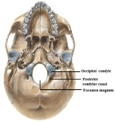
The foramen magnum is the largest bony foramen in the central basal region of the occipital bone. Occipital bone with the foramen magnum and the occipital condyles form the cranial aspect of the craniovertebral junction (Fig.1). Bony malformations at the craniovertebral junction may lead to symptoms secondary to compression of vital structures or may manifest as instability due to malalignment of bones1. Therefore it is of great importance to study the dimensions of foramen magnum and occipital condyles.
The posterior part of the cranial base is largely formed by the occipital bone. The occipital bone is trapezoid, concave internally and invests the foramen magnum. It consists of four parts namely the basilar or basioccipital part, squamous part and two lateral or condylar parts. The basilar part is quadrilateral in shape and lies in front of foramen magnum. The squamous part is an expanded plate and lies posterosuperior to the foramen magnum and the two lateral or condylar or exoccipital parts lie on each side of the foramen magnum.55,14
2
Foramen magnum is unpaired, oval and oriented obliquely. The anteroposterior diameter of the foramen magnum is more than the transverse diameter. The anterior margin of the foramen magnum is encroached on each side by the occipital condyles which project down to articulate with the superior articular facets of the atlas. Anterior and posterior atlanto occipital membranes are attached to the corresponding margins of the foramen magnum.
The structures adjacent to the foramen magnum are the bilateral occipital condyles, jugular foramina, mastoid notches, squamous parts of the occipital bone, hypoglossal canals (anterior condylar canal) and posterior condylar canals. The posterior cranial fossa communicates with the vertebral canal through the foramen magnum.
The following structures traverse through the foramen magnum:
Anteriorly, the upper surface of the basilar part of the foramen magnum gives attachment to apical ligament of dens and membrana tectoria which is the upward prolongation of the posterior longitudinal ligament.
Its wider posterior part transmits the lower end of medulla oblongata which continues down as the spinal cord.
Fig.2. Internal surface of Base of skull
3 Anterior and posterior spinal arteries Spinal accessory nerve
Upper three cervical meningeal nerves
OCCIPITAL CONDYLE
The occipital condyles are oval or reniform in shape, with their long axes converging anteromedially. On the medial aspect of each condyle, a tubercle for the alar or check ligament is present.25
The anterior one third of each condyle extends forwards on to the basilar part of the bone. The site of union between the basilar and condylar parts is marked by the anterior condylar or hypoglossal canal (Fig.2). The hypoglossal canal is directed laterally and slightly forwards, and transmits the hypoglossal nerve, a meningeal branch of the ascending pharyngeal artery and an emissary vein. Behind each condyle there is a condylar fossa. In some cases there is a posterior condylar canal which transmits the emissary vein.
4
BASILAR PART
The basilar or basioccipital part is a bar of bone that extends upwards and forwards from the foramen magnum and fuses with the basisphenoid. Its internal or cerebral surface is concave from side to side. It supports pons and medulla.
In the external surface, pharyngeal tubercle is present which gives attachment to the fibrous pharyngeal raphe.
BLOOD SUPPLY OF FORAMEN MAGNUM
5
AIM OF THE STUDY
The Foramen Magnum (FM) is an oval shaped opening situated at the base of the skull. The surgery for craniovertebral junction anomalies and skull base tumors at FM, poses a challenge for neurosurgeons. Understanding the bony anatomy of FM is essential for any surgery at the craniovertebral junction for safeguarding the vital structures. The primary goal of FM surgery is to decompress the vital neural structures without compromising their function and craniovertebral stability.21
Craniovertebral junction abnormalities can be broadly classified as congenital, developmental, acquired, tumors, infective, inflammatory or traumatic. Meningiomas are the most common primary skull base tumour. About 40% to 50% of meningiomas involves skull base. The incidence of skull base meningioma is 2 per 100,000 per year. The male to female ratio is 1:2.2 in patients aged from 12 to 81 years.60 FM meningioma mostly presents on the anterior margin of FM. It can be diagnosed by CT scan and confirmed by MRI scan. Innovative skull base approaches are now practised to achieve total surgical removal of basal meningiomas.
6
far-lateral, posterolateral or extreme lateral approach.60 In the far-lateral approach craniovertebral stability is not affected due to minimal removal of occipital condyles. It also provides an adequate exposure to ventral brainstem. Many varieties of lateral approaches have been reported including transfacetal approach, partial or complete transcondylar approach, extreme lateral transjugular approach and transtubercular approach.60,28
Hence, neurosurgeons performing posterior or lateral approaches to Craniovertebral junction surgery should be familiar with the normal anatomy and possible variations of the foramen magnum, occipital condyle and hypoglossal canal to reduce the surgical morbidity.
The aim of the present study is to analyse the FM and occipital condyles morphologically and morphometrically. Hopefully the data will be beneficial to neurosurgeons, radiologists and orthopaedicians for preoperative planning and management of Craniovertebral junction surgeries.
The parameters studied are :
1. Shape of the foramen magnum
2. Maximum anteroposterior diameter of the foramen magnum 3. Maximum transverse diameter of the foramen magnum
7 5. Length of the right occipital condyle
6. Maximum width of the right occipital condyle 7. Minimum width of the right occipital condyle 8. Length of the left occipital condyle
9. Maximum width of the left occipital condyle 10. Minimum width of the left occipital condyle 11. Bicondylar distance
12. Anterior intercondylar distance 13. Posterior intercondylar distance 14. Presence of Posterior condylar canal
15. Presence of Septum of the hypoglossal canal
16. Distance between intracranial edge of right hypoglossal canal and anterior margin of right occipital condyle
17. Distance between intracranial edge of right hypoglossal canal and posterior margin of right occipital condyle
18. Distance between intracranial edge of left hypoglossal canal and anterior margin of left occipital condyle
8
REVIEW OF LITERATURE
1) SHAPE OF THE FORAMEN MAGNUM
Khalil Awadh Murshed et al 30(2003) in their study of spiral CT scan of the FM of 110 normal subjects reported that the FM shape was oval in 8.1%, egg shaped in 6.3%, round 21.8%, pentagonal in 13.6%, tetragonal in 12.7%, irregular type(B) in 9.09%.hexagonal in 17.2%, and irregular type(A) in10.9%.
Muthukumar N et al 35(2005) in their study of 50 dry skulls, stated that the FM was found to have round shape in 46%.
P. Chethan et al 41(2011) in their study of 53 skulls, observed that the FM was round shaped in 22.6%, egg shaped in 18.9%, oval in 15.1%, irregular in 15.1%, tetragonal in 18.9%, hexagonal in 5.6% and pentagonal in 3.8% of the cases.
Emel AVCL et al 9(2011) in their study of 30 skulls, found that the FM was oval in 58%.
9
Radhakrishnan P et al 45(2012) in their study of 250 Cranial CT of normal subjects between the ages of 18 and 80 years, stated that the shape of the FM was oval in 35.2%, hexagonal in 24.8%, round in 7.6%, trigonal in 1.6%, pentagonal in 12.4%, tetragonal in 6.8% and irregular in 11.6%.
Gobbur et al 19(2013) in their study of cranial CT of 150 subjects, stated that the FM was found to be round in 40% and oval in 30%.
K. Natsis et al 31(2013) in their study of 143 skulls, reported that the shape of the FM was two semicircles in 25.9%.It was pear shaped in 22.4%, egg shaped in 21%, oval in14.7%, rhomboid in14%, round in 1.4% and irregular in 0.7%.
Jose Aderval Aragao et al 26 (2014) studied about 110 adult dry skulls and observed that the shape of FM was pear in 37%, oval in 5.45%, tetragonal in 10.91%, pentagonal in 2.73%, round in 15.45%, hexagonal in 9.09%, heptagonal in 1.82% , biconvex in 10.91% and irregular in 6.36% of subjects.
2) MAXIMUM ANTEROPOSTERIOR DIAMETER OF THE FORAMEN MAGNUM (FM)
10
Wanebo et al 58(2001) studied about 32 dry skulls and reported that the mean AP diameter of the FM was 36mm.
Khalil Awadh Murshed et al 30(2003) in their study of about 110 cranial CT scans, found that the mean AP diameter of the FM in male and female was 37.2±3.4 and 34.6±3.16 respectively.
Muthukumar. N et al 35(2005) studied about 50 adult dry skulls. They found that the maximum AP diameter of FM was 33.3 mm.
Emine et al 10(2006) studied 59 adult dry skulls and reported that the AP diameter of the FM was in between 29.7mm and 39.7mm with an average of 34.8±2.2mm.
Manoel. C et al 33(2009) in their study of 215 (139 males and 76 females) adult human dry skulls reported that the mean AP diameter of the FM of male and female were 35.7±0.29 mm and 35.1± 0.33 mm respectively.
11
Philipp Gruber et al 43(2009) studied about 111 adult dry skulls and they reported that the mean AP diameter was in the range of 30.1mm to 42.6mm with an average of 36.6mm.
Fatma Hayat Eridil et al 13(2010) studied 54 cranial CT scans and the mean AP diameter of the FM was reported as 35.58±4mm.
P. Chethan et al 41 2011) studied about 53 dry skulls and reported that the mean AP was 31±2.4mm.
Emel AVCL et al 9(2011) in their study of 30 adult dry skulls, recorded the mean AP diameter of the FM as 34.5mm.
Ukoha U et al 57(2011) reported the mean AP diameter of the FM of male and female as 36.26mm and 34.39 mm respectively.
F.Burdan et al 11(2012) observed the mean AP diameter of the FM of male and female as 37.06mm and 35.57 mm respectively in 313 CT scans.
12
Gautam Kanodia et al 17(2012) studied about 100 adult dry skulls and 100 CT scans of posterior fossa. They reported that the mean AP diameter of the FM was 34.1±0.29mm in dry skull group and 33.1±0.35 in CT scan.
Osunwoke E.A et al 38(2012) studied 120 adult human dry skulls and the mean AP diameter of the FM was reported as 36.11±0.24mm.
Radhakrishnan S.K et al 46(2012) studied 100 adult human dry skulls, and reported that the mean AP diameter of the FM of male and female were 34.04±2.36mm and 31.72±2.14mm respectively with an average of 32.88mm.
Radhakrishnan P et al 35(2012) in their study of 250 Cranial CT of normal subjects between the ages of 18 and 80years observed that the AP diameter of FM was in the range of 25.8mm to 45.9mm with the average of 35.76±3.4mm.
13
Fathy Ahmed Fetouh et al 12(2013) studied about 100 adult dry human skulls and recorded that the AP diameter of FM was in the range of 31mm to 40.2mm with the average of 34.94mm.
K. Natasis et al 31(2013) in their study of 143 adult human dry skulls, reported that the mean AP diameter of the FM was 35.53±3.06mm.
Shanthi CH et al 52(2013) studied about 100 adult human dry skulls and reported that the mean AP diameter of the FM of male and female were 37.1mm and 33.8mm respectively.
S.K.Jain et al 53(2013) in their study of 68 skulls, reported that the mean AP diameter of the FM of male and female were 36.9±0.2mm and 32.9±0.3mm respectively.
Surwase Ramdas Gopal rao et al 54(2013) in their study of 100 cranial C.T. scans reported that the mean AP diameter of the FM of male and female were 33.9±2.61mm and 32.35±3.16mm respectively.
14
Yogesh Yadav et al 59(2014) in their study of 96 skulls, reported that the mean AP diameter of the FM of male and female were 35.22± 2.17mm and 33.1±2.04mm respectively.
3) MAXIMUM TRANSVERSE DIAMETER OF THE FORAMEN MAGNUM (FM)
Georges Olivier 18(1975) studied about 125 adult human dry skulls and reported that the mean transverse diameter of the FM was 30.34mm.
Wanebo et al 58(2001) studied about 32 dry skulls and reported that the mean transverse diameter of the FM was 31mm.
Khalil Awadh Murshed et al 30(2003) in their study of cranial CT of 110 normal subjects between the age of 18 and 80 years, observed that the mean transverse diameter FM in male and female were 31.6±2.99mm and 29.3.±2.19mm respectively.
Muthukumar N et al 35(2005) studied about 50 adult human dry skulls. They found that the maximum transverse diameter of FM was 27.9 mm.
15
Manoel. C et al 33 (2009) in their study of 215 (139 males and 76 females) adult dry skulls reported that the mean transverse diameter of the FM of male and female were 30.3±0.2 mm and 29.4± 0.23 mm respectively.
Ivan Claudio Suazo Galdames et al 22(2009) studied 211 skulls and concluded that the mean transverse diameter of the FM of male and female were 30.6 mm and 29.5 mm respectively.
Philip Gruber et al 43(2009) studied about 111 adult human dry skulls and reported that the mean transverse diameter of the FM was in the range of 25.0mm.to 38.9mm with an average of 31.1mm.
Fatma Hayat Eridil et al 13(2010) in their study of 54 cranial CT scans stated that the mean transverse diameter of the FM was 29.84mm.
P. Chethan1 et al 41(2011) studied about 53 dry skulls and reported that the mean transverse diameter was 25.2±2.4mm.
Emel AVCL et al 9(2011) in their study of 30 adult dry skulls, reported that the mean transverse diameter of FM was 29mm.
16
Gagandeep Singh et al 15(2012) studied about 50 skulls (26 males and 24 females). They reported that the mean transverse diameter of the FM of male and female were 27.77mm and 27.21mm respectively.
Gautam Kanodia et al 17(2012) studied about 100 adult dry human skulls and 100 CT scans of posterior fossa. They reported that the mean transverse diameter of the FM was 27.5±o.25mm in dry skull group and 27.6±0.31 in CT scan.
Osunwoke E.A et al 38(2012) in their study of 120 human dry skulls, reported that the mean transverse diameter of the FM was 29.65±0.24mm.
F.Burdan et al 11(2012) in their study of the mean transverse diameter of the FM, reported that the values for male and female were 30.95mm and 32.98 mm respectively in 313 CT scans.
Radhakrishnan S.K et al 46(2012) studied 100 adult human dry skulls. They reported that the mean transverse diameter of the FM of male and female were 28.63±1.89mm and 25.59±1.64mm respectively.
17
A.T.Uthman et al 3(2012)) in their study of 88 cranial CT scans reported that the mean transverse diameter of the FM of male 24mm.to 34.8mm with an average of 29.5mm and that of female was in between 22.3mm and 31.8mm with an average of 27.3mm.
K. Natasis et al 31(2013) in their study of 143 adult human dry skulls, observed that the mean transverse diameter of the FM was 30.31±2.79mm.
Shanthi CH et al 52(2013) studied about 100 adult human dry skulls and reported that the mean transverse diameter of the FM of male and female were 32.0mm and 30.4mm respectively.
S.K.Jain et al 5 (2013) in their study of 68 skulls, reported that the mean transverse diameter of the FM of male and female were 31.5±0.27mm and 29.5±0.28mm respectively.
Surwase Ramdas Gopalrao et al 54(2013) in their study of 100 cranial CT scans reported that the mean transverse diameter of the FM of male was 28.05±2.22mm and that of female was 26.88±2.96mm.
18
Yogesh Yadav et al 59(2014) in their study of 96 skulls, reported that the mean transverse diameter of the FM of male and female were 27.6±2.26mm and 26.71±1.76mm respectively.
4) PROTRUSION OF OCCIPITAL CONDYLE (OC) INTO THE FORAMEN MAGNUM.
Muthukumar N et al 35(2005) in their study of 50 dry skulls, observed that the OC protrude into the FM in 20% of adult dry skulls.
P. Chethan1 et al 41(2011) studied about 53 skulls and found that the OC protruded into the FM in 20.7% of skulls.
Emel AVCL et al 9(2011) in their study of 30 adult dry skulls, observed that the OC protruded into the FM in 57% of skulls.
5) LENGTH OF THE RIGHT OCCIPITAL CONDYLE (ROC)
Georges Olivier 18(1975) studied about 125 adult human dry skulls and stated that the mean length of the ROC was 23.75mm.
19
Muthukumar N et al 35(2005) studied about 50 adult human dry skulls. They found that the mean length of the ROC was 23.6mm.
Sait Naderi et al 49(2005) in their study of 202 adult human dry skulls, found that the length of the ROC was 23.6mm.
Emine et al 10(2006) studied about 59 human adult dry skulls and reported that the length of the ROC was in the range of 19.7mm to 30.7mm with an average of 24.4±2.2mm.
Nehi’r Barut et al 36(2009) studied about 56 dry human skulls. They found that the mean length of the OC was 23.1mm.
Emel AVCL et al 9(2011) in their study of 30 adult dry skulls, reported that the mean maximum length of ROC was in the range of 18.2mm to 28.7mm with an average of 23.7±2.6mm.
J.T.Hong et al 27(2011) studied 13 frozen cadaveric specimens and reported that the mean length of OC was 22.9±2.5mm.
Mehmet Asim Ozer et al 34(2011) studied 144 adult dry skulls and recorded that the length of ROC was 23.9±3.4mm.
20
Tien V et al 56(2011) in their study of 170 cranial CT scans reported that the mean length of ROC was 22.2±2.1mm.
Pereira G.A et al 42(2012) in their study of 111 adult human dry skulls found that the mean length of the ROC was 24±3.6mm.
Fathy Ahmed Fetouh et al 12(2013)studied about 100 adult dry human skulls and recorded that the mean maximum length of ROC varied from 18mm to 31mm with an average of 23.5mm.
K. Natasis et al 31(2013) in their study of 143 adult human dry skulls found that the length of ROC was 25.60±2.91mm.
Pooja Gangrade et al 44(2013) )studied 100 adult dry skulls and recorded that the mean length of ROC of male and female was 25.55mm and 23.1mm respectively.
Bello S.S et al 4(2013) studied about 240 cranial CT scans and reported that the mean length of ROC was 23.5±2.7mm.
21
6) MAXIMUM WIDTH OF THE RIGHT OCCIPITAL CONDYLE Georges Olivier 18(1975) studied about 125 adult human dry skulls and reported that mean width of the ROC was 11.5mm.
Daniel J et al 7(2001) in their study of 522 adult dry skulls, reported that the mean maximum width of ROC of black male and female were 12.8mm and 12mm respectively and white male and female were 12.3mm and 11.7mm respectively.
Muthukumar N et al 35(2005) studied about 50 adult human dry skulls. They found that the mean width of the ROC was 14.72mm.
Sait Naderi et al 49(2005) in their study of 202 adult human dry skulls, reported that the width of the ROC was 10.6mm.
Emine et al 10(2006) studied about 59 human dry skulls and reported that the width of the ROC varied from 10.3mm to 16.9mm with an average of 13±1.5mm.
Emel AVCL et al 9(2011) in their study of 30 adult dry skulls, reported that the maximum width of ROC was in the range of 9 mm to 14.5 mm with an average of 12.2±1.2mm.
22
Mehmet Asim Ozer et al 34(2011) studied 144 adult dry skulls and recorded that the width of ROC was 11.9±2.3mm.
Tien V et al 56(2011) in their study of 170 cranial CTscans reported that the mean width of ROC was 11.2±1.4mm.
di Vasudha V. Saralaya et al 8(2012)studied about 70 adult human dry skulls and reported that the mean width of the ROC was 11.26mm.
Pereira G.A et al 42(2012) in their study of 111 adult human dry skulls, reported that the mean width of the ROC was 13.4±1.4mm.
Fathy Ahmed Fetouh et al 12(2013) studied about 100 adult dry human skulls and recorded that the mean maximum width of ROC varies from 9.5mm to 18mm with an average of 13.58mm.
Bello S.S et al 44(2013) studied about 240 cranial CT scans and reported that the mean width of ROC was 12.8±1.7mm.
Parvindokht Bayat et al 39(2014) in their study of 50 adult dry skulls, reported that the mean maximum width of ROC ranged from 6mm to 13mm with an average of 9.21±1.97mm.
23
7) MINIMUM WIDTH OF THE RIGHT OCCIPITAL CONDYLE K. Natasis et al 31(2013) in their study of 143 adult human dry skulls, found that the minimum width of ROC was 5.71±1.61mm.
8) LENGTH OF THE LEFT OCCIPITAL CONDYLE (LOC)
Sait Naderi et al 49(2005) in their study of 202 human dry skulls, reported that the length of the LOC was 23.2mm.
Emine et al 10(2006) studied about 59 human dry skulls and stated that the length of the LOC was in the range of 18.2mm to 31.1mm with an average of 24.6±2.5mm.
Emel AVCL et al 9(2011) in their study of 30 adult dry skulls reported that the maximum length of LOC was in the range of 18.8 mm to 30.9mm with an average of 24.7±2.7mm.
Mehmet Asim Ozer et al 34(2011) studied 144 adult dry skulls and recorded that the length of LOC was 23.92±3.3mm.
Tien V et al 56(2011) in their study of 170 cranial CT scans reported that the mean length of LOC was 22.5±2.2mm.
24
Bello S.S et al 4(2013) studied about 240 cranial CT scans and reported that the LOC mean length was 23.7±2.8mm.
Fathy Ahmed Fetouh et al 12(2013) studied about 100 adult dry skulls and recorded that the mean maximum length of LOC was in the range of 18.3mm to 29.4mm with an average of 23.75mm.
K. Natasis et al 31(2013) in their study of 143 adult human dry skulls, found that the length of LOC was 25.60±2.70mm.
Pooja Gangrade et al 44(2013) studied 100 adult dry skulls and recorded that the mean length of LOC of male and female were 26.12mm and 22.18mm respectively.
Parvindokht Bayat et al 39(2014) in their study of 50 adult dry skulls, reported that the mean maximum length of LOC varied from 10mm to 26mm with an average of 19.28±3.57mm.
9) MAXIMUM WIDTH OF THE LEFT OCCIPITAL CONDYLE
25
Emine et al 10(2006) studied about 59 human dry skulls and reported that the width of the LOC was from 10.1mm to 17.2mm with an average of 13±1.5mm.
Emel AVCL et al 9(2011) in their study of 30 dry skulls reported that the maximum width of LOC was in the range of 9.3 mm to 15.3 mm with an average of 12.4±1.5mm.
Mehmet Asim Ozer et al 34(2011) studied 144 adult dry skulls and recorded that the width of LOC was 10.7±2.3mm.
Tien V et al 56(2011) in their study of 170 cranial CT scans reported that the mean width of LOC was 11.2±1.5mm.
Pereira G.A et al 42(2012) in their study of 111 adult human dry skulls reported that the mean width of the LOC was 16.4±1.6mm.
Bello S.S et al 4(2013) studied about 240 cranial CT scans and reported that the mean width of LOC was 12.7± 1.4mm.
26
K. Natasis et al 31(2013) in their study of 143 adult human dry skulls, found that the maximum width of LOC was 13.01±1.98mm.
Parvindokht Bayat et al 39(2014) in their study of 50 adult dry skulls, reported that the mean maximum width of LOC varied from 6mm to 13mm with an average of 9.40±1.8mm.
10) MINIMUM WIDTH OF THE LEFT OCCIPITAL CONDYLE K. Natsis et al 31(2013) in their study of 143 adult human dry skulls, found that the minimum width of LOC was 6.25±1.76mm.
11) BICONDYLAR DISTANCE (BCD)
Daniel J et al 7(2001) in their study of 522 adult dry skulls, reported that the BCD of black male and female were 49.6mm and 47.3mm respectively and white male and female were 51.9mm and 49.8mm respectively.
27
12) ANTERIOR INTERCONDYLAR DISTANCE (AICD)
Daniel J et al 7(2001)in their study of 522 adult dry skulls, reported that the AICD of black male and female were 20.1mm and 18.6mm respectively and white male and female were 20.9mm and 19.2mm respectively.
Aynur Emine Cicekcibasi et al 2(2004) studied about 60 skulls (34 male and 26 female). They reported that the AICD of male and female were 16.09±1.93mm and 14.68±1.80mm respectively.
Sati Naderi et al 49(2005) in their study of 202 adult human dry skulls, reported that the AICD was 21.0mm.
Emine et al 10(2006) studied about 59 human adult dry skulls and reported that the AICD was in between 15mm and 32mm with an average of 22.6±3.9mm.
Mehmet AsimOzer et al 34(2011) studied 144 adult dry skulls and reported that the mean AICD was 20.9 ±3.6mm.
di Vasudha V. Saralaya et al 8(2012) reported that the mean AICD was 18.7mm.
28
Fathy Ahmed Fetouh et al 12(2013) studied about 100 adult dry human skulls and recorded that the AICD varied from 11.5mm to 25.5mm with an average of 20.64mm.
K. Natasis et al 31(2013) in their study of 143 adult human dry skulls, found that the mean AICD was 19.30±3.25mm.
Pooja Gangrade et al 44(2013) studied 100 adult dry skulls and recorded that the mean AICD of male and female were 18.32mm and 15.44mm respectively.
Parvindokht Bayat et al 39(2014) in their study of 50 adult dry skulls, reported that the AICD was in the range of 2mm to 42mm with an average of 15.39±7.99mm.
13) POSTERIOR INTERCONDYLAR DISTANCE (PICD)
Sait Naderi et al 49(2005) in their study of 202 adult human dry skulls, reported that the PICD was in the range of 35.1mm to 48.3mm with an average of 41.6mm.
29
di Vasudha V. Saralaya et al 8(2012) studied about 70 adult human dry skulls.They reported that the mean PICD was 38.7mm.
Gagandeep Singh et al 15(2012) studied about 50 skulls (26 male and 24 female). They reported that the maximum intercondylar distance of the FM of male and female were 26.15mm and 24.71mm respectively.
Fathy Ahmed Fetouh 12(2013) studied about 100 adult dry skulls and recorded that the PICD varied from 35.5mm to 48.5mm with an average of 41.4mm.
K. Natasis et al 31(2013) in their study of 143 dry skulls, found that the mean PICD was 51.61±5.01mm.
ParvindokhtBayat et al 39(2014) in their study of 50 adult dry skulls, reported that the PICD varied from 13mm to 44mm with an average of 35.60±8.4mm.
14) PRESENCE OF POSTERIOR CONDYLAR CANAL (PCC)
30
K. Natsis et al 31(2013) in their study of 143 skulls, observed that the PCC was present in 75.5%, out of which 11.9% was present on right side, 16.1% on left side and 47.6% bilaterally.
Jatin Goda et al 24(2013) observed that the PCC was present bilaterally in 70.31% and unilaterally in 20.31 % of the 64 dry human skulls.
Ketu Chauhan et al 29(2013) studied about 82 dry human skulls and found that PCC was present in 6% on left side and bilaterally in 3.6%.
Parvindokht Bayat et al 39 (2014) in their study of 50 adult dry skulls, reported that the PCC was present in 4% on right side, 16% on left side and bilaterally in 40%.
15) PRESENCE OF SEPTUM OF THE HYPOGLOSSAL CANAL (HGC)
Muthukumar N et al 35(2005) studied 50 adult dry skulls. In 30%, HGC was divided into two compartments by a bony septum.
Nehi’r Barut et al 36(2009) studied about 56 adult dry human skulls. They found that 14 HGC (25%) were divided into two parts by a bony septum.
31
Jasbir Kaur et al 23(2012) found division of HGC in 10.5% of male and 9.1% of female adult human skulls.
Jatin Goda et al 24(2013) observed septum of HGC in 3.12% of the 64 human adult dry skulls studied.
K. Natsis et al 31(2013) studied 143 skulls and found that the HGC septum was present in 25.5%.
Roopali et al 48(2013) stated that the septum of HGC was present in 3% of the human dry skulls studied.
Singh Rajani 50(2013) found double condylar canal in 66 dry human skulls.
Siva N R S et al 51 (2013) reported duplicated HGC in 50 dried skulls.
16) DISTANCE BETWEEN INTRACRANIAL EDGE OF RIGHT HYPOGLOSSAL CANAL (RHGC) AND ANTERIOR MARGIN OF RIGHT OCCIPITAL CONDYLE (ROC)
32
Emine et al 10(2006) studied about 59 human dry skulls and reported that the distance of the intracranial end of the RHGC from the anterior margin of the ROC was in the range of 8.1mm to 16.9mm with an average of 11.0±1.6mm.
Pereira G.A et al 42(2012) in their study of 111 adult human dry skulls reported that the intracranial end of the RHGC from the anterior margin of the ROC was 11.0±1.8mm.
17) DISTANCE BETWEEN INTRACRANIAL EDGE OF RIGHT HYPOGLOSSAL CANAL (RHGC) AND POSTERIOR MARGIN OF RIGHT OCCIPITAL CONDYLE (ROC)
Muthukumar N et al 35(2005) studied about 50 adult human dry skulls. They found that the distance of the intracranial end of the RHGC from the posterior margin of the ROC was12.2mm.
33
Nehi’r Barut et al 36(2009) studied about 56 adult dry human skulls. They found that the distance between the intracranial edge of RHGC and posterior margin of ROC was 12.5mm.
Emel AVCL et al 9(2011) in their study of 30 adult dry skulls, reported that the distance between the RHGC and posterior border of ROC varied from 7.9 mm to 12.2mm with an average of 9.8±1.1mm.
Pereira G.A et al 42(2012) in their study of 111 adult human dry skulls reported that the distance of the intracranial end of the RHGC from the posterior margin of the ROC was 10.3±2.5mm.
18) DISTANCE BETWEEN INTRACRANIAL EDGE OF LEFT HYPOGLOSSAL (LHGC) CANAL AND ANTERIOR MARGIN OF LEFT OCCIPITAL CONDYLE (LOC)
Sait Naderi et al 49(2005) in their study of 202 adult human dry skulls, reported that the distance of the intracranial end of the LHGC from the anterior tip of the LOC was 9.6mm.
34
Pereira G.A et al 42(2012) in their study of 111 adult human dry skulls reported that the distance of intracranial end of the LHGC from the anterior margin of the LOC was 10.7±1.8mm.
19) DISTANCE BETWEEN INTRACRANIAL EDGE OF LEFT HYPOGLOSSAL CANAL (LHGC) AND POSTERIOR MARGIN OF LEFT OCCIPITAL CONDYLE (LOC)
Emel AVCL et al 9(2011) in their study of 30 adult dry skulls, reported that the distance between LHGC and Posterior border of LOC varied from 6.6 mm to 12.2 mm with an average of 9.9±1.4mm.
Emine et al 10(2006) studied about 59 human dry skulls and reported that the intracranial end of the LHGC from the posterior margin of the LOC was in between 8.4mm and 17.6mm with an average of 12.4±2.3mm.
Nehi’r Barut et al 36(2009) studied about 56 adult dry human skulls. They found that the distance between the intracranial edge of LHGC and posterior margin of LOC was 12.6mm.
Fig.3. Development of skull bones
Fig.4.The Occipital bone of newborn
35
EMBRYOLOGY
The skull consists of two major anatomical and functional components: the neurocranium and viscerocranium. The neurocranium forms a protective covering around the brain and viscerocranium forms the skeleton of the face.32
The skull develops from neural crest cells, cranial paraxial mesoderm and sclerotome(Fig.3). The neural crest cells form the whole viscerocranium and the rostral portion of neurocranium. The skull base is formed by neural crest rostral to the tip of the notochord and by sclerotome (mesoderm) in the notochordal region.
Neurocranium: It has two portions- membranous and cartilaginous portions.
Membranous neurocranium: It is derived from neural crest cells and paraxial mesoderm. The mesenchyme from both sources covers the brain and undergoes membranous ossification. It forms the cranial vault.
36
The cartilages that are present posterior to the rostral limit of notochord arise from occipital sclerotomes formed by paraxial mesoderm. The central region of occipital sclerotomes contribute to the parachordal cartilage, which enclose the notochord and extends as a flat plate on either side of it and forms the basioccipital component of the occipital bone.40 The exoccipital components chondrify and border the foramen magnum.
Roots of the hypoglossal nerve run between the parachordal and exoccipital cartilages. The fusion of exoccipital and parachordal components forms the foramina for hypoglossal nerve roots bilaterally.
OSSIFICATION
The occipital bone is a compound structure with respect to its origin and type of ossification (Fig.4).
37
of intauterine life, two centres each for the lateral or condylar or exoccipital parts appear. During sixth week one centre for the basilar part appears and it unites with the rest of the bone by sixth year.
The occipital bone is made up of four parts at birth – basilar, two lateral and a squamous part which fuse by cartilage and form a ring around foramen magnum. The squamous part is present posteriorly, the lateral or condylar parts are present on each side, and the basilar part or basiocciput is anterior. These names are retained for the parts of the adult bone also.25,16
38
MATERIALS AND METHODS
STUDY MATERIALS:
Hundred human adult dry skulls of unknown sex. 20 Computerized Tomographic Scan Images. Digital Vernier Calipers.
Flexible wire.
STUDY METHODS: 1. Dry skull Method 2. Radiological Study
SPECIMEN COLLECTION:
Hundred human adult dry skulls of unknown sex available in the Institute of Anatomy, Madras Medical College were used for this study. A. DRY SKULL METHOD:
Inclusion criteria:
1. Adult human dry skull of unknown sex. 2. Third molar tooth erupted.
3. Well defined skull sutures. Exclusion criteria:
Fig.5. Skull showing various shape of Foramen magnum
OVAL EGG SHAPE
PENTAGONAL HEXAGONAL
Fig 6.Protrusion of occipital condyle into the foramen magnum.
Fig 8.Presence of bilateral Posterior condylar canal
Figg. 10. Illustrations showing the measurements of parameters related to Foramen magnum and occipital condyle
1). Maximum AP diameter of the FM
2) Maximum transverse diameter of the FM
3) ROC length 4) ROC maximum width 5) ROC minimum width
6) LOC length 7) LOC maximum width 8) LOC minimum width
Fig.11 Photograph of digital vernier caliper
Fig.12.Maximum anteroposterior diameter of the Foramen magnum
Fig.13.Maximum transverse diameter of the Foramen magnum
Fig.15. Length of the left occipital condyle
Fig.17.Maximum width of the left occipital condyle
Fig.19.Minimum width of the left occipital condyle
Fig.21.Anterior intercondylar distance
Fig.23 Distance between intracranial edge of RHGC and anterior margin of ROC
Fig.25..Distance between intracranial edge of LHGC and anterior margin of LOC
39
The following morphological parameters were observed by gross examination.
1. Shape of the FM (Fig.5)
2. Protrusion of OC into the FM (Fig.6)
3. Presence of Posterior condylar canal (Fig.7 and 8) 4. Presence of Septum of the Hypoglossal canal. (Fig.9)
The following measurements (Fig.10) were made with the use of digital vernier calipers (Fig.11).
1) Maximum anteroposterior diameter of the FM:
Maximum distance between anterior and posterior margins measured along the midsagittal plane of the FM (Fig.12).
2) Maximum transverse diameter of the FM:
Maximum distance between the lateral margins measured along the transverse plane of the FM (Fig.13).
3) Length of the occipital condyle:
40
4) Maximum width of the occipital condyle :
Maximum width of the OC taken along the articular surface perpendicular to the OC length and the parameter is recorded bilaterally(Fig.16 and 17).
5) Minimum width of the occipital condyle:
Minimum width of the OC taken along the articular surface perpendicular to the OC length and the parameter is recorded bilaterally (Fig.18 and 19).
6) Bicondylar distance:
Maximum distance between the lateral margin of right and left condylar articular facets perpendicular to the midsagittal plane (Fig.20).
7) Anterior intercondylar distance:
Distance between the anterior tips of the right and left OC perpendicular to the midsagittal plane (Fig.21).
8) Posterior intercondylar distance:
41
9) Distance between intracranial edge of Hypoglossal canal and anterior margin of OC:
Distance between intracranial edge of HGC and anterior margin of the corresponding occipital condyle and the parameter is recorded bilaterally (Fig.23 and 24).
10) Distance between intracranial edge of Hypoglossal canal and posterior margin of the OC:
Distance between intracranial edge of HGC and posterior margin of the corresponding occipital condyle and the parameter is recorded bilaterally (Fig.25 and 26).
B. RADIOLOGICAL STUDY:
1. Adult Clinical 4 slice cranial CT scan.
The CT images from the archives of the Barnard Institute of Radiology attached to Rajiv Gandhi Government General Hospital and Research Institute were used for the study. Images of patients who had their CT pictures taken for various ailments in head and neck were used for analysis of foramen magnum.
Fig 27. C.T Scan image of base of skull showing Foramen magnum
42
The images were digitized and stored on the Picture Archiving Communication System which was later retrieved for measurement of parameters. The system was incorporated with image enhancement and manipulation tools. The software also had a sensitive measuring tool.
43
OBSERVATIONS
100 adult dry human skulls were studied and the observations were grouped under morphological and morphometric parameters.
TABLE.1 SHOWING THE PERCENTAGE OF FM OF DIFFERENT TYPES IN DRY SKULL
Sl. No Shape of the foramen magnum
Number of skulls N=100
Percentage
1) Oval 40 40%
2) Egg shape 22 22%
3) Round 13 13%
4) Pentagonal 3 3%
5) Hexagonal 7 7%
6) Others 15 15%
40%
22% 13%
3% 7%
15%
CHART 1: SHAPE OF FORAMEN MAGNUM
44
TABLE.2: ANTEROPOSTERIOR DIAMETER OF THE FORAMEN MAGNUM (FM) IN DRY SKULLS
STATISTICAL DATA AP DIAMETER OF FM IN
DRY SKUL (in mm)
No. of skulls 100
Minimum 24.64
Maximum 39.89
Mean 35.12
S.D 02.65
45
TABLE.3: MAXIMUM TRANSVERSE DIAMETER OF THE FORAMEN MAGNUM (FM) IN DRY SKULL
STATISTICAL DATA TRANSVERSE DIAMETER OF
FM IN DRY SKULLS (in mm)
No. of skulls 100
Minimum 24.01
Maximum 35.98
Mean 29.03
S.D 2.15
The whole range of values is shown in the histogram with a bell shaped curve below.
46
TABLE.4: MAXIMUM ANTEROPOSTERIOR DIAMETER OF THE FORAMEN MAGNUM IN CT SCAN
STATISTICAL DATA AP DIAMETER OF FM IN
CT SCAN (in mm)
No. of skulls 20
Minimum 33.13
Maximum 36.54
Mean 35.03
S.D 0.95
47
TABLE.5: MAXIMUM TRANSVERSE DIAMETER OF THE FORAMEN MAGNUM IN CT SCAN
STATISTICAL DATA TRANSVERSE DIAMETER OF
FM IN CT SCAN (in mm)
No. of skulls 20
Minimum 27.65
Maximum 28.04
Mean 28.79
S.D 1.17
The whole range of values is shown in the histogram with a bell shaped curve below.
48
TABLE.6: COMPARISON OF ANTEROPOSTERIOR AND
TRANSVERSE DIAMETER OF FM IN DRY SKULLS WITH RADIOLOGICAL STUDY
STATISTICAL DATA
ANTEROPOSTERIOR DIAMETER OF FM
TRANSVERSE DIAMETER OF FM
DRY
SKULL CT SCAN
DRY
SKULL CT SCAN
No. of skulls 100 20 100 20
Mean 35.12 35.03 29.03 28.79
S.D 02.65 0.95 2.15 1.17
49
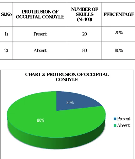
PRESENCE OF PROTRUSION OF OCCIPITAL CONDYLE
[image:81.595.108.538.210.717.2]Of the 100 skulls examined, protrusion of OC was found in 20 skulls and was absent in 80 skulls
TABLE:7 PROTRUSION OF THE OCCIPITAL CONDYLE
20%
80%
CHART 2: PROTRUSION OF OCCIPITAL CONDYLE
Present Absent
Sl.No PROTRUSION OF
OCCIPITAL CONDYLE
NUMBER OF SKULLS
(N=100)
PERCENTAGE
1) Present 20 20%
50
TABLE.8: LENGTH OF THE RIGHT OCCIPITAL CONDYLE(ROC)
STATISTICAL DATA ROC - LENGTH (in mm)
No. of skulls 100
Minimum 18.16
Maximum 32.68
Mean 23.85
S.D 2.12
51
TABLE.9: MAXIMUM WIDTH OF THE ROC
STATISTICAL DATA ROC - MAXIMUM WIDTH
(in mm)
No. of skulls 100
Minimum 9.76
Maximum 16.19
Mean 13.29
S.D ±1.36
52
TABLE 10: MINIMUM WIDTH OF THE ROC
STATISTICAL DATA ROC - MINIMUM WIDTH
(in mm )
No. of skulls 100
Minimum 3.25
Maximum 10.62
Mean 6.86
S.D ±1.34
53
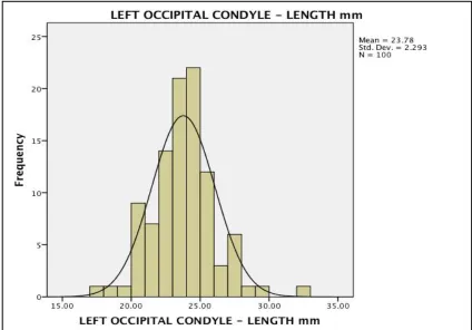
TABLE 11: LENGTH OF THE LEFT OCCIPITAL CONDYLE(LOC)
STATISTICAL DATA LOC - LENGTH
(in mm)
No. of skulls 100
Minimum 17.25
Maximum 32.02
Mean 23.77
S.D ±2.29
54
TABLE 12: MAXIMUM WIDTH OF THE LOC
STATISTICAL DATA LOC - MAXIMUM WIDTH
(in mm)
No. of skulls 100
Minimum 9.85
Maximum 16.78
Mean 13.44
S.D ±1.41
55
TABLE 13: MINIMUM WIDTH OF THE LOC
STATISTICAL DATA LOC - MINIMUM WIDTH
(in mm)
No. of skulls 100
Minimum 4.72
Maximum 10.32
Mean 7.04
S.D ±1.26
56
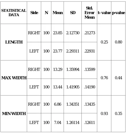
TABLE 14: COMPARISON BETWEEN MEAN LENGTH,
MAXIMUM WIDTH AND MINIMUM WIDTH OF ROC AND LOC OF DRY SKULLS ALONG WITH t AND p-VALUE
STATISTICAL
DATA Side N Mean SD
Std. Error Mean
t- value p-value
LENGTH
RIGHT 100 23.85 2.12730 .21273
0.25 0.80 LEFT 100 23.77 2.29311 .22931
MAX WIDTH
RIGHT 100 13.29 1.35994 .13599
0.76 0.44 LEFT 100 13.44 1.41905 .14190
MIN WIDTH
RIGHT 100 6.86 1.34351 .13435
0.93 0.35 LEFT 100 7.04 1.26114 .12611
‘p’ value ≤ 0.05 is considered to be significant
[image:88.595.106.547.180.646.2]57
TABLE 15: COMPARISON BETWEEN MEAN LENGTH AND MAXIMUM WIDTH OF ROC AND LOC OF CT SKULL
STATISTICAL DATA
ROC LOC
LENGTH BREATH LENGTH BREATH
No. of skulls 20 20 20 20
Minimum 22.12 11.34 22.23 11.43
Maximum 24.33 13.86 24.54 13.98
Mean 23.11 12.92 23.20 12.88
S.D 0.73 0.65 0.74 0.69
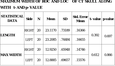
TABLE 16: COMPARISON BETWEEN MEAN LENGTH, AND MAXIMUM WIDTH OF ROC AND LOC OF CT SKULL ALONG WITH t- AND p- VALUE
STATISTICAL
DATA Side N Mean SD
Std. Error
Mean t- value p-value
LENGTH
RIGHT 20 23.1170 .73189 .16366
0.392 0.697
LEFT 20 23.2085 .74484 .16655
MAX WIDTH
RIGHT 20 12.9250 .65948 .14746
0.612 0.866
LEFT 20 12.8885 .69657 .15576
‘p’ value ≤ 0.05 is considered to be significant
[image:89.595.107.550.412.669.2]58
TABLE 17: BICONDYLAR DISTANCE
STATISTICAL DATA BICONDYLAR DISTANCE
(in mm)
No. of skulls 100
Minimum 32.71
Maximum 53.75
Mean 47.23
S.D 3.10
The whole range of values is shown in the histogram with a bell shaped curve below.
59
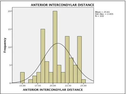
TABLE 18: ANTERIOR INTERCONDYLAR DISTANCE (AICD)
STATISTICAL DATA AICD (in mm)
No. of skulls 100
Minimum 14.87
Maximum 25.16
Mean 20.81
S.D 2.40
The whole range of values is shown in the histogram with a bell shaped curve below.
60
TABLE 19: POSTERIOR INTERCONDYLAR DISTANCE (PICD)
STATISTICAL DATA PICD (in mm)
No. of skulls 100
Minimum 38.02
Maximum 45.43
Mean 41.97
S.D 1.67
61
PRESENCE OF POSTERIOR CONDYLAR CANAL:
Of the 100 skulls examined, posterior condylar canal was found on the right side in 40 skulls and absent in 60 skulls. On the left side posterior condylar canal was found in 49 skulls and absent in 51 skulls and it was present bilaterally in 33skulls.
TABLE 20: SHOWING THE INCIDENCE OF PCC
SL.NO POSTERIOR CONDYLAR
CANAL (N=100) PERCENTAGE
1) Present
Right 40 40%
Left 49 49%
2) Absent Right 60 60%
Left 51 51%
0% 10% 20% 30% 40% 50% 60% Presence of posterior condylar canal Absence of posterior condylar canal Right 40% 60%
Left 49% 51%
40%
60%
49% 51%
62
PRESENCE OF SEPTUM OF THE HYPOGLOSSAL CANAL:
Of the 100 skulls examined, HGC septum was found on the right side in 10 skulls and absent in 90 skulls and on the left side it was found in 20 skulls and absent in 80 skulls. Out of 100 skulls examined HGC septum was found in 24% and absent in 76 %.
TABLE 21: SHOWING THE INCIDENCE OF HGC SEPTUM
SL.NO HYPOGLOSSAL CANAL
SEPTUM (N=100) PERCENTAGE
1) Present
Right 10 10%
Left 20 20%
2) Absent
Right 90 90%
Left 80 80%
0% 10% 20% 30% 40% 50% 60% 70% 80% 90% Presence of septation Absence of septation Right 10% 90%
Left 20% 80%
10%
90%
20%
80%
63
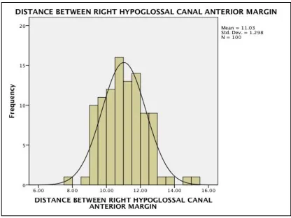
TABLE 22: DISTANCE BETWEEN INTRACRANIAL EDGE OF RIGHT HGC (RHGC) AND ANTERIOR MARGIN OF ROC
STATISTICAL DATA
DISTANCE BETWEEN RHGC AND ANTERIOR MARGIN OF ROC(in mm)
No. of skulls 100
Minimum 7.51
Maximum 15.25
Mean 11.02
S.D 1.29
[image:95.595.107.523.448.760.2]64
TABLE 23: DISTANCE BETWEEN INTRACRANIAL EDGE OF RIGHT HGC (RHGC) AND POSTERIOR MARGIN OF ROC.
STATISTICAL DATA
DISTANCE BETWEEN RHGC AND POSTERIOR MARGIN OF ROC (in mm)
No. of skulls 100
Minimum 9.8
Maximum 14.90
Mean 12.27
S.D 0.6
[image:96.595.117.526.453.757.2]65
TABLE 24: DISTANCE BETWEEN INTRACRANIAL EDGE OF LEFT HGC (LHGC) AND ANTERIOR MARGIN OF LOC.
STATISTICAL DATA
DISTANCE BETWEEN LHGC AND ANTERIOR MARGIN OF LOC(in mm)
No. of skulls 100
Minimum 6.81
Maximum 15.18
Mean 10.93
S.D 1.3
[image:97.595.113.533.440.723.2]66
TABLE 25: DISTANCE BETWEEN INTRACRANIAL EDGE OF LEFT HGC (LHGC) AND POSTERIOR MARGIN OF LOC.
STATISTICAL DATA
DISTANCE BETWEEN LHGC AND POSTERIOR MARGIN OF LOC (in mm)
No. of skulls 100
Minimum 9.6
Maximum 14.5
Mean 12.26
S.D 0.59
The whole range of values is shown in the histogram with a bell shaped curve below.
[image:98.595.110.533.431.720.2]67
TABLE 26: COMPARISON OF THE DISTANCE BETWEEN HGC (RIGHT AND LEFT) AND ANTERIOR MARGIN AND DISTANCE BETWEEN HGC (RIGHT AND LEFT) AND POSTERIOR MARGIN OF ROC AND LOC OF DRY SKULL ALONG WITH t - VALUE AND p - VALUE.
STATISTICAL
DATA SIDE N Mean SD t- value p-value
Distance between HGC
and anterior margin
RIGHT 100 11.02 1.29794
0.537 0.592 LEFT 100 10.93 1.30521
Distance between HGC
and posterior margin
RIGHT 100 12.26 0.59966
0.126 0.900 LEFT 100 12.25 0.59853
‘p’ value ≤ 0.05 is considered to be significant
[image:99.595.106.553.248.583.2]68
DISCUSSION
The findings of the present study were correlated with the findings of other similar studies conducted in different parts of India and in other countries.
1) SHAPE OF THE FORAMEN MAGNUM
Muthukumar N et al 35(2005) reported that the FM was ovoid in 46%.
P. Chethan et al 41(2011) recorded that the FM was observed to be round in 22.6%, tetragonal in 18.9%, oval in 15.1%, egg shaped in 18.9%, pentagonal in 3.8% , irregular in 15.1%, and hexagonal in 5.6% of the cases.
Emel AVCL et al 9(2011) stated that the FM was oval in 58%.
Radhakrishnan S.K et al 46(2012) reported that the FM was oval in 39%, round in 28%, pentagonal in 14% and tetragonal in 19%of the cases.
Radhakrishnan P et al 45(2012) reported that the FM was oval in 35.2%, hexagonal in 24.8%, pentagonal in 12.4%,round in 7.6%,irregular in 11.6% ,trigonal in 1.6%, pentagonal in 12.4%and tetragonal in 6.8% in cranial CT.
69
Khalil Awadh Murshed et al 30 (2003)studied CT images of the FM and recorded that the FM was oval in 8.1%, egg shaped in 6.3%, round in 21.8%, pentagonal in 13.6%, irregular (type A) in 10.9%,hexagonal in17.2%, tetragonal in 12.7%, and irregular (type B) in 9.09%.
Gobbur et al 19 (2013) reported that the FM was round in 40% and oval in30% in CT images.
Comparison was done with various studies showing the shape of the FM and was tabulated.The most common shape of the FM was oval. The present study also showed that the FM was oval in 40% and egg shaped in 22%.
70
TABLE 27: THE INCIDENCE OF FM OF DIFFERENT TYPES IN DRY SKULLS
Sl.NO STUDY
YEAR OF STUDY
SHAPEOF THE FORAMEN MAGNUM O val E gg s h ap e R ou n d P en tagon a l H exagon al O th er s
1) P.Chethan et al 2011 15.1 84.9
2) Emel AVCL et al 2011 58 42
3) Radhakrishnan S.K et al 2012 39 28 14 19
4) Present study 2014 40 22 13 3 7 15
0% 10% 20% 30% 40% 50% 60%
P.Chethan et al Emel AVCL et
al
Radhakrishnan SK et al
Present study 15%
58.00%
39% 40%
CHART 5
:
SHAPE OF THE FORAMEN MAGNUM IN DRYSKULLS
[image:103.595.108.559.172.748.2]71
2) MAXIMUM ANTEROPOSTERIOR DIAMETER OF THE FORAMEN MAGNUM (FM)
Georges Olivier et al 18(1975) reported that the mean AP diameter of the FMwas 35.7mm.
Manoel. C et al 33(2009) stated that the mean AP diameter of the FM of male and female were 35.7±0.29 mm and 35.1± 0.33 mm respectively.
Philipp Gruber et al 43(2009) recorded that the mean AP diameter ranged from 30.1mm.to 42.6mm with an average of 36.6mm.
Fatma Hayat Eridil et al 13(2010) stated that the mean AP diameter of the FM was 35±5.8mm in CT scans.
Emel AVCL et al 9(2011) found that the mean AP diameter of the FM was 34.5mm.
F.Burdan et al 11(2012) recorded the mean AP diameter of the FM in male and female were 37.06mm and 35.57 mm respectively in CT scans.
Gautam Kanodia et al 17(2012) concluded that the mean AP diameter of the FM was 34.1±0.29mm in dry skull group and 33.1±0.35mm in CT scan.
72
Radhakrishnan P et al 45(2012) concluded that the AP diameter of FM varied from 25.8mm to 45.9mm with the average of 35.76±3.4mm in cranial CT scans.
Fathy Ahmed Fetouh et al 12(2013) recorded that the AP diameter of FM varied from 31mm to 40.2mm with the average of 34.94mm.
K. Natasis et al 31 (2013) recorded that the mean AP diameter of the FM was 35.53±3.06mm.
Yogesh Yadav et al 59(2014) reported that the mean AP diameter of the FM of male and female were 35.22± 2.17mm and 33.1±2.04mm respectively.
In the Present study, the AP diameter of FM ranged from 24.64mm to 39.89mm with the average of 35.12±2.65mm. The mean AP diameter of the FM was compared with that found in various other studies and tabulated.
73
TABLE 28: COMPARISON OF AP DIAMETER OF THE FM IN DRY SKULLS
SL No STUDY YEAR OF
STUDY
AP DIAMETER OFFM IN DRY SKULLS
(in mm) 1) Georges Olivier et al 1975 35.70
2) Philipp Gruber et al 2009 36.60
3) Emel AVCL et al 2011 34.50
4) Osunwoke E.A et al 2012 36.11
5) K.Natasis et al 2013 35.53
6) Present Study 2014 35.12
30 31 32 33 34 35 36 37 35.70 36.60 34.50 36.11 35.53 35.12 CHART 6: AP DIAMETER OF THE FM IN DRY
[image:106.595.107.533.272.745.2]74
TABLE 29: COMPARISON OF AP DIAMETER OF THE FM IN CT SCAN IMAGES
Sl. No STUDY
YEAR OF STUDY
AP DIAMETER OF FM IN CT SCANS
(in mm)
1) Fatma et al 2010 35.58
2) Gautam K et al 2012 33.10
3) Radhakrishnan P et al 2012 35.76
4) Present study 2014 35.03
30 31 32 33 34 35 36
Fatma et al Gautam K et al Radhakrishnan
P et al
Present study 35.80
33.10
35.76
35.03
CHART 7: AP DIAMETER OF THE FM IN CT SCANS (in mm)