INDUCED HEPATOTOXICITY IN WISTAR RATS
A dissertation submitted to
THE TAMILNADU DR.M.G.R MEDICAL UNIVERSITY
CHENNAI-600032
in partial fulfilment of the requirements for the award of the degree of
MASTER OF PHARMACY IN
PHARMACOLOGY
Submitted by Reg. No. 261426061 Under the guidance of
Dr. N. JAYASHREE, M.Pharm., Ph.D.,
INSTITUTE OF PHARMACOLOGY
MADRAS MEDICAL COLLEGE
CHENNAI - 600003
This is to certify that the dissertation entitled “EVALUATION OF IN VITRO AND IN VIVO
HEPATOPROTECTIVE ACTIVITY OF Pongamia pinnata Linn., SEED EXTRACT
AGAINST CARBON TETRACHLORIDE INDUCED HEPATOTOXICITY IN
WISTAR RATS” submitted by the Reg. No. 261426061 in partial fulfilment of the
requirements for the award of the degree of Master of Pharmacy in Pharmacology by the Tamil Nadu Dr.M.G.R Medical University, Chennai, is a bonafide work done by her during the academic year 2015-2016 under the guidance of Dr. N. Jayashree, M.Pharm., Ph.D., Professor of Pharmacology, Institute of Pharmacology, Madras Medical College, Chennai-03.
The Dean,
Madras Medical College, Chennai- 600003.
Place: Chennai-03
This is to certify that the dissertation entitled “EVALUATION OF IN VITRO AND IN VIVO
HEPATOPROTECTIVE ACTIVITY OF Pongamia pinnata Linn., SEED EXTRACT
AGAINST CARBON TETRACHLORIDE INDUCED HEPATOTOXICITY IN
WISTAR RATS” submitted by the Reg. No. 261426061 in partial fulfilment of the
requirements for the award of the degree of Master of Pharmacy in Pharmacology by the Tamil Nadu Dr.M.G.R Medical University, Chennai, is a bonafide work done by her during the academic year 2015-2016 under the guidance of Dr. N. Jayashree, M.Pharm., Ph.D., Professor of Pharmacology, Institute of Pharmacology, Madras Medical College, Chennai-03.
Dr. B. VASANTHI, M.D., D.O.,
The Director & Professor Institute of Pharmacology, Madras Medical College, Chennai - 600003.
Place: Chennai-03
This is to certify that the dissertation entitled “EVALUATION OF IN VITRO AND IN VIVO
HEPATOPROTECTIVE ACTIVITY OF Pongamia pinnata Linn., SEED EXTRACT
AGAINST CARBON TETRACHLORIDE INDUCED HEPATOTOXICITY IN
WISTAR RATS” submitted by the Reg. No. 261426061 in partial fulfilment of the
requirements for the award of the degree of Master of Pharmacy in Pharmacology by the Tamil Nadu Dr.M.G.R Medical University, Chennai, is a bonafide work done by her during the academic year 2015-2016 under my guidance.
Dr. N. JAYASHREE, M.Pharm., Ph.D.,
Professor of Phamacology, Institute of Pharmacology, Madras Medical College, Chennai- 600003.
Place: Chennai-03
I am grateful to thank to the Almighty for guiding me with his wisdom and support throughout the project work.
I express my honourable thanks to The Dean, Madras Medical College, Chennai-03 for providing all the facilities and support during the period of my academic study.
I express my heartfelt gratitude and humble thanks to Dr. B. Vasanthi M.D., D.O., Director and Professor, Institute of Pharmacology, Madras Medical College, Chennai-03 for providing the facilities, support and her guidance for the work.
I take this opportunity with profound priviledge and great pleasure in expressing my deep sense of gratitude to my respected guide Dr. N. Jayashree, M.pharm., Ph.D.,
Professor of Pharmacology, Institute of Pharmacology, Madras Medical College, Chennai-03, for her gracious guidance, innovative ideas, constant inspiration, encouragement, suggestion and infinite help throughout my research work. I greatly thank her valuable support and endless consideration for the completion of the project work.
I express my sincere thanks to Dr. K. M. Sudha, M.D., Professor, Institute of Pharmacology, Madras Medical College, Chennai-03 for the support throughout the project work.
I express my thanks and gratitude to Dr. A. Jerad Suresh, M.Pharm., Ph.D.,
M.B.A., Principal and Professor, College of Pharmacy, Madras Medical College, Chennai-03
for providing the facilities to carry out my project work.
I express my sincere thanks to all my staff members Mrs. R. Indumathy, M.Pharm., Mrs. M. Sakthi Abirami, M.Pharm., Mr. V. Sivaraman, M.Pharm., Assistant Professors
Professors in Institute of Pharmacology, Madras Medical College, Chennai-03 for their support throughout the project work.
I take this opportunity to thank Dr. V. Chelladurai, for his efforts in collection identification and authentication of the plant material.
I express my sincere thanks to Dr. R. Radha, M.Pharm., Ph.D., Professor and Head of the Department, Department of Pharmacognosy, College of Pharmacy, Madras Medical College, Chennai-03 for providing the facilities to carry out my project work.
I would like to thank Dr. K. Ramadevi, M.D., The Director and Head of the Department, Institute of Biochemistry, Madras Medical College, Chennai-03 for providing the facilities, support and her guidance for the work.
I would like to thank Dr. M. Saraswathi, M.D., Director, Institute of Pathology, Madras Medical College, Chennai-03, for providing the facilities, support and her guidance for the work.
I am very glad to convey my sincere gratitude and heartfelt thanks to Dr. S. K.
Seenivelan, B.V.S.C., Veterinarian, Animal House, Madras Medical College, Chennai-03 for
providing experimental animals, facilities in the animal house and his valuable ideas to carry out the experimentation on animals.
I express my sincere thanks to Mr. Kandasamy, animal attendent in animal house whose support was very essential to perform experimental procedures on animals.
A special word of thanks goes to the non-teaching staff members Mrs. S. Ramadevi, Mr. Nainaar Mohamed, Mrs. V. Indira Gandhi, Mrs. V. Sivasri, Institute of
Pharmacology, Madras Medical College, Chennai-03 for their help throughout the study.
S. NO. TITLE PAGE NO.
1. INTRODUCTION 1
2. REVIEW OF LITERATURE 13
3. PLANT PROFILE 22
4.
AIM AND OBJECTIVE 29
5. PLAN OF WORK 30
6. MATERIALS AND METHODS 31
7. RESULTS 49
8. DISCUSSION 73
9. CONCLUSION 77
10. BIBLIOGRAPHY
PPE - Pongamia pinnata extract SOD - Superoxide dismutase CAT - Catalase
LPO - Lipid peroxidation GPx - Glutathione Peroxidase
AST - Aspartate Aminotransferase ALT - Alanine Aminotransferase ALP - Alkaline phosphatase TB - Total Bilirubin TP - Total protein ALB - Albumin IFN-γ - Interferon- γ
VCAM-1 - Vascular Cell Adhesion Molecule1 ICAM-1 - Inter Cellular Adhesion Molecule1
PECAM - Platelet Endothelial Cell Adhesion Molecule1 IL - Interleukin
TNF - Tumor Necrosis Factor
CBC - Complete Blood count
ERCP - Endoscopic Retrograde Cholangio Pancreatography MRI - Magnetic Resonance Imaging
MS - Mass spectroscopy
Institute of Pharmacology, MMC Page 1 1. INTRODUCTION
The liver is a major detoxifying organ in the body. It is also one of the largest organs
in the human body and is a intense site for metabolism. It has a major role in the
maintenance, performance and regulation of homeostasis of the body. It is involved with
almost all the biochemical pathways responsible for growth, nutrient supply, energy
provision and reproduction. The major functions of the liver are carbohydrate, protein and fat
[image:11.595.146.487.292.564.2]metabolism, detoxification, secretion of bile and storage of vitamins.
Figure . 1 Functions of liver
Liver diseases are posing as a major health problem around the world. Toxic industrial
chemicals, alcohol, viral infection, water pollutants and aflatoxins are major risk factors of
Institute of Pharmacology, MMC Page 2
There are more than a hundred kinds of liver diseases, some of the most common ones
are2,
1. Hepatitis, inflammation of the liver, is caused by various viruses (viral hepatitis) liver
toxins (e.g. alcoholic hepatitis), autoimmunity (autoimmune hepatitis) or hereditary
conditions.
2. Alcoholic liver disease is a hepatic manifestation of alcohol over consumption. It
includes fatty liver disease, alcoholic hepatitis and cirrhosis.
3. Fatty liver disease is a reversible condition where large vacuoles of triglyceride fat
accumulate in liver cells.
4. Non-alcoholic fatty liver disease is a spectrum of disease associated with obesity and
metabolic syndrome.
5. Hereditary diseases that cause damage to the liver include hemochromatosis,
involving accumulation of iron in the body.
6. Gilbert’s syndrome, a genetic disorder of bilirubin metabolism found in a small
percent of the population, can cause mild jaundice.
7. Cirrhosis is the formation of fibrous tissue (fibrosis) in the place of liver cells that
have died due to a variety of causes, including viral hepatitis, alcohol over
consumption and other forms of liver toxicity. Cirrhosis causes chronic liver failure.
8. Autoimmune disorders Sometimes the immune system may begin to attack the liver
or bile ducts causing inflammation and scarring which leads to a progressive form of
liver disease. Examples of liver diseases believed to be caused by the immune system
are primary biliary cirrhosis (PBS), primary sclerosing cholangitis (PSC) and
autoimmune hepatitis.
9. Fascioliasis, a parasitic infection of liver caused by a Liver fluke of the Fasciola
Institute of Pharmacology, MMC Page 3
Along with the major health complications such as cancer, respiratory diseases,
cardiovascular diseases, the incidence of liver disease is also on the rise along with the
growing population. It is a major cause of death increasing every year and it is the fifth big
killer in countries such as England and Wales.
According to the latest WHO data published in May 2014, liver disease deaths in India
reached 216,865 or 2.44% of total deaths3. The worldwide death from cirrhosis and liver
cancer is 50 million per year. 1.3 million deaths worldwide are due to chronic viral hepatitis.
The harmful use of alcohol results in 2.5 million deaths worldwide each year with over 5,000
deaths in England and Wales in each of the last ten years4. Statistics obtained from birth and
death registration department of Pune Municipal Corporation, India showed that an average
of 35-40 people die every month from liver related problems. Contrary to popular belief, even
non-alcoholics can fall to deadly liver diseases. Alcohol abuse continues to cause maximum
cases of liver cirrhosis 5.
CAUSES OF LIVER DAMAGE6, 7
Chemical induced liver damage: Carbon tetrachloride, alcohol consumption, aflatoxins,
1, 1, 2, 2- tetrachloroethane, carbon tetrabromide, dimethyl formamide,
ethylene dichloride.
Alcohol abuse
Drug induced: More than 900 drugs have been implicated in causing liver injury and it is
the most common reason for a drug to be withdrawn from the market. Drug induced liver
injury is responsible for 5% of hospitalised persons and 50% of all acute liver failures.
Institute of Pharmacology, MMC Page 4
Virus induced: Hepatitis A, B, C, D and E.
Other causes: Non-alcoholic fatty liver, malnutrition, extrahepatic infections, ingestion of
poisonous wild mushrooms, haemochromatosis.
PATHOPHYSIOLOGY OF LIVER DAMAGE
The hepatocellular stress induced by hepatotoxins or by viruses may lead to the activation of liver resident macrophages on one side and to the release of chemokines on other side. Proinflammatory cytokines are Interleukins-1β, Interferon-gamma (IFN-γ), whose tissue concentration increases early after toxins administration, followed by Tumor Necrosis Factor- , Interleukin-6 in a similar kinetics, which are released by natural killer cells as well
as kupffer cells.
Figure. 2 Pathophysiology of liver damage
They induce an increase expression of cell adhesion molecules like ICAM-1 and VCAM-1 on the portal or sinusoidal endothelial cells and a down regulation of PECAM 1.These molecules allow the recruitment and sinusoidal transmigration of inflammatory cells toward their target, the hepatocyte.
Institute of Pharmacology, MMC Page 5 MECHANISM OF LIVER DAMAGE7, 9
75% of blood coming to the liver arrives directly from gastrointestinal organs and then spleen via portal veins that bring drugs and xenobiotics in near-undiluted form. Several mechanisms are responsible for either inducing hepatic injury or worsening the damage process. Many chemicals damage mitochondria, an intracellular organelle that produces energy. Its dysfunction releases excessive amount of oxidants that, in turn, injure hepatic cells. Activation of some enzymes in the cytochrome P-450 system such as CYP2E1 also leads to oxidative stress.Injury to hepatocyte and bile duct cells lead to accumulation of bile acid inside the liver. This promotes further liver damage. Non-parenchymal cells such as Kupffer cells, fat storing stellate cells, and leukocytes (i.e. neutrophil and monocyte) also have a role in the mechanism.
Immune mechanisms involve cell cooperation and are mediated by cytokines, nitric oxide and complement. Pathologic apoptosis is potentially an important mechanism of acute liver injury. Specific attention is paid here to the more frequent causes of acute liver failure, hypoxia/reoxygenation, liver congestion, acetaminophen poisoning, posttransplant acute liver rejection, severe sepsis, viral hepatitis, and alcoholic liver disease. Knowledge of the intimate mechanisms of liver injury at the cellular level may lead to adaptation of therapeutic
Institute of Pharmacology, MMC Page 6 STAGES OF LIVER DAMAGE
Figure. 3 Stages of liver damage
SIGNS AND SYMPTOMS OF THE LIVER DISEASE9, 10
Symptoms partly depend on the type and the extent of liver disease. In many cases, there may be no symptoms. Signs and symptoms that are common to a number of different types of liver disease include,
Jaundice or yellowing of the skin Darkened urine
Nausea, vomiting Loss of appetite
Institute of Pharmacology, MMC Page 7 Diarrhoea
Light-coloured stools
Abdominal pain in the upper right part of the stomach Generalized itching
Varicose veins (enlarged blood vessels) Hypoglycaemia (low blood sugar) Muscle aches and pains
Depression
RISK FACTORS
A number of factors increase the risk of developing liver disease, which can lead to liver failure. Risk factors include,
Alcoholism
Coronary artery disease (due to atherosclerosis or hardening of the arteries, or other
causes) Diabetes
Exposure to certain toxins, such as arsenic Exposure to hepatitis
Institute of Pharmacology, MMC Page 8 DIAGNOSIS
A healthcare professional can determine whether a person symptoms, medical history, and physical exam are consistent with liver disease. Hepatomegaly, an enlarged, firm liver, and other signs of liver disease may be found on examination.
Many further tests may also be used to support the diagnosis. These include blood tests, such as
Liver function tests, which are blood tests that check a wide variety of liver enzymes
and by products.
A complete blood count (CBC), which looks at the type and number of blood cells in
the body.
Abdominal X-rays.
Ultrasounds, to show size of abdominal organs and the presence of masses.
An upper GI study, which can detect abnormalities in the oesophagus caused by liver
disease.
Liver scans with radio tagged substances to show changes in the liver structure. ERCP or Endoscopic Retrograde Cholangio Pancreatography. A thin tube called an
endoscope is used to view various structures in and around the liver.
Abdominal CT scan or abdominal MRI, which provide more information about the
liver structure and function.
Institute of Pharmacology, MMC Page 9 Some of the drugs currently available in the management of liver disease are,
1) Ursodeoxycholic acid (Ursodiol)
Mechanism of action
It is more hydrophilic and hepatotoxic than the major circulating bile acids in humans. The immunomodulatory effects of Ursodeoxycholic acid are to involve decreased immunoglobulin production by B lymphocytes, decreased interleukin-1 and interleukin-2 production by T lymphocytes, decreased expression of hepatocyte cell surface membrane HLA Class I molecules and possibly stimulation of the hepatocyte glucocorticoid receptor. Clinical applications
Ursodeoxycholic acid has been used in the management of chronic hepatic diseases in humans such as primary biliary cirrhosis, biliary disease secondary to cystic fibrosis, non-alcoholic steatohepatitis, idiopathic chronic hepatitis, autoimmune hepatitis, primary sclerosing cholangitis, and alcoholic hepatitis.
Dose rate
It is recommended that Ursodeoxycholic acid be administered for 3-4 months after which the patient should be reassessed for improvement in biochemical markers of hepatocellular pathology. If there has been improvement, treatment is continued, but if there has been no improvement or progression, either treatment should be terminated or additional therapies such as glucocorticoids or colchicine added.
Adverse effects
Institute of Pharmacology, MMC Page 10 2) Penicillamine
Penicillamine is a degradation product of penicillin but has no antimicrobial activity. It was first isolated in 1953 from the urine of a patient with liver disease who was receiving penicillin.
Penicillamine is a monothiol chelating agent which is used in veterinary medicine in the treatment of copper-storage hepatopathy (e.g. Bedlington Terriers), lead toxicity and cystine urolithiasis. It has also been used in the management of rheumatoid arthritis and Wilson’s disease.
Dosage and formulations
For management of copper-associated hepatopathy, a dose of 10–15 mg/kg q12h p.o is given.
Adverse effects
GIT adverse effects are common resulting in nausea and vomiting. Smaller doses on a
more frequent basis may alleviate adverse effects. Alternatively, the drug can be given with food although this will reduce absorption.
Other adverse effects observed infrequently or rarely include, Fever
Lymphadenopathy
Skin hypersensitivity reactions
Institute of Pharmacology, MMC Page 11 Other drugs include
Liver disease treatment will depend on the type and the extent of disease. For
example, treating hepatitis B, hepatitis C and hepatitis D may involve the use of medications such as the antiviral medication alpha interferon. Other medications used to treat liver disease may include ribavirin, lamivudine, steroids and antibiotics.
The currently available drugs, though useful, are also associated with side effects.
IMPORTANCE OF HERBS IN TREATING LIVER DISORDER
Herbal medicines are in great demand in the developed world due to their efficacy,
safety and lesser side effects. Herbal drugs are most widely used than allopathic drugs as
hepatoprotective because they are inexpensive, have better cultural acceptability, better
compatability with the human body and minimal side effects.
A number of plants and traditional formulations are available for the treatment of liver diseases. Around 170 phytoconstituents isolated from 110 plants belonging to 55 families have been reported to possess hepatoprotective activity11. It is estimated that about six hundred (600) commercial herbal formulations are used world over as hepatoprotective drugs.
Institute of Pharmacology, MMC Page 12
Many plants like Solanum xanthocarpum, Adiantun incisum, Ardisis solanacea
Parmelia perlata, etc. have been reported to possess hepatoprotective activity against CCl4
induced hepatotoxicity. Some lichens like Usnea ghattensis have been reported to contain
antioxidant and hepatoprotective activity against ethanol induced liver damage13.
There are many plants which have not been subjected to a through scientific
evaluation. One such plant is Pongamia pinnata L.,In this plant the leaves and stem bark
have been studied and they are known to possess hepatoprotective activity. These are rich in
flavonoids such as furanoflavones, furanoflavonols, chromenoflavones, furanochalcones and
pyranochalcones and it consist of several flavone and chalcone derivatives such as Pongone,
Galbone, Pongagallone A and B14. The presence of flavonoids has been reported as the
reason for their hepatoprotective activity.
The seeds have not been so far subjected to hepatoprotective activity screening. The
seeds of Pongamia pinnata L., are also rich in flavonoids. Hence the present study was
carried out to explore the hepatoprotecive efficacy of seeds of Pongamia pinnata L.,against
CCl4 induced hepatotoxicity in rats model.
Institute of Pharmacology, MMC Page 13 2. REVIEW OF LITERATURE
2.1. PHARMACOLOGICAL STUDIES
Kauret al., 2014 evaluated the hepatoprotective activity of the ethanolic and aqueous
stem bark extract of Pongamia pinnata L.,against paracetamol induced hepatotoxicity.
The efficacy of protection was measured by evaluation of biochemical parameters, such as SGOT (Serum glutamate oxalate transaminase), SGPT (Serum glutamate pyruvate transaminase), ALP (Alkaline phosphatase) and total bilirubin levels, as well as in vivo estimation of GSH (Glutathione) from liver tissue. The results were
observed that the activity of SGOT, SGPT, ALP and total bilirubin was reduced in extract treated rats as compared to the disease control group rats15.
Institute of Pharmacology, MMC Page 14 Selvaraju Kavipriya et al., 2013 studied the antidiabetic activity of methanolic
leaves extract of Pongamia pinnata L., with the dosage of 500mg/kg and 1g/kg was carried out in experimental animals. When the dosage level increased to 1g/kg, results revealed higher increment when compared to 500mg/kg. The methanolic leaf extract showed significant anti-hyperglycaemic activity at higher dose 1g/kg17.
Divya Singh et al., 2013 studied the anti-inflammatory and anti-arthritic activity of
seed extract of Pongamia pinnata L., by in vitro model. Pongamia pinnata L.,hydro alcoholic extract exhibited a concentration dependent inhibition of protein (albumin) denaturation. The stabilization of HRBC membrane showed a concentration
dependent anti-inflammatory activity and the protection increased with increase in the concentration of the Pongamia pinnata L.,hydroalcoholic extract18.
SR Arote et al., 2012 evaluated the hepatoprotective activity of the methanolic extract
Institute of Pharmacology, MMC Page 15 S. Aneela et al., 2011 have reported the acute toxicity studies of crude seed extract of
Pongamia pinnata L., in female Albino Wistar rats. The studies included the gross observation such as changes in body weight and food intake. The rats treated with dose of 2000mg/ kg body weight were safe20.
In a study by Jimidi Bhaskar et al., 2011the methanol extracts of leaves of Pongamia pinnata L.,produced significant (P<0.01) anti-pyretic activity. The 200mg/kg extracts has shown a good anti-pyretic effect (P<0.01) when compared to the control group. The results obtained indicate that the crude leaf extracts of Pongamia pinnata L., possess potent anti-pyretic activity21.
The anticonvulsant effect of 70% ethanol extract of Pongamia pinnata L.,leaf against pentylene tetrazole induced convulsion (PTZ) in rats was evaluated by Ashish
manigauha et al., 2010. The ethanolic extract showed significant anticonvulsant
Institute of Pharmacology, MMC Page 16 The anti-hyperglycaemic and anti-lipidperoxidative activity of flowers of Pongamia
pinnata L., were carried out by Punitha et al., 2006. The oral administration of ethanolic extract of Pongamia pinnata L.,flower shows significant
anti-hyperglycaemic and anti-lipidperoxidative effect and also enhance antioxidant defense system in alloxan-induced diabetic rats23.
Essa et al., 2006 have evaluated the hepatoprotective effects of ethanolic extract of Pongamia pinnata L., leaves in ammonium chloride induced hyperammonimia in rats. It was reported that the ethanolic extract offered protection against hepatotoxicity induced by Ammonium chloride by influencing the levels of lipid peroxidation products and liver marker enzymes. This activity is mediated by the antioxidant property of the leaf extract of Pongamia pinnata L.,24.
Brijesh et al., 2006 have reported the anti-microbial effect of crude leaf extract of
Pongamia pinnata L.,and have evaluated its effect on production and action of enterotoxins. Its extraction has no anti-bacterial, anti-giardial and anti-viral activities but reduces the production of cholera toxin and bacterial invasion to epithelial cells. This indicates that the extract of Pongamia pinnata L.,has selective anti-diarrhoeal action with efficacy against cholera25.
Ethanolic extract of flowers of Pongamia pinnata L.,was studied for its protective effect against cisplatin and gentamicin induced renal injury in rats Shirwaikar et al., 2004. When the extract (300 & 600 mg/kg) was administered orally for 10 days
Institute of Pharmacology, MMC Page 17 mechanism of its protective effect against nephrotoxicity was attributed to its
antioxidant activity26.
Prabha et al., 2003 have reported that the methanolic extract of Pongamia pinnata L.,roots showed significant protection against aspirin and has a tendency to decrease acetic acid-induced ulcer after 10-days treatment. By augmentation of mucosal defensive factors like - mucin secretion, life span of mucosal cells, mucosal cell glycoproteins, cell proliferation and prevention of lipid peroxidation, the extract also shows ulcer protective effect27.
Uddin et al., 2003 investigated the antifilarial potential of the fruits and leaves
extracts of Pongamia pinnata L., on cattle filarial parasite. In their investigation, the aqueous and alcohol extracts of fruits and the alcohol extract of leaves caused an inhibition of spontaneous movements of the whole worm and the nerve-muscle preparation of Setaria. cervi. The concentration required to inhibit the movements of the whole worm preparation was 250μg/mL for aqueous, 120μg/mL for alcohol extract of fruits and 270μg/mL for alcohol extracts of the leaves. The concentrations
of Pongamia pinnata L., extracts required to produce an equivalent effect on the nerve-muscle preparation were 25μg/ml, 5μg/ml and 20μg/ml28.
Srinivasan et al., 2003 evaluated the analgesic activity of the various root extracts of
Pongamia pinnata L., The petroleum ether extract (PEE), n-Butanol extract (BE) and Ethanol extract (EE) of the roots of Pongamia pinnata L.,showed significant
Institute of Pharmacology, MMC Page 18 Srinivasan et al., 2001 evaluated the anti-inflammatory activity of ethanolic extract
of Pongamia pinnata L.,leaves (PPLE) in acute, subacute and chronic models of inflammation was assessed in rats. Oral administration of PPLE (300, 1000 mg/kg) exhibited significant anti-inflammatory activity in acute (carrageenin, histamine, 5-hydroxytryptamine and prostaglandin E2-induced hind paw edema), subcute (kaolin-carrageenin and formaldehyde-induced hind paw edema) and chronic (cotton pellet granuloma) models of inflammation. PLE did not show any sign of toxicity and mortality up to a dose level of 10.125 g/kg, p.o in mice. Both acute as well as chronic administration of PLE (100, 300 and 1000 mg/kg, p.o) did not produce any gastric lesion in rats. These results indicate that PLE possesses significant anti-inflammatory activity without ulcerogenic activity suggesting its potential as an anti-inflammatory agent for use in the treatment of various inflammatory diseases30.
Simonsen et al., 2001 in their study have reported that the Pongamia pinnata L., shows anti-plasmodial activity against Plasmodium falcipar31.
The antibacterial activity of various extracts of Pongamia pinnata L., seeds was evaluated by Dayanand et al., 2013. The methanol extracts of Pongamia pinnata L., L (PPM) showed higher antibacterial activity than ethanol extracts of Pongamia pinnata L., (PPE). Pongamia pinnata L., L has good bactericidal activity against the selected Hospitalized pathogens and the maximum activity was evinced on
Pseudomonas aeruginosa,with a zone of inhibition 20mm by methanol extract and 18.5mm on Pseudomonas aeruginosa in ethanol extract in comparison to
Institute of Pharmacology, MMC Page 19 Singh R K et al., 1997 have reported that the petroleum ether extract (PEE) of the
roots enhanced pentobarbitone sleeping time, probably due to CNS depression. The PEE of the seed of Pongomiapinnata L.,was further tested for nootropic activity in an experimental model of Alzheimer’s disease (created by ibotenic acid induced
lesioning of nuclear basali magnocellularis). It reversed both the cognitive deficits and the reduction in cholinergic markers after 2 weeks of treatment. Reversal of perturbed cholinergic function was considered as the possible mechanism33.
2.2. PHYTOCHEMICAL STUDIES
Prashanth G.K et al., 2014have reported the phytochemical screening of the
aqueous and ethanolic extracts of the leaves of Pongamia pinnata L., revealed the presence of alkaloids, carbohydrates, reducing sugars etc. GC-MS analysis of the ethanolic extract indicated the presence of many constituents in the leaves of
Pongamia pinnata L., such as Alkaloids, Glycoside, Saponins, Tannins and phenolic compounds34.
Institute of Pharmacology, MMC Page 20 Isolation and characterization of five structurally unusual flavonoids pongamones A–
E, along with 16 known flavonoid metabolites were carried out by from the stem of Pongamia pinnata L., by Li et al 2006. Their structures were determined on the basis of spectroscopic analyses and by comparison of their spectroscopic data with those of related compounds reported in the literature36.
Yadav et al., 2004isolated four new furanoflavonoids, pongapinnol A–D and a new
coumestan, pongacoumestan along with thirteen known compounds from the fruits of Pongamia pinnata L., They elucidated the structures of isolated compounds on the basis of spectroscopic data interpretation37.
Kalidhar et al., 2003 investigated the chemical constituents of the roots of Pongamia pinnata L., Four compounds, karanjin, pongachromene, pongapin and
demethoxykanugin were characterized from the methanolic extract of the roots38.
Six compounds (two sterols, three sterol derivatives and one disaccharide) together
with eight fatty acids (three saturated and five unsaturated) have been isolated from the seeds of Pongamia pinnata L., by Shameel S et al., 1996. The metabolites, beta-sitosteryl acetate and galactoside, stigma sterol, its galactoside and sucrose are being reported from this plant. The saturated and unsaturated fatty acids (two monoenoic, one dienoic and two trienoic) were present in exactly the same amount. Oleic acid occurred in highest amount (44.24%), stearic (29.64%) and palmitic (18.58%). Hiragonic and octadecatrienoic acids were present in trace amounts (0.88%).
Institute of Pharmacology, MMC Page 21 pongol. The other flavonoid isolated from the seeds includes glybanchalcone,
isopongachromene39.
2.3. LIST OF REVIEW ON THE MODELS USED FOR HEPATOPROTECTIVE
EVALUATION
Various models used to investigate the heptoprotective activity are,
CCl
4 induced hepatotoxicity40 Ethanol induced hepatotoxicity41 Paracetamol induced hepatotoxicity15 Lead acetate induced hepatotoxicity42
Institute of Pharmacology, MMC Page 22 3. PLANT PROFILE
Pongamia pinnata (Linn.,) belongs to the family Papilionaceae. SYNONYMS
Derris indica(Lam.,)Bennett. Millettia novo - guineensisKane. Pongamia glabra Vent.
Pongamia pinnata Merr
BOTANICAL CLASSIFICATION
Kingdom : Plantae
Subkingdom : Tracheobionta Superdivision : Spermatophyta Division : Magnoliophyta Class : Magnoliopsida Subclass : Rosidae
Order : Fabales
Family : Leguminoseae
Genus : Pongamia
Species : Pinnata BOTANICAL NAME
Pongamia pinnata (L.,) pierre VERNACULAR NAMES52
Institute of Pharmacology, MMC Page 23 Tamil : Ponga, Pongam
Malayalam : Pungu, Punnu Oriza : Koranjo
[image:33.595.79.529.52.809.2]Punjab : Sukhehein, Karanj, Paphri Assam : Karchuw
Figure. 4
A B
C D Figure A : Pongamia pinnata L.,whole plant
Institute of Pharmacology, MMC Page 24 GEOGRAPHICAL DISTRIBUTION
It is widely distributed throughout tropical Asia and the Seychelles Islands, South Eastern Asia, Australia, India and locally distributed throughout the State of Maharashtra (India) along the banks of rivers, very common near the sea-coast in tidal and beach-forests in Konkan along Deccan rivers53.
BOTANICAL DESCRIPTION
Plant type
Medium - sized, evergreen, perennial and deciduous tree Height - 35 to 40 feet
Growth rate - Fast Texure - Medium Growing requirements
Light requirement - Tree grows in full sun.
Soil tolerance - Clay, loam, sandy, slightly alkaline, acidic, well-drained. Macroscopy
Leaf - Alternate, odd pinnately compound, evergreen, hairless.
Flower - Lavender, pink; white, 2- 4 together, short-stalked, pea shaped, 15-18mm long.
Institute of Pharmacology, MMC Page 25 PHYTOCHEMICAL CONSTITUENTS
Phytochemical investigation of Pongamia pinnata L., indicated the presence of abundant prenylated flavonoids such as furanoflavones, furanoflavonols, chromenoflavones, furanochalcones and pyranochalcones.
Karanja (Pongamia pinnata L.,) seed oil contains karanjin a bioactive molecule with important biological attributes.
Pongamia pinnata L., contain 30 to 40% pongam oil and also called pongomol or Hongay oil.
Institute of Pharmacology, MMC Page 26 Table - 1
ECONOMIC AND MEDICINAL IMPORTANCE OF PONGAMIA PINNATA (L.,) 53,54
Economic
value
Used as cattle fodder.
Used in stored grains to repel insects.
Used as manure for rice and sugarcane fields
Leaf
Medicinal
value
Juice of leaves is used for cold, cough, diarrhea, dyspepsia, flatulence, gonorrhea, leprosy.
Leaves are antihelminthitic, digestive and laxative used for inflammations, piles, hepatoprotective and wounds.
As an infusion to relieve rheumatism. As an extract to treat itches and herpes.
Flower
Economic
value
Good source of pollen for honey bees. Flowers are edible.
Medicinal
value
Useful to quench dipsia in diabetes for alleviating vata kapha and for bleeding piles
Root
Economic
value
Root is used as fish poison
Medicinal
value
Juice of roots with coconut milk and lime water used for treatment of gonorrhea.
Used for cleaning gums, teeth and ulcers.
Institute of Pharmacology, MMC Page 27 Juice of the root is used for cleansing foul ulcers and closing fistulous sores.
Fruit
Seed
Economic
value
Fruits are edible
Medicinal
value
Fruits used for abdominal tumors.
Useful in ailments of female genital tract, leprosy, tumour, piles, ulcers
and upward moving of the wind in the abdomen.
Economic
value
After oil extraction the marc has been used as “green manure” as it is rich in protein and nitrogen.
Used as insecticides.
Medicinal
value
Used for keloid tumors.
Used in hypertension, skin ailments and rheumatic arthritis. Seed powder valued as a febrifuge, tonic and in bronchitis and whooping cough.
Useful in inflammations, pectoral diseases, chronic fevers, hemorrhoids and anemia.
Stem
Economic
value
Used for stove top fules. Used as poles and carving Ash of wood used for drying
Agriculture implements, tool handles and combs.
Medicinal
value
Aqueous extracts of stem bark exhibit significant CNS sedative and antipyretic activity.
Economic
value
Institute of Pharmacology, MMC Page 28 Oil
Used as lipids for commercial processes. Used in cosmetics.
Medicinal
value
Oil is styptic, anthelmintic and useful in leprosy, piles, ulcers, chronic fever and in liver pain.
Useful in rheumatism arthritis, scabies, whooping cough. Mixture of oil and zinc oxide used for eczema.
Bark
Economic
value
String and rope can be made from the bark fiber. Used for paper pulp
Medicinal
value
For bleeding piles, for beriberi, reduces swelling of the spleen.
Useful in mental disorder, cough and cold.
Institute of Pharmacology, MMC Page29 4. AIM AND OBJECTIVE
From the literature review it has been found that
Pongamia pinnata L., has been reported to possess antioxidant activity and antioxidants are known to be associated with hepatoprotective activity. Plants containing flavonoids are known to exihibit the hepatoprotective activity.
In this plant, the leaves and stem bark of Pongamia pinnata L., have been shown to
possess hepatoprotective activity. No work has so far been carried out on the seeds of
Pongamia pinnataL.,
Hence, the aim and objective of the present study is
To evaluate the in vitro hepatoprotective activity of the seeds of Pongamia pinnata L., by MTT assay using normal Chang liver cell line.
Institute of Pharmacology, MMC Page 30 5. PLAN OF WORK
Collection of seeds
Authentication of plant material
Processing and extraction of seeds with 90% ethanol
Pharmacological studies
In vivo study (CCl4 model) In vitro studies
MTT (Microculture Tetrazolium Test) assay by using Chang liver cell lines
Evaluation of
antioxidant enzymes Evaluation of bio
chemical parameters
Institute Of Pharmacology, MMC Page 31
6. MATERIALS AND METHODS
6.1. Plant collection and identification55-57
Dried seeds of Pongamia pinnata L., were collected from the forest around Kutralam hills, Tirunelveli District, Tamilnadu (India) in the month of August 2015. The plant was authenticated by Prof. V. Chelladurai, Research Officer- Botany (Scientist - C) (Retd), Central Council for Research in Ayurveda & Siddha, Govt. of India.
6.2. Preparation of plant extract
The freshly collected seeds of this plant was chopped, shade dried. The dried material was powdered and passed through a 10-mesh sieve. The powder was then extracted with ethanol (90%) using a Soxhlet extractor. In this process, the vapour from the solvent is carried to the condenser, where it condenses liquid returns to the flask for continuous extraction.
Procedure
50g of dried powdered seeds was weighed and transferred into thimble of the Soxhlet
apparatus for packing.
While packing, the content was wetted with 90% ethanol (the solvent used) and then
poured until the siphon tube was filled.
A piece of porcelain was added into the round bottom flask to avoid bumping effect.
Institute Of Pharmacology, MMC Page 32
6.3. IN VITRO STUDIES
6.3.1. IN VITRO HEPATOPROTECTIVE ACTIVITY BY MTT ASSAY USING
CHANG LIVER CELL LINE58
The ethanolic extract of seeds of Pongamia pinnata L., was subjected to evaluation of in vitro hepatoprotective activity.
Reagents
MEM (Minimal Essential Media) purchased from Hi Media Laboratories, Mumbai FBS (Fetal Bovine Serum) purchased from Cistron laboratories
Trypsin, MTT [3-(4, 5- Dimethyl thiazol -2-yl) - 5- diphenyl tetrazolium bromide]
was purchased from Sisco Research Laboratory Chemicals, Mumbai.
DMSO (Dimethyl sulfoxide) was purchased from Sisco Research Laboratory
Chemicals, Mumbai. Cell line and culture
Normal Chang liver cell lines were obtained from National Centre for Cell Sciences
(NCCS), Pune. The cells were maintained in Minimal Essential Medium supplemented with 10% FBS, Penicillin (100 U/ml) and Streptomycin (100 μg/ml) in a humidified atmosphere at 37ºC.
Maintenance of cultures was passaged weekly and the culture medium was changed
twice a week. MTT ASSAY
Principle
MTT assay is a standard colorimetric assay used for measuring the viability of cells. It can be used indirectly to evaluate the cytotoxicity of the sample under study.
Institute Of Pharmacology, MMC Page 33
the active mitochondrial reductase (or cellular reductase) present in the viable cells. The colour solution can be quantified by measuring absorbance, the purple colour thus formed is directly proportional to the number of viable cells present.
Procedure
Cytotoxicity evaluation by tetrazolium (MTT) assay
The Chang liver monolayer cells were detached with Trypsin-ethylene diamine tetra
acetic acid (EDTA) to make single cell suspension and the viable cells were counted using a haemocytometer and diluted with medium along with 5% Fetal Bovine Serum (FBS) to give final density of 1×10⁵ cells/ml.
Cells (1×10⁵ cells/ml) were plated in 5ml of medium/well in 96 well plates (Coster
Coring, Rochester, NY).
After 48 hours incubation, the cell reaches the confluence.
Then, cells were incubated with different concentrations of Silymarin, EPPS for 24
hours at 37ºC.
After removal of the sample solution and washing with phosphate-buffered saline (pH
7.4), 200µl of (5mg/ml) of 3-(4, 5 – dimethyl thiozol - 2-yl)-2, 5- diphenyl tetrazolium bromide cells (MTT) phosphate-buffered saline solution was added each well.
After 4 hour incubation, 0.04M HCl/isopropanol were added.
Viability of cells was determined by measuring the absorbance at 570nm using UV
spectrophotometer and wells not containing sample were treated as blank.
Measurements were performed and the concentration required for a 50% inhibition of
Institute Of Pharmacology, MMC Page 34
The effect of the samples on the proliferation of Chang Liver cells was expressed as
the % cell viability, using the formula
OD of treated cells
% cell viability = × 100 OD of control cells
6.3.1.2. IN VITRO HEPATOPROTECTIVE ACTIVITY OF SILYMARIN AND EEPPS
AGAINST CCl4 INDUCED TOXICITY BY MTT ASSAY USING CHANG LIVER
CELL LINE
Since the extract was found to be as safe Silymarin and the CTC50 value of both EEPPS, Silymarin were approximately 100µg/ml, the hepatoprotective activity was determined at various concentration with a maximum of 100 µg/ml.
Reagents
MEM (Minimal Essential Media) purchased from Hi Media Laboratories, Mumbai FBS (Fetal Bovine Serum) purchased from Cistron laboratories
Trypsin, MTT [3-(4, 5- Dimethyl thiazol -2-yl) - 5- diphenyl tetrazolium bromide]
was purchased from Sisco Research Laboratory Chemicals, Mumbai.
DMSO (Dimethyl sulfoxide) was purchased from Sisco Research Laboratory
Chemicals, Mumbai. Cell line and culture
Normal Chang liver cell lines were obtained from National Centre for Cell Sciences
Institute Of Pharmacology, MMC Page 35
MTT ASSAY
Principle
MTT assay is a standard colorimetric assay used for measuring the viability of cells. It
can be used indirectly to evaluate the cytotoxicity of the sample under study.
The assay is based on conversion of the MTT [3-(4, 5 – dimethyl thiozol - 2-yl)-2, 5- diphenyl tetrazolium bromide], a yellow tetrazole to a purple coloured formazan crystal by the active mitochondrial reductase (or cellular reductase) present in the viable cells. The colour solution can be quantified by measuring absorbance, the purple colour thus formed is directly proportional to the number of viable cells present.
Procedure
The Chang liver monolayer cells were detached with trypsin-ethylene diamine tetra-acetic acid (EDTA) to make single cell suspensions and viable cells were counted using Haemocytometer and diluted with medium along with 5% FBS to give final density of 1×10⁵ cells/ml.
Cells (1×10⁵/well) were plated in 5ml of medium/well in 96 well plates (Coster
Coring, Rochester, NY).
After 48 hours incubation, the cell reaches the confluence.
Then, cells were challenged with Hepatotoxicant (CCl4) 125µg/ml and different
concentration (100, 50, 25, 10µg/ml) of extract and the Standard drug (Silymarin) were added. The cells were incubated for 24-48 hours at 37ºC.
After removal of the sample solution and washing with phosphate-buffered saline (pH
7.4), 200µl of (5mg/ml) of 3- (4, 5 - dimethyl thiozol -2-yl)-2, 5- diphenyl tetrazolium bromide cells (MTT) phosphate-buffered saline solution was added each well.
Institute Of Pharmacology, MMC Page 36
Viability of cells was determined by measuring the absorbance at 570nm using UV
spectrophotometer and wells not containing sample were treated as blank.
Measurements were performed and the concentration required for a 50% inhibition of
viability (IC50) was determined graphically. Triplicate was maintained for all concentrations.
The effect of the samples on the proliferation of Chang Liver cells was expressed as
the % cell viability, using the formula
OD of treated cells
% cell viability = × 100 OD of control cells
6.4. IN VIVO STUDIES
Experimental Animals
The present study was conducted after obtaining approval from the Insitutional Animal Ethics Committee, Madras Medical College, Chennai-03 and this protocol met the
requirements of national guidelines of CPCSEA (PROPOSAL NO: 03/243/CPCSEA) The Wistar rats (150-200g) used for this study were procured from, Insitutional Animal Ethics Committee, Madras Medical College, Chennai-03, India.
Quarantine and Acclimatization
Institute Of Pharmacology, MMC Page 37
Housing
The animals were housed in well ventilated animal house which was maintained at a constant temperature and relative humidity of 55 – 65%. The animals were housed in spacious polypropylene cages and paddy husk was utilized as bedding material.
Diet and water
The animals were maintained on standard pellet diet and purified water. The animals were provided with food and water ad libitum expect during fasting. The bedding material was changed frequently.
Drug administration
Drug was administered by oral gavage using oral feeding tube fixed to a syringe needle to administer the required quantity of the drug.
Animal identification
All animal cages used in the study had a proper identification i.e. labels. Each animal in the cage was marked either on head or body or tail with picric acid for their appropriate identification.
6.4.1. IN VIVO HEPATPROTECTIVE EVALUATION OF EEPPS WITH CCl4
MODEL OF HEPATOTOXICITY41,46,59
The ethanolic extract of seeds of Pongamia pinnata L., was subjected to evaluation of in vivo hepatoprotective activity.
Drugs and chemicals
Carbon tetrachloride Silymarin
β - cyclo dextrin (BCD)
Preparation of drug solutions
Institute Of Pharmacology, MMC Page 38
Ethanol extract was dissolved in 1% BCD
Standard drug (Silymarin) was dissolved in distilled water. Experimental method
Carbon tetrachloride induced hepatotoxicity in wistar albino rats was used as a model
for screening for hepatoprotective activity. 6.4.1.1. Acute toxicity study
Acute toxicity studies have been already done and it was found to be safe upto
2000mg/kg p.o. Hence, the doses selected for this study were 200mg/kg and 400mg/kg p.o.23
HEPATOPROTECTIVE EVALUATION
Rats were divided into five groups consisting of 6 animals each.
GROUP 1 : Received a single oral dose of BCD (1ml of 1%, w/v)
GROUP 2 : The animals were injected a mixture of carbon tetrachloride in olive oil in a
dose of 1ml/kg body weight, i.p, once every 72 hours for 14 days and received daily a single oral dose of BCD (1ml of 1%, w/v)
GROUP 3 : The animals received daily oral dose of standard drug Silymarin 25mg/kg for
14 days. The mixture of carbon tetrachloride in olive oil (1ml/kg body weight, i.p, once every 72 hours for 14 days and
received daily a single oral dose of BCD (1ml of 1%, w/v)
GROUP 4 : The animals received daily oral dose of 200mg/kg body weight ofethanolic
extract of Pongamia pinnata L.,seeds (EEPPS) in BCD.
GROUP 5 : The animals received daily oral dose of 400mg/kg body weight of ethanolic
Institute Of Pharmacology, MMC Page 39
S.
NO GROUP
(n=6)
TREATMENT TREATMENT SCHEDULE
1 I
Normal control 1ml of 1% BCD p.o for 14 days
2 II Disease control
1ml/kg CCl4 in olive oil (1:1), i.p, once in every 72h for 14 days
3
III
Standard control
1ml/kg CCl4 in olive oil (1:1), i.p, once every 72 h for 14 days +
25mg/kg p.o of Silymarin, daily for 14 days
4 IV Test group I
1ml/kg CCl4 in olive oil (1:1), i.p, once every 72 h for 14 days +
200mg/kg of EEPPS p.o, daily for 14 days
5
V
Test group II
1ml/kg CCl4 in olive oil (1:1), i.p, once every 72h for 14 days +
400mg/kg EEPPS p.o, daily for 14 days
For all rats, body weight was measured before and after the induction of hepatotoxicity
Institute Of Pharmacology, MMC Page 40
Superoxide dismutase (SOD) levels. The other part of liver was subjected to histopathological study.
6. 5. BIOCHEMICAL PARAMETERS
The blood samples were collected and allowed to clot and centrifuged at 2000 rpm for
15-20 minutes using REMI (412 LAG) cooling centrifuge. The serum ept - 0 C until analysed. Levels of Total protein, Albumin, Total Bilirubin, Alanine aminotransferase (ALT), Aspartate aminotransferase (AST), Serum alkaline phosphatase (ALP) were determined with an Automated Analyser (Hitachi 911, Japan).
6.5.1. Estimation of Aspartate Aminotransferase60 (AST/SGOT)
Aspartate aminotransferase (AST) also referred to as Serum glutamate oxaloacetate transferase (SGOT), is an enzyme involved in amino acids metabolism. AST is widely distributed in liver, RBC, heart, pancreas and kidney. A high level of AST in blood is observed in severe liver disease, kidney disease and lung disease.
Principle
- Ketoglutarate + L – Aspartate SGOT L –Glutamate + Oxaloacetate
Oxaloacetate + NADH + H+ MDH L – Malate + NAD+
The rate of NADH consumption is measured photometrically and is directly proportional to the AST concentration in the sample.
Reagents
L – Aspartate 200mmol/l
Malate dehydrogenase 200mmol/l - Ketoglutarate 35mmol/l
Institute Of Pharmacology, MMC Page 41
Procedure
800µl of L - Aspartate & Malate dehydrogenase and 200µl of - Ketoglutarate are mixed together and incubated at 37ºC for 2 minutes and 100 µl of sample is added. The change in absorbance is measured at 340nm.
Calculation
AST = Abs/ min × 1764(Factor)
6.5.2. Estimation of Alanine Aminotransferase61 (ALT/SGPT)
Alanine aminotransferase/ Serum Glutamate Pyruvate Transferase is an enzyme which is involved in amino acids metabolism.
Principle
- Ketoglutarate + L – Alanine SGPT L – Glutamate + Pyruvate
Pyruvate + NADH + H+ LDH L – Lactate + NAD+
The rate of NADH consumption is measured photometrically and is directly
proportional to the SGPT concentration in the sample. Reagents
L – Alanine 200mmol/l
Lactate Dehydrogenase 1500mmol/l - Ketoglutarate 35mmol/l
NADH 1.05mmol/l
Tris buffer 80mmol/l pH 7.5
Procedure
800µl of L – Alanine and Lactate dehydrogenase and 200µl of - Ketoglutarate are
Institute Of Pharmacology, MMC Page 42
Calculation
ALT = Abs/min × 1764(Factor)
6.5.3. Estimation of Alkaline Phosphatase (ALP)
Principle
When the enzymes incubated with p-nitro phenyl phosphate and Tris buffer (pH 9.6) in
alkaline condition inorganic phosphate and p-nitro phenol are formed by the catalytic action of alkaline phosphatase. Amount of p-nitro phenol liberated by the enzyme is measured at 420nm.
2-amino-methyl-1-propanol+p-nitro phenyl phosphate + H2O
4-nitro phenol+ 2-amino-2-methyl-1-propanol phosphate
Requirements
P-nitro phenyl phosphate (10mM) Tris-HCl pH 9.6 (80mM)
NaOH (0.1N)
Procedure
1ml of p-nitro phenyl phosphate and 1.5ml of buffer were added with 100µl of homogenate. The mixture was incubated at 37ºC for 30mins. Then the reaction was stopped by addition of 0.1 N NaOH. The absorbance of liberated p-nitro phenol was measured at 420nm.
Calculation
Institute Of Pharmacology, MMC Page 43
6.5.4. Estimation of Total Bilirubin (TB)
Principle
Method of Malloy and Evelyn used to estimate serum bilirubin level.
Direct : (Conjucated) Bilirubin couples with diazotized Sulfanilic acid, forming Azobilirubin, a red-purple coloured product in acidic medium.
Indirect : (Unconjucated) Bilirubin is diazotized only in the presence of its dissolving
solvent (Methanol). Indirect fraction and thus represents Total bilirubin concentration. The difference of Total and Direct Bilirubin gives Indirect (Unconjucated) Bilirubin.
The intensity of red-purple colour so developed above is measured calorimetrically and it id proportional to the concentration of the appropriate fraction of Bilirubin. This reaction can be represented as:
H+
Bilirubin + Diazotized Sulfanilic Acid Azobilirubin
Red-purple colour (λ max 540nm)
Reagents
Diazo-A, Diazo-B and Diazo blank Methanol
Artificial standard (100mg bilirubin)
Institute Of Pharmacology, MMC Page 44
Procedure
0.1ml of serum, 0.25 ml of diazo reagent and methanol 1.25ml were mixed well and kept in dark at room temperature for 30mins and read the optical density (O.D) at 540nm against distilled water on a colorimeter with a yellow green filter.
Calculation
O.D.T1 – O.D.T2
Total Bilirubin = × 10 O.D. Standard
6.5.5. Estimation of Total Protein (TP) 62 (Biuret Method)
The serum content of the soluble proteins, those circulating in extracellular and
intracellular fluids, has been used as a marker to aid in clinical diagnosis. Serum total protein including albumin is mainly involved in the maintenance of osmotic pressure of plasma and is used to transport many substances including macromolecules.
Principle
In the biuret reaction, a chelate is formed between the Cu2+ ion and the peptide bonds of the proteins in alkaline solutions to form a violet coloured complex whose absorbance is measured photometrically at 540nm. The intensity of the colour produced is proportional to the concentration of the protein in the sample.
Cu2+ + Serum protein Copper-protein complex
(Violet coloured)
Reagents
Total protein reagent (Biuret reagent)
Institute Of Pharmacology, MMC Page 45
Procedure
1ml of biuret reagent which is stored under 2-8ºC and 10µl of serum sample/standard are mixed well and incubated for 5mins. The intensity of the colour is measured at 540nm.
Calculation
Absorbance of test Total protein in gm/ml =
Absorbance of standard
6.5.6 Estimation of Albumin (ALB)
Principle
Bromocresol-Green (BCG) method
When albumin binds with bromocresol green in a suitable buffer, pH 4.15-4.25, an intense blue coloured albumin-BCG complex is formed
Albumin + BCG (Yellow) Albumin-BCG complex (blue)
Reagents
Bromocresol green reagent Serum sample from animals Procedure
Institute Of Pharmacology, MMC Page 46
6.6 ESTIMATION OF LIPID PEROXIDATION AND SUPEROXIDE DISMUTASE
LEVELS
6.6.1. Estimation of Lipid Peroxidation (LPO) 63
The extent of lipid peroxidation was estimated as evidenced by the formation of Thiobarbituric acid reactive substance (TBARS). The pink colored chromogen formed by the reaction of 2-thiobarbituric acid (TBA) with breakdown products of lipid peroxidation was read at 532 nm.
Reagents
1. 8.1% sodium dodecyl sulphate (SDS)
2. 20 % v/v acetic acid.
3. 0.5% v/v TBA
4. n-butanol and pyridine (15:1 v/v)
5. Stock malondialdehyde solution
1,1,3,3 - tetramethoxy propone (184 µg/ml)
Procedure
To 0.2 ml of liver tissue homogenate, 0.2 ml of 8.1 % SDS, 1.5 ml of 20 % acetic acid, solution (adjusted to pH 2 to 3.5 with NaOH) and 1.5 ml of 0.8 % aqueous solution of TBA were added. The mixture was made up to 4.0 ml with distilled water and then heated in a water bath at 95°C for 60 minutes. After cooling with tap water, 1.0 ml of distilled water and 5.0 ml of a mixture of n-butanol and pyridine were added and shaken vigorously. After centrifugation at 4000 g for 10 minutes, the organic layer was removed and its absorbance at 532nm was measured.
Institute Of Pharmacology, MMC Page 47
6.6.2. Estimation of Superoxide Dismutase (SOD) 64
The assay is based on the inhibition of the formation of NADH-phenazine methosulphate-nitroblue tetrazolium formazon. The reaction was initiated by the addition of NADH. After incubation for 90 sec, addition of glacial acetic acid stops the reaction. The colour developed at the end of the reaction was extracted into n-butanol layer and measured in a spectrophotometer at 520 nm.
Reagents
1. Sodium pyrophosphate buffer: 0.025 M, pH. 8.3
2. Absolute ethanol
3. Chloroform
4. n-butanol
5. Phenazine methosulphate (PMS): 186 mol
6. Nitroblue tetrazolium (NBT) : 300 mol
7. NADH : 780 mol
Procedure
0.5 ml of tissue homogenate was diluted to 1.0 ml with water followed by addition of 2.5 ml of ethanol and 1.5 ml of chloroform (chilled reagents were added). This mixture was
shaken for 90 sec at 4 C and then centrifuged. The enzyme activity in the supernatant was
determined as follows. The assay mixture contained 1.2 ml of sodium pyrophosphate buffer, 0.1 ml of phenazine methosulphate and 0.3 ml of nitroblue tetrazolium and appropriately diluted enzyme preparation in a total volume of 3 ml. The reaction was started by the addition
of 0.2 ml NADH. After incubation at 30 C for 90 sec, the reaction was stopped by the
Institute Of Pharmacology, MMC Page 48
with 4 ml n-butanol. The mixture was allowed to stand for 10 min, centrifuged and n-butanol layer was separated. The colour density of the chromogen in n-butanol was measured at 510 nm. A system devoid of enzyme served as control. The enzyme concentration required to inhibit the chromogen produced by 50% in one minute under standard conditions was taken as one unit.
SOD activity was expressed as Unit/min/mg of protein
6.7. HISTOPATHOLOGICAL STUDIES65
The liver from the animals was rinsed in ice cold 0.9% saline and was fixed in 10% formalin embedded in paraffin and cut into 5µm thick section using a microtome. Sections were mounted on glass slides using standard techniques. The sections were stained with Haematoxylin - Eosin and were examined under a microscope using 100 x magnifications and photographed under a light microscope equipped for photography (Olympus CK 40).
STATISTICAL ANALYSIS
All the values were expressed as mean ± SD. The data was statistically analysed by one y ANOVA follo ed by Dunnet’ te t. One y n ly i of v ri nce (ANOVA)
Institute of Pharmacology, MMC Page 49
7. RESULTS
6.3. IN VITRO STUDIES
The ethanolic extract of Pongamia pinnta L., seed was prepared by Soxhlet extraction. The extract was greenish brown in colour and it was semi solid in nature.
6.3.1. Cytotoxicity evaluation by MTT assay
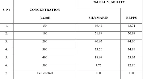
[image:59.595.86.549.423.680.2]The cytotoxicity studies were carried out for the standard drug Silymarin and ethanolic extract of Pongamia pinnata L., seeds (EEPPS) on the Chang liver cell line using different concentration range (50, 100, 200, 300,400, 500µg/ml) and its CTC50 values was determined. (Table 2and Figure. 5)
Table - 2
MTT assay of Silymarin and ethanolic extract of Pongamia pinnata L., seed on Chang
liver cell line
S. No CONCENTRATION
(µg/ml)
%CELL VIABILITY
SILYMARIN EEPPS
1. 50 69.49 63.71
2. 100 51.84 50.84
3. 200 40.67 44.06
4. 300 33.20 34.89
5. 400 18.64 23.03
6. 500 7.77 12.86
Institute of Pharmacology, MMC Page 50
Figure. 5
Cytotoxicity of Standard Silymarin and extract on Chang liver cell line
The cell viability decreased with increase in concentration in the case of Silymarin and EEPPS. The CTC50 values of the EEPPS was found to be 100µg/ml which is similar to that of the standard Silymarin. This indicates that the EEPPS is as safe as Silymarin against the normal Chang liver cell line.
0 20 40 60 80 100 120
Cell control
50 100 200 300 400 500
%
Ce
ll
viab
il
ity
Concentration µg/ml MTT assay
Silymarin
Institute of Pharmacology, MMC Page 51
6.3.1.2. In vitro hepatoprotective activity using Standard Silymarin and extract against
CCl4 induced hepatotoxicity
[image:61.595.86.549.333.657.2]The Chang liver cells were first challenged with the CCl4 at the concentration of 125µg/ml in which 39.92% cells are viable. Then the cells were treated with standard Silymarin and ethanolic extract of Pongamia pinnata L., seed in the concentration of 100, 50, 25, 10µg/ml to assess the percentage cell viability and the values are tabulated in Table 3 and Figure.6
Table - 3
In vitro hepatoprotective activity of Silymarin and EEPPS against CCl4 induced
hepatotoxicity
S. NO TREATMENT
CONCENTRATION (µg/ml)
% CELL VIABILITY
1. Control 100
2. CCl4 125 39.92±1.02
3.
CCl4 + Silymarin
10 56.46±2.0
25 61.9±2.3
50 66.5±2.34
100 72.2±2.1
4.
CCl4
+
EEPPS
10 50.3±0.54
25 57.9±1.89
50 65.2±2.32
100 71.1±1.20


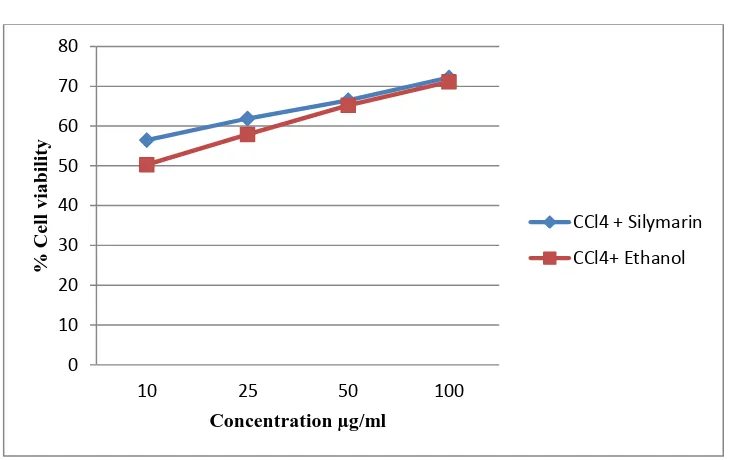


![Bis[μ N (2,6 dimethylphenyl)acetamidato]bis(dimethylaluminium)](data:image/gif;base64,R0lGODlhAQABAIAAAP///wAAACH5BAEAAAAALAAAAAABAAEAAAICRAEAOw==)