Copyright © 2003, American Society for Microbiology. All Rights Reserved.
Expression of the Major Histocompatibility Complex Class I Molecule
Mamu-A*01 Is Associated with Control of Simian Immunodeficiency
Virus SIV
mac
239 Replication
Bianca R. Mothe´,
1,2Jason Weinfurter,
1Chenxi Wang,
3William Rehrauer,
1Nancy Wilson,
1Todd M. Allen,
1David B. Allison,
3and David I. Watkins
1,2*
Wisconsin Regional Primate Research Center, University of Wisconsin, Madison, Wisconsin 537151; Department of Pathology
and Laboratory Medicine, University of Wisconsin, Madison, Wisconsin 537922; and Department of Biostatistics,
University of Alabama at Birmingham, Birmingham, Alabama 352943
Received 18 July 2002/Accepted 7 November 2002
Several HLA alleles are associated with attenuated human immunodeficiency virus disease progression. We explored the relationship between the expression of particular major histocompatibility complex (MHC) class I alleles and viremia in simian immunodeficiency virus SIVmac239-infected macaques. Of the common MHC
class I alleles, animals that expressed Mamu-A*01 exhibited the best control of viral replication.
It is becoming increasingly clear that an individual’s HLA genes can affect the outcome of human immunodeficiency vi-rus (HIV) infection. Carrington et al. demonstrated that HLA-B*35 and -Cw*04 are present at a high frequency in individuals who progress rapidly to AIDS (5). Conversely, there is a het-erozygous advantage in HIV infection (5). Furthermore, two
specific alleles,HLA-B*57andHLA-B*27, are associated with
slow progression to disease (10, 12, 15). HLA-B*57-positive individuals develop a broad repertoire of cytotoxic T-lympho-cyte (CTL) responses, especially against the Gag protein (15). HLA-B*27 binds an immunodominant epitope, KK10, derived from the Gag protein. In some patients, this response has selected for viral escape variants associated with progression to disease (8, 9).
Unique properties of the simian immunodeficiency virus (SIV)-infected rhesus macaque make it an ideal model for HIV infection (11). Both SIV and HIV-1 are primate lentivi-ruses, sharing similar genomic organizations, biological prop-erties, and tissue tropisms (11). Both viruses use the CD4 molecule as the primary receptor and chemokine receptors as coreceptors (6). Persistent disease is the result of infection with HIV and SIV. Individuals experience ongoing viral replication, and set point viremia represents the equilibrium between the ability of the virus to replicate and the ability of the host’s immune responses to limit replication. Virus replication is held in check for an extended period of time (usually years for humans), until viremia increases followed by the onset of AIDS (21). The concentration of viral RNA (vRNA) at the set point in plasma has been inversely correlated with progression
to disease (14). Rhesus macaques infected with SIVmac239
develop symptoms characteristic of human AIDS, including
depletion of CD4⫹T cells followed by development of
oppor-tunistic infections.
We wanted to determine whether a major histocompatibility
complex (MHC) effect existed in a cohort of animals
experi-mentally infected with the molecular clone SIVmac239.
Previ-ous studies have shown that SIVmac251-infected animals that
express the class I Mamu-A*01 molecule (18) control viremia more effectively than Mamu-A*01-negative animals. We there-fore wanted to explore the role of Mamu-A*01 in controlling
replication of SIVmac239.
To determine the effect of different MHC class I alleles on viral replication, we screened for the expression of the follow-ing eight common MHC class I alleles in our cohort of 53
SIVmac239-infected animals by PCR-sequence-specific primers
(SSP): Mamu-A*01, Mamu-A*02, Mamu-A*08, Mamu-A*11,
Mamu-B*01,Mamu-B*03,Mamu-B*04, andMamu-B*17
(Ta-ble 1). These alleles were chosen because they purportedly
bind SIV-derived epitopes (3, 4, 7). The 3⬘-terminal region of
PCR-SSP primers targeted a nucleotide polymorphism unique to these eight Indian rhesus MHC class I alleles.
Approxi-mately 75 ng of genomic DNA was isolated from 200 l of
EDTA-anticoagulated whole blood or buffy coat for each
sample in a total reaction volume of 25 l. Final reaction
mixtures contained 1⫻ PCR buffer (Invitrogen, Carlsbad,
Calif.) composed of 60 mM Tris-HCl (pH 9.5), 2 mM MgCl2,
15 mM ammonium sulfate, 410 M each deoxynucleoside
triphosphate (dNTP; Promega, Madison, Wis.), 0.5M each
PCR-SSP primer, 0.3M each internal control primer, and
0.961 U of Platinum Taq polymerase (Gibco BRL, Life
Technologies, Gaithersburg, Md.). The thermal cycling con-ditions were as follows: an initial 1 min of denaturation at 96°C, followed by 5 cycles of 96°C denaturation for 25 s, 70°C annealing for 50 s, and a 45-s extension at 72°C; and, finally, 4 cycles of 96°C denaturation for 25 s, 55°C annealing for 60 s, and a 120-s extension at 72°C. Subsequently, PCR
products were electrophoresed on 2% agarose gels (0.5⫻
Tris-borate-EDTA [TBE]) at a constant voltage and ana-lyzed for the presence of the required internal control prod-uct and each of the specific allele amplicons relative to a 100-bp DNA ladder (Gibco BRL).
All macaques were infected with a molecularly cloned virus,
SIVmac239 (Table 1). Three sets of viruses were used: SIVmac239/
* Corresponding author. Mailing address: Wisconsin Regional Pri-mate Research Center, 1220 Capitol Ct., Madison, WI 53715-1299. Phone: (608) 265-3380. Fax: (608) 265-8084. E-mail: watkins@primate .wisc.edu.
2736
on November 8, 2019 by guest
http://jvi.asm.org/
nef open and SIVmac239/nef stop, which were both expanded
on rhesus peripheral blood lymphocytes (PBLs); and
SIVmac239/nef open, which was expanded on CEMx174 cells
only. SIVmac239/nef stop differs from SIVmac239/nef open
by a stop codon present in the nefgene (13, 19). There is
[image:2.603.42.540.85.603.2]selection for full-length Nef protein in vivo, and the Nef open reading frame is restored within a few weeks after infection. The third virus stock was expanded in vitro on
TABLE 1. Viruses used, MHC class I alleles, set point viremia, and vaccination information for each SIV-infected animal in our cohorta
Animal Virus strain
Allele type No. of set
point copies of vRNA/ml of plasma
Survivorship (no. of days postinfection)b
Vaccination statusc
A*01 A*02 A*08 A*11 B*01 B*03 B*04 B*17
95061 SIVmac239/PBL ⫹ ⫹ ⫺ ⫺ ⫺ ⫺ ⫺ ⫹ 400 908* V
1937 SIVmac239/CEMx174 ⫹ ⫺ ⫺ ⫹ ⫺ ⫺ ⫺ ⫹ 747 728* C
95096 SIVmac239/CEMx174 ⫹ ⫺ ⫺ ⫹ ⫺ ⫺ ⫺ ⫹ 2,364 728* C
95058 SIVmac239stop/PBL ⫹ ⫺ ⫺ ⫺ ⫺ ⫺ ⫺ ⫺ 11,380 620 V
85013 SIVmac239/PBL ⫹ ⫺ ⫺ ⫺ ⫺ ⫺ ⫺ ⫺ 62,653 407 C
93062 SIVmac239/PBL ⫺ ⫺ ⫺ ⫺ ⫺ ⫺ ⫺ ⫺ 66,264 309 V
2130 SIVmac239/PBL ⫹ ⫺ ⫺ ⫺ ⫺ ⫺ ⫺ ⫺ 79,271 195* C
95084 SIVmac239/CEMx174 ⫹ ⫹ ⫺ ⫺ ⫺ ⫺ ⫺ ⫺ 81,303 460 C
1975 SIVmac239/PBL ⫹ ⫺ ⫺ ⫺ ⫺ ⫺ ⫺ ⫺ 83,522 593* V
2122 SIVmac239/PBL ⫹ ⫺ ⫺ ⫺ ⫺ ⫺ ⫺ ⫺ 86,764 195* V
96135 SIVmac239/PBL ⫹ ⫺ ⫹ ⫺ ⫺ ⫺ ⫺ ⫺ 88,000 295 V
96118 SIVmac239/PBL ⫹ ⫺ ⫺ ⫺ ⫺ ⫺ ⫺ ⫺ 88,464 764 V
2095 SIVmac239/PBL ⫹ ⫺ ⫺ ⫺ ⫺ ⫺ ⫺ ⫹ 102,959 195* C
80035 SIVmac239/PBL ⫹ ⫺ ⫺ ⫺ ⫺ ⫺ ⫺ ⫺ 131,158 311 V
97085 SIVmac239/PBL ⫹ ⫺ ⫺ ⫺ ⫺ ⫺ ⫺ ⫺ 158,859 106 V
96114 SIVmac239/PBL ⫹ ⫺ ⫹ ⫺ ⫺ ⫹ ⫺ ⫺ 185,285 153 V
96111 SIVmac239/PBL ⫹ ⫹ ⫺ ⫺ ⫹ ⫺ ⫺ ⫺ 193,189 398 C
95086 SIVmac239/PBL ⫹ ⫺ ⫹ ⫺ ⫺ ⫺ ⫺ ⫺ 199,904 308 C
94004 SIVmac239/PBL ⫹ ⫺ ⫹ ⫺ ⫺ ⫹ ⫺ ⫺ 205,941 307 V
92080 SIVmac239/PBL ⫺ ⫺ ⫺ ⫹ ⫺ ⫺ ⫺ ⫹ 222,473 427 C
93057 SIVmac239/PBL ⫹ ⫺ ⫺ ⫺ ⫺ ⫺ ⫺ ⫺ 222,473 477 C
2129 SIVmac239/PBL ⫹ ⫺ ⫺ ⫹ ⫺ ⫺ ⫺ ⫹ 279,859 195* C
98018 SIVmac239/PBL ⫹ ⫺ ⫺ ⫺ ⫺ ⫺ ⫺ ⫺ 281,625 195* C
2127 SIVmac239/PBL ⫹ ⫺ ⫺ ⫺ ⫺ ⫺ ⫺ ⫺ 291,335 195* C
2124 SIVmac239/PBL ⫹ ⫺ ⫺ ⫺ ⫺ ⫺ ⫺ ⫺ 315,125 195* V
2126 SIVmac239/PBL ⫹ ⫹ ⫺ ⫺ ⫺ ⫺ ⫺ ⫺ 328,656 195* V
97073 SIVmac239/PBL ⫺ ⫺ ⫹ ⫺ ⫺ ⫺ ⫺ ⫺ 337,034 539* V
96113 SIVmac239/PBL ⫺ ⫺ ⫺ ⫺ ⫺ ⫹ ⫺ ⫺ 387,650 526 C
92077 SIVmac239/CEMx174 ⫹ ⫺ ⫺ ⫺ ⫺ ⫺ ⫺ ⫺ 410,000 219 C
87081 SIVmac239stop/PBL ⫺ ⫺ ⫺ ⫹ ⫺ ⫺ ⫺ ⫺ 411,205 296 V
95045 SIVmac239stop/PBL ⫹ ⫺ ⫹ ⫺ ⫺ ⫹ ⫺ ⫺ 519,350 378 V
95114 SIVmac239stop/PBL ⫹ ⫺ ⫺ ⫺ ⫹ ⫺ ⫺ ⫺ 698,450 424 C
2065 SIVmac239/PBL ⫹ ⫺ ⫹ ⫺ ⫺ ⫹ ⫺ ⫹ 829,674 195* C
96020 SIVmac239/PBL ⫺ ⫹ ⫹ ⫺ ⫺ ⫹ ⫺ ⫺ 881,531 398 C
90069 SIVmac239/PBL ⫺ ⫺ ⫺ ⫺ ⫺ ⫺ ⫺ ⫺ 957,299 539* C
96031 SIVmac239stop/PBL ⫹ ⫹ ⫺ ⫺ ⫺ ⫺ ⫺ ⫺ 1,013,700 1,143 C
95115 SIVmac239stop/PBL ⫹ ⫺ ⫹ ⫺ ⫹ ⫹ ⫺ ⫺ 1,142,204 633 C
87082 SIVmac239/PBL ⫺ ⫹ ⫹ ⫺ ⫺ ⫺ ⫺ ⫺ 1,178,314 227 V
96104 SIVmac239/PBL ⫺ ⫺ ⫺ ⫺ ⫹ ⫺ ⫺ ⫺ 1,367,915 420 C
81035 SIVmac239/PBL ⫺ ⫺ ⫺ ⫺ ⫹ ⫺ ⫺ ⫺ 1,928,291 214 V
96123 SIVmac239/PBL ⫹ ⫺ ⫹ ⫺ ⫺ ⫹ ⫺ ⫺ 2,022,816 316 V
96093 SIVmac239/PBL ⫺ ⫺ ⫺ ⫺ ⫺ ⫺ ⫺ ⫺ 2,573,018 448 C
96072 SIVmac239/PBL ⫺ ⫹ ⫺ ⫺ ⫺ ⫺ ⫺ ⫹ 2,669,679 467 C
95003 SIVmac239stop/PBL ⫺ ⫺ ⫹ ⫺ ⫹ ⫹ ⫺ ⫺ 2,845,750 489 C
96016 SIVmac239/PBL ⫹ ⫺ ⫺ ⫺ ⫺ ⫺ ⫺ ⫺ 3,909,697 271 V
80025 SIVmac239/CEMx174 ⫹ ⫺ ⫺ ⫺ ⫹ ⫺ ⫺ ⫺ 4,100,000 101 C
87108 SIVmac239/CEMx174 ⫹ ⫺ ⫺ ⫺ ⫺ ⫺ ⫺ ⫺ 4,100,000 120 C
97086 SIVmac239/PBL ⫺ ⫹ ⫹ ⫺ ⫺ ⫹ ⫺ ⫺ 6,397,434 553* C
92050 SIVmac239/PBL ⫺ ⫹ ⫺ ⫺ ⫺ ⫹ ⫺ ⫺ 6,547,283 269 C
96081 SIVmac239/PBL ⫺ ⫺ ⫹ ⫺ ⫺ ⫹ ⫺ ⫺ 63,942,700 180 C
90131 SIVmac239/PBL ⫺ ⫺ ⫹ ⫺ ⫹ ⫺ ⫺ ⫺ 177,000,000 115 C
95112 SIVmac239stop/PBL ⫺ ⫺ ⫹ ⫺ ⫹ ⫺ ⫺ ⫺ 149,935,800 153 C
83108d SIV
mac239/PBL ⫺ ⫺ ⫺ ⫺ ⫹ ⫹ ⫺ ⫺ died wk 5 24 V
aAnimals were infected with either SIV
mac239/nef stop, SIVmac239/nef open, or SIVmac239/nef open expanded on CEMx174 cells. Using PCR-SSP, we screened for the presence of eight different MHC class I alleles. We measured viral loads between 12 and 16 weeks when viral replication stabilized in these animals (set point).
b*, animal still alive as of writing. cV, vaccines; C, controls.
dAnimal 83108 was not included in viral load analysis because no set point viremia was available. The animal died of non-AIDS-related complications 5 weeks
postinfection.
on November 8, 2019 by guest
http://jvi.asm.org/
CEMx174 cells rather than on rhesus PBLs. Animals were
infected intrarectally with SIVmac239/nef open (3,000 50%
tissue culture infectious doses [TCID50]) and SIVmac239/nef
stop (1,000 TCID50). Animals infected with SIVmac239/nef
open expanded on CEMx174 cells only received 20 ng of p27
SIVmac239 intrarectally. A subset of animals was vaccinated
according to DNA-modified vaccinia virus ankara (MVA) regimens containing different constructs (Table 1).
We stratified the animals based on their set point viremia. Viral loads were quantitated with the SIV branched DNA assay (Chiron Diagnostics, Emeryville, Calif.) and the Taqman kinetic reverse transcription (RT)-PCR assay. Plasma SIV RNA concentrations were measured every week for the first 4 weeks and then biweekly thereafter. Viral set points were
mea-sured between weeks 12 and 16, when plasma vRNA concen-trations were constant for at least 2 weeks.
Viral load differences between groups of animals infected
with SIVmac239 were tested for statistical significance with t
tests after log transforming the data to improve normality and homoscedasticity. (Animal 83108 was not included in the analysis, because this animal was euthanized 5 weeks postin-fection due to non-AIDS-related complications. No set point viremia was therefore available for this animal.) In addition, Levene’s test for homoscedasticity was conducted, and if a difference was found to be significant, the Welch correction for unequal variances was employed. Finally, to further examine the robustness of the results, a nonparametric test, the
Mann-Whitney U test, was performed. TheP values for the
non-FIG. 1. Average plasma SIVmac239 vRNA concentration for all Mamu-A*01-positive animals versus Mamu-A*01-negative animals. The
[image:3.603.109.476.71.260.2]average plasma vRNA concentration for each of the two groups of animals is shown for the first 28 weeks postinfection. Mamu-A*01-positive animals significantly control viral replication more effectively than Mamu-A*01-negative animals (P⫽0.001).
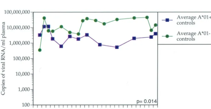
FIG. 2. Average plasma SIVmac239 vRNA concentration for control Mamu-A*01-positive animals versus Mamu-A*01-negative animals. The
average plasma vRNA concentration for each of the two groups of animals is shown for the first 28 weeks postinfection. Control Mamu-A*01-positive animals significantly control viral replication more effectively than Mamu-A*01-negative animals (P⫽0.014).
on November 8, 2019 by guest
http://jvi.asm.org/
[image:3.603.111.479.504.695.2]parametric tests were calculated by exact methods. AllPvalues are two tailed.
We found that the majority of animals with the lowest set point vRNA levels expressed Mamu-A*01. Between weeks 12 and 28 postinfection, Mamu-A*01-positive animals controlled viral replication more effectively than Mamu-A*01-negative
animals (P⫽0.001;ttest) (Fig. 1). To ensure that the
Mamu-A*01 effect is not due to a subset of vaccinated Mamu- Mamu-A*01-positive animals, we removed these animals from the analysis. Indeed, vaccine-naive Mamu-A*01-positive animals restricted viral replication more efficiently than vaccine-naive
Mamu-A*01-negative animals (P⫽ 0.014;ttest) (Fig. 2). We
com-pared the virus loads between vaccinated and nonvaccinated Mamu-A*01-positive animals. We did not find a significant difference in set point viremia between these groups by either
independent t test (P ⫽ 0.58) or Mann-Whitney test (P ⫽
0.43). In addition to viral load analysis, we performed survival analysis by Cox regression (proportional hazards model) with A*01 status as a covariate. The hazard ratio of Mamu-A*01-positive animals to Mamu-A*01-negative animals is 0.76,
which is not significant (P⫽ 0.43). Nevertheless, analysis of
survivorship as a function of vRNA revealed that survivorship
is significantly inversely related to set point viremia (P⬍0.001;
Cox proportional hazards regression).
We have previously shown that Mamu-A*01-restricted epitopes dominate the anti-SIV CTL responses in Mamu-A*01-positive animals early in infection (16). Additionally, we have identified 14 different Mamu-A*01-restricted CTL epitopes, indicating that this MHC class I molecule engenders a broad repertoire of CTL responses (1). This may partially explain why Mamu-A*01-positive animals control viral repli-cation more successfully than Mamu-A*01-negative animals. Furthermore, one of these responses, directed against the Tat28-35SL8 epitope, exerts selective pressure on the virus, as
evidenced by viral escape from this response early in infection (2). This epitope has also been shown to be recognized by CTL with high functional avidity, requiring a low peptide concen-tration to trigger a CTL response (17).
The association of Mamu-A*01 with enhanced control of viral replication has been shown for infection with the
heter-ogeneous biological isolate SIVmac251 (18), but not for
infec-tion with SHIV 89.6p. Our studies extend those findings to
SIVmac239 replication. SHIV 89.6 is a chimeric virus created by
using Env, as well as Tat, Vpu, and Rev from the HIV 89.6
isolate with the backbone of the molecular clone SIVmac239
(20). Mamu-A*01-restricted responses directed against SIV Env, Tat, Vpr, and Rev may therefore be partially responsible
for the Mamu-A*01 effect in both SIVmac239 infection, shown
here, and SIVmac251 infection (18). Tat28-35SL8 and other
Mamu-A*01-restricted responses (1) may contribute to the effect of diminishing viral replication in Mamu-A*01-positive animals. Understanding how particular MHC genes affect vire-mia may provide insight into the correlates of protection from lentiviral infection.
We thank Millie Schultz, Elizabeth Meek, and Christopher Fischer for technical help, in addition to Kevin Kunstman and Steve Wolinsky for plasma vRNA concentration analysis. We also thank Jody Helge-land for assistance with blood processing, Jacque Mitchen for
coordi-nating all animal procedures, and Carol L. Emerson for performing all animal procedures.
This work was supported by NIH grants RR1537, AI46366, AI45461, and RR00167 (awarded to David I. Watkins). David I. Watkins is a recipient of an Elizabeth Glaser Scientist Award.
REFERENCES
1. Allen, T. M., B. R. Mothe, J. Sidney, P. Jing, J. L. Dzuris, M. E. Liebl, T. U. Vogel, D. H. O’Connor, X. Wang, M. C. Wussow, J. A. Thomson, J. D. Altman, D. I. Watkins, and A. Sette.2001. CD8⫹lymphocytes from simian
immunodeficiency virus-infected rhesus macaques recognize 14 different epitopes bound by the major histocompatibility complex class I molecule Mamu-A*01: implications for vaccine design and testing. J. Virol.75:738–749. 2. Allen, T. M., D. H. O’Connor, P. Jing, J. L. Dzuris, B. R. Mothe, T. U. Vogel, E. Dunphy, M. E. Liebl, C. Emerson, N. Wilson, K. J. Kunstman, X. Wang, D. B. Allison, A. L. Hughes, R. C. Desrosiers, J. D. Altman, S. M. Wolinsky, A. Sette, and D. I. Watkins.2000. Tat-specific cytotoxic T lymphocytes select for SIV escape variants during resolution of primary viraemia. Nature407: 386–390.
3. Allen, T. M., J. Sidney, M. F. del Guercio, R. L. Glickman, G. L. Lensmeyer, D. A. Wiebe, R. DeMars, C. D. Pauza, R. P. Johnson, A. Sette, and D. I. Watkins.1998. Characterization of the peptide binding motif of a rhesus MHC class I molecule (Mamu-A*01) that binds an immunodominant CTL epitope from simian immunodeficiency virus. J. Immunol.160:6062–6071. 4. Allen, T. M., and D. I. Watkins.1999. SIV and SHIV CTL epitopes identified
in macaques, p. IV-45–IV-54.InB. T. Korber, C. Brander, B. F. Haynes, R. A. Koup, C. L. Kuiken, J. P. Moore, B. D. Walker, and D. I. Watkins (ed.), HIV Molecular Immunology Database. Theoretical Biology and Bio-physics Group, Los Alamos National Laboratory, Los Alamos, N.Mex. 5. Carrington, M., G. W. Nelson, M. P. Martin, T. Kissner, D. Vlahov, J. J.
Goedert, R. Kaslow, S. Buchbinder, K. Hoots, and S. J. O’Brien.1999. HLA and HIV-1: heterozygote advantage and B*35-Cw*04 disadvantage. Science 283:1748–1752.
6. Chen, Z., A. Gettie, D. D. Ho, and P. A. Marx.1998. Primary SIVsm isolates use the CCR5 coreceptor from sooty mangabeys naturally infected in west Africa: a comparison of coreceptor usage of primary SIVsm, HIV-2, and SIVmac. Virology246:113–124.
7. Evans, D. T., D. H. O’Connor, P. Jing, J. L. Dzuris, J. Sidney, J. da Silva, T. M. Allen, H. Horton, J. E. Venham, R. A. Rudersdorf, T. Vogel, C. D. Pauza, R. E. Bontrop, R. DeMars, A. Sette, A. L. Hughes, and D. I. Watkins. 1999. Virus-specific cytotoxic T-lymphocyte responses select for amino-acid variation in simian immunodeficiency virus Env and Nef. Nat. Med.5:1270– 1276.
8. Goulder, P. J., M. A. Altfeld, E. S. Rosenberg, T. Nguyen, Y. Tang, R. L. Eldridge, M. M. Addo, S. He, J. S. Mukherjee, M. N. Phillips, M. Bunce, S. A. Kalams, R. P. Sekaly, B. D. Walker, and C. Brander.2001. Substantial differences in specificity of HIV-specific cytotoxic T cells in acute and chronic HIV infection. J. Exp. Med.193:181–194.
9. Goulder, P. J., R. E. Phillips, R. A. Colbert, S. McAdam, G. Ogg, M. A. Nowak, P. Giangrande, G. Luzzi, B. Morgan, A. Edwards, A. J. McMichael, and S. Rowland-Jones.1997. Late escape from an immunodominant cyto-toxic T-lymphocyte response associated with progression to AIDS. Nat. Med. 3:212–217.
10. Hill, A. V.1998. The immunogenetics of human infectious diseases. Annu. Rev. Immunol.16:593–617.
11. Johnson, R. P.1996. Macaque models for AIDS vaccine development. Curr. Opin. Immunol.8:554–560.
12. Kaslow, R. A., M. Carrington, R. Apple, L. Park, A. Munoz, A. J. Saah, J. J. Goedert, C. Winkler, S. J. O’Brien, C. Rinaldo, R. Detels, W. Blattner, J. Phair, H. Erlich, and D. L. Mann.1996. Influence of combinations of human major histocompatibility complex genes on the course of HIV-1 infection. Nat. Med.2:405–411.
13. Lewis, M. G., S. Bellah, K. McKinnon, J. Yalley-Ogunro, P. M. Zack, W. R. Elkins, R. C. Desrosiers, and G. A. Eddy.1994. Titration and characteriza-tion of two rhesus-derived SIVmac challenge stocks. AIDS Res. Hum. Ret-rovir.10:213–220.
14. Mellors, J. W., C. R. Rinaldo, Jr., P. Gupta, R. M. White, J. A. Todd, and L. A. Kingsley.1996. Prognosis in HIV-1 infection predicted by the quantity of virus in plasma. Science272:1167–1170.
15. Migueles, S. A., M. S. Sabbaghian, W. L. Shupert, M. P. Bettinotti, F. M. Marincola, L. Martino, C. W. Hallahan, S. M. Selig, D. Schwartz, J. Sulli-van, and M. Connors.2000. HLA B*5701 is highly associated with restriction of virus replication in a subgroup of HIV-infected long term nonprogressors. Proc. Natl. Acad. Sci. USA97:2709–2714.
16. Mothe´, B. R., H. Horton, D. K. Carter, T. M. Allen, M. E. Liebl, P. Skinner, T. U. Vogel, S. Fuenger, K. Vielhuber, W. Rehrauer, N. Wilson, G. Franchini, J. D. Altman, A. Haase, L. J. Picker, D. B. Allison, and D. I. Watkins.2002. Dominance of CD8 responses specific for epitopes bound by a single major histocompatibility complex class I molecule during the acute phase of viral infection. J. Virol.76:875–884.
17. O’Connor, D. H., T. M. Allen, T. U. Vogel, P. Jing, I. P. DeSouza, E. Dodds,
on November 8, 2019 by guest
http://jvi.asm.org/
E. J. Dunphy, C. Melsaether, B. Mothe, H. Yamamoto, H. Horton, N. Wilson, A. L. Hughes, and D. I. Watkins.2002. Acute phase cytotoxic T lymphocyte escape is a hallmark of simian immunodeficiency virus infection. Nat. Med. 8:493–499.
18. Pal, R., D. Venzon, N. L. Letvin, S. Santra, D. C. Montefiori, N. R. Miller, E. Tryniszewska, M. G. Lewis, T. C. VanCott, V. Hirsch, R. Woodward, A. Gibson, M. Grace, E. Dobratz, P. D. Markham, Z. Hel, J. Nacsa, M. Klein, J. Tartaglia, and G. Franchini.2002. ALVAC-SIV-gag-pol-env-based vacci-nation and macaque major histocompatibility complex class I (A*01) delay simian immunodeficiency virus SIVmac-induced immunodeficiency. J. Virol.
76:292–302.
19. Regier, D. A., and R. C. Desrosiers.1990. The complete nucleotide sequence of a pathogenic molecular clone of simian immunodeficiency virus. AIDS Res. Hum. Retrovir.6:1221–1231.
20. Reimann, K. A., J. T. Li, R. Veazey, M. Halloran, I.-W. Park, G. B. Karlsson, J. Sodroski, and N. L. Letvin.1996. A chimeric simian/human immunode-ficiency virus expressing a primary patient human immunodeimmunode-ficiency virus type 1 isolateenvcauses an AIDS-like disease after in vivo passage in rhesus monkeys. J. Virol.70:6922–6928.
21. Sewell, A. K., D. A. Price, A. Oxenius, A. D. Kelleher, and R. E. Phillips. 2000. Cytotoxic T lymphocyte responses to human immunodeficiency virus: control and escape. Stem Cells18:230–244.