STUDY OF EXPRESSION OF CD117 IN GLIAL TUMORS
AND ITS ROLE IN TUMOUR TYPE AND GRADE
Dissertation submitted
in partial fulfilment of therequirements for the degree of
M.D. (PATHOLOGY)
BRANCH – III
GOSHEN INSTITUTE OF PATHOLOGY AND ELECTRON
MICROSCOPY,
MADRAS MEDICAL COLLEGE,
CHENNAI – 600 003.
THE TAMIL NADUDR. M.G.R. MEDICAL UNIVERSITY
CHENNAI
CERTIFICATE
This is to certify that this Dissertation entitled “STUDY OF
EXPRESSION OF CD117 IN GLIAL TUMOURS ANDITS ROLE
INTUMOUR TYPE AND GRADE” is the bonafide original work of
Dr.M.JAYANANDHINI, in partial fulfilment of the requirement for M.D.,
(Branch III) in Pathology examination of the Tamilnadu Dr.M.G.R Medical
University to be held in April 2016
Prof. Dr.VIMALA. R. M.D.,
DEAN.
Madras Medical College, Rajiv Gandhi Government General Hospital,
Chennai – 600003.
PROF. DR.M.SARASWATHI, M.D.,
DECLARATION
I, Dr.M.JAYANANDHINI solemnly declare that the dissertation titled
“STUDY OF EXPRESSION OF CD117 IN GLIAL TUMORS AND ITS
ROLE IN TUMOUR TYPE AND GRADE” is the bonafide work done by
me at Institute of Pathology, Madras Medical College under the expert
guidance and supervision of Prof. Dr.M.SARASWATH., M.D., Director,
Institute of pathology, Madras Medical College.
The dissertation is submitted to the Tamilnadu Dr.M.G.R Medical
University towards partial fulfilment of requirement for the award of M.D.,
Degree (Branch III) in Pathology.
Place : Chennai
ACKNOWLEDGEMENT
I express my sincere thanks to Prof. Dr. R.VIMALA, M.D., Dean,
Madras Medical College and Rajiv Gandhi Government GeneralHospital, for
permitting me to utilize the facilities of the Institution.
I take this opportunity to express my heartfelt sincere gratitude to
Prof.Dr.M.Saraswathy M.D., Professor and Director of Institute of
Pathology, Madras Medical College, Chennai for her keen interest, constant
encouragement, and valuable suggestions throughout the study.
I am truly thankful to Prof. Dr. SHANTHA RAVISANKAR
M.D.D.C.P, Prof.Dr.R.PADMAVATHI.M.D.,D.G.O., Prof.Dr.
V.RAMAMURTHY M.D., Prof.Dr.GEETHA DEVADAS M.D.,D.C.P.,
Prof.Dr.M.P.KANCHANA M.D., Prof.Dr.SUDHA VENKATESH M.D.,
Prof.Dr.K.RAMA M.D., Prof.Dr.RAJAVELU INDIRA M.D., and
Prof.Dr.S.PAPPATHI M.D.,D.C.H for their cooperation and constant
encouragement throughout the study.
I express my heartfelt sincere thanks to all my Assistant Professors for
their help and suggestions during the study.
I am thankful to all my colleagues, senior and junior postgraduates,
friends, technicians and staff of the Institute of Pathology, Madras Medical
College, Chennai for all their help and support they extended for the successful
My sincere and heartfelt thanks to my spouse Mr.P.Ramachandran B.E,
MBA, for his constant encouragement throughhis words of appreciation in all
my career guided activities. I thank my son R.Mohnish Kaarthic and my
daughter R.Jayasupriya for their love, sacrifice and support.
Last but not the least I thank my Parents for making me what I am
now and my in laws for their voluminous love and care. Above all I thank
ABBREVIATIONS
SVZ : Sub ventricular zone
MPE : Myxopapillary ependymoma
GFAP : Glial Fibrillary Acidic Protein
WHO : World Health Organisation
IHC : Immunohistochemistry
ICMR : Indian Council of Medical Research
SEER : Surveillance, Epidemiology and End Result Program
CBTRUS : Central Brain Tumour Registry of the United States
CNS : Central Nervous System
PA : Pilocytic Astrocytoma
DFA : Diffuse Fibrillary Astrocytoma
DA : Diffuse Astrocytoma
ODG : Oligodendroglioma
EPEN : Ependymoma
PXA : Pleomorphic Xanthoastrocytoma
OA : Oligoastrocytoma
AA : Anaplastic Astrocytoma
GBM : Glioblastoma Multiforme
IDEM : Intradural Extramedullary
IDIM : Intradural Intramedullary
CONTENTS
CHAPTER
NO. TITLE
PAGE NO.
1 INTRODUCTION 1
2 AIMS AND OBJECTIVES 3
3 REVIEW OF LITERATURE 4
4 MATERIALS AND METHODS 35
5 OBSERVATION AND RESULTS 39
6 DISCUSSION 79
7 SUMMARY 94
8 CONCLUSION 96
9 BIBLIOGRAPHY
10 ANNEXURES
MASTER CHART
ABSTRACT
INTRODUCTION
Glial tumors are a varied group of neoplasm. CD117 is a protooncogene
encoded tyrosine kinase receptor. It is expressed usually in some normal tissues and
in neoplasms like GIST.(gastrointestinal stromal tumors) . Treatment with inhibitors
of receptor for tyrosine kinase namely imatinib is proved successful in cases like
gastrointestinal stromal tumour which are CD117- positive. Likewise aim of this
study is to evaluate the expression of CD117 in tumors of glial origin,to prove the
marker as useful marker for diagnostic purpose and also therapeutic purpose.This
study also includes analysis of CD117 expression in glial tumor subtypes and
grades.
AIMS AND OBJECTIVES
To study the expression of CD117 In glial tumors , to correlate the percentage
of expression of cd117 with tumor type and grade, to evaluate CD117 as a diagnostic
and therapeutic tool and also to study the age, sex, and site predeliction of different
types of gliomas.
MATERIALS AND METHODS
This is a retrospective study,during the period from jan 2013-dec2014 .A total
of 263 gliomas samples were received.out of these cases 50 representative cases
were selected.Corresponding paraffin blocks were subjected to H&E staining and
immunohistochemical analysis by primary marker CD117 .The results were
tabulated in a master chart ,also recorded with photographs and were statistically
RESULTS:
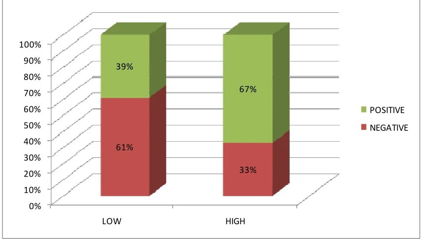
CD117 was positive in 54% of total cases. CD117 expression was seen maximum
in High grade tumours(66.8%) with statistical significance of (P <0.05). Highest
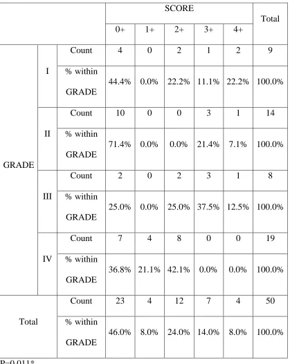
CD117 expression score was seen in WHO grade III tumours with statistically
significance ( P=0.011*).The cells with marked nuclear pleomorphism showed
increased CD117 expression,with statistical significance(P< 0.042). CD117
expression was high in cases with high cellularity showing statistically
significance.(P < 0.041). Highest CD117 positivity was seen in 61-80 years age ,in
females more than males,and in left hemisphere than right.
CONCLUSION
Incidence of gliomas are on the rise.CNS tumours are different from other
tumours as treatment and prognosis depends on the site of the tumour and most
tumours are less amenable for surgery. Our study shows CD117 is expressed
remarkably in high grade tumours than low grade. Hence CD117 can be used as a
perfect tool to identify these tumours and thus make them amenable to chemo therapy
with tyrosine kinase inhibitor, like Imatinib., which is already proven and in vogue in
gastrointestinal stromal tumour.Hence this study opens the vision on targeted therapy
in gliomas for high grade tumours.
KEY WORDS : CD117,gliomas,high grade tumours, GIST, imatinib.
1
INTRODUCTION
Tumours of central nervous system pose a major challenge as they are
less amenable for treatment. As high grade tumours they have short survival
time, and prognosis depends not only on tumour type but also on site of
the tumour .
The major milieu of brain tumours are derived from glial cells called
gliomas. Highly malignant tumours are glioblastomas. In past decade in
elderly people incidence of glioblastoma has increased spuriously due to
advances in neuroimaging as even, the smaller lesions are picked out which
were missed in earlier days. Hence according to Kurland and Schonberg(1) there is an ultimate necessity for a descriptive data on primary brain tumours.
hence this study is designed to get a descriptive data.
Nervous system neoplasms affects both adults and children. The effects
of these tumours were devastating even though they constitute a small
percentage of all cancers. (2) Tumours of the CNS account for less than 2% of all malignancies (About 175,000 cases per year worldwide) (3) .The incidence does not vary markedly between regions or populations (Stewart and Kleihues,
2
Glial tumours are a varied group of neoplasms. CD117 is a
protooncogene encoded tyrosine kinase receptor. . It is expressed usually in
some normal tissues and. It is also expressed in neoplasms like GIST.
(gastrointestinal stromal tumours). Treatment with inhibitors of receptor for
tyrosine kinase namely imatinib is proved successful in cases like
gastrointestinal stromal tumour which are CD117- positive Likewise aim of
this study is to study the expression of CD117 in tumours of glial origin, to
prove the marker as useful tool for diagnostic purpose and treatment. This
study also includes analysis of CD117 expression in glial tumor subtypes and
AIMS
AND
3
AIMS AND OBJECTIVES
1) To study the expression of CD117 in Glial tumours
2) To correlate the percentage of expression of CD 117 with tumor type and
grade.
3) To study the CD117 expression in tumours of glial origin as a useful tool for
diagnostic and therapeutic purpose.
REVIEW
OF
4
REVIEW OF LITERATURE
The central nervous system consists of, neurons, glial cells,
meningothelial cells and endothelial cells of the blood vessels. Neurons form
major part of the cells. The glial cells form supporting cells. They are divided
into macro and microglia. Macroglia are composed of astrocytes,
oligodendrocytes, and ependymal cells, derived from neurectoderm. microglia
are derived from bone marrow and they are the phagocytic system of the
CNS.
Astrocyte is the glial cell found throughout the CNS. They are called so
because of their star shaped appearance, caused by multipolar branching
cytoplasmic processes which have cytoplasmic intermediate filaments, named
as glial fibrillary acidic protein. They are the main cells responsible for repair
and scar formation in brain.
Oligodendrocytes are responsible for myelination in central nervous
system. Ependymal cells are cuboidal cells lining the ventricles of
cerebrovascular system Microglia are phagocytic system of the CNS derived
from mesoderm. They are the fixed macrophage system respond to injury
5
Neoplasms of the CNS was first described by Gupta longati a scientist
from Russia in the year 1873 .They account for 2% of all cancers world
wide. Annual incidence of CNS neoplasms ranges from 10-17/100,000 people
for intracranial tumours and 1 to 2 /100000 people for intraspinal tumours. In
children 70% of tumours occurs in posterior fossa and in adults most common
in the cerebral hemisphere above the tentorium(4-9). CNS tumours are divided into four broad categories namely
(1) gliomas
(2) neuronal neoplasms
(3) neoplasms which are poorly differentiated,
(4) and the last category meningiomas..
Gliomas are derived from glial cells and are composed of
astrocytomas, oligodendrogliomas, and ependymomas. Gliomas are
classified histologically by WHO (10). But recent advances in brain tumor research are complemented with a list of biomarkers for molecular
histopathology, Focus is also increasing on drugs against targeted therapy of
tumour cell population.
Glial cells are derived from adult neural stem cells and glial progenitor
cells. They have been proven to share some traits with gliomas as proposed
6
Human gliomas and glioma derived tumourspheres express stem
cell related genes.(12).
Neural stem cells show high resistance for senescence and apoptosis,
so they need lesser mutation for transforming into malignant when
compared to well differentiated counterpart which require
approximately four to seven mutation as postulated by (Renan1993; Sanai,
Alvarez-Buylla et al. 2005)(11,13).
Adult human SVZ has proliferating adult neural stem cells(14) The biggest germinal areas of adult brain is the sub ventricular zone of
lateral wall of left ventricle,(11,14,15,16) .MRI shows a group of glioblastomas that are associated with SVZ ,which arise as typically
multifocal lesions and recur at sites significantly remote to primary
tumour( 17) .Interestingly showing that some gliomas originate in a mutated stem cell which give rise to transformed progenitor cell that can go away from
the germinal area and multiply vigorously once they reach an amicable
7
EPIDEMIOLOGY OF NERVOUS SYSTEM TUMOURS:
In an analysis made in nation wide registry 2005 in korea studied 5,692
patients WHO were diagnosed as primary brain neoplasms in 2005. It was
seen that CNS tumours occurred in a F:M ratio of 1.43:1. It was found that
meningiomas were the most common tumours (31.2) %.Of all the gliomas
30.7% was glioblastomas, and among the malignant tumours of CNS 19.3%
were glioblastomas. The medulloblastoma and germ call tumours were the
most common tumours below 19 years of age (18). This study showed the epidemiology in korea.
CBTRUS-The Central Brain Tumor Registry of the United States
analysed data from 1990–1994 consisting of all primary CNS tumours
from 11 state cancer registries, 20,765 case data was analysed and showed in
general that the cns tumours annual incidence was at 11.5 cases per 100,000
person-years
• in male patients the incidence was (12.1 per 100,000 person-years)
• in female the incidence was (11.0 per 100,000 person-years)(19)
This study was conducted by Surawicz et al .it had similar results as the
study of arora et al in England, it also showed that whites are affected more
than blacks. Brain cancer accounts for1.4% of all cancer deaths and 2.3% of all
8
Another study conducted by arora et al in 53326 primary brain
tumours showed that overall incidence was 9.21 per 100000 person years
(21). and the peak age affected was 70 -79 years. This study also showed that malignant tumours were 5.64%, benign tumours were 60%.it also showed
supratentorial tumours affected older age group and infratentorial affected
younger age group.
Similar work by Porter et al also shows that incidence of CNS tumours
in united states was 18.1/100,000 person years.(22)
In india CNS tumours account for less than 2% of all malignancies.
According to ICMR statistics from 1982-1983 to 2002-2003 was
studied incidence in males: according to Chennai registry 2.53 to 4.14
according to Delhi registry. Incidence in females according to Bhopal registry
was 1.46 to 2.66 according to Delhi registry(23).
In pediatric age CNS tumours account for 27% according to SEER
programme and national cancer registry(24)
According to study of Linabery et al of pediatric CNS tumours from
1992-2004 incidence in children were found to be 158/1000,000 person years.
9
25.1 per 1000,000 person years (25), during the period of 1992-2004 and found that the incidence of CNS tumours in children was 158 per 1,000,000
years. It was more common in boys than in girls (29.9 per 1,000,000
person-years as opposed to 25.1 per 1,000,000 person-person-years)
According to jain et al study in India paediatric CNS tumours show
14.8% incidence of all tumours.
EPIDEMIOLOGY OF GLIOMAS
• Diffuse astocytomas accounts for 10- 15 % 0f all astrocytic brain tumours
and has incidence of 1.4 new cases per million population an year.(26) It
represents the largest group and most prevalent tumour in adult brain(27). Over all population based survival times of diffuse astrocytoma is 5.6
years.(28)
• Pilocytic astroctoma are slow growing neoplasms constituting 5-6% Of
gliomas( 29) . Follows an incidence rate of 0.37 per 100 persons per year and also commonest glioma of paediatric age group .These gliomas most
commonly occur in cerebellum accounting to 68%.(30)
• Pleomorphic xanthoastrocytoma relatively uncommon astrocytic tumour
10
features of this tumour was first described in 1979 and so far 200 cases are
reported.(31)
• Anaplastic astrocytomas occur in 50-75% of recurrent tumours(32).It is usually present at the age of 45-51 years and male:female ratio of 1.31 to
1.6 :1(29,30)
• Most common brain tumour is glioblastoma multiforme constituting
12–15% of the brain tumours and about 60-75% of all the astrocytic
tumours.(30,33)andfollows incidence rates of 2.96 per 100000 population per year.(29,30,33).Glioblastomas has preference for cerebral hemispheres in adults but in neonatal and pediatric age group brain stem is the most
common site(34)
• Gliosarcoma is a sarcomatous phenotype of glioblastoma constitutes 2-8
% of all cases of glioblastomas.(35,36,37,38)
• Gliomatosis cerebri - usually occurs in between 40 and 50 years of age, but
also seen in neonates. Both sexes are affected equally but at an early age in
males when compared to females.(39).
• Oligodendroglioma accounts for 2% - 4% of all primary CNS
11
to 0.37 /100000 person years(40,41). It also constitutes 71 to 79 % of WHO gradeII neoplasmspeak incidence rate.occurs in 35-44 years..
• Anaplastic oligodendroglioma constitutes minority of oligodendroglioma
and form less than 5% of new malignant gliomas.(42).They also consist of 1.2% of all primary brain tumours with an annual incidence rates of 0.07 to
0.18 per 100000 population have been reported(29,30)
• oligoastrocytoma comprise 35 % of all oligodendroglial neoplasms and
20% of WHO grade 2 tumours,with an incidence rate of 0.1/100000 person
years ( 39,41,43),
• Ependymomas constitutes 5.8 % OF gliomas 2.1% 0f all primary CNS
neoplasms, It comprises for 50–60% of spinal gliomas (44) i.n adults.frequency of progression to anaplastic ependymoma is less common
than astrocytic neoplasms (45)
• subependymomas constitutes 8% of all ependymal tumou.rs (46,47).
• myxopapillary ependymoma COMPRISES 9–13% of all
12
CLINICAL FEATURES:
• The clinical presentation of CNS neoplasms present corresponding to site
and nature. They are epilepsy, focal neurological deficits, symptoms and
signs due to raised intracranial pressure like headache,vomiting, clouding of
consciousness, coma, papilloedema, and hydrocephalus. Signs and
symptoms of brain tumours are usually nonspecific like headache, nausea,
vomiting paresthesia, seizure, paraplegia or hemiplegia. visual disturbance
or increased intracranial pressure. Signs specific to site are behavioural
changes in frontal lobe lesions. most common lesions causing seizures are
oligodendroglioma, anaplastic oligodendroglioma and gliosarcoma.
Neoplasms causing raised intracranial pressure are ependymoma, anaplastic
ependymoma, oligodendroglioma and glioblastoma multiforme. Neoplasms
that present with hydrocephalus are posterior fossa neoplasms, central
neurocytoma, chordoid glioma of third ventricle, subependymal giant cell
astrocytoma,hypothalamic pilocytic astrocytoma and pineal gland
13
RISK FACTORS AND ETIOLOGICAL AGENTS
1) Sex:
Gliomas are more common in men than women with M:F ratio of 60:40.
2) radiation:
There is a strong correlation with exposure to therapeutic ionizing
radiation like treatment of tinea capitis with development of gliomas
according to Bondy et al 1994 and hodges et al 1992(51,52,53). Electromagnetic fields can produce mutation which favours tumorigenesis and causes brain
tumours.This was studied by Gurney and vanwijnagaarden 1999.
Mobile phones: It was thought that there was link between non ionizing
radiation of mobile phones and gliomas but various studies conducted showed
no clear association.(54,55), Interphone international case control study in 13 countries in 2765 gliomas and 2409 meningiomas showed no association
between mobile phone usage and brain tuours
3) Low penetrance gene:
The popularly called GWAS - as genomic wide association studies has
revealed many especially seven chromosomal regions , (TERT), telom erase
reverse transcriptase, 1 (RTEL 1), in the regulator of telomerase longation
helicase 1 (RTEL 1), cyclin dependent kinase inhibitor 2A and 2B
(CDKN2A-CDKN2B), coiled-coil domain containing 26 (CCDC26), EGFR genes, TP53
and PHLDB1 gene (pleckstrin homology-like domain family B member 1) , is
14
4). Genetic s syndromes:
In Literature as per study of Bondy et al , it is observed that there is
strong association of gliomas in families having mentally retarded child. Also
seen in first degree relatives and clustering of cases seen in some families.it
showed association with hereditary syndromes like neurofibromatosis
NF1,NF2,tuberous sclerosis TSC1,TSC2, LiFraumeni syndrome, turcot
syndrome (59-63). In 2007 an international study called Gliogene has started analysing susceptibility loci in families with more than two gliomas.It is an
international study, started in 2007 to workup on the susceptibility loci in the
families with more than two glioma.(63)
5) Viruses and glioma
In literature viruses have seen to be associated with carcinogenesis For
example HPV is associated with cervical cancer. EBV is known to cause
burkitts lymphoma, nasopharyngeal carcinoma and Hodgkin lymphoma
likewise gliomas have been associated with sv 40 –simian 40 virus,
cytomegalovirus, john Cunningham virus and adenovirus(64-70)
6) Occupational hazards
Literature shows increased incidence of brain cancer among farmers
in vineyards exposed to pesticides. Studies reveal strong association. There was
also increased incidence of gliomas in workers of petrochemical industries,
15
The by products in rubber industry, n-nitroso compound also found in
nitrate containing preservative have found to have association with gliomas
is under study.(71)
ROLE OF NEUROIMAGING IN GLIOMAS
MRI, CT SCAN are primary modalities of investigation in brain
tumour, in which CT is very useful when skull bone is involved.
MRI holds good when soft tissue is involved. In case of gliomas brain
tissue with white matter looks expanded. Imaging also gives information
on site, edema, dural attachment , enhancing lesions indicate vascularity and
subsequent neoplastic nature.
Other recent techniques are
PET, positron emission tomography,
MRS-Magnetic resonance spectrophotometry.
FLAIR-fluid attenuation inversion recovery scan.in MRI is done.
SPECT-single photon emission computerised tomography scan and
16
INTRAOPERATIVE ANALYSIS OF SURGICAL SPECIMENS AND
TYPES.
A methodological approach to the diagnosis is important in pathology,
Hence intraoperative examination plays an important role in neuropathology.
The surgeon needs information whether the lesion is non neoplastic, benign or
malignant and the grade of the tumour for malignant lesions .This information
helps them to plan on the extent of resection.
The types of intra operative consultation are
1) frozen section,
2) csf cell cytology
3) crush or squash preparation
4) imprint cytology,
5) open biopsy
6) stereotactic needle biopsy
7) secondary biopsy and
17
MACROSCOPIC SPECIMEN INTERPRETATION AND
PROCESSING:
The specimen should be resected in such a way that, arachnoid,
gray matter and white matter are seen. In order to get such a specimen it
should be sectioned perpendicular to the meninges. On gross examination
tumorous lesions are seen as solid areas which cannot be differentiated as
either gray or white matter. They are usually admixed with haemorrhage and
necroses. Cyst with a mural nodule is seen in pilocytic astrocytoma and some
other astrocytomas .Gelatinous and cystic areas are seen in
oligodendroglioma.
GLIOMAS –AN OVERVIEW
Gliomas in general are infiltrating neoplasms, less amenable for
complete surgical resection. WHO classifies them according to cellularity,
mitoses, pleomorphism, necroses and endothelial proliferation.
WHO grading of gliomas are
GRADE I - Pilocytic astrocytoma
Desmoplastic astrocytoma
18
GRADE II - Diffuse infiltrating astrocytoma ,
a) fibrillary astrocytoma
b) Gemistocytic astrocytoma
c)protoplasmic
Mixed gliomas
Pleomorphic xanthoastrocytoma
Oligodendroglioma
Ependymoma
GRADE III - Anaplastic astrocytoma
Anaplastic ependymoma
Anaplastic oligodendroglioma
Anaplastic oligoastrocytoma
GRADE IV - Glioblastoma multiforme
Giant cell glioblastoma
Gliosarcoma
Pilocytic astrocytomas (WHO grade I) :
It is a different type of astrocytoma which lacks infiltration into
surrounding tissues ,hence carry better outcome than other gliomas. It affects
children, adolescence and sometimes early adulthood. The tumour has
predilection for cerebellum, hypothalamus, anterior optic pathway and third
ventricle.in cases of NF1 it is seen in basal ganglia, thalami, cerebral
19
cerebellar astrocytomas. MRI T1 and T2 images shows microcystic areas
within solid components. Microscopically it is a tumour exhibiting biphasic
pattern. architecturally and cytologically areas of microcystic and fascicular
pattern are seen. Fascicular pattern is seen as bipolar cytoplasmic processes
which are delicate the cells appear spindle or oval embedded in a fibrillary
matrix. The other pattern of microcystic areas appears as neoplastic cells
suspended in pools of myxoid basophilic material. Also seen are Rosenthal
fibres and eosinophilic granular bodies.
DESMOPLASTIC INFANTILE ASTROCYTOMA
It is nothing but tumours having histomorphology similar desmoplastic
infantile glioma but with astrocytic differentiation. With slight female
preponderance.(M:F =0.8:1)(72).This tumour invariably occurs in infancy.it is a special variant consisting the family of neuronal –glial neoplasms .The age
group of the tumour occurs inbetween 2 to 24 months. They are
supratentorial occupying commonly, frontoparietal, combination of sites also
seen.(73). CT shows hyperdense lesion with contrast enhancement. The
diagnostic histopathological feature is abundant desmoplasia seen as deposition
of dense stroma in along with fibroblasts and primitive neuroepithelial
components. These tumours have a favourable prognosis revealing that the
20
Pleomorphic xanthoastrocytoma (WHO grade II)
This tumour comes under family of desmoplastic neoplasms and it
affects late childhood and young adults. It is seen to affect temporal lobes. It
usually presents with epilepsy.Neuroimaging studies show cystic well
demarcated contrast enhancing mural nodule. Grossly it occupies
leptomeninges and subarachnoid space with invasion into brain parenchyma.
Microscopically the cells exhibit marked pleomorphism, the cytoplasm has a
ground glass appearance and filled withlipid vacuoles. Also seen are
pericellular reticulin The cells are seen admixed with spindle-shaped cells in
fascicular array, a few tumor giant cells and lymphoid infiltrates. Eosinophilic
granular bodies which are lysosomes containing autophagic debris may be
seen. It exhibits a favourable prognosis.
Diffusely infiltrating astrocytomas
They forms the largest group of astrocytic tumours and is the most
prevalent brain tumours of the adults. These are unique in having an inherent
tendency for biologic progression due to stepwise accumulation of specific
genetic abnormalities(21).The diffusely infiltrating astrocytomas is subclassified according to cytology of the tumour cells as – fibrillary, gemistocytic, and
protoplasmic, of which diffuse fibrillary astrocytoma is the commonest. They
are predominantly supratentorial lesions affecting frontal and temporal cerebral
lobes but can be located anywhere in the CNS. The spinalcord and brain stem
21
involved. A peak incidence occurs between 30 and 40 years of age. The main
feature of this tumour is its feature of transforming into anaplastic. If the
cytological component of DFA is composed of cells with plump cell body,
abundant eosinophilic cytoplasm, nucleus displaced by the cytoplasm ,named
as gemistocytes comprises 20% cells then the tumour is called gemistocytic
astrocytoma.
Diffuse fibrillary astrocytomas (WHO grade II) :
The above group are the common primary neoplasms of the human
CNS(26)familial syndromes associated cases are seen in type 1 neurofibromatosis, Li–Fraumeni (germline TP53 gene mutation) syndrome,
hereditary nonpolyposis colorectal carcinoma type 1 Turcot syndrome and
multiple enchondromatosis syndromes known as Ollier or Maffucci disease.(26) Neuroimaging shows areas of hypodensity that are non-enhancing by contrast
media which is employed to define foci of blood–brain barrier disruption.
Presence of enhancement indicates anaplastic transformation. Gross appearance
varies from barely visible lesions to large, gelatinous, soft, ill-defined
gray-white areas that cause blurring of the gray-gray-white interface expanding gray-white
matter and the cortex .Histologically these are hypercellular as compared to
normal brain, ill-defined infiltrative, lesions centered in the white matter or
rarely in the cerebral cortex. Neoplastic astrocytes are slightly enlarged,
pleomorphic and has angular hyperchromatic cigar-shaped nucleus. Cytoplasm
22 loose fibrillary glia.
Gemistocytic astrocytoma (WHO grade II) :
These are tumours composed of > 20% of large plump tumor cells with
globose glassy eosinophilic cytoplasm and eccentric nucleus called the
gemistocytes.
Protoplasmic astrocytomas (WHO grade II) :
They affect cerebral cortex of children and young adults. They are
superficially located tumours. ‘protoplasmic’ astrocytes principally reside in
gray matter, rather than white matter . Histology reveals cytologically uniform
cells with moderate eosinophilic cytoplasm. The cells are evenly dispersed in a
matrix of short cytoplasmic fibrils (cobweb-like), myxoid material and
microcysts. Nuclei are monomorphous, round or slightly oval, with minimal
mitotic activity.
Anaplastic astrocytoma (WHO grade III) :
Anaplastic astrocytoma is seen in adults (mean age of 45 years), in
children pontine lesions have been reported. On neuroimaging a hypodense
lesion with contrast enhancement should raise suspicion of anaplastic
astrocytoma. They have a higher cellularity with pleomorphic and
hyperchromatic nucleus. The other nuclear alterations includes angulation,
23
Mitotic figures are readily seen as compared to low grade lesion.
Glioblastoma (WHO grade IV) :
There are two types according to etiology of glioblastoma namely
‘Primary’ and ‘secondary’ variants are recognized. Secondary glioblastomas
affects younger individuals as compared to the primary variants.Mean ages of
secondary GBM is approximately 40 years, and that of primary GBM is 55
years. On neuroimaging shows a typical ring-enhancement is seen also
showing abnormal vascularization, its also has a tendency to undergo
spontaneous necrosis in the centre . Gross inspection shows a relatively
circumscribed,clearly demarcated lesion with hemorrhagic discoloration and
focal yellow softening caused due to coagulative necrosis which gives the
variegated appearance. Histologically, glioblastoma is charecterised by
extreme variation in cell cytology so named as glioblastoma ‘multiforme’.
Foci of differentiated elements may be seen with multinucleated bizarre tumor
giant cells. The tumour cells can be spindle, rhabdoid, epithelioid, signet ring
or rarely small cell. The last often dominates the histologic picture in case of
recurrence, in such case the term small cell glioblastomas is used. The other
known variants of GBM are giant cell glioblastoma, ‘glioblastoma with
oligodendroglioma component’ ‘glioblastomas with primitive-neuroectodermal
24
GLIOSARCOMA
It is a combination of gliomatous areas with sarcomatous region in
varying proportions. The gliomatous area is astrocytic in nature with anaplasia.
sometimes focal carcinomatous areas(74) J38 with gland formations or squamous metaplasia(75,76) can be seen in the glial portions. The sarcomatous component often shows typical fibrosarcoma like areas, occasionally resembles
malignant fibrous histiocytoma(77,78). A subset of cases may show other mesenchymal differentiation like areas of bone, cartilage, smooth and striated
muscle ,osteoid-chondral tissue rarely lipoma like area.(79-,82)
Oligodendrogliomas
It is a another set of tumours included in gliomas. It includes classic and
anaplastic type.
Oligodendroglioma, WHO grade II:
It affects young and middle-aged adults involving the cerebral
hemispheres (the temporal and frontal lobes). It is the well-differentiated mor
that exhibits little mitotic activity and is devoid of proliferative microvascular
alterations or necrosis. Nodular foci of increased cellularity alone are
compatible with this diagnosis Neuroradiologic study shows partial
calcification diagnostic of oligodendroglioma. Oligodendrogliomas primarily
involves the white matter with infiltration into the overlying cortex. It is
25
contains a foci of dense calcification the cells are uniform, nuclei are round and
an artefactual dissolution of cytoplasm accounts for the fried egg appearance.
These cells are seen in sheet-like arrangements. Scattered
calcospherites, may be seen along the advancing edge of the tumor, and with
‘chicken wire’ like thin-walled blood vessels, also seen are 'satellitosis’- which
is explained as perineuronal tumor cell aggregation can be seen
Anaplastic oligodendroglioma, (WHO grade III)
It may evolve in an already - existing oligodendroglial neoplasm of
low-grade character or develop de novo. Dense cellularity, nuclear atypia,
microvascular proliferation, endothelial hypertrophy and increased mitotic
activity – with necrosis is characteristic of these tumours.. Tumour has avery
bad prognosis
Ependymal tumours
The gliomas include tumours from ependymal cells, called
ependymoma, which in turn possesses unique characteristics.
Ependymomas
Ependymomas has age with site predeliction, intracranial lesions are
seen in childhood and itramedullary lesions are seen in adults.it also
26
in the fourth ventricle, and hence it causes obstructive hydrocephalus.in MRI
they are contrast enhancing lesions that are well circumscribed. These lesions
are found arising from floor of the fourth ventricle.
Ependymomas originating in spinal cord commonly arises from the
cervicothoracic segments. except myxopapillary type. They constitute the
position of being the commonest of all neoplasms in adult(83,84).In cases of ependymomas seen in cases of neurofibromatoses -2 (NF-2), characteristic
features are multifocality and intramedullary location(83). They have also seen to arise from cranial nerves(85), retina(86), and the sella turcica(87) .
Microscopically, the perivascular pseudorosettes are seen as dense
meshwork of fibrillary cytoplasmic process of ependymal cells which condense
around blood vessels. True rosettes are tubules resembling normal
ependymocytes can be seen cells have nuclei which are spindly having uniform
granular chromatin without nucleoli. Some cells show cytoplasmic
pseudoinclusions(88). Also lesions which have dense celluarity, narrow pseudorosettes and occasional mitosis are called cellular ependymoma.
Dystrophic calcification is sometimes present. Literature also describes variants
like giant cell xanthomatous and melanotic ependymomas.
Tanycytic ependymomas are a variant of ependymomas in which.
27
tumours which have fascicular growth pattern and also seen are,spindly
cytology admixed with illformed pseudorosettes predominantly seen in
spinal levels .(89) .
Clear cell ependymomas(90 91) are seen especially in supratentorium showing clear cell morphology with calcospherules along with rich
vascularity. Also seen are brisk mitotic activity. The cells show nuclear
features of high grade.
Another variety of ependymomas have papillary processes lined by
neoplastic columnar epithelial cells and is so called papillary ependymomas
Anaplastic ependymoma - grade III WHO
These are a group of poorly differentiated tumours having dense
cellularity, brisk mitotic activity necrosis, and also seen are pseudopalisading
and rich microvascular proliferation). As the name suggest they are highly
aggressive tumours with short survival.
Myxopapillary ependymoma grade I WHO
It is seen most commonly in the conus medullaris(92). They are mostly seen as extradural ependymomas in the region of sacrococcyx in the
subcutaneous tissues(93,94) or, sometimes extremely rare in the sacrum. rarely (95)
28
life. Neuroimaging studies usually shows a contrast-enhancing sharply well
circumscribed mass. Microscopically the tumour cells seem to be cuboidal and
are also seen are mucinous basophilic material which are seen to surround
blood vessels.
Subependymoma grade I –WHO
It mainly affects adults and common site involved are the fourth
ventricles, lateral ventricle, cerebral aqueduct. It can also be seen in third
ventricle, or spinal cord (96,97,98).
Subependymomas are seen to be well lobulated and delimited lesions.
While Small ones are solid lesions larger lesion are cystic. Calcification is seen
most commonly. Histo pathology shows three features they are tumor cells
arranged in multinodular pattern seen in aggregates, dispersed in a voluminous
fibrillary matrix in a disorganized manner, also seen are compact or
microcystic areas. Tumor cell nuclei are oval in shape consisting of punctate
chromatin.
Mixed gliomas
These are infiltrative lesions affecting the cerebral hemispheres of
adults especially the frontal and temporal lobes and are termed as
oligoastrocytomas comes under WHO grade II and anaplastic
29
higher cellularity, nuclear atypia, readily apparent mitotic activity and complex
microvascular hyperplasia rarely.
Prognostic factors in Glial tumours:
1) The age of the patient(99)
2) Karnofsky Performance Status at the time of diagnosis(100) , 3) Extent of surgery(101)
4) Macroscopically radical resection (102),
5) Another prognostic factor is a histological grade and type are known to
affect prognosis.
Glioblastoma patients with promoter region methylation of MGMT
gene are treated with Temozolomide(103,104) and those treated with Bevacizumab(105) have an increased chance of prolonged disease free survival .
CYTOGENETICS:
It was initially thought glioblastoma arise in two different settings
clinically one in elderly, called primary and another seen less commonly in
younger age groups .as transformation of low grade tumours to high grade
ones. Advances in molecular genetics (Data From Cancer Genome Atlas
Network) have classified glioblastomas under four categories namely classic
30
10 deletion., also seen are focal deletions of chr 9p21. mesenchymal type seen
are deletions of the NF1gene on chr 17, also highly expressed are NF-KB
pathway genes, and TNF pathway genes. Neural type shows increased
neuronal marker expression namely, NEFL, GABRA1, additional markers
expressed are SYT1 and SLC12A5.
Proneural Type is most commonly seen in secondary glioblastomas is
associated with TP53 mutation and point mutation of IDH1 and IDH
2 gene. They also show over expression of PDGFRA. Low grade gliomas
have mutations in TP53 and also IDH genes.R tumours.
• Cytogenetic studies in pilocytic astrocytomas show normal karyotype
in majority of cases.(106,107,108), but studies show have postulated some balanced translocations {108}.or minute copy number changes.
• Pleomorphic xanthoastrocytoma also discovered to have gain of
chromosomes 7 and 3 frequently.(109,110,111). Most of them are diploid. (112,113)
.
• Low-gradeastrocytoma which possess TP53 mutation show very less
time for progreession into diffuse astrocytoma {114, 115}.
• TP 53 mutations are also seen in higher frequency in anaplastic
31
• Gliosarcoma possess the following mutations (123,124,125,), PTEN mutations - (38–45%)
p16INK4a deletions - (38%)
TP53 mutations - (23–24%),
Oligodendrogliomasis both classical and anaplastic variety seems to
possse unbalanced translocation in chromosomes 1 and 19 [t(1;19) (q10;p10)],
lesser expression is seen in anaplastic astrocytoma.(125)
Ependymomas, show aberrations involving chromosome 22 very often.
{126} like deletions Monosomy or translocations .
ROLE OF IMMUNOHISTOCHEMISTRY IN NERVOUS
SYSTEM TUMOURS:
The armamentarium of Diagnostic neuropathology has improved more
in recent past due to immunohistochemistry techniques in addition to squash
and histomorphology. New reagents are being developed against specific
antigens associated with stages of cell lineage, oncogene, cell cycle and
suppressor gene product or cell activation. Use of these antibodies will help
us to define the nature of cellular maturation, tissue differentiation, tumour
32
It is of great help in cases of metastases with unknown primary as the
metastases express antibody of parent site Immunohistochemistry helps us not
only to know about the cell of origin ,ability of the tissue to express a particular
antigen but also its exact cellular localization. This method also employs
antibodies to distinguish the antigenic differences between the cells. These
antigens can identify cellular lineage,within particular cell lineage different
subsets,and also some infections.
For immunohistochemistry, Peroxidase – antiperoxidase method,
Avidin-biotin method are commonly used methods. The important step is
selection of antibody particular that antigen.
The most important groups of antibodies are Intermediate filaments,
Neuroendocrine related proteins, Markers with predominant expression in CNS
tumour, Markers associated with suppressor genes, oncogenes and related gene
products,. Markers to detect cell proliferation and cell death in CNS tumours
Immunohistochemical markers most widely used are;
1) Keratin - identifies epithelium
2) Vimentin – identifies Mesenchymal cells, Astrocytes,
3) Glial fibrillary acidic protein (GFAP) identifies. glial cells
33
ROLE OF CD117 IN GLIAL TUMOURS
CD 117 is a proto-oncogene, a C KIT product, tyrosine kinase receptor,
found to be located in chromosome 4 in its long arm. This receptor has many
specialised unique biological functions. The expression of this receptor is seen
in some tumours and also few normal cells. CD117 expression has found its
place in immune histochemistry after the successful treatment of CD117
positive gastrointestinal stromal tumours being treated with tyrosine kinase
inhibitors, namely Imatinib. Hence in this concept the evaluation of CD117
distribution and its functions, the evaluation of this receptor, its encoding gene
in many tumours of central nervous system is very useful. As the fact is only
that very little data is available on immunohistochemical expression of CD117
in glial tumours, this study is aimed at determining CD117 expression in these
tumours in all grades and all histopathological types .It also correlates and
compares the percentage of stained cells and intensity of staining in different
pathological grades . It also analyses the uniqueness in immunoreactivity in
various histopathological type and grades.
According to the study of Parvin et al(127). However, the intensity of CD117 expression as well as the percentage of stained cells shows difference
of considerable significance among low-grade tumours and high- grade
gliomas, suggesting them as helpful parameters in making distinction between
34
straight forward. Study of parvin et al shows that high grade gliomas showing
prominent expression of CD117 can be subjected to chemotherapy with
MATERIALS
AND
35
MATERIALS AND METHODS
This is a retrospective descriptive clinicopathological analysis of
gliomas in the Department of Neuropathology, Madras Medical College and
Rajiv Gandhi Government General hospital, Chennai during the period of jan
2013 to dec 2014
Source of data:
Cases of gliomas reported in the Department of Neuropathology,
Madras Medical College during the period of jan 2013 to dec 2014 from the
Department of Neurosurgery, Rajiv Gandhi Government General hospital. A
total 836 specimens where received during this study period, out of this 263
cases are gliomas.
Inclusion Criteria:
Patients diagnosed as glial tumours astrocytoma, oligod endro glioma,
oligoastrocytoma, ependymoma, their subtypes and combinations. As per the
WHO grading system, tumours of glial origin are grouped as either low or
high grade. WHO Grade I and II are considered as low grade tumours and
WHO grade III and IV are considered as tumours of high grade.
Exclusion criteria:
Nonneoplastic lesions of CNS, benign lesions of CNS, meningiomas,
36
METHOD OF DATA COLLECTION:
This is a retrospective study. Relevant clinical details regarding age, sex,
symptoms, radiological findings including CT and MRI, per operative squash
findings and gross findings were obtained for all the case of glioma from
neuropathology registers from january 2013 to December 2014. and relevant
investigations are collected from the medical records of Institute of pathology,
Madras Medical College, Chennai between the period of 2013-2014. Out of
these cases a total of 50 cases which were representative of the Whole sample
was selected randomly with grade-I(9 cases), grade II(15 cases), grade III
(7cases), grade IV (19 cases).
Corresponding paraffin blocks are collected and histopathological
sections are prepared in a glass slide from formalin fixed paraffin embedded
tissue of resected specimens. They are subjected to H&E staining and
immunohistochemical analysis by primary marker CD117. This is done for a
total of 50 selected cases. The results were tabulated in a master chart and also
recorded with photographs. As far as possible follow up data regarding
adjuvant therapy, recurrence disease free survival, for some patients were got
from MRD –medical records section Follow up data of some of the patients
regarding the adjuvant therapy, recurrence, disease free survival were obtained
37
IMMUNOHISTOCHEMICAL EVALUATION:
Immuno Histochemistry is a molecular technique which was first
described by Dr.Albert Coons in 1941. Immunohistochemical analysis using
CD 117 were done in paraffin embedded tissue samples using poly Excel
detection system protocol. Due to economic constraints, immunohistochemistry
were done only for 50 cases. 4 micron thickness of paraffin embedded sections
are taken and transferred to gelatin coated slides. Antigen retrieval is done
using EDTA buffer, as antigen retrieval solution. And heat retrieval
methodology is used for antigen retrieval.
Sections with a thickness of 4 µ from selected formalin fixed paraffin
embedded tissue samples were transferred onto gelatin coated slides. Heat
induced antigen retrieval was done. The antigen is bound with rabbit
monoclonal antibody (Pathnsitu) CD117 receptor and then detected by the
addition of secondary antibody conjugated with horse radish
Peroxidase-polymer and Diaminobenzidine substrate.
ANTIBODIES IN IMMUNOHISTOCHEMISTRY
Antigen Vendor Clone Species Dilution Positivity
Positive
control
CD117-C-terminus
Pathn
Situ EP10
Rabbit – igG
1:50 To 1:100
Cytoplasm &membrane
38
INTERPRETATION & SCORING SYSTEM:
The immunohistochemically stained slides were analyzed under the
microscope for the presence of reaction which was detected by the brown
colour of DAB chromogen and various parameters are analysed namely
1) Cellular localization - both cytoplasmic and membrane positivity
2) Among the neoplastic cells the percentage of cells stained. IHC stained cells
was graded by a semiquantutative scale which ranges from
score 0 - No immunoreactive cells
score 1+ - 1-10%
score 2+ - 11-50%
score3+ - 51-75%
score4+ - more than 75% (128)
3) The intensity of staining reaction was graded as Weak, moderate and
strong.
STATISTICAL ANALYSIS:
The statistical analysis is performed using statistical package for social
science software version IBM SPSS 20. For analysis for category variables we
OBSERVATION
AND
39
OBSERVATION AND RESULTS
The total number of cases received in our institute of neuropathology
were 836 cases over a period of 2 years, from jan 2013 to dec 2014. Out of
which 274 were non neoplastic, 562 were neoplastic. Among the neoplastic
cases, 263 cases were gliomas, 137 cases were meningiomas and rest 299 were
other neoplasms like, tumour of sellar origin, neurectodermal tumours, germ
cell neoplasms etc.
TABLE 1: Distribution of cases in neuropathology department
Gliomas constitute maximum incidence of 31.4% among all cases.
No of cases Percentage
Non neoplastic 274 32.8
Gliomas 263 31.4
Meningiomas 137 16.4
Other neoplasms 162 19.4
40
CHART NO 1 : INCIDENCE OF GLIOMAS
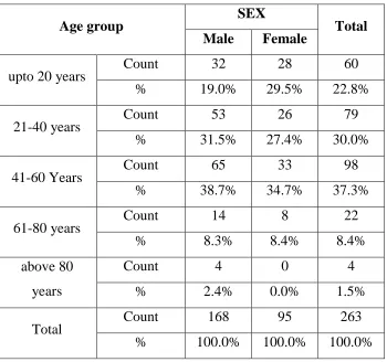
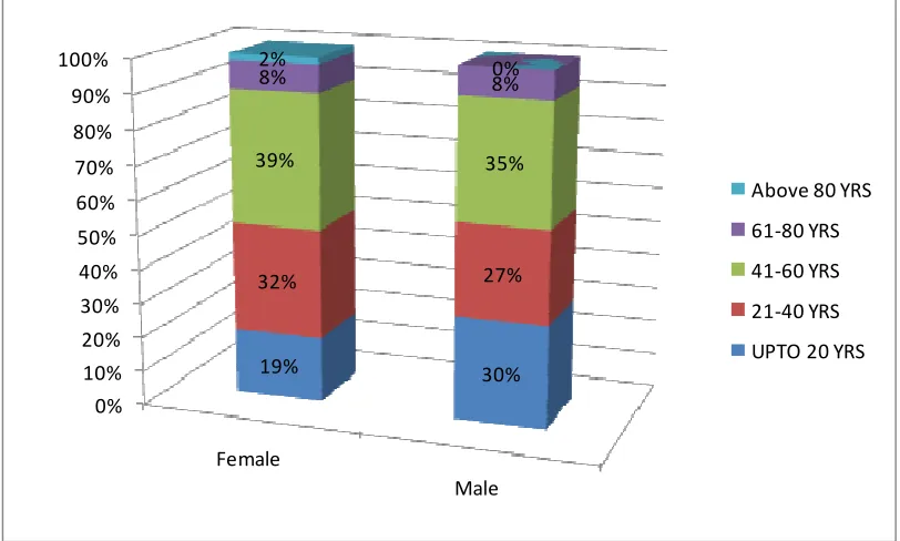
The age distribution of gliomas has been tabulated among the 263 cases.
In this study, the minimum age in which glioma was observed was 6 months.
The maximum age observed was 85 and it showed a mean age of 38 years with
a standard deviation of 19.6.
DISTRIBUTION OF AGE IN THE STUDY
N Minimum Maximum Mean
Std.
Deviation
AGE 263 0.5years 85 37.9224 19.67214
31%
69%
Gliomas
41 23%
30% 37%
8% 2%
AGE WISE DISTRIBUTION OF GLIOMAS
upto 20 years
21-40 years
41-60 Years 61-80 years
[image:57.595.107.524.405.642.2]above 80 years
TABLE NO 2 : AGE WISE DISTRIBUTION OF GLIOMAS
Age_group No of cases Percent
upto 20 years 60 22.8
21-40 years 79 30.0
41-60 Years 98 37.3
61-80 years 22 8.4
above 80 years 4 1.5
Total 263 100.0
42
The above table shows there is no specific increase in incidence of
gliomas with increasing age.
The peak incidence of gliomas was seen in 41-60 years
accounting to 98 cases( 37.3 %) .
It also shows least incidence of gliomas in the age group more than
80 years accounting to 4 cases.(1.5%).
TABLE 3 DISTRIBUTION OF HISTOLOGICAL SUBTYPES OF GLIOMAS
HISTOLOGICAL SUBTYPE NO OF
CASES
Percentage
1) PILOCYTIC ASTROCYTOMA 38 14.4
2) MYXOPAPILLARY EPENDYMOMA 2 .8
3) DESMOPLASTIC ASTROCYTOMA 1 .4
4) GEMISTOCYTIC ASTROCYTOMA 1 .4
5) DIFFUSE FIBRILLARY 18 6.8
6) DIFFUSE ASTROCYTOMA 20 7.6
7) PLEOMORPHIC 3 1.1
8) OLIGO ASTROCYTOMA 5 1.9
9) OLIGODENDROGLIOMA 20 7.6
10) GANGLIOGLIOMA 6 2.3
11) EPENDYMOMA 24 9.1
12) ANAPLASTIC ASTROCYTOMA 21 8.0
13) ANAPLASTIC OLIGO 2 .8
14) ANAPLASTIC 2 .8
15) ANAPLASTIC EPENDYMOMA 6 2.3
16) GLIOBLASTOMA MULTIFORME 94 35.7
43
From the above table it is inferred that out of the 263 gliomas studied in
a two year period in our institute Glioblastoma multiforme was the most
common one accounting for 94 cases (35.7%). Pilocytic astrocytoma was the
second most common glioma accounting for 38 cases (14.4%).
Least common were pleomorphic xanthoastrocytoma (1.1%),
oliogoastrocytoma (1.9%), anaplasticoliogoastrocytoma (0.8%), Anaplastic
oligodendroglioma (0.8)%.
CHART NO 3 : DISTRIBUTION OF GLIOMA SUBTYPES
0.0% 5.0% 10.0% 15.0% 20.0% 25.0% 30.0% 35.0% 40.0% P IL O C Y T IC A S T R O C Y T O M A M Y X O P A P IL L A R Y E P E N D Y M O M A D E S M O P L A S T IC A S T R O C Y T O M A G E M IS T O C Y T IC A S T R O C Y T O M A D IF F U S E F IB R IL L A R Y A S T R O C Y T O M A D IF F U S E A S T R O C Y T O M A P L E O M O R P H IC X A N T H O A S T R O C Y T O M A O L IG O A S T R O C Y T O M A O L IG O D E N D R O G L IO M A G A N G L IO G L IO M A E P E N D Y M O M A A N A P L A S T IC A S T R O C Y T O M A A N A P L A S T IC O L IG O A S T R O C Y T O M A A N A P L A S T IC O L IG O D E N D R O G L IO M A A N A P L A S T IC E P E N D Y M O M A G L IO B L A S T O M A M U L T IF O R M E 14.4%
0.8% 0.4% 0.4%
6.8% 7.6%
1.1% 1.9% 7.6%
2.3%
9.1% 8.0%
44
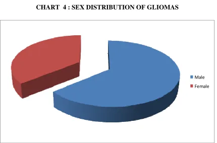
TABLE 4 : SEX DISTRIBUTION OF GLIOMAS
SEX NO OF CASES PERCENTAGE
Male 168 63.9
Female 95 36.1
Total 263 100.0
The above table shows that gliomas have a male preponderance. Out of
the 263 cases studied 63.9 % (168) of cases have occurred in males and 36.1 %
(95) of cases have occurred in females.
CHART 4 : SEX DISTRIBUTION OF GLIOMAS
Male
45
TABLE 5 : DISTRIBUTION OF SIDE INVOLVEMENT OF BRAIN IN
GLIOMAS
SIDE NO OF CASES PERCENTAGE
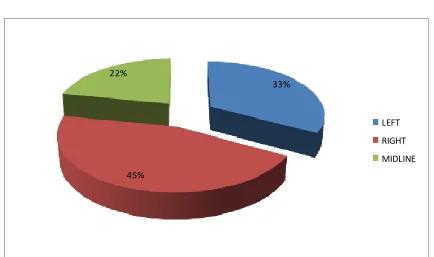
LEFT 87 33.07%
RIGHT 118 44.8%
MIDLINE 58 22.05%
TOTAL 263 100%
In the above table right side of brain was most commonly involved
44.8%, left side 33.07%, and other midline deep structures 22.05%.
CHART NO 5 : DISTRIBUTION OF SIDE INVOLVEMENT
OF BRAIN IN GLIOMAS
33%
45% 22%
LEFT
RIGHT
46
TABLE 6 : SITE WISE DISTRIBUTION OF GLIOMAS
SITE NO OF
CASES
Percentage
1. FRONTAL 52 19.8
2. FRONTOPARIETAL 23 8.7
3. FRONTO TEMPORAL 13 4.9
4. TEMPORAL 30 11.4
5. TEMPOROPARIETAL 34 12.9
6. PARIETAL 17 6.5
7. OCCIPITAL 12 4.6
8. PARIETOFRONTAL 2 .8
9. CEREBELLUM. 7 2.7
10. CPANGLE. 2 .8
11. BRAIN STEM, 5 1.9
12. CORPUS CALLOSUM 7 2.7
13. INTRAVENTRICULAR 14 5.3
14. POSTERIOR FOSSA 19 7.2
15. OPTIC NERVE 1 .4
16. OPTIC CHIASMA 1 .4
17. BASAL GANGLIA 7 2.7
18. SUPRASELLAR 3 1.1
19. ED SOL 1 .4
20. ID IM SOL 10 3.8
21. ID EM 3 1.1
47
The cerebral lobes are most commonly involved by glioma of which
frontal lobe has the highest incidence accounting to 19.8 %( 52 cases). The
second most common is the temporoparietal region accounting to 12.9 % (34
cases). The occipital lobe showed the least common involvement accounting to
4.6% (12 cases).
In the spinal cord out of the 14 cases, intradural intramedullary lesions
were most common accounting to 10 cases (3.8% 0f total cases), intradural
extra medullary was second most common accounting to 3 cases (1.1% of total
cases) and one case was extradural lesions accounted to 0.4% of total cases.
CHART NO 6 : SITE WISE DISTRIBUTION OF GLIOMAS
48
TABLE 7: DISTRIBUTION OF GLIOMAS- GRADEWISE
CHART NO 7 : DISTRIBUTION OF GLIOMAS GRADEWISE
All the 263 cases of gliomas, were grouped according to WHO
classification into four grades namely grade I - IV. WHO Grade I and II are
grouped together as low grade and Grade III and IV are grouped under high
grade in this study.
No of cases Percentage
WHO
GRADE
I 40 15.2
II 97 36.9
III 33 12.5
IV 93 35.4
Total 263 100.0
15%
37%
13% 35%
I
II
III
49
In the above table 40 cases belong to grade I, 97 cases belong to grade
II, 33cases belong to grade III, and 93 cases belong to grade IV, constituting
15.2%, 36.9%, 12.5%, 35.4% respectively.
It was found that most common grade was WHO grade II tumours
[image:65.595.102.532.256.633.2](36.9 %), followed WHO grade IV tumours (35.4%).
TABLE NO 8 : CORRELATION OF AGE AND GRADE OF GLIOMAS.
P<0.001*
The above table is given to analyse if there is any particular age
predilection for different grades of tumour.
WHO GRADE
Total
I II III IV
Age Group
upto 20 years
Count 32 22 4 2 60
% 53.3% 36.7% 6.7% 3.3% 100.0%
21-40 years
Count 6 40 16 17 79
% 7.6% 50.6% 20.3% 21.5% 100.0% 41-60
Years
Count 2 30 10 56 98
% 2.0% 30.6% 10.2% 57.1% 100.0% 61-80
years
Count 0 5 2 15 22
% 0.0% 22.7% 9.1% 68.2% 100.0% above
80 years
Count 0 0 1 3 4
% 0.0% 0.0% 25.0% 75.0% 100.0%
Total
Count 40 97 33 93 263
50
WHO grade I tumour was most common in age group of less than 20
year, (53.3%). WHO grade II was most commonly seen in age group of 21-40
years (50.6 %). WHO grade IV tumours are commonly seen in the age group
above 80 years (75% ).
CHART NO 8 : CORRELATION OF AGE GROUP WITH
GRADE OF GLIOMAS
0% 10% 20% 30% 40% 50% 60% 70% 80% 90% 100%
upto 20 years
21-40 years 41-60 Years 61-80 years above 80 years 53%
8% 2%
0% 0%
37%
51%
31%
23%
0% 7%
20%
10%
9%
25% 3%
22%
57%
68%
75%
GRADE 4
GRADE 3
GRADE 2
51
TABLE NO 9 : CORRELATION OF SUBTYPES OF GLIOMAS WITH AGE
This table shows various percentage of different types of tumours, in different age group. it also show in which age group the
particular tumor predominantly affects.
DIAGNOSIS * age_group correlation
age_group
Total upto 20
years
21-40 years
41-60 Years
61-80 years
above 80 years
PILOCYTIC ASTROCYTOMA Count 32 4 2 0 0 38
% 84.2% 10.5% 5.3% 0.0% 0.0% 100.0%
MYXOPAPILLARY EPENDYMOMA
Count 0 2 0 0 0 2
% 0.0% 100.0% 0.0% 0.0% 0.0% 100.0%
DESMOPLASTIC ASTROCYTOMA
Count 1 0 0 0 0 1
% 100.0% 0.0% 0.0% 0.0% 0.0% 100.0%
GEMISTOCYTIC ASTROCYTOMA
Count 0 1 0 0 0 1
% 0.0% 100.0% 0.0% 0.0% 0.0% 100.0%
DIFFUSE FIBRILLARY ASTROCYTOMA
Count 1 12 5 0 0 18
% 5.6% 66.7% 27.8% 0.0% 0.0% 100.0%
DIFFUSE ASTROCYTOMA Count 1 8 8 3 0 20
% 5.0% 40.0% 40.0% 15.0% 0.0% 100.0%
PLEOMORPHIC XANTHOASTROCYTOMA
Count 1 0 2 0 0 3
% 33.3% 0.0% 66.7% 0.0% 0.0% 100.0%
OLIGO ASTROCYTOMA Count 0 2 3 0 0 5
% 0.0% 40.0% 60.0% 0.0% 0.0% 100.0%
OLIGODENDROGLIOMA Count 0 9 10 1 0 20
52
GANGLIOGLIOMA Count 3 3 0 0 0 6
% 50.0% 50.0% 0.0% 0.0% 0.0% 100.0%
EPENDYMOMA Count 15 7 2 0 0 24
% 62.5% 29.2% 8.3% 0.0% 0.0% 100.0%
ANAPLASTIC ASTROCYTOMA
Count 0 12 7 1 1 21
% 0.0% 57.1% 33.3% 4.8% 4.8% 100.0%
ANAPLASTIC OLIGO ASTROCYTOMA
Count 1 0 0 1 0 2
% 50.0% 0.0% 0.0% 50.0% 0.0% 100.0%
ANAPLASTIC OLIGODENDROGLIOMA
Count 0 0 1 1 0 2
% 0.0% 0.0% 50.0% 50.0% 0.0% 100.0%
ANAPLASTIC EPENDYMOMA
Count 3 2 1 0 0 6
% 50.0% 33.3% 16.7% 0.0% 0.0% 100.0%
GLIOBLASTOMA MULTIFORME
Count 2 17 57 15 3 94
% 2.1% 18.1% 60.6% 16.0% 3.2% 100.0%
Total Count 60 79 98 22 4 263
% 22.8% 30.0% 37.3% 8.4% 1.5% 100.0%
5 3 C H A R T N O 9 : C O R R E L A T IO N O F G L IO M A T Y P E S V S A G E G R O U P 0 % 1 0 % 2 0 % 3 0 % 4 0 % 5 0 % 6 0 % 7 0 % 8 0 % 9 0 % 1 0 0 % PILOCYTIC ASTROCYTOMA MYXOPAPILLARY EPENDYMOMA DESMOPLASTIC ASTROCYTOMA
GEMISTOCYTIC ASTROCYTOMA
DIFFUSE FIBRILLARY ASTROCYTOMA
DIFFUSE ASTROCYTOMA
PLEOMORPHIC XANTHOASTROCYTOMA OLIGO ASTROCYTOMA OLIGODENDROGLIOMA GANGLIOGLIOMA EPENDYMOMA ANAPLASTIC ASTROCYTOMA
ANAPLASTIC OLIGO ASTROCYTOMA
ANAPLASTIC OLIGODENDROGLIOMA
ANAPLASTIC EPENDYMOMA
54
The commonest age group of individual tumour has been inferred from
the table. Pilocytic astrocytoma was found to commonly affect the age group of
0 to 20 years. Myxopapillary ependymoma was found c