DISSERTATION ON
“FACTORS INFLUENCING CONVERSION OF
LAPAROSCOPIC CHOLECYSTECTOMY TO OPEN
CHOLECYSTECTOMY”
submitted in partial fulfillment
of requirements of
M.S. DEGREE EXAMINATION
BRANCH - I
GENERAL SURGERY
GOVERNMENT KILPAUK MEDICAL COLLEGE
THE TAMILNADU
DR.MGR MEDICAL UNIVERSITY
CHENNAI
CERTIFICATE
This is to certify that this dissertation entitled “FACTORS INFLUENCING CONVERSION
OF LAPAROSCOPIC CHOLECYSTECTOMY TO OPEN CHOLECYSTECTOMY”
is the bonafide record work done by Dr.REUEL PAMEI NONGRUM submitted as partial
fulfillment for the requirements of M.S. Degree Examinations Branch I, General Surgery,
April 2015.
Prof P.N.Shanmuga Sundaram M.S Prof V.Ramalakshmi M.S
Head of the Department, Dissertation guide and Unit Chief Dept of General Surgery, Dept of General Surgery,
Govt Kilpauk Medical College. Govt Royapettah Hospital.
Dr N.Gunasekaran MD DTCD DEAN
DECLARATION
I Dr.REUEL PAMEI NONGRUM, solemnly declare that the dissertation submitted on the
topic “FACTORS INFLUENCING CONVERSION OF LAPAROSCOPIC
CHOLECYSTECTOMY TO OPEN CHOLECYSTECTOMY” is a bonafide work done by
me from April 2014 to September 2014, towards partial fulfillment of the requirements of M.S
Degree examinations, General Surgery, April 2015.
ACKNOWLEDGEMENT
I sincerely thank Dr N.Gunasekaran MD DTCD, The Dean, Kilpauk Medical College for
granting me permission to carry out and successfully complete my dissertation work.
I consider it a privilege to have done this study under the supervision and guidance of
Prof.V.RAMALAKSHMI MS who was a constant source of inspiration and guidance.
I would also like to thank Dr.S.THIRUNAVUKKARASU, M.S, Dr.SURESH BABU, M.S, for
their valuable support and encouragement throughout the period of study.
I would like to thank my fellow postgraduates for their suggestions and ideas and finally I would
LIST OF ABBREVIATIONS USED
CHD - Common Hepatic Duct
CBD - Common Bile Duct
LC - Laparoscopic Cholecystectomy
CCK - Cholecystokinin
HDL - High Density Lipoproteins
TPN - Total Parenteral Nutrition
USG - Ultrasonography
YAG - Yttrium Aluminium Garnet
EKG - Electrocardiogram
COPD - Chronic Obstructive Pulmonary Disease
CDCA - Chenodeoxycholic acid
UDCA - Ursodeoxycholic acid
CONTENTS
Sr . Page
1. INTRODUCTION 1
2. AIMS AND OBJECTIVES 2
3. REVIEW OF LITERATURE 3
4. METHODOLOGY 84
5. RESULTS 87
6. DISCUSSION 102
7. CONCLUSION 110
8. SUMMARY 112
9. BIBILIOGRAPHY 113
10. ANNEXURES
1. Proforma
2. Informed Consent Form for Surgery 3. Master Chart
LIST OF TABLES
Serial no Table Page No
1 Variations in the arterial supply to the gallbladder 18
2 Classification of gallstones 25
3 Selection criteria for oral bile acid dissolution therapy 48
5 Comparison of rates of conversion 78
6 Age Incidence 87
7 Sex Incidence 88
8 Sex and Surgery Outcome 90
9 Clinical Presentation 91
10 Associated Symptoms 92
11 Co-morbidities 93
12 Ultrasound findings 95
13 Pre-operative diagnosis 98
14 Duration of surgery 99
15 Duration of hospital stay 100
16 Reasons for conversion 101
17 Average duration of surgery in other studies 106
18 Conversion rate of laparoscopic cholecystectomy into open
cholecystectomy
LIST OF FIGURES
Sn Page
1. Biliary embryology 4
2. Anatomy of the gall bladder 5
3. Anatomy of the biliary tree 8
4. Anatomy of the sphincter of Oddi 9
5. Calot‟s triangle 10
6. Arterial blood supply of the gall bladder 11
7. Arterial blood supply of the extrahepatic biliary tree 12
8. Nerve supply to the extrahepatic bile tree 13
9. Rouviere‟s sulcus 15
10. Anomalies of gallbladder 16
11. Variations of cystic duct anatomy 17
12. Enterohepatic circulation 21
13. The effect of CCK on the gallbladder and the sphincter of Oddi. 22
14. Composition of bile with relationships that result in the formation of gallstones. 26
15. The physical-chemical processes involved in formation of cholesterol stones 29
16. Schematic depiction of the natural history and complications of gallstones. 33
17. Plain radiograph of gall bladder 38
18. Oral cholecystography 39
19. Normal intraoperative cholangiogram. 40
20. Ultrasonographic appearance of cholelithiasis. 42
21. CT image of gall bladder 43
22. Normal MRCP image 44
23. Endoscopic Retrograde Cholangiopancreatography 45
25. Various incisions for open cholecystectomy 53
26. Anterior rectus sheath incised 53
27 Division of rectus muscle 53
28 Division of posterior rectus sheath 53
29 Retraction of gallbladder by sponge holding forceps 54
30 Anterior layer of peritoneum incised 54
31 Ligation of cystic artery and duct 55
32 Division of peritoneal folds lateral to GB 55
33 Fundus first approach 56
34 Ligation of cystic artery after mobilization of GB 56
35 Laparoscopes 60
36 Digital laparoscopic camera unit 61
37 Optic fibre cable 61
38 Insufflator 62
39 Veress needle 63
40 Trocars 64
41 Laparoscopic trolley 65
42 Positions of the surgeon, the camera operator and assistant 67
43 Positions of the surgeon, the camera operator and assistant 67
44 Veress needle insertion 68
45 Port position for laparoscopic cholecystectomy 71
46 Dissection of cystic duct 72
47 Dissection of cystic artery 72
48 Dissection of cystic pedicle 73
49 Dissection of cystic duct by blunt dissection 73
50 Clipping of cystic artery 74
51 Clipping of cystic duct 74
52 Dissection of gallbladder from its bed 74
54 Mobile stack for laparoscopy 83
55 Operative room set-up 83
56 Graphical representation of age incidence 88
57 Graphical representation of sex incidence 89
58 Graphical Representation of age and sex incidence 89
59 Graphical Representation of Sex and Surgery Outcome 90
60 Graph showing clinical presentation 91
61 Graph showing associated symptoms 92
62 Graph showing Co-morbidities 93
63 Graphical representation of conversion among diabetic patients 94
64 Graphical representation of conversion among hypertensive patients 94
65 Graph showing ultrasound findings 95
66 Graphical representation of conversion in patients with single calculi 96
67 Graphical representation of conversion in patients with multiple calculi 96
68 Graphical representation of conversion in patients with peri-cholecystic fluid 97
69 Graphical representation of conversion in patients with GB wall thickening 97
70 Graphical representation of indication for laparoscopic cholecystectomy 98
71 Graphical representation of duration of surgery 99
72 Graphical representation of post-operative hospital stay 100
1
INTRODUCTION
Gallstone disease are a common gastrointestinal illness in the general population which
frequently requires hospitalization . The prevalence is around 11% to 36% . The treatment of
choice for patients with symptomatic cholelithiasis is Laparoscopic cholecystectomy. Previously
Open cholecystectomy was frequently performed but that has given way to a laparoscopic
approach.The advantages of laparoscopic cholecystectomy are the avoidance of large incision,
shortened hospital stay and earlier recovery.
The patients condition, the surgeon‟s level of experience, and technical factors can play a major
role in the decision for conversion. Inability to define the anatomy and difficult dissection are the
leading cause for conversion followed by other complications like bleeding.
The conversion rate for elective laparoscopic cholecystectomy is around 5%, whereas the
conversion rate in the setting of complications like acute cholecystitis is around 30%.
The goal of this study was to determine the conversion rate and identify the factors
responsible for conversion of laparoscopic cholecystectomy to open cholecystectomy. Hence,
these findings will allow us to :-
1. To identify a patient‟s risk for conversion based on preoperative information leading to
more meaningful and accurate preoperative counseling
2. Improved operating room scheduling and efficiency
2
AIMS AND OBJECTIVES
1. To identify the risk factors predictive of conversion of laparoscopic cholecystectomy
to open surgery .
2. To determine the rate of conversion of laparoscopic cholecystectomy to open surgery
3
REVIEW OF LITERATURE
Historical Review
The first to describe calculi in human liverswas Alexander of Tralles (525-605), a physician of
the Byzantine Empire.4
The first clinical description of gallstone disease suggested by Gordon Taylor (1937)
was recorded in the 4th century BC. An accurate description of human gallstones was given by
Vesalius, concluding that they represented a disease process.
Joenisius was the first surgeon to successfully perform cholecystolithotomy in 1676, but the
extracted gallstones was apparently extracted from a biliary fistula of the abdominal wall
following spontaneous drainage of the abscess.
The first successful cholecystectomy was performed by Carl Langenbunch in 1882, a noted
German surgeon and for more than 100 years it was the standard treatment for symptomatic
gallbladder stones 2,1
Laparoscopic Cholecystectomy
The first laparoscopic cholecystectomy was performed in 1987 by Philippe Mouret.6
In 1988 Mc Kernan and Saye also perfomed laparoscopic cholecystectomy in the United States.7
In fact, in 1985, a German professor named Erich Muhe of Boblingen performed the first lap
Cholecystectomy but his technique was not appreciated by his colleagues at the German surgical
Society.8 Only in 1999 he was recognized by SAGES. In India the first laparoscopic
cholocystectomy was performed in Mumbai , JJ Hospital 1990 .Within a span of few years
4
EMBRYOLOGY
The liver arises from the ventral surface of duodenal foregut as a diverticulum in the fourth
week close to its midgut junction. The diverticulum is lined with endoderm , it grows vertically
and cranially into the septum transversum. Two solid hepatic buds diverges from the tip , the
right and left lobes of liver .The buds develops into epithelial and branches and anastomoses to
form a closed meshwork. Sinusoids arises from intervals of meshwork which are filled with
blood. The bile duct arises from the original diverticulum and the cystic duct and gall bladder
arises as an outgrowth from its distal part. It is solid at first but later it canalized.
5
ANATOMY
The gallbladder is a pear-shaped sac lying on the visceral surface of the right lobe of the liver in a fossa between the right and quadrate lobes.
It has:
a rounded end (fundus of gallbladder), which may project from the inferior border of
the liver,
a major part in the fossa (body of gallbladder), which may be against the transverse
colon and the superior part of the duodenum;
a narrow part (neck of gallbladder) with mucosal folds forming the spiral fold.[11]
6
The gallbladder is around 7 to 10 cm in length and varies from 2.5 to 3.5 cm in width.
It has a capacity of 50 to 60 ml of bile.
The Hartmann's pouch is a peculiar bulge of the infundibulum that lies close to
the gallbladder's neck. Here gallstone commonly lodges. The neck joins the cystic duct.
The gallbladder is made up of five layers.
1. The innermost layer is the epithelium,
2. The lamina propria,
3. The smooth muscle,
4. The perimuscular subserosal connective tissue, and
5. The serosa.
The gallbladder is devoid of muscularis mucosa or submucosa.
The lamina propria contains :-
Nerve fibers,
Vessels,
Lymphatics,
Elastic fibers,
Loose connective tissue, and
Occasional mast cells and macrophages.
The muscle layer is composed of loose arrangement of circular, longitudinal, and oblique fibers
7
Rokitansky-Aschoff sinuses are invaginations of epithelium into the lamina
propria, muscle, and subserosal connective tissue. The ducts of Luschka are tiny bile
ducts found around the muscle layer on the hepatic side of the gallbladder. [12]
Anatomy of biliary tract
The right and left hepatic ducts unite outside the liver in the porta hepatis to form the
common hepatic duct (CHD). The cystic duct arises from the gallbladder and joins the CHD to
form the common bile duct (CBD) . The length of the cystic duct is averages between 2 to 4 cm.
The cystic duct contains a number of mucosal folds, similar to those found in the neck of the
GB. These spiral folds are referred to as valves of Heister and they do not have
8
The common bile duct also referred to as CBD runs in between the layers of the lesser omentum,
It lies anterior to the portal vein and to the right side of the hepatic artery. It passes behind the1st
part of the duodenum in a groove on the back of the head of the pancreas, it enters the 2nd part of
the duodenum. The duct runs obliquely through the posterior-medial wall, and it usually joins the
main pancreatic duct to form the Ampulla of Vater . The mucous membranes made to bulge
inwards in the ampulla to form aneminence: the duodenal papilla. The bile and pancreaticducts
open into the duodenum separately in about 10-15% of subjects. The dimensions of the common
bile duct is usually less than 11 mm and anything greater than 18 mm is considered
pathological.[13]
9
The duodenal portion of the CBD is surrounded by thickened longitudinal and circular muscle
fibres. This is called the sphincter of Oddi. [14]
10
Calot’s Triangle
In 1891, Calot described what is now known as The Calot‟s Triangle .
It is a triangular anatomic region formed by the :-
Common hepatic duct medially,
Cystic duct laterally,
Cystic artery superiorly.
The Calot's triangle at present is considered by most to comprise the triangular area with an
upper boundary formed by the inferior margin of the right lobe of the liver, rather than the cystic
artery. During cholecystectomy, clear visualization of the hepatocystic triangle is important for
accurate identification of the structures within the triangle.[12]
11
Blood supply
The cystic artery supplies the Gall Bladder . It is a branch of the Hepatic artery . It is large and
tortuous and variable in its location. Smaller blood vessels enter from the liver through the GB
fossa. The cystic vein drains into the portal venous system and constitutes its venous drainage.
12
The arterial blood supply to the supraduodenal part of bile duct is generally by 2 main(axial)
vessels, which run along the bile duct in 3‟o‟ clock and 9‟o‟ clock position. [15] These are
supplied predominantly by the retroduodenal artery from below, and the right hepatic artery from
above. This pattern of arterial supply therefore would explain why vascular damage results in
bile duct stricturing.
Figure 7: Arterial blood supply of the extrahepatic biliary tree
13
Lymphatics
There are quite a number of lymphatic vessels in the sub mucous and sub peritoneal layers.
They drain through the cystic gland at the neck of the gallbladder to glands along the
CBD, where they anastomose with lymphatic from the head of the
pancreas.
Nerve supply
The gallbladder and bile ducts are innervated by both the parasympathetic and sympathetic
system. [16]
14
Laparoscopic anatomy [17]
With the advent and popularity of LC , a new look and insights into the biliary anatomy began to
emerge especially of the Calot‟s triangle area and the term “laparoscopic anatomy” has actually
found a place even in anatomy texts.
The different anatomical „laparoscopic view‟ of the area during cholecystectomy and especially
in the area around the Calot‟s triangle does contribute to misidentification of structures. During
laparoscopic procedure due to different methods of retraction the Calot‟s triangle tend to be
distorted by actually flattening it rather than opening it out. Also the reluctance to perform (due
to difficulty) a fundus first cholecystectomy during the laparoscopic procedure as opposed to
open procedure also contributes to the some lack of exposure of the Calot‟s triangle.
Finally the most popular approach is the „posterior‟ or „reverse‟ dissection of the Calot‟s triangle,
again gives a different view of the area and since gallbladder is flipped over during this method
it may lead to further anatomical distortion. The Rouviere‟s sulcus is fissure on the liver between
the right lobe and caudate process and is seen clearly during LC while doing a posterior
dissection approach in majority of the patients.
It is at the level of the porta hepatic where the right pedicle enters the liver. It has been
emphasized that all dissection should be kept to a level above (or anterior) to the sulcus to
prevent injury to the bile duct.
Also, very importantly this being an “extrabiliary” reference point it does not get affected by
distortion due to pathology. Similarly, a clear veiw of the junction of cystic duct
15
and the liver and clear of any structure other than the cystic artery is also recommended as a
vital step to prevent bile duct injury.
16
ANOMALIES OF GALLBLADDER [12]
The Gallbladder may be :-
1. Septate, transversely or longitudinally placed.
2. Double with a single cystic duct.
3. Double with separate ducts opening into hepatic or common or both ducts.
4. The serosa may be separate or common.
5. Small ducts may connect gallbladder with liver. Usually these become obliterated.
6. Phrygian cap deformity is the most common congenital abnormality.
7. It may have a mesentery; which can be found on the left of the falciform ligament or
intrahepatic
Phrygian gallbladder. Gallbladder diverticulum
Double gallbladder with separate cystic ducts Double gallbladder with common cystic duct
17
VARIATIONS OF CYSTIC DUCT ANATOMY [18]
Figure 11: Variations of cystic duct anatomy
A. Low junction between the cystic duct and common hepatic duct.
B. Cystic duct adherent to the common hepatic duct.
C. High junction between the cystic and the common hepatic duct.
D. The cystic duct drains into right hepatic duct.
E. Long cystic duct that joins the common hepatic duct behind the duodenum.
F. Absence of the cystic duct.
G. The cystic duct crosses posterior to the common hepatic duct and joins it anteriorly.
18
19
PHYSIOLOGY [19]
Bile Formation and Composition
The bile is continuously produce in the liver and excreted out into the bile canaliculi. The
normal adult produces within the liver 500 to 1000 mL of bile a day. The bile secretion is
controlled by neurogenic, humoral, and chemical stimuli. Vagal stimulation increases bile
secretion, whereas splanchnic nerve stimulation decreases bile flow. Hydrochloric acid, partly
digest proteins, and fatty acids in the duodenum and that stimulate the release of secretin from
the duodenum which in turn, increases bile production and bile flow. Bile flows from the liver
to the hepatic ducts, to the common hepatic duct, through the common bile duct, and finally into
the duodenum.
Bile is composition is that of water, electrolytes, bile salts, proteins, lipids, and bile
pigments. Sodium, potassium, calcium, and chlorine have the same concentration in
bile as in plasma or extracellular fluid. The pH of hepatic bile is usually neutral or
slightly alkaline, but varies with diet; an increase in protein shifts the bile to a more
acidic pH.
The liver synthesizes primary bile salts, cholate and chenodeoxycholate from cholesterol. These
are conjugated there with taurine and glycine, and act as anions within the bile (bile acids) that
are balanced by sodium. Bile salts are excreted into the bile by the hepatocyte and aid in the
digestion and absorption of fats in the intestines. About 80% of the conjugated bile acids in the
intestines, are absorbed in the terminal ileum. The remainder is dehydroxylated (deconjugated)
by gut bacteria which forms secondary bile acids deoxycholate and lithocholate. These are
absorbed in the colon and transported to the liver, conjugated, and secreted in the bile.
Eventually, about 95% of the bile acid pool is reabsorbed and returned via the portal venous
system to the liver, this is known as enterohepatic circulation. 5% is excreted in
20
The principal lipids found in bile which are excreted by the liver are cholesterol and
phospholipids. There synthesis is in part regulated by bile acids. The bile colour is due to
the presence of the pigment bilirubin diglucuronide, which is the metabolic product from the
breakdown of hemoglobin, and its concentrations in bile is 100 times greater than in plasma.
Once in the intestine, bacteria convert the bile into urobilinogen, a small fraction of which is
absorbed and secreted again into the bile.
Gallbladder Function
The gallbladder, the bile ducts, and the sphincter of Oddi act together to store and regulate bile
flow. The gallbladder‟s main function is to concentrate and store hepatic bile and to deliver it
into the duodenum in response to a meal.
Absorption and Secretion
In fasting state, approximately 80% of the bile is stored in the gallbladder. This storage is made
possible because of the absorptive capacity of the gallbladder, its mucosa has the greatest
absorptive power per unit area of any structure in the body. It rapidly absorbs elecytrolytes like
sodium, chloride, and water against a significant concentration gradients, the bile is concentrated
upto 10-fold and leading to a marked change in the composition of bile. This rapid absorption
prevent a rise in biliary pressure within the biliary tree in normal circumstances. Gradual
relaxation and emptying of the gallbladder during the fasting period also plays a role in
21
Figure 12: Enterohepatic circulation
Motor Activity
Gallbladder filling is facilitated by the tonic contraction of the sphincter of Oddi,
22
fasting, the gallbladder does not only fill passively. In association with phase II ofthe
inter-digestive migrating myenteric motor complex in the gut, the gallbladder empties small volumes
of bile repeatedly into the duodenum. This process is mediated partly by the hormone motilin.
During a meal, the gallbladder empties by a coordinated motor response of gallbladder
contraction and sphincter of Oddi relaxation. Cholecystokinin (CCK) is one of the main stimuli
responsible for gallbladder emptying. CCK is released endogenously from within the duodenal
mucosa in response to a meal.
When stimulated by eating, the gallbladder empties 50 to 70% of the bile within 30 to 40
minutes. Over the 60 to 90 minutes following emptying, the gallbladder refills gradually.
Thiscorrelates with a reduced CCK level. Other hormonal and neural pathways also are involved
in the coordinated action of the gallbladder and the sphincter of Oddi. Defects in the motor
activity of the gallbladder are may play an important role in cholesterol nucleation and gallstone
formation.
[image:33.612.143.461.482.644.2]
Figure 13: The effect of cholecystokinin on the gallbladder and the sphincter of Oddi.
23
GALLSTONES
INCIDENCE:
The most common biliary pathology are gallstones . The incidence of gallstone disease varies
throughout the world. By the age of 75, about 35% of women and 20% of men would have
developed gallstones. The incidence of cholesterol gallstones is increasing in Asia which may be
related to environmental and dietary habits. Incidence of asymptomatic gallstones is only about
10% and many patients will have developed symptoms 5 years after discovery. Most of the gall
stones are cholesterol stones in a functioning gall bladder. Gall stone disease is a relatively
common problem in India particularly in North India. It is estimated that more than 60% of these
patients have cholesterol stones. Recent studies from south India have shown pigment and
mixed variety of gall stones to be more common (more than 90 %) as compared to cholesterol
24
RISK FACTOR ASSOCIATED WITH GALLSTONE FORMATION
:[20]25
There is a dual pattern of prevalence in India. North Indians are more prone to cholelithiasis
according to some studies than South Indians. The disease process has a different nature in North
India and South India. Cholesterol stones form the majority of gallstones in North India. In
contrast to South India, where pigment stones are more frequent.
CLASSIFICATION OF GALLSTONES:
26
PATHOGENESIS:[21]
Gallstones are mainly composed of cholesterol, bilirubin, and calcium salts, with small amounts
of protein and other materials. In Western countries cholesterol is the main constituent of more
than three quarters of gallstones, and these stones many of them are more than 80 percent
cholesterol. Non-cholesterol stones are categorized as black or brown pigment stones, consisting
of calcium salts of bilirubin. About 15 percent of gallstones can be seen on a plain abdominal
radiograph, and pigment stones constitutes two thirds of these . Calcification which is visible
only on the rim occurs usually in cholesterol stones. Cholesterol gallstones formation results
when the cholesterol concentration in bile exceeds the ability of bile to hold it in solution i.e.
reaches its saturation point, so that crystals form and grow as stones. The lithogenicity of bile are
affected by various factors.
[image:37.612.191.400.406.577.2]
27
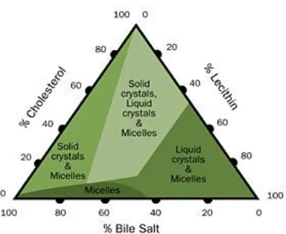
Cholesterol gallstones:
The three major factors that determine the formation of cholesterol gallstones are
1. Altered hepatic bile,
2. Nucleation of cholesterol monohydrate crystals and
3. Impaired function of gallbladder.
Altered hepatic bile composition
Bile is composed 85-95% water. Cholesterol must be maintained in solution, it is secreted from
the canalicular membrane in unilamellar phospholipid vesicles. In unsaturated hepatic bile with
cholesterol and containing sufficient bile acid, these vesicles are solubilised into mixed lipid
micelles.
These have a hydrophobic interior and a hydrophilic external surface . Cholesterolis incorporated
into the hydrophobic interior. Phospholipid grow as they are inserted into the walls of the
micelles. These „mixed micelles‟ are therefore able to hold more cholesterol in a stable
thermodynamic state. This represents the situation with a low cholesterol saturation index .
When bile as a high cholesterol saturation index, i.e. it is supersaturated with cholesterol, or bile
acid concentrations are low the excess cholesterol cannot be transported in mixed micelles and
thus a unilamellar phospholipids vesicles remains which are not stable and can sometimes
aggregate. They fuse to form large multilamellar vesicles from which cholesterol monohydrate
crystals can nucleate. In majority of the patients the primary defect is reduced hepatic secretion
28
synthesis is surprised as a result of a more frequent circulation within the entero hepatic
circulation.
Cholesterol nucleation
The most crucial step in the process leading to gallstone formation is nucleation of cholesterol
monohydrate crystals from multilamellar vesicles . The differentiating feature between those
who form gallstones and those who do not is the ability of the bile to promote or inhibit
nucleation rather than the degree of cholesterol supersaturation.
The time taken for this process (nucleation time) is significantly shorter in those with gallstones
than in those without and in those with multiple as opposed to solitary stones.
Ursodeoxycholic acid decreases cholesterol saturation and prolongs the nucleating time, which
may help in the prevention of gallstones recurrence. The gallbladder is filled with hepatic bile
during fasting, concentrates the bile and excretes the concentrated bile into the duodenum during
meals. It must empty itself of sludge and debris that might initiate stone formation, especially in
the patient with supersaturated bile with cholesterol and a short nucleation time. Hepatic bile is
stored in the gallbladder and concentrated by the absorption of sodium, chloride, bicarbonate and
water. The biliary concentration of bile salts, bilirubin and cholesterol, may rise 10 fold or more.
Gallbladder contraction is under cholinergic and hormonal control. Cholecystokinin (CCK),
secreted from the intestine, stimulates contraction and empties the gallbladder and increases fluid
secretion and dilution of gallbladder contents. Other hormones that influence the gallbladder
includes motilin (stimulatory) and somatostatin (inhibitory). The relationship between impaired
29
parenteral nutrition has pointed out that gallbladder stasis plays an important role in the
formation of gallstones.
Figure 15: The physical-chemical processes involved in formation of cholesterol stones
Biliary sludge
It consists of calcium, bilirubin and cholesterol. It can occurs in the presence or absence of
gallstones. It can also cause biliary-type pain.
Role of infection
Infection has little importance in cholesterol gallstone formation. The bile is usually sterile.
Biliary infection can lead to brown-pigment stone formation. These stones may contain bacteria
30
Age
The prevalence of gallstone disease increases with advancing years, which maybe to increased
cholesterol content in bile. Pigment and cholesterol type stones are reported in childhood.
Genetics
There is a familial predisposition to the incidence of gallstones in families with known history of
gallstone disease irrespective of their age and weight.
Sex and oestrogens
Gallstones are two times as common in women as in men. There is higher incidence in
multiparous than in nulliparous women. Incomplete emptying of the gallbladder in late
pregnancy leads to retention of cholesterol crystals; this favours gallstone formation.Women
younger than 30 years, gallbladder stones are commonly associated with pregnancy and obesity.
When women are placed on birth control pills the bile becomes more lithogenic. Women on
long-term OCP‟s have a twice the incidence of gallbladder disease over controls.
Post-menopausal women on oestrogen containing drugs have 2.5 times increase in gallbladder
disease.
Obesity
There is increased incidence of gallstone disease in obese individuals than in the general
population and is particularly an important risk factor in women less than 50 years old (fat,
fertile, female of forty). Obesity is associated with increased cholesterol synthesis and excretion.
31
Dietary factors
Dietary fibre deficiency is associated with increased incidence of gallstone disease. This is due
to increase in secondary bile acids, and render it more lithogenic, carbohydrate in refined form
increases saturation of biliary cholesterol. A moderate amount of alcohol has a protective action
against gallstones. Vegetarians have fewerincidence ofgallstones .
Serum factors
Low HDL levels and high triglyceride levels have the highest risk of gallstones (both cholesterol
and pigment) .
Pigment gallstones:
This term is refers to stones containing less than 25% cholesterol. They may be irregular or
crystalline or smooth and amorphous on cross-section. They represent 25% of gallstones
removed at cholecystectomy.
There are two types of pigmented stones: black and brown.
Black pigment stones are largely composed of an insoluble black pigment polymer mixed with calcium phosphate and carbonate. They are usually seen inside the gallbladder. They are
associated with chronic hemolysis, like hereditary spherocytosis or sickle cell disease, and
mechanical prostheses in the circulation.
Brown pigment stones have calcium bilirubinate and are also composed of calcium palmitate and cholesterol as their other major constituents. They are usually radiolucent. They are usually
found in the gallbladder,intra-hepatic and extra-hepatic bile ducts. They have a 10% association
with stricture, sclerosing cholangitis and Caroli‟s syndrome. Recurrent bile duct stones are
32
In Oriental countries, parasitic infestations of the biliary tract by Clonorchis sinensis or Ascaris
Lumbricoides are associated with these stones.
CLINICAL FEATURES:
33
Figure 16: Schematic depiction of the natural history and complications of gallstones.
The percentages indicate the approximate frequencies of complications that occur in
untreated patients, based on natural history data. The most frequent outcome is for the
patient with a stone to remain asymptomatic throughout life (1). Biliary pain (2),
acute cholecystitis (3), cholangitis (5), and pancreatitis (5) are the most common
complications. Mirizzi's syndrome (4), cholecystoenteric fistula (6), Bouveret's
34
SYMPTOMATIC GALLSTONES: [23]
Chronic Calculous Cholecystitis
Chronic cholecystitis is referred to as an ongoing inflammation with recurrent episodes of biliary
colic due to cystic duct obstruction . Histologically, increase in subepithelial and subserosal
fibrosis and a mononuclear cell infiltrate is characteristic of chronic cholecystitis.
The main symptom of chronic symptomatic cholecystitis is pain, usually referred to as biliary
colic. It is constant pain and lasts 1 to 5 hours which may be accompanied by nausea, vomiting ,
bloating and belching which are present in 50% of cases. Rarely fever and jaundice is seen in
simple biliary colic.During a biliary colic episode the patient might have mild right upper
quadrant tenderness.
The standard diagnostic exam for gallstones is an abdominal ultrasound. Cholecystectomy is
indicated if there is recurrent attacks of biliary colic and on ultrasound sludge is detected on two
or more occasions. Other causes for typical biliary attack are cholesterolosis and
adenomyomatosis of the gallbladder. Cholesterolosis is caused by the accumulation of
cholesterol in macrophages in the gallbladder mucosa, either locally or as polyps. It is also
known as “strawberry gallbladder.”
Elective laparoscopic cholecystectomy is the standard treatment for patients with symptomatic
cholelithiasis. Patients are advised to avoid dietary fats and heavy meals while awaiting surgery.
Diabetic patients have a higher risk for acute cholecystitis or even gangrenous cholecystitis
35
Acute Calculous Cholecystitis:
Gallstone obstruction of the cystic duct causing biliary colic is the initial event in acute
cholecystitis in 90%-95% of cases. Obstruction of the cystic duct causes the gallbladder to be
distended, and the gallbladder wall then becomes inflamed and edematous. In the most cases, the
gallstone dislodges, and the inflammation resolves by itself.
The most common symptom of acute cholecystitis is right upper quadrant pain, similar in
severity but has a much longer duration than pain from previous biliary colic episodes. Other
common symptoms are fever, nausea, and vomiting. On physical exam, right hypochondrium
tenderness and guarding inferior to the right costal margin are usually elicited, distinguishing the
episode from simple biliary colic.
The most useful radiographic test for diagnosis is ultrasonogram. It has good sensitivity for
identifying gallstones. Radionuclide scanning is used less frequently and mostly in atypical
cases. Lack of filling of the radiotracer (99mTc-HIDA) in the gallbladder after 4 hours indicates
obstruction in cystic duct.
After the diagnosis is confirmed treatment includes, IV fluids, antibiotics, and analgesia.
Antibiotics should cover gram-negative aerobes and anaerobes.Laparoscopic cholecystectomy is
the gold standard treatment for patients with acute cholecystitis. Early cholecystectomy within 2
to 3 days of presentation is preferred over interval or delayed cholecystectomy which is
36
Choledocholithiasis
Common bile duct stones are classified into primary and secondary depending on their point of
origin. Primary stones are those that are formed in the CBD itself and secondary stones are those
that have been formed in the gallbladder and have migrated in to the CBD, 6% to 12% of
patients with stones in the gallbladder. The secondary stones are of the brown pigment type. The
primary stones are associated with biliary stasis and infection .They are commonly seen in Asian
populations. Common duct stones are also defined as retained stones if they are found within 2
years of cholecystectomy, recurrent if they have detected more than 2 years after
cholecystectomy.
Common bile duct stones are usually silent and are often incidentally discovered. Biliary
obstruction in these patients are transient, and laboratory tests are usually normal. Clinical
features of CBD obstruction include biliary colic associated with progressive jaundice, clay
colour stools, and darkening of the urine. Fever and chills may be present in patient‟s cholangitis
and constitute charcots triad. Ultrasonography, is the first test to be done,it can detect stones in
the gallbladder and measure the diameter of the common bile duct. A dilated bile duct (>8 mm in
diameter) in a patient with gallstones presenting with jaundice and biliary pain is highly
suggestive of choledocholithiasis. MRCP is a uselful imaging modality and it provides excellent
anatomic detail with sensitivity of 95% and specificity of 98%. ERCP is the diagnostic and also
therapeutic test of choice in patients with distal common bile duct stones. Endoscopic
cholangiography helps in confirmation of diagnosis in patients with suspected common bile duct
37
cholecystectomy. We should consider prompt cholecystectomy after endoscopic clearance of the
common bile duct during the hospital admission if the patient is fit for surgery.
A single sitting procedure can be performed where laparoscopic common bile duct exploration
through the cystic duct after cholecystectomy or with formal choledochotomy and insertion of
T-tube which allows retrieval of stone during the same procedure. The purpose of the T T-tube is to
aid in postoperative radiologic stone extraction as it allows easy access to the biliary system.
Gallstone Pancreatitis
Another common complication of CBD stones is the obstruction of the main pancreatic duct by
an impacted stone or temporary obstruction of the stone passing through the ampulla leading to
gallstone pancreatitis. Treatment in acute cases includes ERCP with sphincterotomy and stone
extraction. Once the pancreatitis has subsided, cholecystectomy is advised during the same
admission.
INVESTIGATIONS:
There are many different array of investigations available for the diagnosis of gallstones:
38
Blood investigations:
Serum transaminases will be elevated in nearly 50% of patients with symptomatic gallstones.
White blood cell count can be elevated in acute cases. Serum lipase and amylase are useful in
patients with suspected gallstone pancreatitis. In patients with liver dysfunction due to severe
jauncie, prothrombin time (PT) and activated partial thromboplastin time (aPTT) might be
abnormal.
Plain X-ray abdomen:
X-ray abdomen is considered a preliminary test. Only 10% of gallstones are radio opaque.A
porcelain gallbladder due to its extensive wall classification can be seen on a plain x-ray film.It
is an indication of urgent cholecystectomy due to its pre malignant potential.
Figure 17A: Plain radiograph show Figure 17B – Porcelain Gallbladder
[image:49.612.104.274.349.619.2]39
Oral Cholecystography:
40
Cholangiography:
Cholangiography is useful in proper assessment of the anatomy of the biliary system and helps
avoid CBD injury during cholecystectomy. An intra-operative cholangiography aids in the
dissection of the function between cystic and common bile ducts which is very important in
cases where anatomic land marks are not clearly identified or in ductal anatomy variations .
41
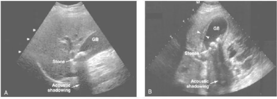
Ultrasonography:
It is the initial investigation in a suspected case of any disease involving the biliary tree. Its
advantage lies in the fact that it is non-invasive, no radiation exposure and it can be done on
critically ill patients [25]. It can detect stones larger than 1mm in diameter. It can also rule out
other causes of right hypochondrium pain like abscess and malignancy. It can also detect
dilatation in CBD diameter in cholidocholithiasis .
In ultrasound gallstones are seen as moveable echogenic spot that produces a shadow. They are
acoustically dense and produce an acoustic shadow. Ultrasound can also detect gallbladder
sludge which does not produce shadows but have tiny echogenic spots. The only disadvantage is
42
Figure 20(A): Typical ultrasonographic appearance of gallstones casting an acoustic shadow.
Figure 20(B): Cholelithiasis in the setting of acute cholecystitis. Multiple gallstones can be seen
within the gallbladder lumen with associate gallbladder wall thickened (arrowheads).
Computed Tomography
It is not the first-line test. Gallstones are frequently missed by routine CT. It has low sensitivity
but it does provide important information regarding the nature, extent, and anatomy of the biliary
tree and the associated surrounding structures. In general, this test provides more useful
information than ultrasound in the setting of extra hepatic obstruction which may be due to
causes other than cholidocholithiasis. Disadvantage for CT scanning include ionizing radiation
[image:53.612.76.541.74.241.2]43
44
Magnetic Resonance Cholangio Pancreatography (MRCP)
Recently,(MRCP) Magnetic Resonance cholangio-pancreatography is another alternative to
(ERCP) endoscopic retrograde cholangiography .It is non- invasive and non-ionizing but lacks
therapeutic capability. In MRCP native high signal intensity of fluid on T2-weighted images
permits imaging of the biliary tree. Sensitivity of MRI cholangiography for detecting gallstones
and cbd stones are over 90 %. [27]
45
Endoscopic Retrograde Cholangio Pancreatography (ERCP): [28]
Here a side-viewing endoscope is used, by fluoroscopy the common bile duct can be cannulated
and a cholangiogram perform. The procedure is done under i.v sedation for the patient. The
advantages of ERCP is that you can directly view the ampullary region and have direct access to
the CBD. It also has a therapeutic role. It is particularly useful for distal CBD stones specially
when it is associated with cholangitis or obstructive jaundice or gallstone pancreatitis. Over here
it is the procedure of choice. Once the distal cbd stone is visualized, sphinterotomy is done and
extraction of the stone with dormia basket is done.
[image:56.612.198.407.318.498.2]
Figure 23: Endoscopic Retrograde cholangiopancreatography demonstrating stone in the CBD.
Hepatobiliary Scintigraphy:
HIDA scan is the most sensitive and specific test for acute cholecystitis – calculus and
acalculous. It helps in rapid assessment of gallbladder function. HIDA - hydoxyiminodiacetic
acid is a short lived technetium – 99m bound to immunodiacetic acids which are excreted into
46
an image is produce. Failure to detect these rays suggest obstruction at the neck of the
gallbladder as in acute cholecystitis. HIDA scan is also useful in the diagnosis of biliary
dyskinesia. HIDA scan is not useful in providing anatomical details of the biliary tree.
Figure 24: Cholescintigraphy demonstrating an obstructed cystic duct characteristic of acute
cholecystitis. The failure of the gallbladder to be visualized as a hot spot within 30 to 60
47
MANAGEMENT OF GALLSTONE DISEASES[30]
Cases have been reported that contact dissolution of gallstones have been used using solvents
and percutaneous cholecystolithotomy technique but it has been observed that these modalities
are not superior to oral dissolution , laparoscopic cholecystectomy technique or shock wave
lithotripsy. Thus they were abandoned. Now the mainstay medical treatment for gallstones is oral
dissolution using ursodeoxycholic acid , which may or may not be used with shock wave
lithotripsy.
1. Dissolution Therapy
Here the rationale is reversing the supersaturation of bile with cholesterol. The main mechanism
is making the cholesterol stones more soluble by addition of certain agents . The two most
commonly used agents ar chenodeoxycholic acid and ursodeoxycholic acid which dissolve
gallstones by desaturating bile and decreasing biliary cholesterol secretion. These agents via
micellar solubilization encourage the removal of cholesterol from stones or encourage the
formation of a liquid crystalline phase, or both.
The first bile acid used for gallstone dissolution was Chenodeoxycholic acid but because of its
side effects like diarrhea and altered liver function it has been abandoned. Ursodeoxycholic acid
is more commonly used now and is well tolerated. Patients with uncomplicated cholilithiasis
should be considered for oral dissolution therapy. It is also important that the gallbladder
function should be normal and the cystic duct is patent to allow unobstructed passage of
unsaturated bile and stones to pass out from the gallbladder. Oral dissolution therapy is used only
48
Table 3: Selection Criteria for Oral Bile Acid Dissolution Therapy
The preferred drug for oral dissolution therapy is Ursodeoxycholic acid (ursodiol) . Dosage is 10
to 15 mg/kg of body weight per day. Night time dosing is preferred. Treatment should continue
until documentation of stone dissolution by two consecutive negative ultrasonograms one month
49
2. Extracorporeal shock wave lithotripsy (ESWL)
In 1985 Sauerbruch was the first to apply the application of extracorporeal shock-wave
lithotripsy to gallstone disease.Diminishing the surface-to-volume ratio of a stone is the rationale
behind this, thereby increasing the efficacy of oral dissolution. This in turn decreases stone size
and allows it and the debris to pass directly unhindered from the gallbladder into the intestine. It
involves delivery of focused high-pressure sound waves to the gallstones. Passage of the shock
wave causes cavitation at the anterior surface of the stone by liberating compressive and tensile
forces on the stone, thereby leading to stone fragmentation. Size, architecture and
microcrystalline structure are the factors that influence fragmentation.
Criteria for selection of patients for ESWL are :
1. Normal gallbladder function
2. Cystic duct patency
3. Mild uncomplicated biliary pain
4. Cholesterol stones
Contraindications:
1. Pregnant patients
2. Bleeding disorders
3. Patients on anti-coagulants
4. Large stones
Side effects are :
1. Petechiae over the skin at the site of shock-wave delivery (8%)
2. Hematuria (4%), and
50
4. Biliary pain
5. Cystic duct obstruction ( 5%)
6. Biliary pancreatitis (< 2%).
OPERATIVE MANAGEMENT
Cholecystectomy can done by 1.Open and 2. Laparoscopic methods
The indications are the same for both techniques.[31] These are:-
1. Symptomatic gallstones causing
Mucocele of the gallbladder
Repeated episodes of biliary pain
Biliary pancreatitis
Choledocholithiasis with extra-hepatic cholestasis
Gallstone ileus.
2. Cholecystitis and its complications like:-
Acute acalculous cholecystitis
Acute calculous cholecystitis
Chronic cholecystitis
Gangrenous cholecystitis
Gallbladder perforation
51
3. Asymptomatic cholelithiasis : only for selective indications, like :
Diabetics.
Patients undergoing bariatric surgery.
Children.
Renal transplantation
Those with hemolytic diseases.
4. Gallstone dyspepsia
5. Gallbladder polyps.
OPEN CHOLECYSTECTOMY
Although laparoscopic techniques have largely superseded open cholecystectomy , there is still a
role for open cholecystectomy in complicated cases of gallstone disease.
There are a number of clinical situations where if present difficulty might be encountered in
laparoscopic approach and open cholecystectomy should be considered. Clinical conditions like
for example morbid obesity, cirrhosis, previous surgery, portal hypertension, severe obstructive
lung disease and pregnancy are factors for which laparoscopic cholecystectomy might be
difficult and associated, with increased risk. [33]
In addition, open cholecystectomy should preferred in patients with severe cholecystitis, acute
cholangitis, gallbladder perforation, empyema of gallbladder or in suspected gallbladder
52
Open cholecystectomy continues to be a perfectly acceptable method for cholecystectomy if
circumstances like unavailability of facilities for laparoscopic surgery arises or if the surgeon is
not adequately trained.
Operative technique:
Incision:
Four incisions can be used for cholecystectomy:
1. A right subcostal incision gives the best exposure of the biliary tract .
2. A transverse incision gives a better cosmetic result at the expense of exposure.
3. A midline incision is useful when the diagnosis is not definite.
4. A right paramedian incision.
A mini-cholecystectomy is performed through a very short subcostal incision. Choice of incision
depends partly on surgeons preference as well as patient factor like patients built and expected
53
Dissecting Calot’s triangle
The operative field is properly exposed by retraction of the liver upwards using an appropriate
retractor , the neck of the gallbladder is retracted anteriorly using a suitable forcep while the
assistant retracts the colon and the duodenum inferiorly using a damp pack[34]. Using sharp
dissection the peritoneum over the neck of the gallbladder is incised and the contents of Calot‟s
triangle displayed by a combination of blunt and sharp dissection.
The operation may be made easier by aspiration of the gallbladder contents if the gallbladder is
54
its junction with the CBD and the cystic artery are absolutely crucial and reduces incidence of
bile duct injuries significantly.
After identification of the cystic duct and the artery , these structures are ligated in continuity
and divided . Adequate length of the cystic duct is left for easy cannulation if operative
cholangiography is planned. Any stones present in the cystic duct are milked back into the
gallbladder .The cystic duct is then ligated close to the gallbladder. Cholangiography if planned
is to be performed at this stage. [35]
Gallbladder dissection can begin from the fundus or in the cystic duct region. We should try our
best to dissect as closely to the gallbladder wall as possible and proper use of diathermy to
[image:65.612.350.539.352.547.2] [image:65.612.81.309.353.544.2]achieve adequate hemostasis. A drain can be placed at the gallbladder bed if required.
Fig 29: Retraction of gallbladder using sponge holding Fig 30: Anterior layer of peritoneum
forceps incised
55
Complications:
1. Arterial hemorrhage during cholecystectomy from a torn cystic artery.
2. Pulmonary complications ( most common).
3. Wound infections
4. Deep-vein thrombosis,
5. Cardiovascular problems
The mortality of open cholecystectomy is 1% and the morbidity about 5%.
OTHER PROCEDURES: Fundus first cholecystectomy:
A „fundus-first‟ or „retrograde‟ cholecystectomy is performed when in doubt about the anatomy.
Dissecting gall bladder wall down in this manner to the cystic duct can be helpful. As we
proceed retrogradely and keeping close to the gallbladder wall, the cystic artery
and cystic duct are eventually exposed making ligation of these structures much easier. Operative
56
gallbladder bed can at times obscure the view of the Calot‟s triangle.
Mini cholecystectomy:
This procedure is performed via a subcostal incision not more than 5 cm over the right
hypochondrium just above the gallbladder. The fundus is dissected out first. There is minimal
postoperative pain and patients can be discharged early. Controlled trial have shown that the
results of mini cholecystectomy are comparable to those of the laparoscopic operation. [38]
Cholecystostomy:
For patients who develop complications of acute cholecystitis drainage procedure is required.
Ultrasound guided percutaneous drainage using a pigtail catheter is the procedure of choice. This
allows the inflammation to settle down and laparoscopic cholecystectomy is planned for a later
57
Partial cholecystectomy:
Sometimes a partial cholecystectomy is required if it becomes obvious that it is too dangerous
to remove the entire gallbladder. It is then wiser to excise a part or as much of the gallbladder as
possible and to remove any remaining stones in the lumen. The gallbladder lumen is then closed
with a suture and a drain is left in place. If necessary a further operation may be planned later to
remove the residual gallbladder, usually with some difficulty when the acute inflammation has
subsided.
LAPAROSCOPIC CHOLECYCTECTOMY [39,40] Indications:
The indications for laparoscopic cholecystectomy is the same as for open procedure.
Contra-indications:
1. Patients unfit for general anesthesia.
2. Uncorrectable coagulopathy.
3. Significant portal hypertension.
4.. Surgeon inexperienced in laparoscopic surgery.
5. Patients with proven or suspected gallbladder malignancy.
Patients likely to require conversion:
It is better to identify conditions in which the surgeon should expect a difficult laparoscopic
58
very important that the surgeon should realize when he or she has reached their limit of expertise
and recognize early on the proper time to convert from laparoscopic cholecystectomy to an open
cholecystectomy.
1. Acute severe cholecystitis- difficult dissection due to inflammation and adhesion.
2. Multiple prior operations- difficulty in safe access to peritoneal cavity.
3. Abnormal anatomy - higher likelihood of biliary/ vascular injury.
4. Acute pancreatitis - difficult visualisation due to edematous pancreatic head
5. Third trimester pregnancy - higher chance of uterine injury during access.
6. Cirrhotic liver - higher likelihood of liver injury and hemorrhage.
7. Evidence of generalized peritonitis.
8. Morbid obesity - Difficulty in access and dissection.
9. Septic shock from cholangitis.
Pre-operative Work-up:
1. Routine blood investigations, including liver function tests.
2. Ultrasonography / CT scan of the abdomen.
3. Upper GI endoscopy - to rule out acid peptic disease or hiatus hernia.
4. DVT prophylaxis in patients with high risk.
Disadvantage of laparoscopic cholecystectomy:
1. The incidence of bile duct injuries is higher as compared to open procedure.
59
Advantages:
1. Postoperative pain is less.
2. Hospital stay is shorter.
3. Post-operative pulmonary function was less impaired after laparoscopic procedure than after
open procedure.
4. Laparoscopic cholecystectomy has a lower risk of surgical site infection than open procedure.
Equipment and Instrumentation:
Laparoscopes:
Rigid instruments that use the Hopkins rod lens are the most commonly used laparoscopes
system of optics. The basic components of the rod lens system are:-
A series of quartz rod lenses
Image reversal system
Optical fibers for light transmission ,
Objective lens
eyepiece.
These features allow superior color reproduction ,enhanced light transmission and image
resolution. Rigid laparoscopes come in variety of sizes ranging from 3 - 10 mm diameter and
viewing angles. Recently, flexible laparoscopes have been developed that allows even greater
60
Figure 35: Laparoscopes
Video Imaging Systems
It consist of :-
1. Laparoscope,
2. Light source (A high intensity light source (usually xenon)
3. Video camera,
4. Camera control unit
5. Video monitor.
The light source is necessary to provide optimum illumination of the peritoneal cavity. The light
source is connected to the laparoscope via fibreoptic cable .The image is displayed on the video
61
Figure 36: Digital laparoscopic camera unit
62 Insufflators:
Pneumoperitonem is accomplished by using CO2. It is delivered to the patient via an automatic,
high-flow, pressure-regulated insufflators.
Carbon dioxide is used because of :-
1. Lack of toxicity to peritoneal tissues,
2. The low risk of gas embolism
3. Rapid rate of reabsorption,
4. Ease of use
5. Low cost
6. Suppresses combustion, making it safe for use with the electrocautery .
The insufflator should deliver CO2 at a flow rate of up to 8 to 10 L/min.The insufflators must
also monitor intra-abdominal pressure . It must stop delivery of CO2 whenever the pressure goes
beyond a predetermined level. This pressure limit is set usually around 12 to 15 mm Hg.
Risk includes acidosis, hypercarbia, acidosis, and adverse haemodynamic and pulmonary
effects at higher pressure.
63 Trocars and Insufflation needles:
Two types of instruments are used to gain access to the peritoneal cavity in laparoscopic surgery:
1. The Veress needle ( for pneumoperitoneum in a closed fashion)
2. The laparoscopic trocar-sheath assemblies (laparoscopic ports).
Figure 39: Veress needle
The basic laparoscopic port consists of :-
1. Outer hollow sheath or cannula with a valve to prevent CO2 gas escaping,
2. A side port for instillation of gas,
3. A portal for instrument access.
The most commonly used trocars are 5 mm and 10 mm in diameter. An inner removable trocar
fits through the outer sheath . It is used when inserting the port through the abdominal wall.there
are also reusable and disposable trochars. Reusable trocars are radiopaque and have rotational
trumpet valves and gaskets to prevent air leaks.
Another instrument is the Hasson‟s cannula which is used for gaining initial access to the
64
Figure 40: Trocars
Surgical Instruments:
Many special instruments have been specifically designed for laparoscopic surgery. These
instruments are special modifications of standard open-surgical instruments.. They are around 30
to 40 cm in length with diameter of the shafts being around 3 to 10 mm.The working tips are
metallic to allow use with electrocautery and the shafts are insulated with a non-conductive
material.
Other instruments include a variety of graspers, scissors, dissectors and tissue manipulators
which are available, in both disposable and reusable forms.[41] Clip appliers are used for ligating
blood vessels and other tubular structures. The clips are titanium based and range from 7 to 11
mm in length. Probes for irrigation / aspiration are also available and are used to maintain a clear
operative field.
Laparoscope cautery probes are available in a variety of tips, including spatula, hook,
and right angle configurations. Special care must be taken when using electrocautery during
65
visualized endoscopically to avoid inadvertent contact with other structures causing unnecessary
burns and tissue injury.
Figure 41: Laparoscopic trolley
Anesthesia:
General anesthesia is the anaesthetic method of choice for patients undergoing
laparoscopic surgical procedures. Two advantages of general anesthesia are:-
1. There is complete control of the patient‟s ventilation, which due to increased diaphragmatic
pressure from the pneumoperitoneum.and systemic absorption of CO2 and can be compromised
2. It also enables complete abdominal wall relaxation necessary for maintaining adequate
66
Patient Position and Room Set-up:
North American Approach: The patient is supine in anti-Trendelenburg position (150 degrees
head up tilt) with left lateral tilt (15-200 degrees). This allows the bowel and omentum to fall
down and medially, away from operative site. The operating and camera surgeon stands on the
left of the patient and the assistant surgeon stands to the right of the patient. The monitor is kept
facing the operating surgeon beyond the right shoulder of the patient.
The (10 mm) camera port is placed in the midline through the umbilicus.The remaining trocars
are: 10 mm trocar placed in the epigastric region, 5 mm trocar placed in the mid-clavicular
line sub-costallv and 5 mm trochar placed in the anterior axillary line subcostally.
French/European Approach: The patient here is in semi-lithotomy anti-Trendelenburg
Position. The legs are placed in Allen stirrups such that the thighs are almost parallel to the
ground . This helps to avoid interference with the manipulations of the operating instruments.
The operating surgeon here stands in between the legs of the patient and the camera surgeon to
the right of the patient with the assistant to the left of the patient.
67
Figure 42: (a) North American positioning and (b) European positioning.
Figure 43: (a) North American practice (b) Typical European practice with respect to the port
[image:78.612.85.499.72.279.2] [image:78.612.79.514.322.555.2]68
Technique:
1. Pneumoperitoneum and port placement:
In the absence of operative scar, periumbilical site which is thinnest site is the most preferred site
for insertion of Veress needle. Using a number 15 or 11 knife either a transverse or vertical stab
is made. Using the r
![Table 1: VARIATIONS IN THE ARTERIAL SUPPLY TO THE GALLBLADDER [18]](https://thumb-us.123doks.com/thumbv2/123dok_us/341558.65799/29.612.81.538.108.718/table-variations-arterial-supply-gallbladder.webp)