ERGONOMICS OF FREE FIBULA FLAP IN
MANDIBLE RECONSTRUCTION
Dissertation submitted to partial fulfilment of the requirements for
the degree of
M.Ch. (Plastic & Reconstructive Surgery) – Branch III
THE TAMILNADU DR. M.G.R. MEDICAL UNIVERSITY
CHENNAI
CERTIFICATE
This is to certify that the dissertation entitled “ERGONOMICS OF FREE FIBULA FLAP IN MANDIBLE RECONSTRUCTION” is a bonafide work done DR. K. SENDHIL NATHAN, post graduate (2011-2014) in the
Department of Plastic, Reconstructive & Faciomaxillary Surgery, Madras
Medical College & Rajiv Gandhi Government General Hospital, Chennai – 03,
in partial fulfillment of the University rules and regulations for award of Master of Chirurgiae, Plastic & Reconstructive Surgery (branch III) degree under
my guidance and supervision during the academic year 2011-2014.
Signature of Dean Signature of Guide
Dr. R.VIMALA M.D., PROF. R.GOPINATH M.CH.,
The Dean, Professor and Head of Department,
Madras Medical College & RGGGH, Plastic & Reconstructive Surgery,
Chennai – 600003 Madras Medical College & RGGGH,
CERTIFICATE
This is to certify that Dr. K. SENDHIL NATHAN, post graduate (2011-2014)
in the Department of Plastic, Reconstructive & Faciomaxillary Surgery,
Madras Medical College & Rajiv Gandhi Government General Hospital,
Chennai-03, has done dissertation titles, “ERGONOMICS OF FREE
FIBULA FLAP IN MANDIBLE RECONSTRUCTION”, under my guidance and supervision in partial fulfilment of the regulations laid down by THE
TAMILNADU DR.M.G.R. MEDICAL UNIVERSITY, GUINDY, CHENNAI-32 for the degree of MASTER OF CHIRURGIAE, Plastic & Reconstructive Surgery (branch III) degree examination.
Signature of Dean Signature of Guide
Dr. R.VIMALA M.D., PROF. R.GOPINATH M.CH.,
The Dean, Professor and Head of Department,
Madras Medical College & RGGGH, Plastic & Reconstructive Surgery,
Chennai – 600003 Madras Medical College & RGGGH,
DECLARATION
I solemnly declare that this dissertation “ERGONOMICS OF FREE FIBULA FLAP IN MANDIBLE RECONSTRUCTION” was done by me in the Department of Plastic, Reconstructive & Faciomaxillary Surgery, Madras Medical College & Rajiv Gandhi Government General Hospital, Chennai-03 between 2011 and 2013.
This dissertation is submitted to THE TAMILNADU DR.M.G.R. MEDICAL UNIVERSITY, GUINDY, CHENNAI-32 in partial fulfilment of the university requirements for the award of degree of M.Ch. PLASTIC & RECONTRUCTIVE SURGERY.
Signature of the Candidate
ACKNOWLEDGEMENT
I gratefully acknowledge and sincerely thank the Dean, Madras Medical College & Rajiv Gandhi Government General Hospital, Chennai – 03, for granting me permission to utilize the facilities of the Institute for my study.
I am extremely grateful to my teacher and guide Prof. R. Gopinath, M.Ch., Professor and Head of the Department of Plastic, Reconstructive & Faciomaxillary Surgery, Madras Medical College & Rajiv Gandhi Government General Hospital, Chennai – 03, for helping me in all stages of my study. I am thankful to him for his timely suggestions, unending patience, constant encouragement and scholarly guidance.
I am extremely grateful to Prof. Udesh Ganapathy, M.Ch., for his guidance and support.
I am extremely grateful to Prof. K. Gopalakrishnan, M.Ch., for his constant support and guidance.
I am extremely grateful to Assistant Professor Dr. T.M.Balakrishnan for his constant support, timely suggestions and encouragement.
I would like to extend my sincere gratitude to Prof. Sudha Seshayyan, M.S., Director and Head of Department of Anatomy, for giving me permission for cadaver dissections.
CONTENTS
1. Introduction - 1
2. Aim of the Study - 3
3. Surgical Anatomy - 4
4. Review of Literature - 16
5. Materials and Methods - 28
6. Surgical Technique - 36
7. Observations and Results - 42
8. Discussion - 52
9. Conclusion - 60
10. Bibliography - 63
11. Annexures - 71
Proforma
Master Chart – Dissections Master Chart - Clinical Cases
INTRODUCTION
Composite defects in the oro-mandibular region commonly resulting from trauma and cancer surgeries pose a great challenge for the reconstructive surgeon. It envisages the surgeon’s planning abilities and abstractive thinking about the defect that has to be reconstructed.
Seeing that Mandibular defects occur more commonly secondary to wide local excision for carcinoma in the oro-mandibular region, they produce significant morbidity by affecting both the function and aesthetics of the face. Also most carcinoma patients may already have had a course of radiotherapy or might need radiotherapy post-surgically. This imposes further burden on any of the flaps designed to cover the cancer post-excisional defect.
Though various local and loco-regional options, like, pectoralis major myocutaneous flap, lattismus dorsi flap, forehead flap, bilobed forehead flap (Narayanan’s flap) are available for covering the defect, they fail to produce optimal functional and aesthetic results and add to the morbidity of the patient. Hence vascualrized free fibula osteomyocutaneous flaps, with better functional and aesthetic outcome have become the mainstay of treatment for defects in the oro-mandibular region.
flap, the muscles to include in the flap to obliterate the dead space, length of the pedicle needed for a tensionless anastamosis, length of the bone needed to replace the mandible lost in surgery, the number and sites of osteotomies needed to get the desired curvature and the positioning of the pedicle in the graft for anastomosis without kinks in the vessels.
Previous radiotherapy and post-radiational vascular diseases impose further challenges in choosing a healthy recipient vessel in the neck for anastamosis.
Reconstruction of the mandible not only brings back the contour of the chin, it also acts as base for fixing prosthetic dentition and thus aids in various functions like chewing, deglutition, breathing and speech.
AIMS/OBJECTIVES
Primary Objectives:
To do cadaveric dissections with the objectives of,
• To find the location of perforator with good size, diameter and length to
serve as the single best perforator for chimeric free fibula osseocutaneous/ osseomyocutaneous flap for reconstruction of mandible
• To find the safe site for ostectomy and application of contoured plates and
screws
Secondary Objective:
Application of the cadaveric study findings in reconstructive surgeries of oro-mandibular defects and assessing the outcome, and
• To find the best position of pedicle in the graft without any kinking
• To ascertain whether the eccentric location of the perforator with respect to
SURGICAL ANATOMY
Compartments of the leg:
The lower leg framework is composed of two long bones – Tibia and fibula. They are arranged parallel to each other and are attached along most of their length by a strong fibrous membrane called the interosseus membrane. Tibia and fibula with the connecting interosseus membrane divide the leg into anterior and posterior compartments. Two fibrous septa – anterolateral and posterolateral divide the anterior compartment into anterior and peroneal (lateral) compartments. Both of them pass from the fibula to the deep fascia. The anterolateral septum occurs between the extensor digitorum longus and the peroneus longus muscles. The posterolateral septum lies between the peroneus longus/brevis and the soleus muscles.
The anterior compartment is made up of 4 muscles – extensor digitorum longus, extensor hallucis longus, tibialis anterior and the peroneus tertius. All of these muscles are innervated by the deep peroneal nerve and supplied by anterior tibial vessels.
Fibula:
The fibula (Latin clasp/pin) does not participate in the formation of the knee joint. It forms a mortice of the ankle joint. It articulates with the tibia at the upper end by a small plane synovial joint enclosed by a capsule and forms a syndesmosis with the tibia at the lower end. The fibula is connected with the tibia throughout the length by the interosseus membrane.
Medial surface: extensor digitorum longus, peroneus tertius and extensor hallucis longus.
Posterior surface: Soleus, flexor hallucis longus and tibialis posterior Lateral surface: peroneus longus and peroneus brevis
Arterial Anatomy:
The femoral artery as it emerges from the hiatus magnus, continues as the popliteal artery. The popliteal artery branches into anterior and posterior tibial arteries at the lower border of the popliteus muscle.
the nutrient foramen located about 15 cms proximal to the styloid process of fibula, posterior to the intermuscular septum. The nutrient artery is about 1-2 cms and 1mm diameter. Within the fibula, the nutrient artery divides into ascending and descending branches, the descending branch is usually longer. In 5% of the population there is no nutrient vessel.
In 1% of the people, the peroneal artery originates from the anterior tibial artery. It is absent in 0.1% of the population. In 8% of population, the anterior and posterior tibial arteries are hypoplastic and the peroneal artery becomes the main supply of the foot. It is then called as the peroneal arteria magna.
Venous Anatomy:
The venous system of the leg is divided into deep and superficial sets. The superficial veins are the great and small saphenous veins and are located superficial to the deep fascia. The deep veins accompany the arteries as venae commitantes. Though both sets are provided with valves, they are more robust in the deep system.
Nerve Innervation:
The sciatic nerve (L4, L5, S1, S2 and S3) divides at the superior angle of
the popliteal fossa into tibial nerve (ventral divisions of the anterior primary rami of L4, L5, S1, S2 and S3) and common peroneal nerves (dorsal divisions of
The peroneal nerve winds around the posterolateral surface of the neck of fibula and enters the lateral compartment of the leg. It then pierces the peroneus longus before dividing into superficial and deep branches. The superficial peroneal nerve run deep to peroneus longus proximally and becomes superficial more distally and ends up as medial and intermediate dorsal cutaneous nerves. The superficial peroneal nerves supplies muscles of the lateral compartment, and the dorsum of the foot (except the first web space). The deep peroneal nerve runs in the anterior compartment and supplies the muscles of the anterior compartment and the first web space.
Choice of fibula for vascularized free flap:
• Reliable blood supply
• A good length of solid and sizeable bone stock can be harvested
• A relatively expendable bone, removal of which does not cause instability
of the knee joint.
• The ability to take as a composite free flap – bone, muscle, fascia and
Flap Anatomy:
Arterial Supply of the free fibula Flap:
Peroneal Artery is the main vascular supply of the flap. It measures 1.5-2.5mm in external diameter. The length of the pedicle is variable and depends on the amount of the proximal being dissected.
The fibula flap also has minor pedicles in the form of periosteal and muscular branches of the peroneal artery. They are segmental and found between 4 and 27 cm from the fibular head. Their length is about 0.8 to 1.7 cm and external diameter is about 0.8 to 1.6 cm. They are mainly musculoperiosteal and musculocutaneus (flexor hallucis longus and soleus). The maximum amount of these periosteal vessels is found in the middle third of the leg.
Venous drainage:
Flap innervations:
REVIEW OF LITERATURE
Classification of Mandibular Defects:
Chalian (1985)8 has arranged mandibular defects in a classification system as
follows
Class I Resection of the ipsilateral condyle
Class II Resection of the ipsilateral condyle and ascending ramus
Class III Resection of the ipsilateral condyle and body to midsymphysis Class IV Resection of the ipsilateral condyle to the contralateral body Class V Total mandibulectomy
Class VI Resection of the midsypmphysis Class VII Segmental resection of the body
Class VIII Marginal or coronal resection of the body.
Boyd JB, Gullane PJ, Rotstein LE, et al. (1993)19 classified mandibular defects
as follows:
H - lateral defects of any length up to the midline that include the condyle L - lateral defects that exclude the condyle
C - defects involve the central segment containing the four incisors and two canines.
The three lowercase letters in this classification system describe the associated soft tissue components:
s - skin m - mucosa
sm - skin plus mucosa
Loss of the central segment poses problems with restoration of stomal competence, restoring the lip height for cosmesis and restoration of an anterior gingivolabial sulcus for fitting prosthetics for dental rehabilitation
Urken ML, Weinberg H, Vickery C, et al. (1991)17Reported of 71 cases of
oro-mandibular reconstruction using microvascular composite free flaps and a new classification scheme for bony, soft tissue and neurologic defects.
C - Condyle R - Ramus B - Body
S - Total Symphysis SH - Hemisymphysis P - Palate
Surgical Anatomy:
Carriquiry C, Aparecida Costa M, Vasconez LO. (1985)10 found in their
4.8mm. They also found that these perforators are found predominantly in the middle ⅓rd of the lower leg, at approximately 13-18 cm proximal to lateral malleolus. The musculocutaneous perforators were found to pierce through soleus and/or peroneus longus muscles. The septocutaneous perforators emerge between flexor hallucis longus and peroneus brevis.
Yoshimura M, Shimada T, Hosokawa M. (1990)16 in their stuidy of the
vasculature of the peroneal tissue found that of the peroneal perforators, 71% were musculocutaneous and 29% were septocutaneous
Beppu M, Hanel DP, Johnston GH, et al. (1992)18 in their clinical studies
found that 38% of the peroneal perforators were musculocutaneous and 62% were septocutaneous.
Wu WC, Chang YP, So YC, et al. (1993)20 found from their clinical experience
that the perforators are mostly septocutaneous, but musculocutaneous perforators were also found in the medial, posterior, and lateral aspects of the soleus.
Heitmann C, Khan FN, Levin LS. (2003)34 found in their studies of the
vasculature of the peroneal artery that 34% of the peroneal perforators are musculocutaneous and 66% are septocutaneous.
Geddes CR, Tang M, Yang D, et al. (2006)37found in their studies that the skin
They found five vascular territories, arranged as a series of four longitudinal rows in the intermuscular septum of the lower leg
Scheverien, M. and Saint-Cyr, M. (2008)40 found in their studies that the
perforators of the lower leg were clustered in three distinct levels.
• First group of perforators were 4-9 cm proximal to intermalleolar line.
• second group of perforators were 13-18 cm proximal to intermalleolar
line
• Third group of perforators were 21-25 cm proximal to intermalleolar line.
• The peroneal perforators were found exiting between the flexor hallucis
longus muscle and the peroneus brevis muscle.
• The perforators with the largest diameter are in the proximal two-thirds,
Diego Ribuffo et al (2010)43 did a Clinical study of peroneal artery perforators
with CT-angiography and concluded that the vascular anatomy of peroneal artery perforators is highly variable and hence pre-operative imaging can be used to demonstrate cases where there is aberrant or non-preferred anatomy, or to select the limb of choice for harvest.
Their study was conducted using 82 limbs in which about 171 cutaneous perforators of the peroneal artery >0.8 mm were identified. Of these,
• 59.6% of the perforators were septocutaneous, running in the
• 29.2% of the perforators were musculocutaneous, passing either through
the soleus or the extensor hallucis longus, or both. They were found more in the upper third of the fibula.
• 11.1% of the perforators were septomusculocutaneous, passing throught
the muscle proximally but more distally emerging from the posterolateral intermuscular septum to course superficially.
• The external diameter of the perforators was from 0.8 mm to 3.2 mm.
• The length of perforators were from 8.32 to 13.71 cm (mean 9.95 cm)
It was notable that every extremity had at least one septocutaneous perforator
Purushothaman R, Balakrishnan TM, Alalasundaram KV (2013)45 found in
their studies 3-5 constanst septocutaneous perforators. The most proximal was about 3 cm from tip of lateral malleolus and the most distal was found at the level of lateral malleolus tip. All were Septofasciocutaneous perforators directed downwards and laterally.
Management of Mandibular Conditions:
Bataineh AB, al Qudah M. (1998)27 discussed the treatment of mandibular
odontogenic keratocysts and found mandibular resection of the involved segment produced complete cure.
Zhao YF, Wei JX, Wang SP. (2002)30in their follow-up studies on 255 chinese
or curettage produces greater recurrence rates and concluded complete excision is the only treatement for cure.
Nakamura N, Mitsuyasu T, Mitsuyasu Y, Taketomi T, Higuchi Y, Ohishi M.
(2002)32 showed by their studies showed that only enucleation produced more
recurrence rates, hence resection of the affected jaw is the only effective treatment for prevention of recurrences.
Van Rensburg LJ, Paquette M, Morkel JA, Nortje CJ. (2003)33 came to the
conclusion, in cases of ameloblastoma, enucleation alone is not adequate treatment due to large recurrence rates and it should combined with peripheral ostectomy to improve results.
Methods of Mandibular Reconstruction:
Ariyan S. (1979)4 advocated the usage of PMMC as a versatile flap for
reconstruction in the head and neck region.
Pogrel MA, Podlesh S, Anthony JP, Alexander J. J (1997)24 did a comparison
study of vascularised and nonvascularized bone grafts for reconstruction of mandibular defects and concluded that vascularized bone grafts had better long term results.
Klotch (1974-1986) studied a series of 60 patients, and described the
Hidalgo DA (1989)14 in his seminal article advocated the usage of vascularized
fibula for reconstruction of the mandible.
Eppley BL (1996)22 did a presentation on vascular methods of mandible
reconstruction. He also compared the usage of metal plates, pectoralis major muscle flaps and other alloplastic materials.
Ribeiro RF, Tallents RH, Katzberg RW, Murphy WC, Moss ME, Magalhaes
AC, Tava-no O. (1997)25 concluded from their clinical studies that the
magnitude of the reconstruction is dictated by the size of the defect.
Ferrari R, Leonard MS. (1998)28showed by their article that non-vascularized
autogenous bone grafts can also be harvested from the patient’s fibula, rib, ilium, calvarium or tibia.
Chepeha DB, Annich G, Pynnonen MA, Beck J, Wolf GT, Teknos TN, et al.
(2004)35 did a comparative study between the effectiveness of Pectolaris major
myocutaneous flap and revascularized free tissue transfer and concluded that revascularized free tissue transfer is the best of the two options.
Koh KS, Eom JS, Kirk I, et al.(2006)38 showed that Pectoralis major
musculocutaneous flap can be used successfully in the oropharyngeal reconstruction
Reconstruction of Mandibular defects by microvascular Technique:
Taylor GI, Miller GD, Ham FJ (1975)1 was the first persons to advocate the
Conley J. (1976)3 showed that composite pedicled rib flap can be used for
reconstruction of the oro-mandibular region.
Harashina T, Fujino T, Aoyagi F. (1976)2 presented the reconstruction of the
oral cavity with a free flap
O’Brien BM, Morrison WA, MacLeod AM, et al. (1979)5 showed the usage of
groin flap with iliac crest bone and the dorsalis pedis flap with second metatarsal for microvascular osteocutaneous transfer.
Cuono CB, Ariyan S. (1980)6 concluded that any composite mandibular defect
can reconstucted immediately with a regional osteomusculocutaneous flap
Green MF, Gibson JR, Bryson JR, et al. (1981)7 showed that single stage
reconstuction of mandibular defects can be done using a split sternum pectoralis major osteo-musculocutaneous transfer.
Robertson GA. (1986)11 advocated the usage of sternum in osteomyocutaneous
reconstruction of major mandibular defects
Evans HB, Lampe HB. (1987)13advocated the usage of free radial forearm flap
in head and neck reconstruction.
Jewer, D.D., et al., (1989)15 studied the usage of iliac crest free flap for
Orofacial and mandibular reconstruction
Kroll SS, Evans GR, Goldberg D, Wang BG, Reece GP, Miller MJ, et al.
(1997)26 did a comparative study of resource cast and reconstruction with free
flaps have better functional and aesthetic outcome and have better patient acceptance.
Osseocutaneous Free Fibula Reconstruction:
Hidalgo D.A. (1989)14in his landmark paper first reported the successful usage
of free fibula osseous flap for mandibular reconstruction. With this paper he revolutionized the surgical treatment of mandibular reconstruction with good reconstructive and aesthetic results, which can also be harvested without significant donor site morbidity.
Jones NF, Monstrey S, Gambier BA (1996)23 showed by anatomical and
surgical confirmation the consistant reliability of the fibular osteocutaneous flap in mandibular reconstruction
Cordeiro PG, Disa JJ, Hidalgo DA et al (1999)29 showed by his follow-up
study of 150 consecutive patients over 10-year that reconstruction of the mandible with osseous free flaps produced better functional and aesthetic results.
Hidalgo DA, Pusic AL. (2002)31 did a 10 year follow-up study of his cases of
free flap mandibular reconstruction and showed that the results were consistant even in long term follow-up and firmly established the usage of vascularized free fibula flap in mandible reconstruction.
Cheng MH, Saint-Cyr M, Ali RS, et al. (2009)42 shared his clinical experience
reconstruction of composite mandibular defects. He also used soleus muscle with the free fibula septocutaneous flap as chimeric flap based on separate musculocutaneous perforators. This study contributed significantly allowing for better 3 dimensional reconstruction of complex mandibular and maxillary defects. This also produced a single stage surgery with better functional and aesthetic outcomes than previous reconstructive options.
Surgical Planning:
DeSanto LW, Beahrs OH, Holt JJ, O’Falon WM. (1985)9 showed after
extensive clinical studies that any head and neck cancer should be treated vigorously with tumor resection, immediate reconstruction and postoperative radiotherapy.
Yagi S, Kamei, Y., Torii, S. (2006)39 highlighted the importance of respecting
the geometry of the fibula osteomyocutaneous flap to obtain good outcomes in mandibular reconstruction.
Wei FC, Chen HC, Chuang CC, et al. (1986)20 defined and mapped the
septocutaneous perforators of the peroneal artery thereby developing a new concept and technique of elevation of the fibular osteoseptocutaneous flap. This also expanded the usage of the fibula flap to complex composite tissue defect reconstruction, especially in head and neck reconstruction.
Chang SY, Huang JJ, Tsao CK, et al. (2010)44 found by clinical experience
osteocutaneous flaps in order to reduce the partial flap loss and other complication rates.
Complications:
Anthony JP, Rawnsley JD, Benhaim P, Ritter EF, Sadowsky SH, Singer MI.
(1995)21 studied the donor leg morbidity and function disturbances after free
fibula flap for mandible reconstruction. The study concluded that post operative donor site morbidity was the least in free fibula flap compared to other vascularised free bone flaps.
Peled M, El-Naaj IA, Lipin Y, Ardekian L. J (2005)36compiled his experiences
MATERIALS AND METHODS
Cadaver Studies:
A total of 21 legs were dissected and the peroneal perforator system studied by mercurochrome injection studies.
Procedure for Cadaver Dissection:
Incision was made on the medial aspect of the popliteal fossa and brought down along the anteromedial border of tibia. The soleus muscle attachement to the soleal line is exposed. The Sartorius, Semimembranosus and gracilis attachements to the upper medial aspect of tibia excised and released. Then soleus muscle was detached from the soleal line exposing the posterior tibial vessels and tibial nerve lateral to the flexor digitorum longus. The posterior tibial artery traced to its site of branching into peroneal artery. A syringe containing mercurochrome was injected pushing it distally into the peroneal artery.
DISTRIBUTION OF PERFORATORS
INK INJECTION STUDY
3.5/1.3mm/2.5cm
5.5/1.2mm/2.7cm
The posterolateral septum was marked on the surface by line extending from the head of fibula to the lateral malleolus. Then 2 incisions were made 2.5 cm anterior and posterior to the posterolateral septum. The dissection was started from posterior incision and carried towards postero-lateral septum. The peroneus longus was stripped anteo-laterally, soleus and flexor hallucis longus posteriorly and medially. Dissection was carried towards the source vessel – the proneal artery. The stained perforators were then studied from the source vessel peroneal artery to the skin paddle.
We used scales and ordinary calipers to measure the location of the perforators from bony points and the position, size and length of the perforators. The size of the perforator was measured at its origin from the source vessel. The course of the perforators and the course of the mucoperiosteal vessels which are stained with mercurochrome was studied. Most of the mucoperiosteal vessels were found to be directed laterally. The safe window for ostectomy was studied in relation to the mucoperiosteal and nutrient arteries of the fibula, which arises from peroneal artery was also studied. The safe window was marked and measured in relation to proximal and distal ends.
CADAVER DISSECTION: OSTECTOMY WINDOWS
CADAVER DISSECTION: WEDGE OSTECTOMY CUTS GIVEN
Clinical Case Studies:
These were conducted in our department, with the cases from Surgical Oncology, ENT, Dental Surgery and Trauma cases in our own department over a period of 30 months from August 2011 to March 2013 after obtaining approval from institutional ethics committee.
Inclusion Criteria:
• For reconstructive surgeries:
o Patients with composite oro-mandibular defect o Patients with loss due to trauma or surgery
o Patients relatively strong enough to withstand long duration of
surgery
o All patients post radiotherapy or chemotherapy are also included o patients who give consent for the use of fibula for mandible
reconstruction
Exclusion Criteria:
• Patients with pulmonary or cardiac complications
• Patients who are emaciated and cannot withstand the long duration of
surgery
• Patients with co-morbid conditions like, diabetes mellitus, connective
tissue disorders, peripheral vascular diseases, less than 3 months of abstinence from smoking or tobacco usage in any form
Pre-Operative Investigations:
• Careful medical history to mainly look for any possibility of unreliability
of the peroneal vasculature like – Deep vein thrombosis, previous trauma to the legs, arteritis, peripheral vascular diseases, atherosclerosis, etc.,
• History of co-morbid conditions which might affect the early recovery of
the patient like diabetes mellitus, hypertension
• Assessment or range of motion for knee and ankle joint to look for any
stiffness or laxity which might indicate any previous trauma or co-morbid conditions.
• Foot Allen’s test was performed, palpating dorsalis pedis artery and
posterior tibial artery.
• The perforators in the legs were identified with hand Doppler (8 Hz) and
marked with permanent markers on the day of the surgery. The recipient site was also examined to locate possible vessels for anastamosis.
• Facial photography was taken for all patients in front and both profiles.
• Oro-mandibular region was examined for mouth opening, facial
• The dimensions of the tumour/defect was assessed. Apparent and real
defects marked and the approximate amount of bone and soft tissue loss post-excision was calculated and provisional surgical plans were made.
• The following radiological investigations were done:
o OPG (Orthopantomogram) o PNS x-ray
o CT facial bones (with 3D reconstruction) and neck regions (to look
for nodal involvement)
o X-ray – both legs with knee and ankle joints
o In cases of suspected vascular pathology, duplex USG of both the
arterial and venous systems in both legs were done.
Clinical Study:
MOCK MANDIBLE SURGERY PLANNING SESSION
All the patients had two teams operating on them. The wide local excision with/without modified radical neck dissection was carried out by surgical oncologists/ENT surgeons/Dental surgeons while our team was involved in the harvest of the vascularized free fibula graft. The fibular ostectomy was done as bench surgery in accordance with the mandible defect.
Once the wide local excision was completed, the mandible defect and soft tissue defect confirmed. The pre-fabricated mandible with the skin paddle was taken to the recipient site and vascularized free fibula was fixed to the recipient site using stainless steel miniplates and screws.
Operating microscopes with 10x magnification was used for microvascular anastamosis.
Flap monitoring:
The flap was monitored in the post-operative period by assessing the following parameters:
• Flap colour – pallor indicates arterial block, darker colour indicates
venous block, while pink colour indicates optimal flow
• Capillary bleed
• Flap temperature
Post-op Protocol:
• Keeping the head of the patient immobilized by keeping two small
pillows on either side of the head.
• Inj. Lomodex (LMW Dextran 40) 20/hr for 24-48 hrs. in the immediate
post-op period.
• Ryle’s Tube feeding started on 2nd day starting with clear liquid followed
by high viscous fluids (kanji, coconut water, etc.,)
• Ryle’s Tube removed on the 10th day if there is no complication
• Patient was ambulated after 48 hrs. with partial weight bearing on the
harvested leg.
• Inj. Heparin 10000 units BD was not routinely used and is reserved only
following re-exploration for venous thrombosis.
Follow-up:
RTA WITH SEGMENTAL MANDIBLE LOSS
AMELOBLASTOMA MANDIBLE
CA. LOWER LIP
CARCINOMA CHEEK AND ALVEOLUS
SURGICAL TECHNIQUE
Fibula Flap Harvest:
Position:
Patient placed in supine position. The parts painted and draped with sterile cloth. Pneumatic tourniquet applied over the midthigh region after exsanguinating the lower limb by keeping it lifted for around 10 mins. and inflated to about 100-120 mm Hg pressure above the systolic pressure. The tourniquet time was recorded.
With the patient in supine position, the hip joint was flexed (~60°) and internally rotated and the knee was flexed (~130°) . This position helps in dissection without compromising the position for the wide local excision.
Flap markings:
Flap Dissection:
The dissection was usually started anteriorly (but can be started posteriorly also). The skin incision was given in the anterior marking of the flap extending both proximally and distally depending on the size of bone stock required. The incision deepened progressively cutting through the skin, subcutaneous tissue and finally entering the deep fascia. The muscles of the lateral compartment – peroneus longus and peroneus brevis once encountered were retracted medially to view the interosseus membrane. The muscles were detached from their attachement to the fibula leaving a thin cuff of tissue attached to the fibula to preserve its periosteal blood supply.
The anterolateral septum was identified and opened to gain access to the anterior compartment. The dissection continued further keeping close to the fibula, the attachements of extensor hallucis longus and extensor digitorum longus were then dissected off the fibula till the interosseus membrane is seen clearly. The anterior tibial aretery and vein with the deep peroneal nerve was seen in the anterior compartment in close relation to the inteosseus membrane and protected.
Taking care not to injure the vessels or damage the septum attached to the skin paddle, dissection was carried down to the fibula along the septum. The muscles of the posteior compartment visualised and lateralized to view the interosseus membrane. The muscles are separted from their fibular attachement leaving a cuff of tissue to protect the periosteal blood supply.
Now an incision was made on the periosteum of the fibula anteriorly and using Howarth’s periosteal elevator a plane was created taking care to protect the pedicle. The mucoperiosteal vessels were seen on either side and separated without damaging them. This helps in visualising the planned ostectomy sites. Ostectomies were then made using oscillating saw – first distally and then proximally. If needed further dissections were done to visualise the entire course of the peroneal artery with its venae commitantes. The distal end of the vessels were ligated and cut. Now the entire fibula with the skin paddle hangs free from the pedicle.
Now leaving about 8 cm of bone proximally and 6 cm distally the required length of fibula was cut using gigly saw. The harvested bone is preserved immediately.
recipeient site. The tourniquet removed and the donor site closed primarily if the margins could be approximated, else it was grafted.
Flap fixation:
The free fibula osteomyocutaneous flap with its pedicle was then transferred to the oro-mandibular region and the bone ends fixed with 1.5mm mini-plates and 2/8 mm screws to the segment left behind. L-plates were used for stabilization of the ostectomy sites. Symphyseal and parasymphyseal sites were fixed with at least two 4-hole plates with gap.
Now the microvascular anastomsis was first done between the peroneal artery and the recipient artery (usually facial artery) and then between the peroneal vein and recipient vein (usually facial vein). 9-0 nylon sutures with M.E.T. needles were used for anastamosis under 10x magnification, using operating microscopes, micro intruments and approximating clamps.
On completion of the anastamosis the flap perfusion is confirmed. Only then flap inset is given. The skin island is usually used for lining the mucosal defect.
PMMC Flap:
Donor Defect:
The post-harvest defect in the donor site if small is closed primarily if possible else grafted with split thickness graft if large. A below knee POP slab applied to the donor limb with ankle in neutral position.
Oro-mandibular resection:
The resection of the tumour was always done by the primary surgeon (surgical oncologist/ENT surgeon/Dental surgeon). The neck was extended with interscapular pillow and the head rotated laterally to the contralateral side. The parts were cleaned with betadine and draped with sterile sheets. Usually Risdon’s incision for approach to oral carcinoma and modified schobinger’s incision for neck dissection were used.
OBSERVATIONS AND RESULTS
Cadaver Studies:
Injection studies performed in the 20 peroneal artery systems with the following objectives,
1. To find out the location is the single best perforator for the skin paddle 2. To study the configuration of the single best perforator in terms of
location, course, size and length.
3. To study the course of the musculoperiosteal vessels of the fibula, to find a safe window for ostectomy without injuring the vessels.
In total 20 peroneal arterial systems were studied. We found that,
The middle third of the leg had the largest number of perforators with an average of 5.5 perforators; the upper third of the leg had an average of 3.5 perforators while the lower third had an average of 2.5 perforators
With the size measured at the origin of the perforator from the main vessel using ordinary calipers, we found that the upper third of the leg has the perforators with average external diameter of 1.3 mm, followed by the middle third of the leg with an average diameter of 1.2 mm, while the lower third had an average diameter of 1 mm.
the upp lower th Leg Upp Mid Low 0 1 2 3 4 5 6
[image:66.612.93.518.381.653.2]per third w hird with a
Table 1
g segment
per third
ddle third
wer third
Chart 1: B
Upper thir 3.5
with an ave an average
: Cadaver
Avg. per
Bar Chart c
rd M
Av
erage perf vessel leng
Study – Lo
number o rforators 3.5 5.5 2.5 comparing
Middle third 5.5 vg. numbe
forator len gth of 1.8 c
ocating the of Avg. the pe 2 2 1
g the averag
Lowe er of perfo
ngth of 2.5 cm.
e single be
length of erforators .5 cm .7 cm .8 cm ge number
er third 2.5 rators
5 cm follow
st perforat s Avg. d of perfo 1.3 1.2 1
r of perfora
wed by th
or diameter f the forators 3 mm 2 mm mm ators
Upper third
Middle third
C 0 0.5 1 1.5 2 2.5 3 0 0.2 0.4 0.6 0.8 1 1.2 1.4 Chart 2:
Chart 3: C Upper thi 2.5
Upper th 1.3 Avg.
: Chart Com
hart Comp ird M Avg. len ird M 3 diameter mparing th
paring the A Middle third
2.7 ngth of the
Middle third 1.2 of the per
he Average Average D Lowe e perforato Lowe forators (i
e number o
Diameter of er third
1.8
ors (in cm
er third 1 in mm)
of Perforato
f the perfor .)
ors
rators Upper third
Middle third
Lower third
Upper third
Middle third
Chart 4: Scatter Diagram comparing all three parameters
During our cadaveric injection studies we also found that about 90% of the single best perforators were musculocutaneous in origin while only 10% of the peforators were of septocutaneous origin. We also found that almost all of the single best musculocutaneous perforator passes through flexor hallucis longus and sometimes through the soleus. Comparing this finding of ours differs to various other studies; we found that our study showed a greater percentage of musculocutenous perforators than any other study. Our cadaveric study findings were later validated in our clinical studies where we found similar findings.
3.5 2.5 1.3 5.5 2.7 1.2 0 1 2 3 4 5 6
Avg. number of
perforators
Avg. length of the
perforators (in cm.)
Avg. diameter of the
perforators (in mm.)
Upper third
Middle third
Perf Musc septof Cha T standard We foun they are 0 0.1 0.2 0.3 0.4 0.5 0.6 0.7 0.8 0.9 forator Ty culocutane fasciocutan
art 5: Bar
The second dization of nd out that e the final
[image:69.612.95.509.280.538.2]Our S 9
Table 2:
ype
eous 9
neous 1
Chart com
d aspect f safe wind t contoured site anast Study 90% 10% Muscu
Type of th
Our Stud 90% thro F
and soleu 10% proxim to fhl mparing our of p studied in dows for o d plates can tomosis of
Yoshimu 71
ulocutaneous Typ
he single b
dy Yos HL
us mal
r study to o perforator
n cadaver ostectomie n be fixed f musculo ura et al 1%
29%
septofasc e of Perfo
est perfora shimura e al16 71% 29% other studie ric dissect es and plat at the pero periosteal Heitmann Ch
34% iocutaneous orator ator t Heit Christ 34% up 66% lo es regardin
tion, is lo te and scre oneal surfa
vessels w hristopher % 66% mann topher34 pper 2/3 ower 1/3
ng the type
ocation an ew fixation ace safely a which run i e
the anteromedial and posteromedial surface in the peroneal surface of fibula. That means that apex of wedge osteotomy at the peroneal surface and base at interosseous border does not cause any kinking on the pedicle vessel. As all of the musculoperiosteal vessels runs downwards and laterally they are safe widows for osteotomies as seen above is available after incising Periosteum and pushing the Periosteum cranially and caudally.
Clinical Studies:
We applied the knowledge obtained through the cadaveric studies to our clinical cases. 20 cases requiring oro-mandibular reconstruction were taken up for the study.
The general etiologies of the cases were: Trauma, Dental Cysts and malignancy. Sex distribution: Males 18, females 2. Of the 20 clinical cases, 2 cases of ameloblastoma and 2 cases of road traffic accidents did not require skin cover. They involved only mandibular reconstrution with lining. The rest of the 16 cases were malignant conditions, necessitating wide local resection. All the 16 cases required reconstruction of the mandible with both mucosal lining and skin cover.
muscle was used to give padding to the floor of the mouth and to fill the cavity created by the excision procedure.
In cases where there is composite deficit in the peri-commissural and lip area, skin paddle was harvested in adequate dimensions as part of the vascualarized free fibula chimeric osteocutaneous flap and folded to provide both the lining and the cover.
In all malignant condition modified radical neck dissection was performed as protocol by the surgical oncologists and ENT surgeons.
The maximum size of skin paddle that was harvested with the pedicle in eccentric location was 15x8 cm. (120 cm2). In all the cases the free fibula flap was harvested with the single best perforator to the skin paddle placed eccentric in location.
In our study, safe skin paddle with a maximum dimension 120 cm2 was safely harvested in chimeric configuration with eccentric location of perforator to the skin paddle which allowed us to better manipulate the skin paddle three dimensionally during positioning of flap in composite reconstructions.
We found that it was advantageous to harvest from middle third as it contains the lengthy perforators. If there is need for long pedicle and robust perforator with good length next best choice is to choose from lower third of fibula
The next aspect studied is optimal positioning of peroneal vessel in relation to graft during fixation of ostectomies. The posteromedial surface of the fibula which contain peroneal vessel, when positioned inferiorly and posteriorly in mandible reconstruction site, is the optimal position without causing any kinking on vessel.
Complications:
for whic not surv Compl Early Hemat Infectio wound Flap L Late Hardw Recurr Malocc ch re-explo vive, result lications toma on d dehiscenc oss ware exposu rence clusion Chart 6 5% 0 0 oration wa ting in the
Ta
ce
ure
6: Pie chart
5% 5% Early
as done, an loss of flap able 3: Fla
No. of c
t showing
and Late
nd the throm p. ap Complic cases 1 2 1 1 0 0 1
early and l
5% Complica mbus evacu cations Perc late compli 10% tions Rate uated, but centage 5% 10% 5% 5% 0% 0% 5% ications ra e Hemat Infecti wound
Flap Lo
Hardw
Recurr
Maloc
the flap di
%
ate
toma
on
d dehiscence
oss
ware exposure
rence
clusion
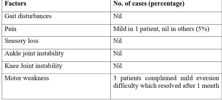
Morbitiy at the donor site was assessed after 1 month after the reconstruction surgery and the findings showed that the patients were generally symptom free except for some of the complication during the early post-op period like, mild pain, motor weakness. 3 patients who complained of mild eversion difficutly had the complaint spontaneously resolved in few months. All other complaints also resolved spontaneously during further follow-ups.
Table 4: Donor site morbidity
Factors No. of cases (percentage)
Gait disturbances Nil
Pain Mild in 1 patient, nil in others (5%)
Sensory loss Nil
Ankle joint instability Nil Knee Joint instability Nil
Motor weakness 3 patients complained mild eversion difficulty which resolved after 1 month
DISCUSSION
Mandible is an important structure which not only forms the lower jaw but also gives character to the face. Loss of the mandible and the soft tissue of the oro-mandibular region, either due to trauma or post-surgical, cause great distress to the patients – both functionally and aesthetically. Hence it needs to be reconstructed to provide optimal functional and aesthetic outcome to the patient. With many regional, loco-regional and distant flap options available, the vascularized free fibula chimeric osseomyocutaneous flaps have become the gold standard for reconstruction of the oro-mandibular defects. With the increasing importance to the vascularizsed free fibula flaps, there arises a need to standardize as much as possible the marking, dissection of the soft tissue, ostectomy sites of the fibula and its positioning in the recipient site. With this in mind, we conducted anatomical study of the peroneal perforator system in cadavers with dye injection and studied various parameters with the objectives of,
• Location of the single best peroneal perforator in the leg,
• Location of the best site surface and site for ostectomy of the fibula
• Best position of the flap in the recipient site that does not cause strain
in the pedicle.
• If the eccentric location of the pedicle of the skin paddle affect the
survival of the flap.
Classification of Mandibular Defects:
Classification that combines the loss of bone and soft tissue is useful for determining the reconstructive method. Various classification schemes have been proposed for quantifying the segmental mandibulectomy defects, which helps with planning the reconstruction.
Initially there was Pavlov’s classification, but it did not take into account the loss of condyle and mentum. But the loss of condyle makes reconstruction of articular surface difficult.
Later, Boyd19 and colleagues modified Pavlov’s classification by
including the loss of condyle and mandible in their classification. In their classification,
H - lateral defects of any length up to the midline that include the condyle L - lateral defects that exclude the condyle
C - defects involve the central segment containing the four incisors and two canines.
o - no skin or mucosal component s - skin
m - mucosa
sm - skin plus mucosa
Loss of the central segment poses problems with restoration of stomal competence, restoration of an anterior gingivolabial sulcus to fit prosthetics for dental rehabilitation, and restoring the lip height for cosmesis.
Urken17 and associates described another classification scheme is based
on functional considerations due to detachment of different muscle groups and difficulties with cosmetic restoration. This reconstruction scheme has similar anatomic designations,
C - Condyle R - Ramus B - Body
S - Total Symphysis SH - Hemisymphysis P - Palate
This classification system also includes a detailed description of soft tissue and neurologic deficits.
osseous free flaps, outcomes are not dependent on the length of the graft required to bridge the defect.
Goals of Mandibular Reconstruction:
The primary objective of mandibulectomy in tumor resections is cure, but the functional and aesthetic rehabilitation are necessary for the psychologic and physiologic recovery of patients.
The ideal reconstruction for a segmental mandibulectomy should have the following goals:
1. Restoration of oral competency
2. Maintainence of the occlusal relationships with left over teeth
3. Give allowance for prosthetic dental restoration in the future if the patient desires
4. Restoration of bone continuity
5. Restoration of facial symmetry and the contour to the lower third of the face.
In addition,
• Wound closure should be immediate and complete with early functional
recovery
• Avoidance of complications like orocutaneous fistula, infection, etc.,
• Creation of a safe wound that can undergo post-operative radiation later
Cadaver and Clinical Studies:
We found that from our cadaveric studies and clinical cases, single best perforator with good length is available in the middle third of the leg. This is because the posterolateral septum is proportionately larger in dimension in relation to to mid-calf muscle mass. Therefore longer septofasciocutaneous and musculocutaneous perforators can be harvested from the middle third of the leg. When lining or skin cover flap needed to be planned for wider mobility in the reconstructed mandible site, for eg., lip reconstruction, cheek reconstruction, upper neck reconstruction, etc., we harvest the chimeric free fibular osteocutaneous flap with skin island based on the perforators from the middle third of the leg. Thereby we were able to obtain a lengthy single best perforator facilitating the wide three-dimensional positioning of lining or cover.
In both cadaveric and clinical studies, we observed that almost 70% of the single best perforator in the middle third and 80% in the lower third of the leg were musculocutaneous (pass through flexor hallucis longus and/or soleus).
We have studied both in the cadaver dissections as well as in clinical cases, the distribution of mucoperiosteal vessels. It is possible to find the safe window for wedge ostectomies to match the curvature of the mandible. A wedge shaped ostectomy can be safely made in the window between adjacent mucoperiosteal vessels, which runs downwards and laterally, with the base on the interosseus surface and the apex in the peroneal surface. This can be fashioned in a way that neither causes damage to the mucoperiosteal blood supply nor causes kinking of the peroneal artery, which runs in relation to the median crest of the posterior medial surface of the fibula.
Incision is placed in the periosteum in the oblique direction, parallel to the direction of the mucoperiosteal blood vessels. The mucoperiosteal vessels are elevated on either sides, enough to visualize the ostectomy site clearly. Now ostectomy is done with power instruments with copious cold saline continuoes irrigation to avoid damage to the osteoblasts and its progenitor cells. This sleeve of mucoperiosteal muscular cuff elevated allows enough space for fixing the contoured plates and screws.
mandible, then we design the paddle so that the single best perforator is placed eccentric in location to the skin paddle. This provides a wider arc of rotation and three dimensional manipulations.
Here we incorporate the biogeometry of the properllar flaps. This is designed by an exploratory single incision to judge the length of the perforator before completing the total delineating incision of the skin paddle. From our clinical experience, we found that all the flaps harvested in the propeller fashion with the eccentric location of the pedicle survived without any complications.
CONCLUSION
Vascularized free fibula osteocutaneous flap is an established procedure for reconstruction of the oro-mandibular defects but several questions arises like; what is the location of the single best perforator for the skin paddle? What is the safe skin paddle dimension that can be harvested on a single eccentrically located pedicle? Where are the safe windows for ostectomy located on the harvested free fibula? What is the best position of the skin paddle in the recipient site which does not cause kinking of the vessel? etc.,
We have done cadaver studies and clinical studies to answer some of these questions. It was found from our studies that the perforators of good length and calibre are best found in the middle third of the leg, and most of the perforators were musculocutaneous travelling through the flexor hallucis longus muscle. Hence if a larger skin paddle is needed, it can reliably be harvested from the middle third of the leg with the pedicle placed eccentrically. But if the defect requires a longer bone stock and a smaller pedicle, it is better to harvest from the lower third of the leg.
Also from our clinical studies we found that a single eccentrically located perforator of good length and calibre can support a skin paddle of 120 cm2.
We also found from our studies that the safe window for ostectomies can be found in the peroneal surface of the fibula as the musculoperiosteal vessles runs in the anteromedial and posteromedial surface of fibula. Hence ostectomies can be performed with the apex of the wedge located at the peroneal surface and base at the interosseus border.
We found from our studies that the posteromedial surface of the fibula which contains the peroneal vessel, when positioned inferiorly and posteriorly in mandible reconstruction site, gives better manoeuverability during anastamosis of the flap pedicle to the recipient vessels and also does not cause kinking of the vessel.
BIBLIOGRAPHY
1. Taylor GI, Miller GD, Ham FJ (1975) The free vascularized bone graft. A clinical extension of microvascular techniques. Plast Reconstr Surg 55(5):533–544
2. Harashina T, Fujino T, Aoyagi F. Reconstruction of the oral cavity with a free flap. PlasticReconstr.Surg. 1976;58:412–414.
3. Conley J. Composite pedicled rib flap for reconstruction of the mandible and face. Trans.Sect.Otolaryngol.Am.Acad.Ophthalmol. Otolaryngol. 1976;82:ORL 447–451.
4. Ariyan S. The pectoralis major myocutaneous flap. A versatile flap for reconstruction in the head and neck. Plast.Reconstr.Surg. 1979;63:73–81. 5. O’Brien BM, Morrison WA, MacLeod AM, et al. Microvascular
osteocutaneous transfer using the groin flap and iliac crest and the dorsalis pedis flap and second metatarsal. Br.J.Plast.Surg. 1979;32:188–206.
6. Cuono CB, Ariyan S. Immediate reconstruction of a composite mandibular defect with a regional osteomusculocutaneous flap. Plast.Reconstr.Surg. 1980;65:477–484.
7. Green MF, Gibson JR, Bryson JR, et al. A one-stage correction of mandibular defects using a split sternum pectoralis major osteo-musculocutaneous transfer. Br.J. Plast.Surg. 1981;34:11–16.
9. DeSanto LW, Beahrs OH, Holt JJ, O’Falon WM. Neck dissection and combined therapy. Arch Otolaryngol 1985;111:366–70.
10.Carriquiry C, Aparecida Costa M, Vasconez LO. An anatomic study of the septocutaneous vessels of the leg. Plast Reconstr Surg 1985;76:354-63. 11.Robertson GA. The role of sternum in osteomyocutaneous reconstruction
of major mandibular defects. Am.J.Surg. 1986;152:367–370.
12.Wei FC, Chen HC, Chuang CC, et al. Fibular osteoseptocutaneous flap: anatomic study and clinical application. Plast.Reconstr.Surg. 1986;78:191– 200.
13.Evans HB, Lampe HB. The radial forearm flap in head and neck reconstruction. J.Otolaryngol. 1987;16:382–386
14.Hidalgo DA. Fibula free flap: a new method for mandible reconstruction. Plast Reconstr Surg 1989;84(1):71–9
15.Jewer, D.D., et al., Orofacial and mandibular reconstruction with the iliac crest free flap: a review of 60 cases and a new method of classification. Plast Reconstr Surg, 1989. 84(3): p. 391-403; discussion 404-5.
16.Yoshimura M, Shimada T, Hosokawa M. The vasculature of the peroneal tissue transfer. Plast Reconstr Surg 1990;85: 917-21.
18.Beppu M, Hanel DP, Johnston GH, et al. The osteocutaneous fibula flap: an anatomic study. J Reconstr Microsurg 1992;8:215-23
19.Boyd JB, Gullane PJ, Rotstein LE, et al. Classification of mandibular defects. Plast Reconstr Surg. 1993;92:1266-1275.
20.Wu WC, Chang YP, So YC, et al. The anatomic basis and clinical applications of flaps based on the posterior tibial ves-sels. Br J Plast Surg 1993;46:470-9.
21.Anthony JP, Rawnsley JD, Benhaim P, Ritter EF, Sadowsky SH, Singer MI. Donor leg morbidity and function after fibula free flap mandible
reconstruction. Plast Reconstr Surg 1995; 96: 146-52.
22.Eppley BL. Non vascular methods of mandible reconstruction. Oper Tech Plast Surg 1996;3(4):226–32
23.Jones NF, Monstrey S, Gambier BA (1996) Reliability of the fibular osteocutaneous flap for mandibular reconstruction: anatomical and surgical confirmation. Plast Reconstr Surg 97(4):707–716
24.Pogrel MA, Podlesh S, Anthony JP, Alexander J. A comparison of vascularised and nonvascularized bone grafts for reconstruction of mandibular continuity defects. J Oral Maxillofac Surg. 1997 Nov;55(11):1200-6.
symptomatic and asymptomatic volun-teers aged 6 to 25 years. J Orofac Pain 11:37-47, 1997
26.Kroll SS, Evans GR, Goldberg D, Wang BG, Reece GP, Miller MJ, et al. A comparison of resource cast and reconstruction with free and
pectoralis major flaps. Plast Reconstr Surg 1997; 99: 1282-6. 27.Bataineh AB, al Qudah M. Treatment of mandibular odontogenic
keratocysts. Oral Surg Oral Med Oral Pathol Oral Radiol Endod. 1998; 86(1):42-47.
28.Ferrari R, Leonard MS. Whiplash and temporomandibular disorders: a critical review. J Am Dent Assoc 129:1739-1745,1998
29.Cordeiro PG, Disa JJ, Hidalgo DA et al (1999) Reconstruction of the
mandible with osseous free flaps: a 10-year experience with 150 consecutive patients. Plast Reconstr Surg 104(5):1314–1320
30.Zhao YF, Wei JX, Wang SP. Treatment of odontogenic keratocysts: a follow-up of 255 Chinese patients. Oral Surg Oral Med Oral Pathol Oral Radiol Endod. 2002;94(2): 151-156.
31.Hidalgo DA1, Pusic AL. Free-flap mandibular reconstruction: a 10-year follow-up study. Plast Reconstr Surg. 2002 Aug;110(2):438-49; discussion 450-1.
follow-up analysis of the effects and changes in growth characteristics. Oral Surg Oral Med Oral Pathol Oral Radiol Endod. 2002;94(5):543-553.
33. van Rensburg LJ, Paquette M, Morkel JA, Nortje CJ. Correlative MRI
and CT imaging of the odontogenic keratocyst: a review of tweny-one cases. Oral Maxillofacial Surg Clin N Am. 2003; 15(3):363-382.
34.Heitmann C, Khan FN, Levin LS. Vasculature of the peroneal artery: an anatomic study focused on the perforator vessels. J Reconstr Microsurg 2003;19:157-62.
35.Chepeha DB, Annich G, Pynnonen MA, Beck J, Wolf GT, Teknos TN, et al. Pectolaris major myocutaneous flap vs. revascularized free tissue transfer: complications, gastotomy tube dependency, and hospitalization. Arch Otolaryngol Head and Neck Surg 2004; 130:181-6.
36. Peled M, El-Naaj IA, Lipin Y, Ardekian L. The use of free fibula flap for
funtional mandibular reconstruction. J Oral Maxillofac Surg 2005; 63:220-4.
37.Geddes CR, Tang M, Yang D, et al. Anatomy of the integument of the lower extremity. In: Blondeel PN, Morris SF, Hallock GG, et al., editors. Perforator flaps: anatomy, technique & clinical applications. St. Louis: Quality Medical Publishing, Inc.; 2006. p. 541-78.
38.Koh KS, Eom JS, Kirk I, et al. Pectoralis major musculocutaneous flap in oropharyngeal reconstruction: revisited. Plast.Reconstr.Surg.
39.Yagi S, Kamei, Y., Torii, S. Donor side selection in mandibular
reconstruction using a free fibular osteocutaneous flap. Ann.Plast.Surg. 2006;56:622–627
40.Schaverien M, Saint-Cyr M. Perforators of the lower leg: anal-ysis of perforator locations and clinical application for pedicled perforator flaps. Plast Reconstr Surg 2008;122:161-70.
41.Weibull L, Widmark G, Ivanoff CJ, Borg E, Rasmusson L. Morbidity after chin bone harvesting--a retrospective long-term follow-up study. Clin Implant Dent Relat Res. 2009 Jun;11(2):149-57. Epub 2008 Jul 24
42.Cheng MH, Saint-Cyr M, Ali RS, et al. Osteomyocutaneous peroneal artery-based combined flap for reconstruction of composite and en bloc mandibular defects. Head Neck. 2009;31:361–370.
43.Diego Ribuffo et al. Clinical study of peroneal artery perforators with computed tomographic angiography: implications for fibular flap harvest Surg Radiol Anat (2010) 32:329–334
44. Chang SY, Huang JJ, Tsao CK, et al. Does ischemia time affect the
outcomes of free fibula flaps for head and neck reconstruction? A review of 116 flaps. Plast.Reconstr.Surg. 2010;126:1988–1995.
45. Purushothaman R, Balakrishnan TM, Alalasundaram KV. Anatomical
PROFORMA
ERGONOMICS OF FREE FIBULA FLAP IN MANDIBLE RECONSTRUCTION
Patient’s Name : ………...
Age/Sex : ………...
IP No : ………...
Contact Address : ………... ………... ………... Contact Number : ………...
Admitted Under : Plastic Surgery/Surgical Oncology/ENT/Dental Surgery
Pre-Operative Details:
1. Diagnosis :
2. Aetiology :
3. Expected Bone Defect (Boyd’s Classification) :
4. Expected Soft Tissue Defect (Boyd’s Classification) :
5. Tissue Diagnosis :
Co-Morbid Conditions : ……… Risk Factors : ……… Donor Site Assessment : ……… Operative Details:
1. Procedure Done : ………
2. Date : ………
3. Incision for Harvest: ………
4. Pedicle: Length: ……….. Diameter: ……….
5. Ostectomies Done: ………
6. Fibula Length Harvested: ………..
7. Skin Paddle Dimensions Harvested: ……….
8. Recipient Artery: ………. Vein: ………...
9. Suture Materials Used: ………..
10. Duration of Surgery: ……….
Post‐Op Drugs Used : ……….
……….
……….
Post‐Op Complications: ……….
Follow‐up Findings : ……….
……….
……….
……….
Investigator(s) Name & Signature: ………
MASTER CHART OF CADAVER DISSECTION
Parameters Number of perforators
Avg. vessel diameter Avg. vessel length
Type of Perforators
(in mm) (in cm)
Cadaver No. Upper third Middle third Lower third Upper third Middle third Lower third Upper third Middle third Lower
third Septocutaneous Musculocutaneous
1 2 6 2 1.2 1.2 0.9 2.5 2.2 1.9 1 9
2 3 6 2 1.5 0.9 0.8 2.3 3 2.1 1 10
3 5 4 3 1.2 1.3 1.1 3.2 3.3 1.6 1 11
4 2 6 2 1.4 1.4 1.2 2.6 2.6 1.6 1 9
5 5 6 3 1.2 1.1 0.9 2.2 3.1 2.2 2 12
6 4 5 2 1.3 0.9 1 3.1 2.5 1.5 2 9
7 4 5 3 1.1 1.2 1.1 2.2 2.5 1.6 1 11
8 2 7 2 1.2 1.3 0.9 2.3 3.4 1.8 1 10
9 5 5 2 1.5 1.3 0.9 2.5 2.5 1.7 1 11
10 3 6 3 1.4 1.2 1.1 2.6 2.7 1.7 1 11
11 4 4 3 1.4 1.3 1 2.5 2.5 1.6 1 10
12 2 5 2 1.2 1.1 1.1 1.9 2.4 1.6 2 7
13 2 6 1 1.3 1.2 0.9 2.6 3.2 2.1 1 8
14 5 5 3 1.3 1.3 0.9 2.9 3.3 1.9 1 12
15 3 6 2 1.4 1.2 1.1 2.3 2.5 1.8 1 10
16 4 5 4 1.2 1.3 1.1 2.4 2.3 1.8 2 11
17 4 6 3 1.3 1.2 0.9 2.5 2.6 1.7 1 12
18 5 6 3 1.2 1.2 0.9 2.7 2.6 1.7 1 13
19 4 6 3 1.4 1.1 1.1 1.9 2.4 2.2 1 12
20 2 5 2 1.3 1.3 1.1 2.8 2.4 1.9 1 8
Average 3.5 5.5 2.5 1.3 1.2 1 2.5 2.7 1.8 10.44% 89.56%