A STUDY ON LYMPHOMA
DissertationSubmitted in partial fulfillment of the regulations of
M.S. DEGREE EXAMINATION
BRANCH I GENERAL SURGERY
Department of General Surgery
GOVT. STANLEY MEDICAL COLLEGE AND HOSPITAL
CHENNAI - 600001
THE TAMILNADU DR.M.G.R MEDICAL UNIVERSITY
CHENNAI
CERTIFICATE
This is to certify that this dissertation titled A STUDY ON
LYMPHOMA is the bonafide work done by Dr.VENGATESAN.S
Post Graduate student (2011– 2014) in the Department of General
Surgery, Government Stanley Medical College and Hospital, Chennai
under my direct guidance and supervision, in partial fulfillment of
the regulations of The Tamil Nadu Dr. M.G.R Medical University,
Chennai for the award of M.S., Degree (General Surgery) Branch - I,
Examination to be held in April 2014.
Prof. K. KAMARAJ, M.S.,
Professor and Head of surgery, Dept. of General Surgery, Stanley Medical College, Chennai-600001.
PROF. S. GEETHA LAKSHMI, M.D., PhD, The Dean,
DECLARATION
I, Dr.VENGATESAN.S solemnly declare that this dissertation
titled
A STUDY ON LYMPHOMA
is a bonafide work done by me in the Department of General Surgery, Government Stanley MedicalCollege and Hospital, Chennai under the guidance and supervision of
my unit chief.
Prof. Dr. J.VIJAYAN M.S
Professor of Surgery
This dissertation is submitted to The Tamilnadu
Dr. M.G.R. Medical University, Chennai in partial fulfillment of the
university regulations for the award of M.S., Degree (General Surgery)
Branch - I, Examination to be held in April 2014.
Place: Chennai.
ACKNOWLEDGEMENT
I am grateful to Prof. S. Geethalakshmi, Dean, Govt. Stanley
Medical College for permitting me to conduct the study and use the
resources of the College.
My sincere thanks to Prof. K. Kamaraj, Professor and HOD,
Department of General Surgery, for his valuable guidance throughout
the study.
I am highly indebted to my guide Prof. J.VIJAYAN Professor
of Surgery for his constant help, inspiration and valuable advice in
preparing this dissertation.
I express my deepest sense of thankfulness to my Assistant
Professors Dr.THIRUMURUGANAND, Dr.CHANDRASEKAR for
their valuable inputs and constant encouragement without which this
dissertation could not have been completed.
I am extremely thankful to my patients who consented and
TABLE OF CONTENTS
S. NO. CHAPTER PAGE NO
1. INTRODUCTION 1
2. AIMS AND ODJECTIVES 2
3. MATERIALS & METHODS 3
4. REVIEW OF LITERATURE 4
5. OBSERVATION AND RESULTS 62
6. DISCUSSION 73
7. CONCLUSION 75
8. BIBLIOGRAPHY
9. ANNEXURE
(i) PROFORMA
(ii) INSTITUTIONAL ETHICAL
COMMITTEE APPROVAL
CERTIFICATE
(iii) TURNITIN SCREEN SHOT
(iv) PATIENT INFORMATION
SHEET
(v) CONSENT FORM
INTRODUCTION
Lymphoma , involves cancers of the lymphatic system..The two
main types of lymphoma are hodgkins lymphomas also known as
hodgkins disease, and the non hodgkins lymphoma.
In hodgkins lymphoma` ,cells of the lymphatic system multiplying
fastly. The cells of HL grow with out any order or without any control.
HL can occur almost anywhere in the lymphatic system.HL may occur in
lymphnodal site. They may also affect the other parts of the lymphatic
system[ bone marrow, spleen]
HL spread from one group of LN to next group of LN.
Non hodgkins lymphoma, accounts,comprises 3 percent of all
malignancies.In NHL cells of lymphatic system become abnormal.They
undergo division and multiplying without any control .they do not die
normally .
They spreads to other group of lymph nodes and lymphoid organs
in a non contigious manner.
The objective of this study is to evaluate clinical presentation
AIM:
To evaluate
• the spectrum of clinical presentations in lymphoma
patients
• organs involved and
• incidence among specific age groups
• the incidence/distribution of different subtypes of
MATERIALS AND METHODS:
PLACE OF STUDY:
Department of General Surgery,
Govt. Stanley Medical College and Hospital.
DURATION:
MAY 2011 TO DEC 2013
STUDY DESIGN:
Prospective and retrospective
SAMPLE SIZE: 51
METHODOLOGY
Clinical data, imaging studies and biopsy material of the all the
REVIEW OF LITTERATURE
LYMPHOMAS
EPIDEMOLOGY
Nonhodgkins kymphomas and hodgkins lymphoma are the most
common haematological malignancies in the world.they account for 4 to
55 of new cancer cases . fifth leading cause of cancer deaths and second
leading cause of cancer mortality.united states ,Europe and Australia are
associated with high incidence rates . asia is generally associated with
low incidence rates.
A dramatic increase in the incidence of non hodgkins lymphoma
has been reported in the last five decades.although there is increase in the
incidence of most histologies ,largest increase is seen in aggressive
lymphomas . This increase is due to occurrence of primary cns
lymphomas in patients with AIDS. Increased incidence is also seen in
non-aids patients also. Difference in histological sub types has also been
attributed geographically. For example, endemic burkitt lymphomas are
common in children of equatorial Africa.gastric lymphomas in Italy
.nasal t cell lymphomas in china.small intestinal lymphomas in middle
follicular lymphomas are lower in asia and Asian immigrants to the
united states.Inspite of this the reason for increase in incidence of Nhl
NON HODGLINS LYMPHOMAS
ETOLOGY
The cause is mostly unknown. Although several genetic disease,
environtmental agents and infectious agents have been implicated
.familial occurrence of lymphomas has also been described. Increased
risk is seen in siblings and relatives of lymphomas and other
haematological malignancies.
Several inherited immunodeficiency states are associated with risk of
developing lymphomas .Some of this include
• severe combined immunodeficiency disease • hypogammaglobulinemia,
• common variable immunodeficiency, • wiskott Aldrich syndrome, and
• ataxia telengectasia.
Development of lymphomas in these are associated with Epstein barr
virus.
Acquired conditions such as Aids and post organ transplant
conditions are also associated with development of lymphomas .A variety
and psoriasis are also associated with lymphomas .In sjogrens most of
this lymphomas are marginal lymphomas . They occur in salivary glands
and other extra nodal sites such as the stomach and lung. Celiac sprue is
associated with poor prognosis lymphomas classified as enteropahty type
intestinal T cell lymphomas
INFECTIOUS AGENTS
Ebv Dna both type A and type B are associated with 95% of
endemic burkitt lymphomas . Less commonly they are associated with
sporadic type.the actual mechanism of development is unknown .it
appears that early Ebv infection and environemental factors increase the
number of infected precursors and the risk of genetic errors.
Ebv is also linked to
• post transplant lymphoproliferative disorders , • some Aids associated lymphomas and
• some lymphomas associated with immunodeficiency disease.
Ebv is associated with almost all cases of Aids associated primary cns
lymphomas .Normal immune responses by T cell supress Ebv infected
lymphomas.The pattern Ebv associated proteins in patients with Aids
associated lymphomas differ from that in other lymphomas.c-myc
activation in the absence of ebv infection can also occur in aids associated
lymphomas. The Ebv latent membrane protein 1 is a viral analog of the
tumor necrosis factor receptor. In ebv positive aids associated lymphomas
LMP 1 binds to members of the TNF receptor. It then activates Nfkb
transcription factor leading to cellular proliferation.
The human T cell lymphotropic virus type 1 was the first human retro
virus associated with development of malignancy. It is a type c rna virus
responsible for the development of ALT in addition to myelopathy and
other disorders.it is primarily transmitted mainly by breast feeding ,sexual
contact and blood transfusion.the latent period takes several decades for
the development of lymphomas.Htlv associated ALT is most prevalent in
southern japan,south America ,Africa and the carribean.The Htlv 1
genome has a regulatory tax gene which is a potent transcriptional
activator. It is responsible for the transforming feature of the virus. The
binding domains of the virus glycoproteins inhibit glucose transport by
interacting with Glut1 . This contributes to the virus associated disorders.
A third virus associated is HHv8.It was discovered from Kaposi
herpes virus.It is also associated with multicenteric castleman’s disease.
In an analysis of 193 patients with lymphomas the virus was found in
only 8 patients. All of these are primary effusion lymphomas.HHV 8
associated lymphomas lack c-myc expression and has different phenotype
and expression. The actual mechanism of stimulation is unknown . It has
been proposed that this virus is necessary for Ebv induced transformation.
Hepatitis c is also linked to lymphomas . Hcv infection is strongly
associated with essential mixed cryoglobulinemia,which is associated low
grade lymphomas.several analyses show higher incidence of Hcv
infection in patients with B-cell NHL.The association appears strongest
for patients with monocytoid lymphomas and lymphoplasmacytoid
lymphomas .Neoplastic transformation is related to chronic antigenic
stimulation of Bcells by HCV.
Simian virus 40 , a potential contaminant of salk polio vaccine may
also be linked to the developement of NHL.
Several bacterial infection is also related to development of
lymphomas .Helicobacter pylori is linked to the development os gastric
mucosa associated lymphoid tissue(MALT) lymphomas. The
of gastric mucosa ,chronic antigenic stimulation leading to gastritis and
subsequent development of malignant B cell clones.
Campylobacter jejuni is associated with small intestinal
mmunoproliferative disease some cases of ocular adnexal lymphomas is
associated with chlamydia psittacci infection.
ENVIRONEMENTAL AND OCCUPATIONAL EXPOSURES
There is increased risk of NHL in several occupation in farmers
forestry and agricultural workers.Exposure to herbicides especially
phenoxy herbicides like 2,4,-dichlorophenoxyacetic acid.NHL has been
associated with organic solvents and high levels of nitrates in drinking
water.
DIET AND OTHER EXPOSURES
Studies suggest that the risk of lNHL is increased in association
with higher intake of meat and dietary fat. Recreational drug use has been
associated with increased NHL risk and tobacco use has also been
associated with increased risk . Risk of NHL in association with ionising
radiation is minimal.solar ultra violet exposure has also been associated
ANATOMY AND MORPHOLOGY OF NORMAL LYMPHOID
TISSUES
Lymphoid tissues can be divided in to two major categories
1.central or primary lymphoid tissues,in which lymphoid precursor cells
mature .
2.periphery or secondary lymphoid tissue ,in which antigen specific
reactions occur
PRIMARY (CENTRAL) LYMPHOID TISSUES
BONE MARROW (bursa equivalent)
These are the antibody producing cells found in the bone marrow .
they are equivalent to the bursa of fabricus found in the avian species.
THYMUS
Thymus is the site at which immature T-cell precursors which
migrate from the bone marrow matures in to naïve T cells capable of
responding to antigen. The thymus is divided in to cortex and medulla
SECONDARY (PERIPHERAL)LYMPHOID TISSUES
The lymph nodes are placed throughout the body to process the
antigen present in the lymphdrained from tissues and lymphatics drained
through lymphatics.lymph nodes have a capsule ,a cortex ,a medulla and
sinuses(subcapsular ,cortical and medullary). The sinuses contain the
macrophages which take up the antigens and process them in to peptides .
these peptides are then presented to the lymphocytes in the major
histocompatibility antigens.the cortex contains B cell follicles and
paracortical T-0cell zones and the medulla contains medullary cords and
sinuses.the paracortex contains endothelial venules through which T and
B cells and the antigen presenting cells which present antigen to the T
cells.the follicles also contain follicular dendritic cells that bind antigen –
antibody complexes and help in regulating the differentiation of B cells in
response tio antigens.
SPLEEN
The spleen has two major areas
1. Red pulp, which functions as a filter for particulate antigens and for
2. White pulp, which is identical to the lymphoid tissue of the lymph
node .
Follicle and germional centres are found in the malphigian
corpuscles,
T cells and dendritic cells in the peri arteriolar lymphoid sheath . plasma
cells are concentrated in the red pulp.
MUCOSA ASSOCIATED LYMPHOID
These are the specialised lymphoid tissue found in association with
the epithelium in nasopharynx ,oropharynx as the waldeyers ring
,adenoids and tonsils . in the gastrointestinal tract as peyer patches in the
distal ileum ,colon and the rectum. In the lung as bronchus associated
lymphoid tissue . they have prominent B cell follicles and also discrete T
cell zones .they function in response to intraluminal antigens . it can also
be acquired in sites such as stomach ,thyroid in response to chronic
B AND T CELL DIFFERNTIATION
It includes 2 phases antigen dependent and antigen independent.
Antigen independent differentiation in primary lymphoid organs
without exposure to antigens. The early stages of lymphocytes are called
stem cells and lymphoblasts where as the later stages are differentiated
cells. On exposure to antigens the naïve lymphocyte undergoes blast
transformation and proliferates to hive rise to a progeny against the
inciting antigen.the early stages are composed of proliferating cells and
later stages are composed of differentiated cells.the neoplasms composed
of proliferating cells are aggressive and neoplasms composed of
differentiated cells tend to be indolent. Proliferatin g cell neoplasm such
as luekemia/lymphomas are more common in children and those
composed of mature cells such as lymphoplasmocytic lymphomas are
common in adults .
ANTIGEN –DEPENDENT B-CELL DIFFERENTIATION
IMMUNOBLASTIC OR PLASMA CELL REACTION
When interacting with antigen the naive B cell transforms into a
proliferating cell, which transforms into an antibody-secreting plasma
response, naive B cells transform into IgM positive blast cells in the
T-cell zones, proliferate, and differentiate into IgM-secreting plasma T-cells,
producing the IgM antibody in response to the antigen. Surface IgD is
lost during blast transformation, and antigens associated with activation
are up-regulated. With maturation to plasma cells, most surface antigens
are lost and secretory cytoplasmic IgM are concentrated . The
immunoblastic reaction occurs in the lymph node paracortex, and
IgM-producing plasma cells accumulate in the medullary cords.
Lymphoplasmacytic lymphoma, associated with Waldenstroms
macroglobulinemia, may correspond to the IgM-producing plasma cell.
However, they express low levels of variable region gene mutation and
IMMUNOPHENOTYPING
Antigens Useful in the Classification of Lymphoid Neoplasms
CD Expression on Normal Cells Useful Diagnostic
Applications in These
Neoplasms
1a Cortical thymocytes (strong),
Langerhans cells Precursor T-lymphoblastic lymphoma/leukemia, Langerhans cell neoplasms
2 T cells, NK cells T- and NK-cell
neoplasms
3 T cells (surface and cytoplasmic), NK cells
(cytoplasmic epsilon chain
only)
T- and NK-cell neoplasms
4 T subset (MHC class II
restricted), monocytes
Some T-cell neoplasms
5 T cells, naive B cells T-cell neoplasms,
CLL/SLL, mantle cell
lymphoma
7 T cells, NK cells T- and NK-cell
neoplasms
8 T subset (MHC class I
restricted), NK subset,
Some T- and NK-cell
splenic sinus lining cells
10 Precursor B cells, germinal
center B cells, granulocytes,
fibroblasts, kidney epithelium
Precursor B or T
lymphoblastic
lymphoma/leukemia,
Burkitt lymphoma,
follicular lymphoma,
diffuse large B-cell
lymphoma
11c T-cell and B-cell subsets, NK
cells, dendritic cells
Hairy cell leukemia,
CLL/SLL, splenic
marginal zone lymphoma
15 Granulocytes, monocytes Reed-Sternberg cells of
classic HL
16 NK cells, granulocytes,
macrophages
NK cell neoplasms,
some T-cell neoplasms
19 B cells in all stages of maturation
B-cell neoplasms
20 Mature B cells (not plasma
cells), T-cell subset
Mature B-cell
neoplasms, some
classic HL
21 Mature B-cell subset, FDCs Mature B-cell
neoplasms, groups of
background FDCs in
some lymphomas, FDC
22 Nearly all stages of B cells
(cytoplasm), B-cell subset
(surface)
B-cell neoplasms,
especially hairy cell
leukemia
23 IgE Fc receptor: activated B
cells, monocytes, FDC
CLL/SLL, other B-cell
lymphomas, groups of
background FDCs in
some lymphomas
25 IL-2 receptor: activated T
cells, activated B cells,
activated monocytes
Hairy cell leukemia,
adult T-cell
leukemia/lymphoma,
other T-cell neoplasms
30 Activated T, B, and NK cells,
monocytes
Reed-Sternberg cells in
classic HL, ALCL,
primary cutaneous
CD30+ LPD
35 Follicular dendritic cells,
myeloid cells, B cells, T
cells, monocytes, erythroid
cells, glomerular podocytes
Follicular dendritic cell
sarcoma
38 Activated T and B cells, NK
cells
Plasma cell neoplasms,
B- and T-cell
lymphoma
43 T cells, B subset, NK cells,
monocytes, plasma cells, and
myeloid cells
T-cell neoplasms, some
B-cell neoplasms,
myeloid neoplasms
45 Leukocyte common antigen,
all leukocytes except plasma
Lymphoid and myeloid
cells
45RA B cells, naive T cells, NK
cells
B-cell neoplasms,
T-cell neoplasms
45RO T cells (most), granulocytes,
monocytes
T-cell neoplasms
56 Neural cell adhesion
molecule: NK cells, activated
T cells
NK-cell neoplasms,
T-cell neoplasms, plasma
cell neoplasms
57 T-cell and NK cell subset,
neural tissue
NK-like T-cell
neoplasms, T-cell
neoplasms, NK-cell
neoplasms
68 Monocytes, macrophages,
activated T cells
Myeloid and histiocytic
neoplasms
79a B cells, including precursor B
and plasma cells
B-cell neoplasms, rare
T-lymphoblastic
neoplasms, rare classic
HL
95 Fas (apoptosis receptor):
activated T cells, B cells
Some B- and T-cell
neoplasms, HL
99 Cortical thymocytes Precursor B- and T-cell
neoplasms, Ewing's
sarcoma
103 Mucosal intraepithelial
lymphocytes
Hairy cell leukemia;
enteropathy-type T-cell
138 Syndecan-1 (stromal
binding): plasma cells
Plasma cell neoplasms,
plasmablastic
lymphomas
Recognising these antigens have become vital to diagnosis and
residual disease monitoring in lymphoid neoplasms.
PRINCIPLES OF MANAGEMENT OF NONHODGKINS
LYMPHOMAS
The phases of patient management include
1. obtaining an adequate biopsy for an accurate diagnosis,
2. a careful history and physical examination,
3. appropriate laboratory studies, imaging studies, and
4. possibly further biopsies to determine an accurate stage and to plan
therapy.
Finally, taking into account factors related to the patient, type of
HISTORY AND PHYSICAL EXAMINATION
It determine the extent of the disease and a key factor in the
therapeutic decision. The duration of symptoms and the pace of
progression of the illness should be documented. waxing and waning
lymphadenopathy could possibly be related to the lymphoma. Especially
in follicular lymphomas, spontaneous regressions are not infrequent. The
presence of specific symptoms known to have an adverse prognosis in
some lymphomas. These include fevers, night sweats, and unexplained
weight loss. Symptoms , such as pain in the chest, abdomen, or bones,
might indicate specific sites of involvement. For example, symptoms like
seizures,focal nuerological deficit is seen in CNS lymphoma .
concurrent illness such as diabetes or congestive heart failure might
modify therapeutic decisions. Obviously, examination of all lymph node
and search for hepatomegaly or splenomegaly are important. m Sites such
LABAROTARY EVALUATION
Laboratory studies should include
• complete blood count
• renal and hepatic function studies, • serum glucose, calcium, albumin,
• Serum lactate dehydrogenase (LDH), Ig and microglobulin
level.
Serum protein electrophoresis is frequently appropriate.
The purpose of these studies is
• determining the prognosis
• identifying abnormalities in other organ systems (e.g., renal or
hepatic dysfunction).
Almost all patients should have a bone marrow aspirate and biopsy
performed. The chance of finding bone marrow involvement varies
considerably among different subtypes of lymphoma. It is present in
approximately 70% of patients with SLL, lymphoplasmacytoid
lymphoma, and mantle cell lymphoma. Patients with follicular lymphoma
have bone marrow involvement approximately 50% of the time. Bone
In certain situations, cytologic evaluation of the cerebrospinal fluid
is indicated. Patients with paranasal sinus, testicular involvement,
epidural lymphoma, should have a diagnostic lumbar puncture. lumbar
puncture is also recommended for highly aggressive histologies and in
immunocompromised patients.
IMAGING STUDIES
CHEST RADIOGRAPHY AND COMPUTED TOMOGRAPHY
Chest radiography and computed tomography (CT) scans of the chest,
abdomen, and pelvis should be performed . Identification of hilar or
mediastinal adenopathy, parenchymal lesions, or pleural effusions is
important a. CT scanning can identify nodal and extranodal sites of
involvement. Thus help in monitoring the response to therapy.
Involvement of intra-abdominal organs, such as kidney, ovary, spleen,
can be identified on CT scans.
MAGNETIC RESONANCE IMAGING
MRI is particularly useful in identifying bone and CNS involvement.
MRI can suggest leptomeningeal involvement when gadolinium has been
NUCLEAR MEDICINE STUDIES: POSITRON EMISSION
TOMOGRAPHY IMAGING
Nuclear scintigraphy may improve staging, through the detection of
occult abdominal or splenic disease. nuclear scintigraphy may detect a
residual mass after therapy as either fibrosis or residual active lesion.
gallium-67, binds to transferrin receptors in the tumor,is used as nuclear
tracer. Bone scans can be useful for looking vertebral involvement and
spinal cord compression.
Positron emission tomography (PET) is a functional imaging technique.
It uses a glucose analog (2-fluoro-2-deoxy-D-glucose [FDG])
radiolabeled with the positron emitter fluorine-18. It evaluate the
glycolytic activity, which is increased in malignancies, including
lymphoma. PET provides several advantages compared with other
nuclear imaging techniques. The short half-life of FDG allows patient
convenience and improved imaging characteristics. With modern PET
machines, a resolution of approximately 5 mm can be achieved.The
majority of studies evaluating FDG-PET in NHL includes patients diffuse
large cell NHL. Limited data are available on the role of PET in other
Persistently positive PET scans during and after chemotherapy
indicates subsequent relapse of aggressive lymphoma. Therefore,
persistently positive PET scans at the end of therapy, warrant close
follow-up or additional diagnostic procedures .
PET is most useful in assessing response to therapy of curable
lymphomas, specifically DLBCL and Hodgkin's lymphoma.
PET after completion of therapy should be performed
• at least at 3 weeks, and preferably at 6 to 8 weeks, after
chemotherapy or chemoimmunotherapy,
• 8 to 12 weeks after radiation or chemoradiotherapy.
STAGING AND PROGNOSTIC SYSTEMS
The goal of initial evaluation of a patient with lymphoma is to
provide information that allows
• intelligent planning of therapy,
• imparting the prognosis to the patient, and
• making possible comparisons between patients in clinical trials.
The studies to accomplish these goals can be aimed at
• characteristics of the patient (i.e., age, performance status, and so
forth), or
• characteristic of the lymphoma (serum LDH, serum
microglobulin) that predict treatment outcome.
Ann Arbor Staging System
Stage Descriptiona
I Involvement of a single lymph node region or a single
extralymphatic organ or site (IE)
II Involvement of two or more lymph node regions on the same
side of the diaphragm (II) or localized involvement of an
extralymphatic organ or site (IIE)
III Involvement of lymph node regions on both sides of the
diaphragm (III) or localized involvement of an extralymphatic
organ or site (IIIE) or spleen (IIIS) or both (IIISE)
IV Diffuse or disseminated involvement of one or more
extralymphatic organs with or without associated lymph node
involvement. Bone marrow and liver involvement are always
The Ann Arbor Staging System was developed for patients with
Hodgkin's lymphoma. This system has a significant effect on prognosis
and is important in treatment planning.’’
HODGKINS LYMPHOMAS
ETIOLOGY AND EPIDEMIOLOGY
There are approximately 7,500 new cases of Hodgkin lymphoma
diagnosed each year in the United States. Slightly more men than women
develop this malignancy (1.4:1). In economically developed countries,
there is an age-related bimodal incidence for Hodgkin lymphoma. The
first peak occurs in the third decade of life with a much smaller peak
occurring after the age of 50. The incidence of Hodgkin lymphoma by
age also differs by histologic subtype.
A number of studies have suggested that there appears to be a
genetic predisposition for Hodgkin lymphoma. There is an increased
incidence in Jews and also among first-degree relatives. Siblings appear
to have a increased risk of developing the disease. There is an increased
risk among parent-child pairs but not among spouses. Also, Hodgkin
There is less support for most other potential causes of Hodgkin
lymphoma. Hodgkin lymphoma is rarely seen as a second malignancy. It
does not appear to be increased in patients with illness or
treatment-related chronic immunosuppression. In AIDS patient with hodgkins there
is lack of evidenceto correlate with immunosuppresion. It seems that
under antiretroviral therapy the incidence of Hodgkin lymphoma seems to
increase with CD4 cells. There is increasing evidence to suggest a viral
etiology for Hodgkin lymphoma. There is association between Hodgkin
lymphoma and c decrease exposure to infectious agents at an early age
has led investigators to propose that the epidemiologic features of
Hodgkin lymphoma appear to mimic those of a viral illness that has an
age-related host response to infection.
BIOLOGY LINEAGE AND CELL OF ORIGIN
SPECIFIC MORPHOLOGIC FEATURES OF HODGKIN
LYMPHOMA
Lymph nodes affected in Hodgkin lymphoma contain a mixture of
lymphocytes, , plasma cells, fibroblasts, and other cells. The malignant
mononuclear cells in hodgkin’s are called hodgkin’s cells. Their
multinucleate counterparts are called Reed-stenberg cells. Except in
cell population .In nodular and lymphocyte-predominant type the
lymphocytic and histiocytic (L&H) cells represent only a small minority
of the total cell population. This scarcity of the tumor cells makes it
difficult to understand their nature.
HRS cells express
• B-cell specific surface antigens (CD19, CD20), in
lymphomcyte-predominant type
• the activation marker CD30, and in the CD15, in classic
hodgkins lymphomas. most cases lack B-cell or T-cell-lineage
antigens in classic hodgkins lymphomas.
CELL LINES AND ANIMAL MODELS
outgrowth of a cell line is extremely rare in Hodgkin lymphoma.
The first two permanent cell lines are designated as L428 and L540 .
These cell lines grew out from a pleural effusion and bone marrow. So
far, only 15 cell lines have been established that may be regarded as
Hodgkin lymphoma-derived. Analysis of immunophenotype, karyotype,
or TCR gene rearrangements of these cell lines revealed heterogeneous
results. Recently, a novel Epstein-Barr virus (EBV)-negative cell line
(PCR) it could be shown that the genomic sequences were identical to
those detected in L1236 cells.25 Thus, the derivation from the primary
HRS cells could definitely be proved on the molecular level in this cell
line.
HL-derived cell lines were successfully used for
• discovery of HRS cell-associated antigens, which include CD30
(Ki-1), CD70, and Ki-27
• for cloning the CD30 gene28 and for studying the CD30 signal
transduction pathway.
They also enabled the in vitro testing of new immunotherapeutic
modalities such as
• Ricin A-linked anti-CD30 immunotoxins, • anti-CD16/CD30 bispecific antibodies and • CD30- anti-idiotype vaccine.
The Results of Single-Cell Analysis shows HRS Cells Are Clonal B Cells
Derivation of HRS Cells from Preapoptotic Germinal Center B Cells
The site of contact between a specific antigen (Ag) and a B
results in somatic mutations accumulating in the Ig genes. This leads to
the expression of antibodies with higher affinity for the respective Ag due
to amino acid exchanges. However, somatic mutations often result in a
lower affinity of the antibody or even in generation of a stop codon. B
cells which lose their ability to express surface Ig or express sIg with low
affinity undergo apoptosis . It is mediated through activation of the
CD95/Fas cell surface receptor. B cells that accumulate favorable
mutations clonally expand and c improve the affinity of their sIg for
antigen. After leaving the GC, selected B cells differentiate into B
memory cells or plasma cells. The clonal L&H cells revealed ongoing
mutations, indicating they are are GC-derived B cells .Their whose
survival depends on antigen binding and selection. L&H cells are thus
similar to follicular lymphoma (FL) cells.
HRS cells of classic Hodgkin lymphoma differ from FL as well as
from LPHL . They accumulate crippling somatic mutations that prevent
expression of sIg.25 These crippling mutations do not necessarily have to
be located within the coding region of Ig genes. In addition to crippling
mutations,HRS cells lack B-cell specific transcription factors. Thus they
HRS cells. These features suggest that HRS cells can grow independently
from antigen selection and even antibody expression.
The mechanisms that prevent negative selection in the GC are
therefore important in understanding the transformation process. Inherited
Fas gene mutations in the autoimmune lymphoproliferative syndrome
increase risk for the development of HL. Interestingly, c-FLIP protein has
recently been shown to be strongly expressed by HRS . c-FLIP may
interrupt transmission of the Fas death signal, thereby preventing negative
selection.
GENETIC ALTERATIONS IN HODGKIN LYMPHOMA
CHROMOSOMAL INSTABILITY
In karyotype analyses , the percentage of abnormal karyotypes
varied considerably, between 22% and 83%. Although a specific
chromosomal marker of Hodgkin lymphoma has not yet been defined. In
a study from untreated patients in about half of the cases numerical and/or
structural aberrations were found.64 Among the abberations, aneuploidy
(100%) with hyperdiploidy (70%) is the most frequent. Trisomies of
chromosomes 1, 2, 5, 12, and 21 are often present. Chromosomal
chromosomal aberrations in cHL were either clonal66 or differed from
metaphase to metaphase. several studies were able to show that specific
gains (and less prominently losses) of chromosomal regions are a typical
feature of cHL.Among the regions affected were loci containing the
JAK2 and the REL genes. Both genes are involved in important
stimulatory signaling pathways.A region that was recurrently affected in
more than cHL cases was identified on chromosome 6q25. This region
suspected to harbour a tumor suppressor gene for a long period of time.
The causes underlying the genetic instability in cHL remain, however,
elusive.
MOLECULAR GENETIC ANALYSES
In several studies, the t(14;18) translocation was found in 0% to
39% of Hodgkin lymphoma cases. The retinoblastoma tumor suppressor
gene, is also not mutatedin most of the Hodgkin lymphoma
cases.7Mutations in the p53 tumor suppressor are also not a typical
feature of HRS cells as. Additionally, mutations in the BCL10 gene could
not be detected in cHL. Importantly, the functionality of the most
important mismatch repair system was recently demonstrated in HRS
CONSTITUTIVELY ACTIVE SIGNALING PATHWAYS IN
CLASSIC HODGKIN LYMPHOMA
NFKB
The transcription factor NFkB was shown to be constitutively active
in cultured as well as in primary HRS cells.80,81Moreover, NFkB activity
lead to down-regulation of a highly antiapoptotic and proproliferative
gene expression program.5 Thus, NFkB seems to be a central modulator
of survival and proliferation in cHL. NFkB may thereby directly lead to
disruption of the principal apoptosis pathway that is needed for negative
selection in the GC. This allowing the preapoptotic HRS cell precursor to
survive. Several mechanisms were identified that cause constitutive
activation of NFkB. Examples are
• Constitutively active CD30- or CD40-signaling,
• autonomous RANK signaling, and EBV-encoded LMP1 or
LMP2a expression.
They lead to nuclear translocation of NFkB and to induction of
transcription of its target genes. NFkB is retained in the cytoplasm by its
inhibitors. Activation of distinct signaling pathways leads to activation of
This NFKB translocates to the nucleus and induces the transcription of its
target genes.
In the mutated cases, cytoplasmic retention of NFkB is abolished.
This facilitating its constitutive transcriptional activity. These findings
suggest that these mutations may be important transforming events in
cHL . Thus constitutively active NFkB is a central mediator of survival
and proliferation of HRS cells of cHL.. Multiple mechanisms were
identified that may contribute to its constitutive activation.
STATS AND AP-1
The signal transducer and activator of transcription (STAT) family
includes several members. Among them, STAT3, STAT6, and STAT5a
were found to be constitutively active in HRS cells of cHL. STAT3
activity was found to be disrupted from its physiological regulatory
circuits. It did not depend on interleukin (IL)-6 receptor signaling and the
subsequent activation of Janus kinases (Jaks). Importantly, the Jak2
genomic locus was shown to be recurrently amplified in cHL. STAT6
was, however, dependent on IL-13 signaling. As IL-13 and its adequate
receptor are expressed by HRS cells, this may account for the observed
EPSTEIN-BARR VIRUS INFECTION IN HODGKIN
LYMPHOMA
ASSOCIATION OF HODGKIN LYMPHOMA AND
EPSTEIN-BARR VIRUS INFECTION
Individuals with a history of infectious mononucleosis have a
two-to threefold increased risk of developing Hodgkin lymphoma.61 Elevated
IgG and IgA titers against the viral capsid antigen in predisease sera were
also shown . Weiss et al were the first to detect EBV DNA in total lymph
nodes affected by Hodgkin lymphoma u. EBER1- and 2-RNAs are small
EBV-encoded, nonpolyadenylated transcripts of high abundance . In
several developing countries more than 90% of case carry the virus in
their tumor cells.HRS cells show a specific expression of LMPs (latent
membrane protein) 1 and 2a and EBNA (Epstein-Barr nuclear antigen) 1.
This pattern is identical to that found in nasopharyngeal carcinoma
endemic .It differs from that of other EBV-associated neoplasias like
endemic Burkitt's lymphoma. Except EBNA1, all latent viral proteins
represent targets for cytotoxic T lymphocytes. Thus, EBV-infected
lymphoma cells in immunocompromised hosts may express the complete
set of latent viral genes. In immunocompetent hosts they down-regulate
The functional relevance of expression of LMPs in HRS cells is
undoubted. LMP1 has transforming potential for epithelial cells. In
lymphocytes, apoptosis can be prevented by LMP1 via up-regulation of
the bcl-2 gene. In addition, LMP1 up-regulat numerous cellular genes,
like CD23, CD30, CD39 and ICAM-1, LFA-3. Thus, it may render a cell
indirectly more susceptible for a T-cell response.38Knecht et al. described
some mutations in the carboxyterminal part of the LMP1 gene . Both
LMP1 and LMP2a, can activate NFkB, a transcription factors that is
constitutively active in cHL. LMP1 mimick a constitutively active CD40
receptor and LMP2a,, shuts down B-cell receptor expression. Thus, EBV
may hide the infected B cell from immune recognition. EBV is present in
the HRS cells of only about 50% of the cHL cases in the Western world.
Integration of fragments of the EBV genome into the nuclei of HRS cells
might prevent its detection..
IMMUNOLOGY OF HODGKIN LYMPHOMA
CELLULAR IMMUNE DEFICIENCIES
Hodgkin lymphoma is characterized by the predominance of a
reactive infiltrate. The infiltrate consists of T cells, B cells, neutrophils,
immune system. Although HRS cells express several molecules necessary
for efficient antigen presentation immune response is not mounted. The T
cells, lack CD26 and CD25, the IL-2 receptor. This may be due to the
concerted interplay of various chemokines and cytokines secreted by
HRS cells. The predominant secretion of Th2-favoring cytokines and
chemokines may inhibit an effective cytotoxic Th1 response. Moreover,
secretion of IL-10 and TGF by the HRS cells and inability of T cells to
secrete IL-2 suppresses immune reaction. Thus HRS cells escape the host
immune system by modulating the immune response. Immune response is
directed towards an impaired Th2 response.
PATHOLOGY
DEFINITION OF HODGKIN LYMPHOMA
The clinical features and responses to treatment of Hodgkin
lymphoma differ from those of NHLs. In nodular lymphocyte
predominance Hodgkin lymphoma (NLPHL), the RS cell variants
express B-cell-associated antigens. while those of nodular sclerosis (NS)
and mixed cellularity (MC) Hodgkin lymphoma lack these antigens.
NLHPL differ in immunophenotype, and have a more indolent clinical
CYTOKINES AND CHEMOKINES EXPRESSED IN HODGKIN-REED-STERNBERG (HRS) CELLS AND THE SURROUNDING T CELLS
Cytokine/Chemokine
Expression in HRS
Cell Lines
Expression in HRS
Cells (%)
IL-4 2/8 2
IL-13 4/5 93
IL-5 2/6 95
IL-6 5/7 75
IL-9 0/1 58
IL-10 2/7 32
IL-12 ND 85
IL-2 0/7 22
IFN 2/3 47
TARC 4/4 88
MDC ND 87
Eotaxin 1/5 63
IP-10 ND 100
MIP ND 100
IL-8 ND 61
TNF 7/7 69
LT 5/6 77
CD40L 0/4 100
CD30L 0/3 100
RANKL 2/2 100
IL-1 3/6 58
TGF alpha 1/1 61
IL-3 0/6 25
IL-7 ND 77
GM-CSF 2/6 0
IL, interleukin; ND, no data; IFN, interferon; TARC, thymus and
activation-regulated chemokine; MDC, macrophage-derived chemokine;
IP, interferon-inducible protein; Mig, monokine induced by interferon;
MIP, macrophage inflammatory protein; TNF, tumor necrosis factor; LT,
TGF, transforming growth factor; GM-CSF, granulocyte-macrophage
colony-stimulating factor.
Current classifications of Hodgkin lymphoma include:
• classic Hodgkin lymphoma (nodular sclerosis Hodgkin
lymphoma [NSHL],
• mixed cellularity Hodgkin lymphoma [MCHL],
• lymphocyte-rich [LRCHL] and lymphocyte depletion [LDCHL])
and NLPHL.
The Hodgkin lymphomas are defined as lymphomas containing one
of the characteristic types of Reed-Sternberg (RS) cells. The RS cells are
CLASSIFICATIONS OF HODGKIN LYMPHOMA (HL) Jackson and Parkera Lukes and Butlerb Rye Classificationd REAL Classificationd WHO Classificatione Paragranuloma Lymphocytic and/or histiocytic, nodular Lymphocytic and/or histiocytic, diffuse Lymphocyte predominant Nodular lymphocyte predominant Classic HL Lymphocyte-rich
classic HLf
Lymphocyte
predominant,
nodular
Classic HL
Lymphocyte-rich classic H
Granuloma Nodular sclerosis Mixed cellularityg Nodular sclerosis Mixed cellularityg Nodular sclerosis Mixed cellularity Nodular sclerosis Mixed cellularity Sarcoma Diffuse fibrosis Reticular Lymphocytic depleted Lymphocyte depleted Lymphocyte depleted Unclassifiable classic HL
NODULAR LYMPHOCYTE PREDOMINANCE HODGKINS
LYMPHOMA
MORPHOLOGIC FEATURES
NLPHL is defined as having at least a partially nodular growth
variants differ from mononuclear and classic RS cells. They have
vesicular, polylobated nuclei and distinctperipheral,nucleoli without
perinucleolar halos; These have been called L&H cells (lymphocytic
and/or histiocytic of Lukes and Butler) or popcorn• cells. They are called
so because of resemblance of their nuclei to an exploded kernel of
corn.110 In fact, they resemble exploded centroblasts. A better name for
these cells is LP cells for lymphocyte-predominant type. Although LP
cells may be very numerous, usually no classic, diagnostic RS cells are
found. In occasional cases, however, the neoplastic cells may resemble
classic or lacunar types. In such cases, immunophenotyping may be
essential in establishing the diagnosis and excluding lymphocyte-rich
cHL. The background is predominantly lymphocytes. clusters of
epithelioid histiocytes may be numerous. plasma cells, eosinophils, and
neutrophils are rarely seen . Occasionally, sclerosis may cause some cases
MORPHOLOGIC AND IMMUNOPHENOTYPIC FEATURES OF
NODULAR LYMPHOCYTE-PREDOMINANT HODGKIN
LYMPHOMA (NLPHL) AND CLASSIC HODGKIN LYMPHOMA
(HL)
Classic HL NLPHL
Pattern Diffuse, interfollicular,
nodular
Nodular, at least in
part
Tumor cells Diagnostic RS cells;
mononuclear or
lacunar cells
L&H or popcorn cells
Background Lymphocytes,
histiocytes,
eosinophils, plasma
cells
Lymphocytes,
histiocytes
Fibrosis Common Rare
CD15 + +
CD30 + +
CD20 ± +
CD45 - +
EMA - +
EBV (in RS cells) + (approximately 50%)
-Background
lymphocytes
T cells > B cells B cells > T cells
CD57+ T cells - +
Ig genes (single-cell
PCR)
Rearranged, clonal,
mutated, crippled•
Rearranged, clonal,
mutated, ongoing
Progressive Transformation of Germinal Centers
A distinctive type of follicular lymphoid hyperplasia is seen in
NHPHL. It is known as progressive transformation of germinal centers
.PTGCs are enlarged follicles that contain numerous small B cells of
mantle zone type. These follicles may closely resemble the nodules of
NLPHL. This phenomenon has given rise to speculation that NLPHL
may arise from PTGCs. PTGCs are usually seen as single or only a few
enlarged follicles in a setting of nonspecific reactive follicular lymphoid
NODULAR LYMPHOCYTE PREDOMINANCE HODGKIN
LYMPHOMA AND LARGE B-CELL LYMPHOMA
Patients with NLPHL have a slightly higher risk of development of
NHL than patients with other types of HL. The DLBCL may consist of
typical L&H cells, but usually resembles other DLBCLs.1 In some cases,
a clonal relationship between the LP and the DBCL has been shown by
molecular genetic analysis. The prognosis of these patients appears to be
similar to that for usual DLBCL.
Immunophenotype
Atypical cells in NLPHL are
• CD45+
• express B-cell associated antigens (CD19, 20, 22, 79a, PAX5,
the transcription factors Oct2 and BOB.1,
• and the GC-associated protein Bcl6)50and EMA • but lack CD15 and CD30.
Immunoglobulin J-chain and in some cases, light chain mRNA can
be detected,.1 Recently, IgD expression has been reported occurring
predominantly young males. LP cells also express CD40 and CD86,
The nodules of NLPHL are actually altered follicles or GCs.The T
cells in reactive or transformed follicles, are scattered singly and often
concentrated in the light zone. The T cells in NLPHL form small
aggregates, often giving the follicle a broken up, moth-eaten, or irregular
contour.They typically surround the neoplastic B cells, forming rings or
rosettes. A prominent concentric meshwork of CD21+ FDC is present
within the nodules. CLINICAL FEATURES
NLPHL accounts for 4% to 5% of the cases of Hodgkin lymphoma
in most series. The median age is in the mid-30s, but cases may be seen
both in children and the elderly. The male-to-female ratio is 3:1 or
greater. NLPHL usually involves peripheral lymph nodes, with sparing of
the mediastinum. About 80% of the patients in most series are stage I or
II at the time of the diagnosis. but rare patients may present with stage III
or IV disease, with a concomitantly worse prognosis. More than 90% of
the patients have a complete response to therapy, and 90% are alive at 10
years. The cause of death is often NHL, other cancers, or complications
CLASSIC HODGKIN LYMPHOMA
cHL is defined by the presence of classic, diagnostic RS cells. The
RS cells are in a background of either nodular sclerosis, mixed cellularity,
or lymphocyte depletion. They have the immunophenotype of cHL
(CD15+ CD30+). cHL includes nodular sclerosis, mixed cellularity,
lymphocyte-rich, and lymphocyte-depleted types.
NODULAR SCLEROSIS HODGKIN LYMPHOMA
Morphologic Features
NSHL by definition has at least a partially nodular pattern. The
characteristic cell is the lacunar type RS cell.It has multilobated nuclei,
small nucleoli, and abundant, pale cytoplasm. The cytpoplasm retracts in
formalin-fixed sections, producing an empty space, or lacune. Diagnostic
RS cells may be rare. The background usually contains lymphocytes,
histiocytes, plasma cells, eosinophils, and occasionally neutrophils.
Grading of Nodular Sclerosis Hodgkin Lymphoma
The British National Lymphoma Investigation (BNLI) developed a
system for grading NSHL (grade 1 and grade 2). It is based on the
number and atypia of the RS cells in the nodules.139About 75% to 85% of
(NS2) tumors were associated with a worse prognosis than grade 1 (NS1)
tumors. NS2 tumors having an increased rate of relapse, shorter survival
and worse response to initial therapy . The impact of NS2 on survival is
most evident in patients who relapse. Those with NS2 have significantly
shorter survival postrelapse than those with NS1. Taken together, these
results suggest that more aggressive therapy benefits grade 2 patients.
They also suggest the possibility that patients with NS1 could be treated
less aggressively and still do as well.
Differential Diagnosis of Hodgkin Lymphoma (HL)
Diagnosis Morphology (Large Cells) Immunophenotype (Large Cells) T-Cell Rings Genetics (Southern Blot)
NLPHL Popcorn cells CD20+, EMA+,
CD15-, CD30-+ Ig polyclonal Classic HL, lymphocyte-rich Classic RS cells CD20-, EMA-, CD15+, CD30+ + Ig polyclonal
PTGC Centroblasts CD20+, EMA-,
CD15-,
CD30-- Ig
polyclonal
lymphoma monoclonal) monoclonal
T-cell,
histiocyte-rich large
B-cell lymphoma Centroblasts, immunoblasts, popcorn cells CD20+, EMA+,
CD15-, CD30- (Ig
monoclonal ±) - Ig monoclonal Anaplastic large-cell lymphoma (T cell) Horseshoe-shaped nuclei, paranuclear hof CD20-, EMA±, CD15-, CD30+,T-Ag± - TCR monoclonal Large B-cell lymphoma, anaplastic subtype Bizarre, large cells, RS-like cells CD20+, EMA±, CD15-, CD30+ - Ig monoclonal
More recently, the GHSG has reported that increased tissue eosinophilia
is an adverse prognostic factor in advanced-stage NSHL,.
Clinical Features
NSHL is the most common subtype of Hodgkin lymphoma in
but can occur at any age; affected females equal or exceed males. The
mediastinum and other supradiaphragmatic sites are commonly
involved.’’
MIXED CELLULARITY HODGKIN LYMPHOMA
MORPHOLOGIC FEATURES
In MCHL, the infiltrate is usually diffuse or at most vaguely
nodular, without band-forming sclerosis, . RS cells are of the classic,
diagnostic type, and are usually easily identified. Many mononuclear
variants are usually also present. rarely lacunar cells may be seen.
Diagnostic RS cells are large cells with
• bilobed, double, or multiple nuclei,
• a large, eosinophilic, inclusionlike nucleolus .
The infiltrate typically contains lymphocytes, epithelioid
histiocytes, eosinophils, and plasma cells.110
MCHL comprises 15% to 30% of Hodgkin lymphoma cases in
most series. It may be seen at any age, and lacks the early adult peak of
NSHL.abdominal lymph node and splenic involvement are more
common.
LYMPHOCYTE DEPLETED HODGKIN LYMPHOMA
MORPHOLOGIC FEATURES
Lymphocyte-depleted Hodgkin lymphoma (LDHL) produces a
diffuse and often hypocellular infiltrate. There is presence of diffuse
fibrosis and necrosis. There are large numbers of RS cells, and bizarre
sarcomatous• variants. There is a paucity of other inflammatory cells.
Confluent sheets of RS cells and variants may occur and rarely
predominate. They are called reticular• variant or Hodgkin sarcoma.
Clinical Features
LDHL is the least common variant of Hodgkin lymphoma,
comprising less than 1% of the cases in recent reports. It is most common
in older people, in (HIV+) individuals,98 and in nonindustrialized
countries. LDHL frequently presents with abdominal lymphadenopathy,
spleen, liver, and bone marrow involvement. It presents without
peripheral lymphadenopathy.145 The stage is usually advanced at
diagnosis; however, response to treatment is reported not to differ from
IMMUNOPHENOTYPE OF CLASSIC HODGKIN LYMPHOMA
The tumor cells are CD 15+, CD30+, CD45-. The frequency with
which CD15 and CD30 is detected varies. 83% of cases positive for
CD15, 96% positive for CD30, and 5% for CD20. Expression of CD20
occurs in a variable number of cases, usually weakly and not in all of the
cells.147Nonetheless, expression of CD20 does not exclude a diagnosis of
Hodgkin lymphoma if the morphologic features are typical. Other B-cell
antigens such as CD79a and OCT2 and BOB.1 are typically absent.
The diagnosis of Hodgkin lymphoma is still made on routine
sections, and immunophenotyping studies are an adjunct to the diagnosis.
In a morphologically typical case, immunophenotyping studies are not
absolutely needed.. Failure to detect CD15 or expression of a B-cell
associated antigen does not preclude a diagnosis of Hodgkin lymphoma.
Absence of both CD15 and CD30 and expression of CD20 should prompt
re-examination of the slides.In these situation consideration of either
NLPHL or LRCHL is considered. Expression of T-cell antigens is di
unusual, and should prompt both re-review of the slides and molecular
genetic analysis .
LYMPHOCYTE-RICH CLASSIC HODGKIN LYMPHOMA
MORPHOLOGIC FEATURES
It has RS cells of classic type, have a background infiltrate that
consists predominantly of lymphocytes.They contain rare or no
eosinophils. Some cases have a nodular pattern, mimicking NLPHL. This
has been termed follicular Hodgkin lymphoma, or nodular
lymphocyte-rich classic Hodgkin lymphoma.
426 cases initially diagnosed as LPHL, were reviewed and by the
European Task Force on Lymphoma (ETFL) . It revealed that only 51%
were confirmed as NLPHL, while 27% were LRCHL with a nodular
pattern.
Thus, cases of LRCHL may very closely resemble NLPHL, and
require immunophenotyping for differential diagnosis.
IMMUNOPHENOTYPE
The cells express CD15 and CD30 similarly to other types of
cHL.CD20 is coexpressed in 3% to 5% of cases.151 In nodular areas, the
background lymphocytes are predominantly B cells, similar to LPHL.
Staining for FDC often reveals a small, dense aggregate of FDC with
mantle area or at the junction of the mantle and interfollicular regions.
CD57+ T cells may also be present and may rim the RS cells. Thus, it is
really the immunophenotype of the RS cells that distinguishes this from
NLPHL.
Clinical Features
The clinical features at presentation of LRCHL seem to be
intermediate between those of LPHL and cHL. similar to NLPHL,
patients had early stage disease and lacked bulky disease or symptoms.
They lacked mediastinal disease and had a predominance of males. They
had an older median age than either NLPHL or NSHL.. However, cases
of NLPHL had an increased frequency of multiple relapses and better
survival after relapse.
ASSOCIATION OF CLASSIC HODGKIN
LYMPHOMA WITH OTHER LYMPHOMAS
Classic HL may be associated with other lymphomas, most often of
B-cell type. They either before, simultaneously with, or after Hodgkin
lymphoma. Patients treated for Hodgkin lymphoma are at risk for
lymphoma.The estimated risk ranges from 1% to 5%. Numerous cases of
cHL associated with follicular lymphoma or DLBCL have been reported.
The Hodgkin lymphoma may precede, follow, or occur simultaneously
with the NHL.,
Rare cases of B-cell chronic lymphocytic leukemia may contain
classical RS cells. patients with typical chronic lymphocytic leukemia
may go on to develop Hodgkin lymphoma. The condition is called
Hodgkin lymphoma variant of Richter's syndrome. These cases may be
clonally related or unrelated to the chronic lymphocytic leukemia.Finally,
cases of mycosis fungoides or lymphomatoid papulosis may be associated
with Hodgkin lymphoma .
THE GREY ZONE BETWEEN CLASSIC HODGKIN
LYMPHOMA AND LARGE B-CELL LYMPHOMA
There are lymphomas that seem to have features of both cHL and
DLBCL. This phenomenon is most common in the mediastinum, and the
term mediastinal grey-zone lymphoma is used. Recent gene expression
profiling suggest that mediastinal large B-cell lymphoma may be more
closely related to. Thus, this phenomenon may reflect true biological
DIAGNOSIS AND STAGING
NATURAL HISTORY AND PATTERNS OF SPREAD
Hodgkin lymphoma spread by contiguity from one lymph node
chain to adjacent chains. The development of new radiographic studies
and staging laparotomy improved understanding of the presentation and
evolution of Hodgkin lymphoma. Hodgkin lymphoma begins in a single
group of lymph nodes and then spreads to contiguous lymph nodes.
Eventually the malignant cells may become more aggressive, invade
blood vessels, and spread aggresively. This is more likely to occur in
patients with stage III than with stage I-II Hodgkin lymphoma.
Evidence for contiguous spread was most convincing for patients
with NS or MC histology. The mediastinum, left side of the neck, and
right side of the neck were involved in more than 60% of patients . There
was a negative association between the right and left neck if the
mediastinum was not involved. This suggests that spread from one neck
to the other occurred through the mediastinal nodes. It appears that when
the spleen is involved there is a high risk of extranodal involvement. This
suggests that spread from diaphragm to the spleen, is perhaps through the
Clinical Presentation
In general, Hodgkin lymphoma patient present with peripheral
lymphadenopathy. The nodes usually are not tender, and changes in the
overlying skin are unusual. Otherwise, tenderness and skin changes are
thought to reflect rapid growth with stretching of nodal capsules. In most
cases, the nodes are discrete and freely movable. Occult presentation with
central (chest and abdomen) lymphadenopathy, visceral involvement, or
with systemic symptoms of the disease is more uncommon. The most
characteristic clinical presentation of Hodgkin lymphoma is enlarged
superficial lymph nodes in young adults. The most frequent locations
being cervical/supraclavicular (60% to 80%), high in the neck, or
axillary. Less often it is found in the inguinal-femoral region.
A mediastinal involvement is discovered often by routine staging
chest radiography. Even fairly large masses may occur without producing
local symptoms. Otherwise, symptoms of retrosternal chest pain, cough,
or shortness of breath may be present
Involvement of the liver in a newly diagnosed patient is
uncommon. IT occurs almost always with concomitant splenic
involvement. Hodgkin lymphoma limited to the spleen is rare. Patients
splenomegaly or, with ascites. Infradiaphragmal lymphadenopathy may
give cause discomfort and pain in the retroperitoneum, the paravertebral,
or loin regions. Symptoms are particularly in the supine position by
nodular compression of nerves or nerve roots. Advanced intra-abdominal
disease may cause obstruction of the ureters or compression of the renal
vein, and/or ascites.
Bone marrow infiltration is usually focal.In most cases is
associated with extensive disease including systemic symptoms.
Laboratory findings like
• leukopenia, anemia, thrombocytopenia, • an elevated alkaline phosphatase level
may give indications of bone marrow infiltration.
Involvement of the central nervous system is rare, although invasion
of the epidural space can occur. This by nodular extension from
para-aortic region through the intervertebral foramina. Several paraneoplastic
neurologic syndromes have been reported, but all are very rare’’.
Complaints from extranodal manifestations of disease may occur, such as
• jaundice from hepatic involvement, or
• abdominal pain from disease adjacent to the bowel.
Gastrointestinal involvement is extremely rare and occur as
infiltration from mesenteric lymph nodes.
A significant proportion presents systemic symptoms prior to
enlarged lymph nodes. Typical symptoms are fever, drenching night
sweats, and weight loss. They are called B-symptoms, relating to the Ann
Arbor classification. fever occurs intermittently and recurs at variable
intervals for several days or weeks. Fever and drenching night sweats are
found in 25% of all patients at first time of presentation. It increases to
50% in patients with more advanced disease. Other nonspecific
symptoms are pruritus, fatigue, and pain shortly after drinking alcohol.
This pain is usually transient at the site of nodal involvement and may be
severe. Pruritus, may be an important systemic symptom of disease. It
often occurs months or even a year before the first diagnosis of Hodgkin
lymphoma.1 The underlying pathophysiologic mechanisms leading to
pruritus may be due to an autoimmune reaction. The reaction activates a
TREATMENT METHODS
CHEMOTHERAPY
A number of different drugs including chlorambucil,
cyclophosphamide, procarbazine, vinblastine, and vincristine were
developed and showed efficacy in Hodgkin lymphoma.
Combined Modality
It has the advantage of interaction and summing of effects when
they are combined. The purpose of adding a second modality is to
overcome resistance to the first.
HIGH-DOSE CHEMOTHERAPY PLUS STEM CELL SUPPORT
High-dose chemotherapy (HDCT) has been used extensively in
patients with relapsed and refractory Hodgkin lymphoma.
CONDITIONING REGIMENS
The most commonly used are 1.CVB (cyclophosphamide, carmustine
[BCNU] and etoposide) or
• BEAM (carmustine [BCNU], etoposide, cytarabine and
RESULTS AND OBSERVATION
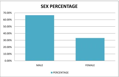
TABLE .1 SEX INCIDENCE
SEX NO OF PATIENTS PERCENTAGE
MALE 34 66.6%
FEMALE 17 33.3%
In my study out of 51 patients 34 were male and 17 were female
the ratio of male to female is 2:1.sex incidence is almost similar to the
western studies.
0.00% 10.00% 20.00% 30.00% 40.00% 50.00% 60.00% 70.00%
MALE FEMALE
SEX PERCENTAGE
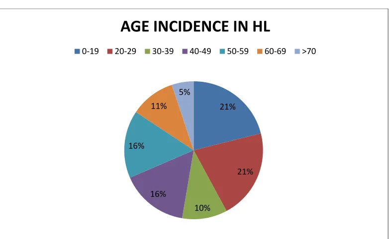
TABLE .2 AGE INCIDENCE IN HL
AGE in years NO OF PATIENTS PERCENTAGE% IN
HODKINS
<19 4 21%
20-29 4 21%
30-39 2 10.5%
40-49 3 15.8%
50-59 3 15.8%
60-69 2 10.5%
>70 1 5.1%
In this study I have found that the most commonly involved people
in Hodkins belong to the age group of between 20-29(21%) and
<19(21%) YEARS
21%
21%
10% 16%
16% 11%
5%
AGE INCIDENCE IN HL
AGE INCIDENCE IN NHL
AGE NO OF PATIENTS %OF PATIENTS
IN NHL
<19 3 9.3%
20-29 1 3.1%
30-39 5 15.6%
40-49 8 25%
50-59 6 18.7%
60-69 4 12.5%
>70 5 15.6%
In this study I have found that the most commonly involved people
in NHL belong to the age group of between 40-49(25%) YEARS.
<19 9%
20-29 3%
30-39 16%
40-49 25% 50-59
19% 60-69
12%
>70 16%
SUB TYPE DISTRIBUTION IN HL
Among 19 cases most common type of HL is mixed cellularity-8
cases(42.1%) ,nodular sclerosis found in 3 cases(15.7%) ,lymphocyte
predominant found in 2 cases(10.4%),lymphocyte depleted in
1case(5.2%) and type not specified in 5 cases(26.3%).. MIXED CELLULARITY 42.1%(8 cases)
NOT KNOWN 26.3%(5 cases)
NODULAR SCLERSOSIS 15.7%(3cases)
LYMPHOCYTE PREDOMINANT 10.4% (2cases)
LYMPHOCYTE DEPLETED 5.2%(1case)
SUB TYPE DISTRIBUTION IN HL
Among 19 cases most common type of HL is mixed cellularity-8
cases(42.1%) ,nodular sclerosis found in 3 cases(15.7%) ,lymphocyte