“COMPARA
SARCOMA O
THE TAMILN
with
COIM
A Dissertation on
PARATIVE STUDY OF SOFT T
MA ON EXTREMITIES AND TR
Dissertation Submitted to
MILNADU Dr.M.G.R. MEDICAL UNIV CHENNAI - 600 032
with partial fulfillment of the regulations
for the award of the degree of
M.S. GENERAL SURGERY (BRANCH 1)
COIMBATORE MEDICAL COLLEGE, COIMBATORE
APRIL 2015
FT TISSUE
ND TRUNK’’
UNIVERSITY
CERTIFICATE
Certified that this is the bonafide dissertation in “COMPARATIVE STUDY OF SOFT TISSUE SARCOMA ON EXTREMITIES AND TRUNK’’ was a work done by Dr.M.SRIDHAR and submitted in partial fulfilment of the requirements for the Degree of M.S. GENERAL SURGERY, BRANCH I of The Tamilnadu Dr. M.G.R Medical University, Chennai.
Date:
Professor and Unit Chief
Department of General SurgeryCoimbatore Medical College.
Date: Professor and HOD
Department of General Surgery Coimbatore Medical College.
Date: The Dean
DECLARATION
I Solemnly declare that the dissertation titled “COMPARATIVE STUDY OF SOFT TISSUE SARCOMA ON EXTREMITIES AND TRUNK’’ was done by me at Coimbatore Medical College and Government General Hospital during the academic year 2012-2015 under the guidance of Prof. Dr. V. ELANGO M.S. This dissertation is submitted to the TamilnaduDr.MGR Medical University towards the fulfilment of the requirement for the award of M.S. Degree in GENERAL SURGERY (BRANCH I).
PLACE : Dr. M. SRIDHAR
ACKNOWLEDGEMENT
First I thank my wife and parents for showering their blessings on me and making me Determined and dedicated to complete this venture in a successful manner.
I express my gratitude to Dr. S. Revwathy M.S OG., the Dean, Coimbatore Medical College Hospital for providing facilities to carry out my project work successfully.
I sincerely thank Prof Dr. V. Elango M.S, Chief And HOD Department of General surgery For his constant guidance and encouragement throughout the period of my study.
I Would like to express my gratitude to my guides Prof. Dr.Swaminathan M.S, D.O, Prof. Dr.Ranganathan M.S, Prof. Dr.Natarajan M.S, Prof.Dr.Ravindharan M.S, Prof.Dr.Saratha M.S, Prof. Dr.Balasubramanian M.S, for their valuable guidance and support throughout the period of my study.
And also my special thanks to ward patients and Nursing staff support throughout the period of my study.
COMPARATIVE STUDY OF
SOFT TISSUE SARCOMA ON
TABLE OF CONTENTS
S.NO CONTENT PAGE NO
1 INTRODUCTION 1
2 AIMS AND OBJECTIVES 6
3 REVIEW OF LITERATURE 7
4 MATERIALS AND METHODS 78
5 RESULTS AND ANALYSIS 81
6 DISCUSSION 105
7 SUMMARY 111
8 CONCLUSION 112
9 BIBILIOGRAPHY 114
10 ANNEXURES A) PROFORMA B) CONSENT FORM
LIST OF TABLES
S.No Table Page No
1. Surgical Staging System (SSS) of soft tissue sarcoma 27
2. AJCC TNM classification 28
3. AJCC TNM classification 29
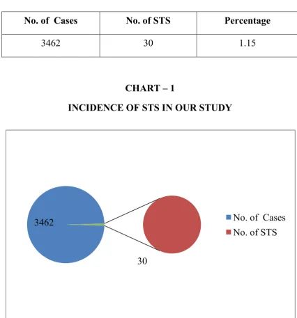
4. MS-KCC sarcoma prognostic factors 51 5. Incidence of STS in our study 83
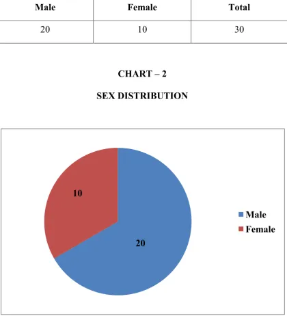
6. Sex distribution 84
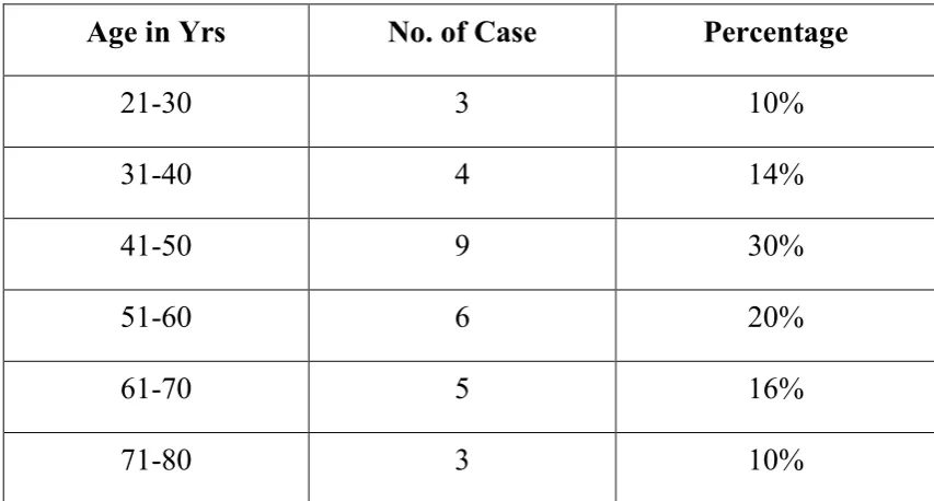
7. Age distribution 85
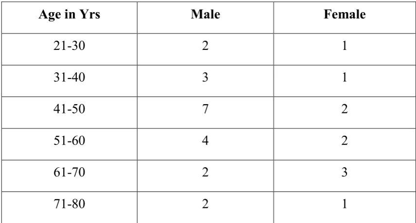
8. Age & sex distribution 87
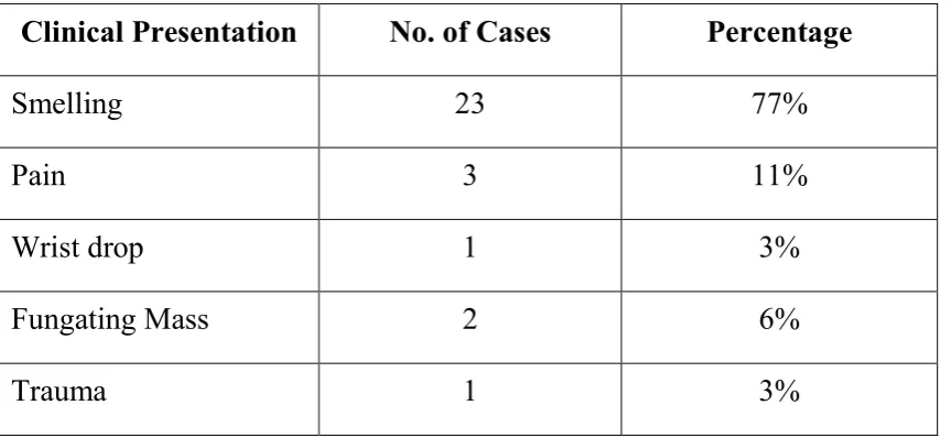
9. Clinical presentation 89
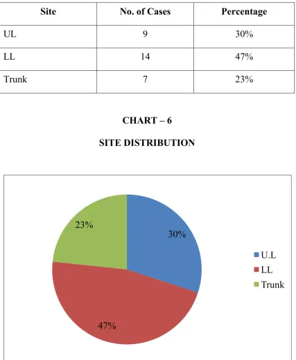
10. Site distribution 91
11. Depth of tumour 92
12. Histological distribution 93 13. Histological grading in our study 95 14. Surgical management given to our patients 96
15. Treatment modalities 98
16. Recurrence 100
17. 6 months survival rate 101
18. Mets free survival rate 102
LIST OF CHARTS
S.No Chart Page No
1. Incidence of STS in our study 83
2. Sex distribution 84
3. Age distribution 86
4. Age & sex distribution 88
5. Clinical presentation 90
6. Site distribution 91
7. Depth of tumour 92
8. Histological distribution 94
9. Histological grading in our study 95 10. Surgical management given to our patients 97
11. Treatment modalities 99
12. Recurrence 100
13. 6 months survival rate 101
14. Mets free survival rate 102
1
INTRODUCTION
Soft tissue sarcomas (STS) are originating mainly from the
embryonic mesoderm. STS are group of malignant tumour, anatomically
and histologically diverse neoplasms that share a common embryonic
origin arising primarily from mesenchymal tissue with notable exception
of neurosarcoma, primitive neuro–ectodermal tumors and Ewing
sarcoma, which are arise from neuro-ectodermal tissues. It is common in
paediatric age group accounting for 6.5% of all childhood malignancy but
rare in adult accounting for 1% of all adult malignancy and 2% of cancer
death.
The incidence of STS is very less and may be 2-4 cases/ 100,000
per annum.
Mortality, is high; the mean five-year survival rate is about 60%.
Because of multiple causes the STS occurs in multiple forms still etiology
remains unknown.
The most common sites are limb about 40%-50% of these are
lower extremities, 25% are upper extremities, and 15-20% are trunk.
2
Hematogenic metastases decide the prognosis of poorly
differentiated soft tissue sarcomas ,and but loco regional recurrence not
decide much about prognosis. In the past three decades, the combined
modality treatment of soft tissue sarcomas may be considerably.
Although surgery is the cornerstone of the sarcoma treatment, there is
definitive treatment by adjuvant radiotherapy after narrow surgical
excision of the tumour. On the other hand, there is no definitive need for
(neo) adjuvant chemotherapy after surgical treatment of soft tissue
sarcomas, with the exception of rhabdomyosarcoma, extraosseous
Ewing’s sarcoma / primitive neuroectodermal tumor (PNET), and
Extraosseous osteosarcoma.
Surgical resection with radiotherapy treatments have mainly
involved to improved locoregional tumour control and only to a lesser
degree to survival. Conversely, the ‘aggressive’ surgical approach in the
treatment of metastasized tumours has contributed to improved
disease-free and overall survival.
Surgery is the main corner stone in the multimodality treatment of
the disease –which involves various methods including pathology,
radiotherapy, medical oncology, rehabilitation medicine, genetics, and
3
Amputation was the treatment of choice for STS of extremity
because of high rate of recurrence after local excision. With advent of
combination treatment (surgery and radiotherapy (RT)), limb sparing
surgery become treatment choice7,5 .
Isolated limb perfusion5,9 (ILP) treatment was the local administration of a cytostatic agent at the highest dosage without
introducing systemic side effects. The extracorporeal circulation system
used was similar to the one employed in open heart surgery. The
technique was first applied to patients with in-transit metastases of
melanoma of the lower extremities who refused amputation.Melphalan
was the cytostatic agent of choice.
Oldhoff and Schraffordt Koops successfully operated the perfusion
program of Groningen in the Algemeen Provinciaal Stads enAcademisch
Ziekenhuis (APSAZ).
Hyperthermia was introduced in 1967 in the perfusion treatment
by Cavalière. Recently, mild hyperthermia(39-40 °C) is widely used,
because this causes less local toxicity than higher temperatures. As the
search for the optimum perfusion technique, other cytostatics such as
dacarbazine (DTIC), adriamycin and cisplatinum all are tried but
4
When Lejeune and colleagues added tumor Necrosis Factor alpha
(TNFα) to melphalan as a perfusion agent in the isolated regional
perfusion for locoregional advanced soft tissue sarcomas and melanomas.
This resulted in a high local response rate with acceptable locoregional
and systemic toxicity and a high percentage of limb salvage. Medical
centers in Lausanne, Amsterdam, Rotterdam, Berlin, Brussels, Tel Aviv,
and Groningen participated in this trial. They Used high dose of TNFα
in combination with interferon and melphalan.
This technique useful for the salvage of the affected extremity in
84% of the cases. The perfused tumours were large size, around 20 cm
on average. Resection was performed to many weeks after post
perfusion. In 87% of the cases, Tumor response was well documented. It
was then clear that ILP in combination with TNFα , interferon gamma
and melphalan was safe and highly effective and constituted important
treatment of locally advanced soft tissue sarcomas of the extremities.
Another important multimodality treatment was radiotherapy.
Rosenberg was the first demonstrate the value of adjuvant radiotherapy in
the limb-saving treatment of sarcomas. Yang and colleagues updated the
5
They identified that maximum reduced in locoregional recurrence after
radiation, without affecting patients survival rate.
Greater stiffness and increased edema of the involved extremity,
6
AIM OF THE STUDY
1.
To compare 6 months survival rate in extremities and
trunk STS.
2.
To compare metastasis free survival rate in extremities
7
REVIEW OF THE LITERATURE
SOFT TISSUE SARCOMA
15
Soft tissue sarcomas (STS) are a heterogeneous group of tumours
arising mainly from the cells of the soft supporting tissues of the body
(also known as mesenchyma): fat, muscles, nerves, fibrous tissues, blood
and lymph vessels. The most commonly STS arises from extremities
(30-52%), lower extremity (29-49%), upper extremity (12-21%),
retroperitoneum (8-15%),head and neck (4-13%), abdomen (10-12%),
pelvis (7-12%), and thorax (9-11).Soft tissue sarcomas are rare tumours,
and globally account for less than 1% of all malignancies.
Soft tissue sarcomas are therefore a heterogeneous group of
tumours. They currently include more than 50 different histological
subtypes. The classification of these tumours is complex and review of
the histopathologic specimen by experienced pathologists is generally
recommended for clinical practice and for clinical research.
Progress in the understanding of sarcoma has frequently resulted in
identification of new histological subtypes or reclassification of existing
8
The development of immunochemistry, cytogenetic and molecular
genetic has brought major progress in this field in the last 10 years. The
World Health Organization classification of soft tissue tumours has been
issued in 2002 .
Several histological grading systems have been proposed, with
either 3 or 4classification levels. Most have been validated as prognosis
level for metastases free and overall survival in untreated soft tissue
sarcoma patient.
The two most widely used grading systems are the one of the us
National Cancer Institute (3 levels, based on the histological subtype, and
tumour necrosis level, mitotic index, cellularity rate and/or pleomorphism
for some subtypes), and the one of the French “Fédération Nationale des
Centres de Lutte Contre le Cancer” (3 levels, based on tumour necrosis,
mitotic rate and tumour differentiation, giving an equal weight to these 3
factors). The French system has been shown to better predict outcome,
but both systems are still used. The two systems do overlap in
9
Soft tissue sarcomas are often diagnosed accidentally, because they
generally develop without pain. Less than 1% of soft tissue tumours are
malignant. Other soft tissue tumors are either benign or of intermediate
malignancy (locally aggressive or rarely metastasizing), according to the
last classification of soft tissue tumours [1]. The probability of
malignancy of a newly diagnosed soft tissue tumour is related to size and
depth, but, so far, malignancy cannot be accurately predicted by clinical
examinations and imaging techniques. A tumour biopsy is needed to
confirm the histology, and this should preferably be performed in a
specialized sarcoma center to avoid compromising the results of the
subsequent surgery.
ETIOLOGY7,9,15
Eventhough several factors are identified as causative factors no
specific etiological factors can be identified. Bone and soft tissue sarcoma
may be caused by previous radiation exposure. Cutaneous lymphangio
sarcoma may caused by chronic lymphedema.
Chronic lymphedema after mastectomy surgery and radiotherapy
may produce Angiosarcoma known as Stewart-Treves syndrome . Vinyl
chloride, phenoxyacetic acid herbicides, and chlorophenols and their
10
Certain genetic conditions such as neurofibromatosis1,caused by a
mutation in 17q11, Aggressive malignant peripheral nerve sheath
tumours (MPNST) may occur in 1-5% patient. Li-Fraumeni syndrome,
mutation of p23,may be common relation with so many malignancies,
including STS. Mutations in the retinoblastoma gene RB1, also increases
risk of sarcoma.
Desmoids tumour is associated with phenotypical variant of
familial adenomatous polyposis (FAP).Benign enchondromas,
hemangiomas and lymphangiomas may undergone into their malignant
sarcomatous changes in Maffucci syndrome.
Kaposi’s sarcoma may be produced by human herpes virus 8
(HHV 8). Epstein-Barr virus (EBV) infection has been found to cause
leiomyosarcoma during therapeutic immunosuppression treatment stage.
Even though no specific etiologic agent is identified in the
overwhelming majority of patient with soft tissue sarcoma the risk factors
11
1 . Previous radiation exposure
The risk of post radiotherapy saracomas arise more than 3 yrs after
radiation therapy exposure and some decades later saracomas occurring
after radiation exposure are most commonly malignant fibrous
histiocytoma.
2. Environmental factors
Chemical exposure to phenoxy acetic acid found in herbicides,
chlorophenol found in wood preservatives are associated with STS.
Thorotrast, vinyl chloride, arsenic are associated with ‘hepatic
angiosaracoma”.
3. Chronic lymphedema
Chronic lymphedema such as that experience after axillary
dissection has been associated with lymphangio sarcoma (stewart-treves
syndrome)
4. Chronic inflammatory process may a risk factor
Agents such as sharpnel, bullets, intra muscular iron injections and
12
5. Genetic predisposition
Specific inherited genetic alterations have been associated with an
increased risk of STS. Patients with gardener’s syndrome (familial
polyposis) have a higher than normal incidence of desmoids.
Patients with germline mutations in the tumour suppressor gene
p53(Li-fraumeni syndrome) have high incidence of sarcomas. Patients
with von recklinhausen’s disease who have abnormalities in the
neurofibromatosis type I tend to develop neurofibrosarcoma.
Soft tissue sarcoma can occur in patients with hereditary
retinoblastoma as a second malignancy.
6 . Chromosome rearrangements
A number of soft tissue tumours both benign and malignant have
been found to have consistent chromosomal abnormalities which in many
cases may be diagnostic. Chromosomal translocations are the most
13
CLASSIFICATION AND HISTOPATHOLOGY7,9,12
Because of the rare variety and heterogeneity of STS tumours
histology examination may be done by an experienced pathologist.
The World Health Organization Classification of STS Tumours
into > 50 subtypes. Undifferentiated pleomorphic sarcoma (malignant
fibrous histiocytoma (MFH)), is the most common type in adults,
representing 28-39% of overall STS. Liposarcoma (14-22%), synovial
sarcoma (11-12%), leiomyosarcoma (6-12%), fibrosarcoma (8-9%) and
MPNST (6-7%), are also among the more common subtypes.
Rhabdomyosarcoma is the most commonly occur histologic
finding in children, incidence occur > 50% of paediatric STS.
FIBROUS TUMOURS
Fibro Sarcoma
a) adult fibro sarcoma.
b) congenital or infantile fibro sarcoma intermediate tumours.
c) Inflammatory fibrosarcoma (inflammatory myofibroblastic
14
FIBROHISTIOCYTIC TUMOURS
1. Intermediatetumours
Dermato fibrosarcoma protuberans.
DERMATO FIBROSARCOMA PROTUBERANS
2. Malignant fibrous histiocytoma.
a. Storiform-plemorphic fibrous histiocytoma
b. Myxoid fibrous histiocytoma
c. Giant cell fibrous histiocytoma(malignant giant cell tumor of soft
parts)
15
LIPOMATOUS TUMOURS
Liposarcoma
a) well-differentiated liposarocoma
i. lipoma-like liposarcoma
ii. Sclerosingliposarocoma
iii. Inflammatory liposarcoma
b) Myxoidliposarcoma
c) Round cell (poorly differentiated myxoid) liposarcoma
d) Pleomorphic liposarcoma
e) De-differentiated liposarcoma
SMOOTH MUSCLE TUMOURS
1. Rhabdomyosarcoma
a) Embryonal rhabdomyosarcoma
b) Botryoid rhabdomyosarcoma
c) Spindle cell rhabdomyosarcoma
16 e) Plemorphic rhabdomyosarcoma
2. Rhabdomyosarcoma with ganglionic differentiation
(etomesenchymoma)
17
TUMOURS OF BLOOD AND LYMPH VESSEL
1. Angiosaracoma and lymphangiosarcoma
2. Kaposi’s saracoma
3. Follicular dendritic cell sarcoma
PERIVASCULAR TUMOURS
1. Malignant glomustumour
2. Malignant hemangio pericytoma
SYNOVAL TUMOURS
1. Synovial sarcoma
a. Biphasic (fibrous and epithelial) synovial sarcoma
b. Monophasic (fibrous or epithelial) synovial sarcoma
18
EXTREMITIES SYNOVIAL SARCOMA
MESOTHELIAL TUMOURS
1. Malignant solitary fibrous tumour pleura and peritoneum
2. Diffuse mesothelioma
a. Epithelial
b. Fibrous
c. Diffuse
NEURAL TUMOURS
1. MPNST (malignant schwannoma , neurofibrosarcoma)
a. Malignant Triton tumour (MPNST with rhabdomyosarcoma)
b. Glandular MPNST ( malignant glandular schwannoma)
19 2. Malignant granular cell tumour
3. Clear cell sarcoma(malignant melanoma of a soft parts)
4. Malignant melanocytic schwannoma
5. Gastrointestinal autonomous nerve tumour(plexosarcoma)
6. Primitive neuroectodermal tumour
a. Neuroblastoma
b. Ganglioneuroblastoma
c. Neuroepithelioma
d. Extraskeletal Ewing’s sarcoma
PARAGANGLIONIC TUMOURS
Malignant Paraganglioma
EXTRASKELETAL CARTILAGINOUS AND OSSEOUS
TUMOURS
1. Extraskeletalchondrosarcoma
a) well differentiated
b) myxoid
20
PLURIPOTENTIAL MESENCHYMAL TUMOURS
Malignant mesenchymoma
MISCELLANEOUS TUMOURS
1. Alveolar soft part sarcoma
2. Epitheloid sarcoma
3. Malignant extra rena rhaboidtumor
4. Desmoplastic small cell tumor
FIBROSARCOMA
The term fibrosarcoma conclude to its cells resemple normal
fibroblast. The incidence and behaviour of this neoplasm greatly vary.
Recently the incidence of fibrosarcoma is declining due to refinement of
histopathological categorisation of sarcoma. The advent of
immunohistochemistry, cytogenetics and molecular genetic techniques
also contributing.
Classification of fibrosarcoma
Classified as adult type and juvenile/infantile type. Adult type
further classified as classic, myxoid, fibromyxoid and sclerosing
21
extremities,17% in trunk and 10% in head and neck. Commonly present
as painless mass predominantly involve deep structure.
PATHOLOGY
The histologic picture is uniform fasciculated growth pattern
consisting of fusiform or spindle- shaped cells. It is histologically graded
as high grade fibrosarcoma and low grade fibrosarcoma by appearance
and collagen content.It express vimentin which is used for immune
histochemical identification. This tumour compossed of elongated
fibroblastic cells with indented nuclei predominantly rough endoplasmic
22
MALINGNANT FIBROUS HISTIOCYTOMA SLIDE
MALIGNANT FIBROUS HISTIOCYTOMA(PLEOMORPHIC
UNDIFFERENTIATED SARCOMA)5,7,9:
It is common in late adult life range of 50 to 70 years. Has range of
histological types as storiform pleomorphic, myxoid,giant cell and
inflammatory. It is most common type soft tissue sarcoma, consisting
mixure of pleomorphic and storiform areas. Frequently occur in lower
23
inflammatory fibrous histiocytoma frequently occurs in
retroperitoneum.
Previous radiation is an important etiologic factor. Inflammatory
fibrous histiocytoma may develop subsequently second neoplasm.
PATHOLOGY
The term sarcoma is Greek for “Fish flesh”,reffering tumour’s
tendency to feel fleshy when palpated,Mesodermal cells give rise to the
connective tissues distributed throughout the body including
pericardium,pleura,blood vessel, endothelium, smooth and striated
muscle, bone, cartilage and synovium are the cells from which all
sarcomas orginate. Consequently sarcomas develop in a wide variety of
anatomic sites.
Approximately one half of all STS occurs in extremities, most
common histopathologies are malignant fibrous histiocytoma,followed
by liposarcoma. Several histologic types of sarcoma have been
characterized. This characterisation can be difficult and is aided by
Vimentin , S
actin are the common
often facilitates reliable histo typing
STS distinguishing it from carcinoma is the tendency of STS
hematogenous means.
Metastases are uncommon in patients with low grade STS.
node metastasis is rare
24
S-100, desmin , factor VIII, keratin , myoglobin and
are the common immunohistochemical staining used for proteins.
cilitates reliable histo typing one of the pathologic hallmark of
STS distinguishing it from carcinoma is the tendency of STS
hematogenous means.
Metastases are uncommon in patients with low grade STS.
node metastasis is rare,occurs in less than 3% of patients.
100, desmin , factor VIII, keratin , myoglobin and
histochemical staining used for proteins.
one of the pathologic hallmark of
STS distinguishing it from carcinoma is the tendency of STS to spread by
Metastases are uncommon in patients with low grade STS. Lymph
25
The histologic sub types with highest incidence of lymph node
metastasis are:
a) Epitheloid sarcoma 16.7%
b) Embryonal rhabdomyosarcoma
c) Angiosarcoma
d) Malignant fibrous histiocytoma
e) Synovial sarcoma
Grading5,9
Broders identify the first grading system for STS when he analysis
of squamous cell carcinoma. He demonstrate the 4-tiered system,That
was based on mitotic rate, percentage of giant cells, and number of
fibrous stroma in fibrosarcomas.
Low Grade tumours includes grade 1-2 (G1-G2) and High
grade tumours includes grade 3-4 (G3-G4).The American Joint
Committee on Cancer Union Against Cancer (AJCC/UICC) staging
26
Tumour necrosis, mitotic rate,and cellular differentiation are
considered in the French Federation of Cancer Centers (FNCLCC)
system,whereas the histologic variety, cellular differenciation, cellular
pleomorphism, and mitotic activity are considered in 3-tiered National
Cancer Institute (NCI) system .In both 3-tiered systems low grade is
(G1) and high grade tumors are(G2-G3).
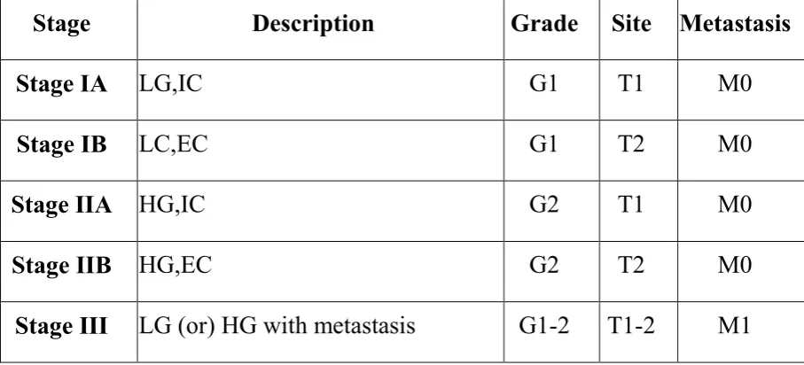
Staging7,9
Outcome not only based on histological grade.Staging systems
uses other factors for prognosis.The Musculoskeletal Tumour
Society(MSTS) staging system, called Surgical Staging System (SSS),
That includes malignancy grade, atypical histology, and mitotic index, all
three defined tumour may be grade Low (G1) or grade High
(G2).Intracompartmental(T1) and extracompartmental (T2) difference
also made.
Tumours which confined to a particular compartmental site is
called as Intracompartmental tumours, other hand tumours which
infiltrate into, or extend beyond the particular compartments is known as
extracompartmental. The absence metastasis (M0) or presence of
27
TABLE 1.Surgical Staging System (SSS) of soft tissue sarcoma
Stage Description Grade Site Metastasis
Stage IA LG,IC G1 T1 M0
Stage IB LC,EC G1 T2 M0
Stage IIA HG,IC G2 T1 M0
Stage IIB HG,EC G2 T2 M0
Stage III LG (or) HG with metastasis G1-2 T1-2 M1
LG-Low grade, HG-High grade,
IC-Intracompartmental, EC-Extracompartmental,
The accurate prediction of STS prognosis is a problem. An attempt
towards better result of outcome has been made in nomograms, which
28
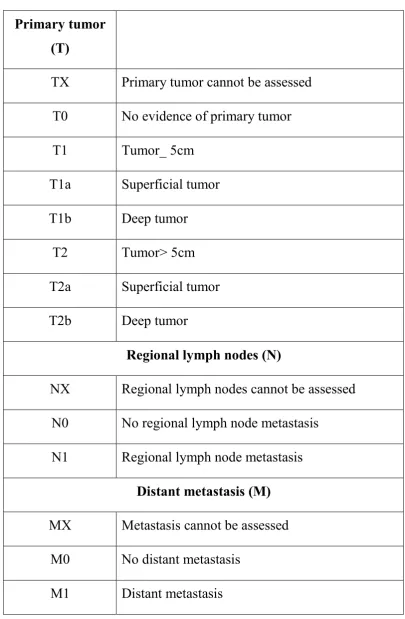
TABLE 2.AJCC TNM classification
Primary tumor (T)
TX Primary tumor cannot be assessed
T0 No evidence of primary tumor
T1 Tumor_ 5cm
T1a Superficial tumor
T1b Deep tumor
T2 Tumor> 5cm
T2a Superficial tumor
T2b Deep tumor
Regional lymph nodes (N)
NX Regional lymph nodes cannot be assessed
N0 No regional lymph node metastasis
N1 Regional lymph node metastasis
Distant metastasis (M)
MX Metastasis cannot be assessed
M0 No distant metastasis
29
Histologic grade (G)
GX Grade cannot be assessed
G1 Well differentiated
G2 Moderately differentiated
G3 Poorly differentiated
[image:39.595.105.513.70.283.2]G4 Poorly differentiated or undifferentiated
TABLE 3.AJCC TNM classification
Stage Tumor (T) Node (N) Metastasis (M) Grade (G) Description
StageI T1a-T2b N0 M0 G1-2 LG
StageII T1a-T2a N0 M0 G3-4 superficial HG
StageIII T2( T2b) N0 M0 G3-4 Large deep HG
StageIV
Any T
Stage N1 M0
Any
grade Lymph node Mets +
Any T
Stage N0 M1
Any
grade Lung metastasis +
30
Surgical margins
Enneking reffered margins clearance of surgery based on depth
and length of healthy tissue surrounding the resected tumour.Surgical
clearance margins separated to radical, lenth,
marginal.Intracompartmental tumours removal called as radical
excision.Intracompartmental resection, with normal tissue excision
surrounding the mass called as Wide margin clearance.The plane of
dissection is within tumour zone or false capsule of the tumour called as
marginal Resection.
Surgical margins classification is defined by the UICC, they are
R0,R1,R2. R0 means no inresidual disease, R1 means microscopic
disease,R2 means macroscopic disease.
It clearly demonstrated that positive surgical margins have negative
prognosis.A recent gernals defined the clearence of 2-3 cm tumour
margins is not possible, but it is followed in so many hospitals. so the
need for ablative procedures when using a 2-3 cm margin instead of 1 cm.
31
GENETIC ASPECTS OF SOFT TISSUE SARCOMAS (STSs)
Natural course and genetic features of STSs
The term sarcoma originates from the Greek language and means
"fleshy growth".
3,7
Sarcomas are malignant tumours of mesenchymal origin that can
arise almost anywhere in the body. The most common location is in the
extremities (the arms, legs, hands, or feet) where about 50% of the
tumours are found. The trunk occurrence may be 40% (chest, back, hips,
shoulders, and abdomen), and Head and neck occurrence may be 10%.
The Sarcomas account for approximately 1% of all adult malignancies
and 20% of pediatric malignancies.
One of the clinical features of sarcoma is that in the early stages
they do not usually cause clinical symptoms. This is related to the fact
that soft tissues are relatively elastic, and the tumours may grow large,
pushing the normal tissue, before they are felt or cause any problems.
Sarcomas generally are capable of invasive or destructive growth and the
patients frequently develope recurrence and distant metastasis forming
secondary tumours. Radical surgery is usually required to ensure total
32
The natural course of sarcomas is highly variable. For example,
some sarcomas such as dermatofibrosarcoma protuberans rarely
metastasise, while other types such as malignant fibrous histiocytoma
(MFH) do so with alacrity. Because of the general aggressiveness of
STSs and the frequent use of extensive surgery, development of
prognostic and diagnostic markers is advocated. Until today, parameters
such as tumour size, location, necrosis, intra-vascular invasion,
histopathological malignancy grade, and the treatment of the tumour have
been shown to have prognostic value.
The histopathological classification is a challenging task. More
than 50 subtypes of proliferative soft tissue lesions are presently defined.
Until today, the most common types of STS diagnosed have been
malignant fibrous histiocytoma (MFH) and liposarcoma (LPS) that
together account for 35% to 45% of all sarcomas.
The classification of STSs has not stayed constant over the years,
but is regularly re-evaluated and re-formulated. For example, the MFH
entity that was introduced forty-two years ago by Ozzelloetal 51 has
recently been challenged to its mere existence 16 by Fletcher and
co-workers 52. Similarly, the gastrointestinal stromal tumor (GIST) entity
33
several abdominal STSs are classified as GIST that would have been
diagnosed as e.g. leiomyosarcoma a few years earlier 55. Nevertheless,
many of the tumours still lack clear-cut diagnostic foundation, especially
when the tumours exhibit an undifferentiated morphology. Therefore,
new specific molecular genetic markers are become very useful in the
clinical evaluations of STSs.
STS tumours constitute a highly heterogenous group of tumours,
for which genetic characterization is still limited. Therefore, the aims of
this thesis has been to extend the knowledge of genetic and molecular
alterations involved in the progression and metastasis formation of this
tumour group.
2.2 Chromosomal events in STSs
The value of cytology and molecular genetics already well
developed in pratical and clinical investigation of hematological
malignancies, and specific gene dearrangement are used for diagnostic,
prognostic and therapeutic purposes. Same method is presently applied
for sarcomas, where so many chromosomal alterations, mostly reciprocal
translocations, have been associated with distinct histopathological
entities. In some status, different oncogenic fusion genes defect (or)
34
Specific translocations result in fusion genes in STSs
On the genomic level fusion genes commonly result from breakage
within introns of the two partner genes whereby exons with the same 5´-
3órientation are joined in frame, enabling the translation of a functional
new protein from the resulting fusion transcript. A series of specific
translocations and fusion genes have already been reported and associated
with certain STS subtypes (Figure 4). For example, a translocation
t(12;16)(q13;p11) is found in more than 90% of myxoid LPS. Through
this rearrangement the fusion gene FUS-CHOP is created and expressed
specifically in the tumour cells. Although the same genes are involved in
each case, variations on the base-pair level are frequently seen which are
mainly attributed to varying 17 breakpoints in the FUS (TLS) gene
(Figure 3) 56-59. Furthermore, myxoid LPS without a typical
t(12;16)(q13;p11) may instead carry a EWS-CHOP fusion gene
resulting from a t(12;22)(q13;q12). However, both these fusions lead to
the same sarcoma subtype displaying indistinguishable histopathological
features 60.
Altogether at least 25 fusion oncogenes have been described in
STSs (Figure 4). Most recently, a novel fusion gene FUS-BBF2H7
35
sarcoma 61. Notably, it is common that the same partner gene is involved
in different fusions, which can each be associated with different tumor
phenotypes.
For example, the EWS gene has been found fused to one of nine
partner genes, giving rise to five different types of STS (Figure 4) 62-70.
Some of the EWS-fusions are associated with very aggressive clinical
tumour progression, e.g. desmoplastic small round cell tumor 69. The
FUS gene is reported to be involved in three different fusion genes 61,
71, 72, that are each associated with a different type of STS.
Obviously, these specific fusion genes and their phenotypes are
tightly linked with each other, but the exact mechanism behind the
specificity is still unclear. The expected pathogenic importance of the
fusion genes is supported by the observations of chromosome
translocations as the sole cytogenetic anomaly in a significant proportion
of STSs (Figure 4). However, in several instances it still needs to be
established whether STS fusion genes represent the first tumour initiating
events or are proceded by other events not detectable on the chromosome
36
Amplicons are common findings in STSs
In addition to chromosome translocations, other recurrent
aberrations are also found in STSs such as double minutes, ring
chromosomes and giant rod chromosomes.
These alterations are non-randomly distributed and commonly
involve amplifications and over-expression of genes in the target regions.
Ring or giant rod marker chromosomes with amplification of 12q13-15,
play a key role in lipomatous tumor development 73, 74.
Well-differentiated LPS frequently involve several genes known to be
amplified in human STSs, e.g. MDM2, SAS, GLI, CHOP and CDK4 in
the chromosomal region 12q13-15. Recently, Tallini et al. showed that
the HMGA gene is commonly over-expressed in well-differentiated LPS
with ring or rod chromosomes and amplification of 12q13-15.
The region 1q21-23 is also commonly involved in amplifications,
and includes the COAS2 gene as reported by Nilsson et al. By FISH, the
most common localization of extra COAS2 signals in lipomatous tumours
was demonstrated to be in supernumerary ring and giant marker
37
In malignant fibrous histiocytoma (MFH), the MASL1 gene has
been suggested to be the oncogenic event driving the amplifications of
the chromosome region 8p23.1 78.
Mutation of the C-KIT and PDGFRA genes in GIST
In addition to specific alterations that can be revealed at the
cytogenetic level, STSs also demonstrate recurrent genetic alterations of
more discrete types. A good example is provided by the gastrointestinal
stromal tumours (GISTs) that show mutations and/or over-expression of
the C-KIT and PDGFRA genes.
GISTs are the most common mesenchymal tumors of the
gastrointestinal tract, representing approximately 20-30% of all STS in
this location. The majority of GISTs exhibit mutations in C-KIT that
cluster in four hot spot exons (9, 11, 13 and 17) and are especially
frequent in exon 11. In GIST, the C-KIT mutations regularly alter or
delete one or more amino acids, but are always in frame. This then leads
to changes in the juxta membrane domain of the c-kit protein and tyrosine
kinase activation without binding of the stem cell factor (SCF) ligand.
The resulting constitutive expression of c-kit in turn results in altered cell
proliferation and tumorigenesis . GISTs with C-KIT mutation are more
38
frequent recurrence and a higher mortality rate than tumours with
wild-type C-KIT only.
GIST tumours with a C-KIT mutation are also responsive to
treatment with Imatinib, a drug that inhibits the c-kit tyrosine kinase, and
which is applied to patients with inoperable or metastatic disease.
However, acquired resistance to Imatinib may develop after a period of
treatment. Additional mutation of C-KIT is one possible explanation for
the observed resistance. Most recently, Heinrich and co-workers found
that approximately 35% of GISTs lacking C -KIT mutations carried
activating mutations in the related receptor tyrosine kinase gene,
platelet-derived growth factor receptor alpha (PDGFRA).
Tumours showC-KIT or PDGFRA oncoprotein spect to activation
of were found to be indistinguishable with respect to activation of
downstream signalling intermediates and cytogenetic changes associated
with tumour progression. Therefore and C-KIT,PDGFRA mutations
appear to be alternative and mutually exclusive oncogenic mechanisms in
GISTs.
There are two hot spot exons for PDGFRA mutations, i.e. exons 12
and 18. Taken together both C-KIT and PDGFRA mutations contribute to
39
PROGNOSTIC ASPECTS OF SOFT TISSUE SARCOMAS
Histopathological markers
Clinical and histopathological markers of documented prognostic
value include e.g.malignancy grade (high grade III or IV), tumor size (>8
cm or >11 cm), tumor depth (deep location), tumor localization and
surgical margin. Presence of necrosis and high mitotic count are similarly
established markers.
As shown by us and others one of the best parameters is tumour
size, which in turn is also related to the location of the tumour. For
example, STSs located in the distal extremities are often small and
superficial when diagnosed. On the other hand, the prognosis is usually
better compared to the tumours located intra-abdominally, where the
tumours are usually rather large already at the initial diagnosis.
The value of malignancy grade as prognostic variable for STSs has
been reported. The features that define the grade are strongly linked with
the degree of cellularity, differentiation,necrosis as well as the number of
40
Immunohistochemical markers
Vascular invasion, metastasis and local recurrence are features of
an aggressive tumour phenotype. Many immune hiostochemical markers
studied are therefore chosen to reflect the three cornerstones of tumour
growth, i.e. cell proliferation, apoptosis, and angiogenesis. For example,
factor VIII measures vessel density, Ki-67 is a marker of proliferation,
and the p53, p27 and Bcl2 proteins are all related to the regulation of the
cell cycle and hence linked to apoptosis.
Increased expression of IGF1-R was seen in some malignancies in
cases with metastatic disease ,while in high grade STS the IGF-1R
expression was associated with favourable outcome.
Furthermore, expression of CD44 and ezrin are associated with cell
adhesion, and related to the cell migration and metastasis.
Over-expression of ezrin is related to poor outcome.
The tumour phenotypes that will usually lead to metastastic
behaviour include the capacity of tumor cells to migrate within tissues,
transmigrate through vessels and to adhere to the metastatic organs. Since
metastasis are the main cause of death in cancer the identification of
genes that regulate tumour cells migration may therefore lead to
41
Recently, ezrin was identified as a key component in the metastasis
of tumors as reported by several authour. In general, the role of ezrin in
tumour metastasis was based on two of the major reasons: A) ezrin is best
known to connect membrane proteins to the actin cytoskeleton, through
the adhesion molecules that are known to depend on the ezrin-mediated
linkage to actin, such as CD44, and are directly related to the invasion
and metastasis of tumors B) The ability of ezrin to confer metastatic
capabilities to tumors, which has been proved in experimental models, for
example, mouse model of osteosarcoma and osteosarcoma in dogs.
The over-expression of ezrin in malignant tumours and its relation
to poor outcome have been reported in carcinomas, such as, prostate
cancer, glioma and melanoma. Concerning mesenchymal tumors,
involvement of ezrin has been discussed for gastrointestinal stromal
tumors, osteosarcoma and rhabdomyosarcoma . In this thesis the impact
42
Genetic markers
Genetic markers represent a field of increasing importance in STS
diagnostics and prognostics. In general, the oncogenes, which can induce
malignant transformation and cell proliferation, have been implicated in
the development of STSs. In the majority of cases, oncogene activation
result from chromosomal rearrangements or gene amplifications. Changes
of the microenvironment of the gene, for example following epigenetic
modifications, must also be considered. Examples of oncogenes linked to
STSs are C-KIT and PDGFRA mutations in GIST, and fusion oncogenes
such as SS18-SSX in 90% of synovial sarcoma. In contrast, the tumour
suppressor genes (TSG) play a critical role in cell growth and dictate the
cell program to apoptosis. In contrast to oncogene activation, loss or
change of the TSG function commonly result from deletions or discrete
mutations. Two major TSGs that are relevant to STS are the RB1 and the
TP53 genes.
Approximately 30%-60% of STS have been reported to harbour
aberrations of the TP53 gene, including a subset of patients with
43
Clinical Presentation
Most of the patient came with c/o painless mass, although the pain
is noted half of the patient. No specific abdominal discomfort and GI
symptoms in intra abdominal or retroperitoneal STS.
The most common differential diagnosis for extremities and trunk
lesion being bleed collection or muscle mass leads to delay in diagnosis.
Diagnosis
Physical examination
Assess tumour size and movement of the mass, fascia intact or
not,and near by neurovascular and bony infiltration present or not, and
44
FUNGATING MASS OVER THE LOWER EXTREMITIES
INVESTIGATIONS
1.Biopsy-The Primary thrust is to obtain adequate tissue for
histopathologic examination, to assess the grade, and to define the
prognostic factors that may help the patient for definitive treatment.
• Indications
Any soft tissue mass in an adult that is symptomatic or enlarging is
greater than 5cm in size and any new soft tissue mass that persists beyond
45
• Technique
Small superficial mass (<5cm) –excisional biopsy with clear
margins in the preferred approach.
Large mass- incisional biopsy with a longitudinal incision
(extremity lesions) to facilitate subsequent wide locall excision. Incision
carried out by center of the mass with most superficial location.
Hemostasis should be adquate to prevent dissemination of tumour
cells into other normal tissue planes.
Wide local resection of a previously biopsied sarcoma, the previous
scar may be excised with the tumour. Biopsy specimens should be sent
fresh, sterile and anatomical site orientation marked for pathologic
studies..
FINE NEEDLE ASPIRATION CYTOLOGY
FNAC has been examined by a number of authors but is usually
confirmed to the diagnosis of recurrence first, then for the primary
46
Imaging9,15
For appropriately assess the staging of STS,Radiological imaging
are required and surgical approach as well as RT/CT treatment. Aim are
to Identify anatomical site, tumour size, homo/hetrogeneity, and
calcification present or not. Infiltration into, nearby vital structures
present or not is essential for treatment approach.
X-ray is the first-line imaging method. Because of inexpensive and
readily available, underlying bone involvement can be assessed. Also
plain xray identify involvement of phleboliths of hemangiomas.
Extremity STS the main imaging modality are MRI.MRI have
specific morphological images data, multiple-plane imaging, and no
ionizing radiation load make the MRI for detection,delineation,
differential diagnosis, and Treatment response monitoring, and also for
postoperative follow-up. MRI used for guided needle-biopsy.fascial
involvement presents or not and invasion of a tumour to adjacent
structures have better idea for surgical planning.
CT is preferred imaging modality for the trunk and retroperitoneal
STS, and also tumour bony invasion. CT is also the imaging of choice for
47
be used for solid and cystic masses. It’s most useful in guided needle
biopsy. vascular supply of tumors and vascular lesions Magnetic
resonance angiography gives better information.
Positron emission tomography (PET) with 18F-fluorodeoxyglucose (FDG) has advanced tool in STS imaging.Tissue glucose metabolism
activity can be identified by FDG. Functional imaging PET scan used
for STS tumour identification, differential diagnosis, imagine guided
biopsy, for identify the difference between recurrences and
therapy-related changes, and treatment responce monitoring.preoperative
TNM-staging can be assessed by PET with CT.Assessment of tumor cell
proliferation may be done using18F-fluorothymidine (FLT), but only experimental use.
Plain chest radiographs essential of choice for all STS patients to
ruleout mets,Most STS distant metastases are found in the lungs.Chest
CTscan for all patients as the primary modality to R/O lung mets.with
primary CT recommended for >5cm high grade lesions only. Plain chest
radiographs diagnosis most lung mass and recommending chest
radiographs for all patients, and primary CT only for patients with an
48
with large, deep, or grade 2 (or) 3 tumors, and other biologically
aggressive histological subtypes.
CT SCAN
Provide information on the size of the lesion and its relationship to
adjacent structure and organs.
MRI
• Examination of choice for imaging of soft tissue masses
• Enhances the contrasts b/w tumor and muscle, and b/w tumor
mass and adjacent blood vessel.
• Provides superior 3 dimensional definition of fascial planes
(These substantial advantages allow for improvement assessment
of respectability with pre operative MRI evaluation)
NEWER INVESTIGATORY TECHNIQUES
Phosphorus magnetic resonance spectroscopy
• It is a form of noninvasive resonance spectroscopy that detects
49
• Employed to evaluate sarcoma inorganic-to-organic phosphate
rations.
• In- vivo metabolic assessment of response to anti-neoplastic
therapy.
Positron Emission Tomography (Pet Scan)
• By evaluating tumour metabolic activity PET scan may permit
noninvasive assessment of tumour grade.
RadioNuclide Scintigraphy
• Thalium- 201 and gallium-67 in osseous sarcoma
• Gallium-67 in STS and in the assessment of patients with
metastatic and recurrent STS.
RadioImmunoScanning
• I radioloabelled anti- sarcoma localizes foci of metastatic disease.
NATURAL HISTORY AND SURVIVAL
Painless mass is the characteric feature of STS, has a tendency to
grow in anatomical compartment and along fascial planes take longer
50
compresses the surrounding tissues. The tumour usually infiltrate into the
surrending region beyond the falsecapsule, and simple “shellout” occur.
STS metastasis,Primarly occur in the Lung. with 17-20% of the
patients developing pulmonary metastases . Median survivalis <12
months only after detection of lung metastases.
3-5% of STS only had Lymph node metastasis.Regional lymph
nodes metastasis usually occur in rhabdomyosarcoma, epitheloid
sarcoma, clear cell sarcoma, angiosarcoma, and possibly synovial
sarcoma. Sentinel node biopsy for extremely rare sarcomas has been
considered.
PROGNOSTIC FACTORS
1. A small disease-free peroid and inadquade pulmonary resection
are important prognostic factors for survival of patients with
pulmonary metastasis.
2. The presence of multiple metastatic pulmonary nodules (>3) is an
adverse prognostic factor.
3. Tumor doubling time (TDT)
4. The most important prognostic factor is the possiblities to resect
51
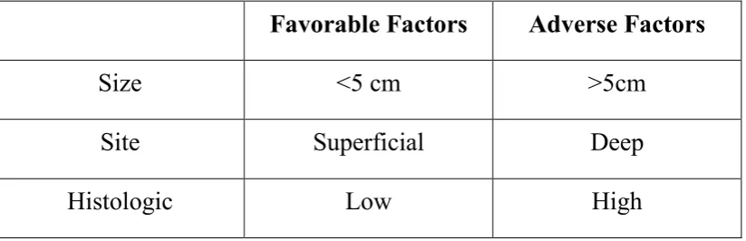
Table 4 MS-KCC sarcoma prognostic factors
Favorable Factors Adverse Factors
Size <5 cm >5cm
Site Superficial Deep
Histologic Low High
Prognostic markers in soft tissue sarcoma
• tumour size
• malignancy
• grade
• high mitotic rate
• tumour necrosis
• surgical margin
• tumour depth
• proliferation
• growth pattern
• vascular invasion
• metastasis
52
TRETMENT7,9
Primary treatment consists of radical surgery, eventually associated
to radiotherapy.Unfortunately, a large proportion of the patients will
subsequently relapse locally or develop metastases. Adjuvant
chemotherapy has not been proved to increase overall survival, but a
meta-analysis or adjuvant trials has demonstrated a significant
improvement of Disease free survival (10% at 5 years), both in terms of
control of the local tumor (5% at 5 years) and of the metastatic spread
(9% at 5 years) [3].
Advanced disease and clinical trials
Inoperable locally advanced or metastatic disease at presentation,
inoperable locoregional relapse and development of metastases are
generally called “advanced disease”.
Systemic therapy is used for patients with advanced disease.
Although responses and prolongation of progression free survival have
been observed, this therapeutic approach is generally considered as
palliative.
Cytotoxic agents that have demonstrated activity against soft tissue
53
randomized clinical trials have been carried out to optimize the
combination of these drugs as first treatment for advanced disease
(generally called first line therapy). So far, no combination has been
shown to significantly improve survival when compared to doxorubicin
administered as a single agent at a dose of 75 or 80 mg/m2 every 3 weeks.
Investigational new drugs are generally considered as therapeutic
options after failure of the first line combination therapy. Most of the
cytotoxic agents tested in the clinic against other malignancies have also
been tested against soft tissue sarcoma in phase II trials, but none of them
has showed any substantial activity in terms of objective response.
Targeted therapies have recently brought new expectations for the
treatment of soft tissue sarcoma. Mechanisms of carcinogenesis and
tumour growth have been extensively studied for sarcoma, making them
ideal candidates for targeted therapies. These expectations have been
substantiated by the documentation of the activity of imatinib mesylate
for Gastro-Intestinal Stromal Tumors (GIST), a sarcoma entity that has
been identified relatively recently and is known to be insensitive to
chemotherapy. In a trial including 946 advanced GIST patients, the
2-years survival estimate is close to 70%, as compared to 20% with
54
therapy for GIST is an encouraging example of the possibilities that can
be offered by targeted therapies.
Specific histological subtypes are, so far, rarely addressed in
clinical trials, and are not even often used for stratification. The difficulty
to conduct histology specific studies resides in the limited potential
accrual (in a subgroup of a rare disease), associated to the complexity and
constant evolution of the histopathological classification. Inclusion of this
heterogeneous group of diseases in clinical trials may have restricted the
discovery of new agents to “large spectrum” drugs active against most of
the frequent histological subtypes. Additionally, the referral pattern of
individual centers and even of clinical research groups may be largely
affected by the quality of the collaboration between medical oncologists
and organ specialists at the institutional level. This may lead to “selection
biases” and irreproducible results in clinical trials.
As an example, trials conducted in uterine leiomyosarcoma have
shown a promising activity of the combination of docetaxel and
gemcitabine, two agents that had failed “mixed histology” phase II
studies .
Objective response to therapy has been used to document activity
55
is defined as an objectively documented decrease in the size of cancer
lesions (subsequent to the administration of the drug), which translates a
biological activity of the drug. WHO response criteria may not have been
an optimal screening tool for new drugs in sarcoma: they used a complex
response evaluation algorithm that was often misunderstood or
misinterpreted; they ignored modern imaging techniques like
computerized tomographic scans and magnetic resonance imaging, both
largely used for the staging of soft tissue sarcoma; more importantly, they
are not appropriate to document the activity of cytostatic agents (expected
to stop tumor growth) that do not necessarily result in an immediate
decrease of the size of the lesions. A few active agents may have been
missed because of the use of inappropriate criteria.
Long disease stabilizations have been observed in recent phase II
trials with trabectedin [6] and brostallicin [7], despite a limited number of
objective responses. Both agents that are currently awaiting confirmation
of their activity in controlled clinical trials.
The Soft Tissue and Bone Sarcoma Group (STBSG) of the
European Organization for Research and Treatment of Cancer (EORTC)
has conducted multiple clinical trials in softtissue sarcoma, with a
56
all been managed at the EORTC Data Center, using similar data
collection forms, and similar database formats. A central pathology
review by a panel of experts is mandatory for all trials subjects. The
group has consistently used the French grading system since its first
publication. As results, the group has accumulated a database of over
2000 patients treated with first line therapy for advanced soft tissue
sarcoma, 380 patients from phase II trials in second or third line therapy,
and 946 advanced GIST patients treated with imatinib in the largest
clinical trial conducted so far.
SURGERY- PRIMARY THERAPY AND RESULTS
For patients with localized diseases, surgical treatment is the corner
stone of treatment. The surgical approach to sarcomas is predicted on
one pathologic fact and its clinical correlate. STS used to expand and
infiltrate tissue line, introduce a pseudo capsule composed of normal
tissue inter locullated in filmbrise of tumour.
For patients with limb sparing option a multimodality approach
proceed for limb-sparing surgery joint with adjuvant chemo or adjuvant
radiotherapy produce local control and disease survival rates to
57
SURGICAL APPROACH
Pre-operative imaging studies (CT scan/MRI) have enabled
accurate prediction respectability. Wide resection should emcomposs the
skin, subcutaneous tissue and soft tissues of tumour, including proir
biopsy site and associated drain sites or wound complications.There are
no data to support compartmental or large muscle group resections over
wide local excision with negative margins.
Tumour should be excised with a 1-2 cm margin of normal
surrounding tissue since there are good approaches to facilitate local
control this ideal target margin is frequently compromised as opposed to
attempting major vascular or bony section.
There is no role in regional lymph node biopsy. Therapeutic lymph
node biopsy results in a 40% actual survival patients with regional lymph
node involvement who has no evidence of extra nodal disease.
Amputation became the treatment of choice for extremity STS
because local recurrence developed in 90% of patients after simple
excision. Today extremity STS limb-sparing multidisciplinary treatment
58
SURGICAL TREATMENT OF EXTREMITY SOFT TISSUE SARCOMA
Amputation vs. limb7,9.
In limb-sparing surgery recurrence rate was high about 26% but
amputation group its low about 4%. A recent study on osteosarcoma
patients gives result as decreased outcome in amputees, but better results
in limp sparing surgery.There was, no apparente difference in quality of
life.
Age of limb-sparing treatment, amputation is reserved treatment
modality for extremity STS. Amputation reserved with advanced
reconstructive techniques combined with RT and/or chemotherapy,
patients with intractable pain, uncontrollable local bleeding,
ulcerated skin, local infection, or neurovascular infiltration, surgery of
choice is amputation.
Amputation and prosthetic reconstruction is the fastest and most
reliable method to gave the result of acceptable functional results. This
may suitable for lower leg, ankle and foot tumours,wide range of
59
anatomic sites, particularly hand, the situation is quite different, and
follow limb-sparing protocols.
Reconstructive surgery5,7,9
Large soft tissue and bone defects,usually not affect the blood
vessels, nerves, joints, or bone is the character of Extremity STS.
Normal tissue moved one place to a different site with a vessel
pedicle or vascular rearrangment known as’ graft’. “Flap” either regain
its blood supply by flap pedicle (or) new vascularisation has blood supply
reestablished at the recipient site.
Classification of Flaps can be done according (local, pedicle or
free flap) their blood supply , The flap reconstruction may be unipedicled,
bipedicled or both. Flaps may be includes of skin, fat, fascia ,or bone as
well as nerve, intestine or omentum. Examples are fasciocutaneous,
myocutaneous, osseous flap. Combinations of these, three flaps also used.
Traditionally, reconstructive ladder” have used the method of
reconstruction,by reconstructive surgeons. Usually doing simplest and
safest method in attempting(i.e. lower down the “ladder”).The
60
flap,pedicleflap,localflap,skin graft. The less complex reconstructive
method is direct closure.
Because of well-vascularized tissue in myocutaneous flaps that
with stands in RT and chemotherapy well,They promote wound healing
very well after enbloc resection. There were fewer complication, more
limb salvage rate, and hospitalization time less .
Microsurgical reconstruction in extremity soft tissue sarcoma
Pedicled vs. free flaps7.9
Pedicled flaps have advantages than free flaps are less procedure
time, and less need of experence surgeon required .Blood and lymphatic
flow disrupted by both local and pedicled flaps, pedicled flaps may be
dearrange the remaining muscles in the affected limb, leading to further
disrup of function. Excisions of large, deep seated STS,free flaps have
been considered better suitable.
Free flaps have several advantages in extremity sarcoma resection
and reconstruction.Free flap have without the limitations of rotational
61
Skin, fascia, muscle, bone, and tendons, blood vessels, and nerves
can be included in Composite flap and the well-vascularized tissue of free
flaps is highly tolerable to wound complications, as well as to RT and
chemotherapy.Chemotherapeutic agents delivery enhanced by high
vascularity in the resection site.
FLAP RECONSTRUCTION IN EXTREMITYSTS
Choice of flap
After tumour resection factors influencing free flap choice mainly
recipient site-dependent factors includes site, size, and depth,Which types
62
considerations. In addition to these, flap reliability and Previous surgery
or RT, are the donor site-dependent factors and donor site morbidity all
may be considered.
Upper extremity
Pedicle vessel LD or thoraco dorsal vessel perforator (TAP) flap
commonly used for reconstruction of shoulder region and upper
arm.Pedicled or free functional flap LD commonly used in the arm
reconstruction surgery.
Pedicled radial forearm flap commonly recommented for elbow
region. Repair of finger and wrist flexion or extension free gracilis
muscle is well suited. Radial forearm, lateral arm, scapular, temporo
parietal fascia flaps are Fascial or fasciocutaneous flaps, They are smaller
and may be higher to muscle flaps. Fascial flaps produce well gliding
surfaces for tendons.Finger reconstruction free toe transfer is a useful
method.
Lower extremity
In the proximal thigh defects, pedicled rectus abdominis flaps can
be used for reconstruction. Free flaps are commonly used for