City, University of London Institutional Repository
Citation
:
Huntjens, B. and O’Donnell, C. (2006). Refractive error changes in Diabetes Mellitus. Optometry in Practice, 7(3), pp. 103-114.This is the accepted version of the paper.
This version of the publication may differ from the final published
version.
Permanent repository link:
http://openaccess.city.ac.uk/6185/Link to published version
:
Copyright and reuse:
City Research Online aims to make research
outputs of City, University of London available to a wider audience.
Copyright and Moral Rights remain with the author(s) and/or copyright
holders. URLs from City Research Online may be freely distributed and
linked to.
City Research Online: http://openaccess.city.ac.uk/ [email protected]
Refractive
Error
Changes
in
Diabetes Mellitus
Byki Huntjens BSc, MSc
Clare O’Donnell, PhD, MCOptom, FAAO
From The Faculty of Life Sciences, The University of Manchester, Manchester, United Kingdom.
Correspondence and reprint requests to: Miss Byki Huntjens, Faculty of Life Sciences, The University of Manchester, Moffat Building, PO Box 88, Manchester, M60 1QD, United Kingdom.
INTRODUCTION
An estimated 1.8 million people in the United Kingdom have diabetes mellitus (DM), which reflects around 3% of the population (Watkins 2003). Statistics of the World Health Organisation suggest that there were around 171 million diabetic patients worldwide in 2000 (an estimated prevalence of 3%). It is predicted that this number will increase to around 366 million patients worldwide by the year 2030 (an estimated prevalence of 5.2%) (WHO 2004). These figures suggest that the number of diabetic patients (either diagnosed or undiagnosed) attending optometric practice is also likely to increase significantly over the next two decades. It has been well documented that fluctuations in refractive power often accompany changing blood glucose levels (Okamoto et al. 2000; Giusti 2003; Sonmez et al. 2005); however, the nature and the aetiology of refractive fluctuations in diabetes mellitus is poorly understood. The aim of this paper is to review the scientific literature relating to refractive error changes associated with diabetes and to discuss the implications of these changes for the optometrist.
Multiple organs or organ systems are affected by the disease process of diabetes mellitus. Some effects are acute (e.g. ketoacidosis, hypoglycaemia), and other effects are more chronic, taking a minimum of five years to develop (e.g. retinopathy). The chronic effects of diabetes on the retina are well known and are seen by ophthalmoscopic inspection and fluorescein angiography. The effects of diabetes on the anterior segment of the eye are less well reported in the literature but the effects of diabetes on the cornea and crystalline lens are thought to play a key role in the refractive error changes sometimes associated with the disease.
established, then eye care practitioners would be better placed to advise patients on expected visual signs and symptoms during periods of altering diabetic metabolic control.
In the first part of this review, the effects of diabetes on the ocular structures that contribute to refractive power will be described. The second part of the paper reviews the scientific literature relating to refractive power shifts in diabetes. The third part considers the main hypotheses as to how myopic and hypermetropic shifts could occur with changing blood glucose concentration. The final part of the paper considers the implications for the optometrist in practice.
THE ANTERIOR OCULAR MANIFESTATIONS OF DIABETES
Cornea
Curvature and thickness
The cornea contributes approximately two-thirds of the refractive power of the human eye. Variations in curvature of either the anterior or the posterior corneal surface or changes in corneal thickness can alter the corneal refractive power.
Diabetes can lead to structural changes in corneal epithelial cells (Tsubota et al. 1991a; Tsubota et al. 1991b; Hosotani et al. 1995; McNamara et al. 1998) and endothelial cells (Schultz et al. 1984; Larsson et al. 1996). However, changes in central corneal curvature do not appear to be associated with altering blood glucose levels in diabetic patients (Saito et al. 1993; Okamoto
Planten and co-workerscarried out several studies to investigate the cause of refractive error changes in newly-diagnosed diabetic patients. Twenty-three patients had reported that they experienced fluctuations in their vision. The authors speculated that the refractive changes occurring in these patients was due to a change in the refractive index of the cornea or crystalline lens as previous studies had found no significant acute changes in lens curvature or position (Planten 1975; Planten et al. 1979; Planten 1981).
In several studies of corneal thickness in Type I diabetic patients, corneal thickness was found to be increased compared to that of age-matched controls (Busted et al. 1981; Pierro et al. 1993; Weston et al. 1995; Larsson et al. 1996). This is in contrast with the findings of other workers (Schultz et al.
1984; McNamara et al. 1998). Generally researchers have failed to demonstrate significant differences in corneal thickness between Type II diabetic patients and age-matched control subjects (Schultz et al. 1984; Larsson et al. 1996).
Refractive index and corneal endothelial cells
In the human eye, the refractive index of the corneal epithelium is 1.401, the underlying stroma is approximately 1.38 and the corneal endothelium is 1.372 (Patel et al. 1995). There are relatively few experimental measurements of human corneal refractive index described in the literature. These values tend to be based on histological and mathematical data (Fatt and Harris 1973; Laing et al. 1976). Nevertheless, corneal refractive index may be an important parameter to consider in diabetes, since the influx of metabolites (including glucose) from the aqueous humour may change the corneal refractive index significantly and thus affect corneal refractive power. Using the formula:
Fcornea = (ncornea –1) * rcornea -1;
where Fcornea is corneal power, ncornea is corneal refractive index, and rcornea is
the radius of curvature of the central cornea (Ellis et al.) and assuming Gullstrand constants, where ncornea = 1.376 and rcornea= 7.7mm: a change of
power. Additionally, a change in central corneal curvature of 0.2mm would give a change of approximately 1 D in corneal power.
Recent evidence linking elevated glucose concentration to reduced corneal endothelial Na+, K+-ATPase activity is of interest since this enzyme is an important component of the endothelial pump which is thought to be responsible for maintaining corneal hydration control (Whikehart et al. 1993). In light of this finding, it is perhaps not surprising that corneal thickness and time to recover from induced oedema are often increased in diabetic humans and animal models (Herse 1990a, 1990b). Oedema occurs when there is a significant reduction in the amount of oxygen reaching the cornea. The total corneal swelling induced by contact lens induced hypoxia has been shown to decrease with increasing blood glucose concentration in diabetic patients. (McNamara et al. 1998).
Crystalline lens
Thickness
Lens thickness is often greater in diabetic patients than in non-diabetic subjects, mainly due to cortical thickening (Huggert 1953; Brown and Hungerford 1982). Other biometric changes include steepening of the anterior and posterior surfaces of the lens and shallowing of the anterior chamber (Sparrow et al. 1990). After adjusting for age, these changes are more pronounced in Type I diabetic patients and in patients with increased disease duration. Age-related increases in axial lens thickness, cortical thickness and nuclear thickness are accelerated after the onset of diabetes (Sparrow et al.
1990). These findings could explain the increased prevalence of myopia found in diabetic patients. The expansion of the sagittal width of the lens is also accelerated in diabetic patients (Sparrow et al. 1990). Neither age nor disease duration appears to be responsible for these changes; however, the presence of diabetic retinopathy in Type 1 patients does seem to have an effect.
It may be that small changes in refractive index in the centre of the lens could induce an acute shift in refractive error in diabetic patients. We would expect a 3D change in refraction with a 0.02 change in refractive index (Planten 1981).
The size of the expected change in the crystalline lens in diabetes mellitus can be calculated with the help of Le Grand’s theoretical eye (Wyszecki and Stiles 1982). To obtain a change in refraction of 2D with an anterior chamber depth of 3.1mm, the anterior surface of the lens would have to shift by 0.5mm, and change its radius from 10.2 to 14.1mm. Alternatively the entire lens would need to shift by approximately 1.5mm to obtain a 2D change in refraction. However, only a small change in refractive index (e.g. from 1.42 to 1.41) would be required for the same refractive change, making refractive error changes more likely to explain these shifts (Planten et al. 1979).
Accommodation
An aspect of ocular performance that seems to be affected in young diabetic patients is accommodation. The amplitude of accommodation in Type I patients is lower than that of age-matched control subjects (Moss et al. 1987; Mantyjarvi and Nousiainen 1988; Braun et al. 1995). It appears that even when there is no detectable retinal damage, the amplitude of accommodation in Type I patients is lower than that of age-matched healthy subjects. Furthermore, it appears that the latency of the response is unusually long (Braun et al. 1995).
Cataract
The intracellular accumulation of sorbitol (a by product of glucose metabolism) during poor metabolic control (hyperglycemia) will not only create an osmotic gradient leading to the uptake of water and lens swelling (Gabbay 1973), but will also cause a change in solubility (precipitation) of lens proteins which can lead to metabolic cataract formation (Kinoshita and Merola 1964; Caird et al.
1969; Ehrlich et al. 1987).
Rotimi et al. 2003; Hennis et al. 2004). Hypoglycemia induced by strict control of blood glucose in diabetic patients may induce cataract, since glucose is the major energy source of the lens (Vinding and Nielsen 1984). Cataract formation is associated with lens swelling, vacuole formation, and increased membrane permeability. Different forms of cataract in diabetic patients are juvenile cataract (opacity shaped like a snowflake, affecting the anterior and posterior cortical layer of the lens in young diabetics) (Nielsen and Vinding 1984) and age-related cataract (nuclear, cortical, and posterior subcapsular) cataract (Leske et al. 1991). Studies of young type I diabetic patients (with no detectable cataract on slit lamp) suggest that there is increased light scatter in diabetic patients that appears to correlate with HbA1c and the degree of diabetic retinopathy present (Kato et al. 2000; Kato et al. 2001).
Aqueous humour
Patients with type 1 diabetes have reduced aqueous flow rates (Hayashi et al.
1989). This does not appear to occur in patients with type 2 diabetes (Larsson
et al. 1995). One of the determinants of aqueous humour production is ocular blood flow (Bill 1975), which has been shown to increase with acute elevations in glucose concentration in type 1 diabetic patients (Bursell et al.
1996). Therefore, an increase of blood flow might be expected to induce an increase in aqueous flow. Lane and co-workers studied aqueous flow, ocular blood flow, and intra-ocular pressure in type 1 diabetic patients and control subjects, controlling for glucose and insulin concentration (Lane et al. 2001). They found a decrease in aqueous flow in type I patients versus fasting controls; however, aqueous production did not appear to be affected by insulin or glucose concentrations.
REFRACTIVE ERROR AND DIABETES
Axial Length
retinopathy compared to the group without retinopathy). No difference in axial length was found between the diabetic patients without retinopathy and the control subjects. Refractive power was not measured during this study.
Refractive error
Since the 19th century, it has been recognised that changes in blood glucose concentration can influence vision in patients with diabetes (Da Costa 1890; Duke-Elder 1925). Optometrists are trained to consider the possibility of undiagnosed diabetes if a patient complains of a bilateral, unexpected or rapid change of vision or prescription. If diabetes is suspected, the eye care practitioner may postpone prescribing spectacles until the refractive error has stabilised, which generally occurs when the patient’s diabetes is better controlled. Refractive changes associated with diabetes can be both acute (transient) and chronic (sustained). However, relatively little is known about the biochemical changes, which accompany these refractive events.
Authors who have investigated the effect of more acute changes in plasma glucose concentration have reported that an acute decrease in plasma glucose levels causes a (transient) hyperopic change (Keller 1973; Marmor 1973; Gwinup and Villarreal 1976; Fledelius and Miyamoto 1987; Saito et al.
1993; Willi 1996). Conversely other authors report the possibility of increasing hyperopia under hyperglycemic conditions (Huggert 1954; Rosen 1956; Vaughan and Asbury 1980). Rosen (1956) described a case-report where a hyperopic change was observed with increasing blood glucose levels, and Huggert (1954) studied the crystalline lens of 23 diabetic subjects. Ten subjects were myopic and 14 subjects were hyperopic (in one subject hyperopia was preceded by myopia, and was therefore counted twice). Vaughan and Asbury (1980) described both myopic and hyperopic refractive error changes in diabetes with changing blood glucose concentration.
Thus both myopic and hyperopic shifts have been reported in diabetic patients and the underlying mechanism responsible remains to be established. Table 1 summarises the results of various studies published in the scientific literature, describing acute refractive error changes with induced changes in blood glucose levels.
Author Year No of
subjects
Key findings
Huggert 1954 23 Myopia before and hyperopia after the onset of treatment
Gwinup et al. 1976 10 Myopia after induced hyperglycaemia Fledelius 1987 72 Myopia and hyperopia (before and after
the onset of treatment)
Saito et al. 1993 5 Hyperopia after the onset of treatment Furushima et
al.
1999 7 Myopia after induced hyperglycaemia in non-diabetic subjects
Table 1. Conflicting results presented in the literature comparing acute refractive changes in the eye to changing blood glucose levels.
Different approaches have been adopted in the above studies, which may explain the lack of agreement in findings. It is important to define the words ‘acute effect’, since investigators describe the refractive shifts at different time points after the commencement of the study. The majority of investigators have been interested in the response of the eye when elevated blood glucose levels were reduced to near normal values. This reduction could take several weeks to months to occur and might be considered a sub-acute response rather than a true acute response.
Although our knowledge about the time required for glucose to enter the eye after blood plasma glucose concentration is increased is limited to animal models, it is thought that the latency is between 5 minutes (Cameron et al.
2001) and 30 minutes (Chou and Lin 2000).
Researchers (Caird et al. 1969; Saito et al. 1993) have examined the refractive shifts occurring after instigating (insulin) treatment for the first time. This may not reflect blood glucose changes as they occur on a day-to-day basis in treated diabetic patients. To investigate refractive error shifts in diabetic patients, the conditions require more ‘regular’ glucose fluctuations. The response of the eye to the body’s first experience with oral diabetes medications or injected insulin has not been thoroughly investigated. Insulin acts on cells throughout the body to stimulate the uptake, utilisation and storage of glucose. A sudden drop in extra-cellular glucose levels or an increase in intra-cellular glucose (as would occur in undiagnosed diabetes) might change the refractive index more significantly than when this change occurs on a hourly basis.
decrease (Figure 1). As a consequence, the osmotic balance along the lens capsule or epithelium will increase the influx of water from the aqueous humour into the lens, and induce a refractive shift. This effect may be dramatic when treatment for diabetes mellitus is first initiated and may be different to the sorts of changes that occur when the diabetes is under control.
Figure 1. Aqueous humour is produced by the ciliary body and secreted into the posterior chamber. It flows through the pupil into the anterior chamber, where it is drained by the trabecular meshwork and Schlemm’s canal (Reprinted from Forrester et al. 2002, with permission of Elsevier).
The initial blood glucose concentration in all 6 subjects was below 150mg/dl (8.4mM/l), but no measurements of blood glucose concentration were taken afterwards.
[image:13.595.97.418.415.686.2]Two of the diabetic subjects were aphakic and both aphakic subjects became more hypermetropic (rather than more myopic) after blood glucose concentration increased. These data also appear to suggest that the cause of the myopic refractive shift in the phakic subjects was likely to be due to the crystalline lens. The data from the aphakic subjects suggest that the cornea and the posterior lens capsule also appear to have an influence on refractive error fluctuations. However, since the refractive change was opposite in sign in the phakic versus the aphakic eyes, the anterior lens capsule, subcapsular epithelium and lens fibres may act to reverse any change induced by the cornea or by the lens capsule. Additionally, the data support the theory that the latency of refractive changes after blood glucose change is small.
Figure 2. Changes in refraction of the eyes of aphakic diabetic subjects (n=4) and diabetic with intact lenses (n=12) following induced hyperglycemia (mean ± SD) (Adapted from Gwinup and Villarreal 1976).
-1 -0.75 -0.5 -0.25 0 0.25 0.5 0.75 1
0 15 30 45 60 75 90 105
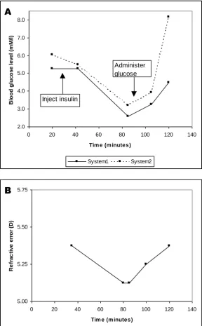
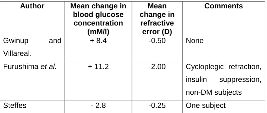
Steffes (1999) obtained similar results to those of Gwinup and Villarreal (1976). In a fasting Type I diabetic patient with a blood glucose concentration of about 95mg/dl (5.3mM/l), the administration of insulin caused the blood glucose concentration to reduce to about 50mg/dl (2.8mM/l) (Figure 3).
Figure 3. Comparison of (A) finger prick measurements of whole blood glucose taken with two different systems (circles and squares); and (B) measured refractive error during induced hypoglycemia (Adapted from Steffes 1999). 2.0 3.0 4.0 5.0 6.0 7.0 8.0
0 20 40 60 80 100 120 140
Tim e (m inutes)
Bl o o d g lu c o s e l e v e l (m M /l ) System1 System2 A Inject insulin Administer glucose 5.00 5.25 5.50 5.75
0 20 40 60 80 100 120 140
Tim e (m inutes)
[image:14.595.93.382.176.642.2]Upon administration of dextrose (glucose), the patient’s blood glucose increased to about 5mM/l with system 1 and >8.0mM/l with system 2. Concurrent measurements of refractive error were made, and the changes in refractive error were closely tracked those of the blood glucose concentration. To the best of our knowledge, this is the only in vivo evidence in the literature of refractive error changes due to induced hypoglycaemia. The results of this study concur with Gwinup and Villarreal’s finding that the latency is short.
A novel approach to the question of whether myopic or hypermetropic changes are caused by hyperglycemia was reported by Furushima et al. (Furushima et al. 1999). Somatostatin, which is known to suppress insulin secretion, was administered to a group of seven non-diabetic volunteers (mean age 23 years), with normal vision. Together with a glucose-load, the somatostatin induced hyperglycemia in all subjects. A statistically significant average change of –1.93±0.39 D in refractive error was observed 150 minutes after the glucose load despite the instillation of cycloplegic eye drops (p<0.01). The subjects’ refractive error changed concurrently with their blood glucose level, which increased from 3.9±0.6mM/l to 15.6±2.3mM/l. The time taken to reach the maximum refractive power was the same as the time taken to reach the maximum blood glucose level. Ultrasound measurements showed significant changes in the dimensions of both the anterior chamber depth which became shorter, and the crystalline lens, which became thicker. The axial length of the eye remained unchanged.
Aetiology of refractive error shifts in diabetes
Thus there is still some uncertainty about how refractive error in humans is influenced by acute and chronic changes in blood glucose concentration.
Myopia
A myopic shift with decreasing blood glucose concentration in diabetes could be explained by the accumulation of metabolites such as sorbitol. The intracellular glucose levels in the crystalline lens are not regulated by insulin, and therefore the concentration of glucose within the lens increases with increasing blood glucose concentration (Olansky 2004).
Aldose reductase is an enzyme that converts any excess of intracellular glucose into sorbitol, which can then be further metabolised into fructose. Cell membranes are relatively impermeable to sorbitol allowing this compound to accumulate within the lens. This is paralleled by an influx of water from the aqueous humour, producing lenticular swelling (Gabbay 1973). This leads to an increase of lens curvature and induced myopia.
Hyperopia
A decrease in blood glucose concentration in the aqueous humour could also cause a transient difference in osmotic pressure between the aqueous humour, crystalline lens and vitreous body. As a result, a decrease in refractive index might develop in the lens, leading to a hyperopic shift. A decrease in lenticular refractive index from 1.42 to 1.40 has been shown to produce a hyperopic change of 3.20D (Planten 1981). Therefore, small changes in refractive index could produce significant changes in refractive power as stated above.
(when the solution has an almost equal glucose concentration to the lens), the already swollen lens experienced hypo-osmotic shock and swelled still further with increased K+ permeability. This increase in K+ efflux (outward flow) is presumably used by cells to increase their volume in response to osmotic stress (Beebe et al. 1990).
Author Mean change in
blood glucose concentration
(mM/l)
Mean change in
refractive error (D)
Comments
Gwinup and Villareal.
+ 8.4 -0.50 None
Furushima et al. + 11.2 -2.00 Cycloplegic refraction, insulin suppression, non-DM subjects Steffes - 2.8 -0.25 One subject
Table 2. Results from three studies showing refractive error changes after induced hypo- or hyperglycemia.
Table 2 summarises the results of the three human studies described above. It is perhaps surprising that cycloplegic refraction results in such a large refractive change. However, insulin secretion was completely suppressed in these subjects, resulting in very high blood glucose levels. Glucose would have been building up into the aqueous humour and extra-cellular space of the cornea and crystalline lens. As a result, the refractive index of the aqueous humour would be increased significantly causing a large refractive change.
Implications for the optometrist
[image:17.595.85.514.215.397.2]disturbances, restlessness, irritability, inability to concentrate, mental confusion, and personality changes among others (National Diabetes Information Clearinghouse 2003). Thus diabetic patients may take immediate action to correct for hypoglycaemia (usually with an intake of glucose), and any associated refractive error shifts may therefore be avoided. Consequently, the response of the crystalline lens to untreated hyperglycaemia may be of more immediate concern to the optometrist involved in carrying out eye examinations on diabetic patients.
Thus, refractive error change can be influenced by fluctuating blood glucose concentration. Therefore it seems appropriate to enquire about blood glucose concentration when carrying out eye examinations on diabetic patients. If blood glucose concentration is uncharacteristically high or low at the time of refraction, then it may be wise to repeat the refraction prior to prescribing, as well as advising patients about the possible implications for diabetic retinopathy and other complications associated with the disease.
In hyperglycemia, glucose can accumulate in the lens causing an increase in curvature and a shift towards myopia (Mantyjarvi 1988). However, hyperopic changes have also been shown to occur during hyperglycemia and this is thought to be due to a decrease in refractive index in the crystalline lens. If the geometric effect dominates (swelling of the lens altering lens curvature), the refraction will shift towards myopia. If the refractive effect dominates, the refraction will shift towards hypermetropia.
CONCLUSIONS
in which case either a myopic or hyperopic response may occur, depending on the individual’s physiology. However, from a clinical point of view, fluctuating blood glucose concentration has an influence on short-term changes in refraction, and these changes may be large enough to measure. In the majority of cases, people with diabetes appear to become more myopic as their blood glucose concentration increases, but a significant minority become more hypermetropic. Where acute changes have been reported in the literature, the latency of these changes appears to be short, being of the order of a few minutes. This implies that a hypo- or hyperglycemic state could influence refractive findings during the eye-examination. When taking a diabetic patient’s history, it seems appropriate to enquire about the most recent blood glucose concentration, or even suggest taking a finger stick test before the eye-examination. It should be borne in mind that (consistently) high blood glucose readings could induce transient myopia.
Acknowledgments
REFERENCES
Beebe, DC, Parmelee, JT and Belcher, KS (1990). "Volume regulation in lens epithelial cells and differentiating lens fiber cells." J Cell Physiol 143(3): 455-9.
Bill, A (1975). "Blood circulation and fluid dynamics in the eye." Physiol Rev
55(3): 383-417.
Braun, CI, Benson, WE, Remaley, NA, Chew, EY and Ferris, FL, 3rd (1995). "Accommodative amplitudes in the Early Treatment Diabetic Retinopathy Study." Retina 15(4): 275-81.
Brown, N and Hungerford, J (1982). "The influence of the size of the lens in ocular disease." Trans Ophthalmol Soc UK 102 Pt 3: 359-63.
Bursell, S, Clermont, A, Kinsley, B, Simonson, D, Aiello, L and Wolpert, H (1996). "Retinal blood flow changes in patients with insulin-dependent diabetes mellitus and no diabetic retinopathy." Invest. Ophthalmol. Vis. Sci.
37(5): 886-97.
Busted, N, Olsen, T and Schmitz, O (1981). "Clinical observations on the corneal thickness and the corneal endothelium in diabetes mellitus." The British Journal Of Ophthalmology 65(10): 687-90.
Caird, F, Pirie, A and Ramsell, T (1969). Transient visual symptoms in diabetes. Diabetes and the Eye. Oxford, Blackwell Scientific: 127-39.
Cameron, BD, Baba, JS and Cote, GL (2001). "Measurement of the glucose transport time delay between the blood and aqueous humor of the eye for the eventual development of a noninvasive glucose sensor." Diabetes Technol Ther 3(2): 201-7.
Chou, C and Lin, PK (2000). "Noninvasive glucose monitoring with optical heterodyne technique." Diabetes Technol Ther 2(1): 45-7.
Da Costa, J (1890). Medical Diagnosis. 7th Edition. Philadelphia, J.B Lippincott.
Duke-Elder, S (1925). "Changes in refraction in diabetes mellitus." Br J Ophthalmol 9: 167-87.
Ehrlich, RM, Kirsch, S and Daneman, D (1987). "Cataracts in children with diabetes mellitus." Diabetes Care 10(6): 798-9.
Ellis, K, Pirnazar, JR, Chuck, RS and McDonnell, PJ (2001). "Corneal Topography." www.ophthalmic.hyperguides.com.
Fledelius, HC (1983). "Is myopia getting more frequent? A cross-sectional study of 1416 Danes aged 16 years+." Acta Ophthalmol (Copenh) 61(4): 545-59.
Fledelius, HC (1986). "Myopia and diabetes mellitus with special reference to adult-onset myopia." Acta Ophthalmol (Copenh) 64(1): 33-8.
Fledelius, HC (1987). "Refractive change in diabetes mellitus around onset or when poorly controlled. A clinical study." Acta Ophthalmol (Copenh) 65(1): 53-7.
Fledelius, HC and Miyamoto, K (1987). "Diabetic myopia - is it lens-induced? An oculometric study comprising ultrasound measurements." Acta Ophthalmol (Copenh) 65(4): 469-73.
Forrester, JV, Dick, AD, McMenamin, PG and Lee, WR (2002). The eye; Basic sciences in practise. Second edition. London, WB Saunders.
Furushima, M, Imaizumi, M and Nakatsuka, K (1999). "Changes in refraction caused by induction of acute hyperglycemia in healthy volunteers." Jpn J Ophthalmol 43(5): 398-403.
Gabbay, KH (1973). "The sorbitol pathway and the complications of diabetes." N Engl J Med 288(16): 831-6.
Giusti, C (2003). "Transient hyperopic refractive changes in newly diagnosed juvenile diabetes." Swiss Med Wkly 133(13-14): 200-5.
Gullstrand, A (1909). Appendix II. Helmholtz's Handbuch de Physiologischen Optik. Volume 1., (English translation edited by JP Southall, Optical Society of America. Dover Publications 1962: 351-2).
Gwinup, G and Villarreal, A (1976). "Relationship of serum glucose concentration to changes in refraction." Diabetes 25(1): 29-31.
Harding, JJ, Egerton, M, van Heyningen, R and Harding, RS (1993).
"Diabetes, glaucoma, sex, and cataract: analysis of combined data from two case control studies." Br J Ophthalmol 77(1): 2-6.
Hayashi, M, Yablonski, ME, Boxrud, C, Fong, N, Berger, C and Jovanovic, LJ (1989). "Decreased formation of aqueous humour in insulin-dependent
diabetic patients." Br J Ophthalmol 73(8): 621-3.
Hennis, A, Wu, SY, Nemesure, B and Leske, MC (2004). "Risk factors for incident cortical and posterior subcapsular lens opacities in the Barbados Eye Studies." Arch Ophthalmol 122(4): 525-30.
Herse, PR (1990a). "Corneal hydration control in normal and alloxan-induced diabetic rabbits." Invest Ophthalmol Vis Sci 31(11): 2205-13.
Hosotani, H, Ohashi, Y, Yamada, M and Tsubota, K (1995). "Reversal of abnormal corneal epithelial cell morphologic characteristics and reduced corneal sensitivity in diabetic patients by aldose reductase inhibitor, CT-112." Am J Ophthalmol 119(3): 288-94.
Huggert, A (1953). "The appearance of the band of disjunction of the lens in diabetes mellitus." Acta Ophthalmol (Copenh) 31(3): 227-34.
Huggert, A (1954). "The appearance of the crystalline lens during different stages of transitory changes of refraction. II." Acta Ophthalmologica 32(4): 375-89.
Jacob, TJ and Duncan, G (1982). "Glucose-induced membrane permeability changes in the lens." Exp Eye Res 34(3): 445-53.
Kato, S, Oshika, T, Numaga, J, Kawashima, H, Kitano, S and Kaiya, T (2000). "Influence of rapid glycemic control on lens opacity in patients with diabetes mellitus." American Journal Of Ophthalmology 130(3): 354-5.
Kato, S, Shiokawa, A, Fukushima, H, Numaga, J, Kitano, S, Hori, S, Kaiya, T and Oshika, T (2001). "Glycemic control and lens transparency in patients with type 1 diabetes mellitus." American Journal of Ophthalmology 131(3): 301-4.
Keller, JT (1973). "Hyperopia in Diabetes." Arch Ophthalmol 90(6): 511-2.
Kinoshita, JH and Merola, LO (1964). "Hydration of the lens during the development of galactose cataract." Invest Ophthalmol Vis Sci 3: 577-84.
Laing, RA, Sanstrom, MM, Berrospi, AR and Leibowitz, HM (1976). "Changes in the corneal endothelium as a function of age." Exp Eye Res 22(6): 587-94.
Lane, JT, Toris, CB, Nakhle, SN, Chacko, DM, Wang, Y-L and Yablonski, ME (2001). "Acute effects of insulin on aqueous humor flow in patients with type 1 diabetes." American Journal of Ophthalmology 132(3): 321-7.
Larsson, LI, Bourne, WM, Pach, JM and Brubaker, RF (1996). "Structure and function of the corneal endothelium in diabetes mellitus type I and type II." Arch Ophthalmol 114(1): 9-14.
Larsson, LI, Pach, JM and Brubaker, RF (1995). "Aqueous humor dynamics in patients with diabetes mellitus." Am J Ophthalmol 120(3): 362-7.
Leske, MC, Chylack, LT, Jr. and Wu, SY (1991). "The Lens Opacities Case-Control Study. Risk factors for cataract." Arch Ophthalmol 109(2): 244-51.
Mantyjarvi, M (1988). "Myopia and diabetes. A review." Acta Ophthalmol Suppl 185: 82-5.
Marmor, MF (1973). "Transient accomodative paralysis and hyperopia in diabetes." Arch Ophthalmol 89(5): 419-21.
McNamara, NA, Brand, RJ, Polse, KA and Bourne, WM (1998). "Corneal function during normal and high serum glucose levels in diabetes." Invest Ophthalmol Vis Sci 39(1): 3-17.
Moss, SE, Klein, R and Klein, BE (1987). "Accommodative ability in younger-onset diabetes." Archives of Ophthalmology 105(4): 508-12.
National Diabetes Information Clearinghouse (2003). Hypoglycemia, Department of Health and Human Services. National Institute of Health.
Nielsen, NV and Vinding, T (1984). "The prevalence of cataract in insulin-dependent and non-insulin-insulin-dependent-diabetes mellitus." Acta Ophthalmol (Copenh) 62(4): 595-602.
Okamoto, F, Sone, H, Nonoyama, T and Hommura, S (2000). "Refractive changes in diabetic patients during intensive glycaemic control." Br J Ophthalmol 84(10): 1097-102.
Olansky, L (2004). Advances in Diabetes for the Millenium: Chronic
Microvascular Complications of Diabetes. Medscape General Medicine. 6.
Patel, S, Marshall, J and Fitzke, FW, 3rd (1995). "Refractive index of the human corneal epithelium and stroma." J Refract Surg 11(2): 100-5.
Pierro, L, Brancato, R, Robino, X, Lattanzio, R, Jansen, A and Calori, G (1999). "Axial length in patients with diabetes." Retina 19(5): 401-4.
Pierro, L, Brancato, R and Zaganelli, E (1993). "Correlation of corneal thickness with blood glucose control in diabetes mellitus." Acta Ophthalmol (Copenh) 71(2): 169-72.
Planten, JT (1975). "Physiologic optic approach of lens and cataract." Ophthalmologica 171(4-5): 249-53.
Planten, JT (1981). "Changes of refraction in the adult eye due to changing refractive indices of the layers of the lens." Ophthalmologica 183(2): 86-90.
Planten, JT, Kooyman, A, de Vries, D and Wolderingh, JH (1979). "Pathologico-optic approach to cataract and lens." Documenta Ophthalmologica. Advances In Ophthalmology 46(2): 237-9.
Rosen, M (1956). "Diabetes mellitus with relative hyperopia; a case report." American Journal Of Ophthalmology 41(4): 680-1.
"Prevalence and determinants of diabetic retinopathy and cataracts in West African type 2 diabetes patients." Ethn Dis 13(S2): 110-7.
Saito, Y, Ohmi, G, Kinoshita, S, Nakamura, Y, Ogawa, K, Harino, S and Okada, M (1993). "Transient hyperopia with lens swelling at initial therapy in diabetes." Br J Ophthalmol 77(3): 145-8.
Schultz, RO, Matsuda, M, Yee, RW, Edelhauser, HF and Schultz, KJ (1984). "Corneal endothelial changes in type I and type II diabetes mellitus." Am J Ophthalmol 98(4): 401-10.
Smith, G (2003). "The optical properties of the crystalline lens and their significance." Clin Exp Optom 86(1): 3-18.
Sonmez, B, Bozkurt, B, Atmaca, A, Irkec, M, Orhan, M and Aslan, U (2005). "Effect of glycemic control on refractive changes in diabetic patients with hyperglycemia." Cornea 24(5): 531-7.
Sparrow, JM, Bron, AJ, Brown, NA and Neil, HA (1990). "Biometry of the crystalline lens in early-onset diabetes." Br J Ophthalmol 74(11): 654-60.
Steffes, PG (1999). "Laser-based measurement of glucose in the ocular aqueous humor: an efficacious portal for determination of serum glucose levels." Diabetes Technol Ther 1(2): 129-33.
Tsubota, K, Chiba, K and Shimazaki, J (1991a). "Corneal epithelium in diabetic patients." Cornea 10(2): 156-60.
Tsubota, K, Yamada, M and Naoi, S (1991b). "Specular microscopic
observation of human corneal epithelial abnormalities." Ophthalmology 98(2): 184-91.
Vaughan, D and Asbury, T (1980). General Ophthalmology. California, Lange.
Vinding, T and Nielsen, NV (1984). "Two cases of acutely developed cataract in diabetes mellitus." Acta Ophthalmol (Copenh) 62(3): 373-7.
Watkins, PJ (2003). ABC of Diabetes. London, BMJ Books.
Weston, BC, Bourne, WM, Polse, KA and Hodge, DO (1995). "Corneal hydration control in diabetes mellitus." Investigative Ophthalmology & Visual Science 36(3): 586-95.
Whikehart, DR, Montgomery, B, Angelos, P and Sorna, D (1993). "Alteration of ATPase activity and duplex DNA in corneal cells grown in high glucose media." Cornea 12(4): 295-8.
WHO (2004). "World Health Organisation (WHO)." http://www.who.int/en/.
Willi, MJ (1996). "Hyperopia and hyperglycemia." Survey of Ophthalmology