1
Isometric Hip and Knee Torque measurements as an outcome
measure in Robot Assisted Gait Training
Sujay S Galen1,2,3 Celia J Clarke1,2, Alan N Mclean2, David B Allan2, Bernard A Conway1,2
1 Bioengineering Unit, University of Strathclyde, Glasgow, UK.
2 Scottish Centre for Innovation in Spinal Cord Injury (SCISCI), Queen Elizabeth National Spinal Injuries Unit, Southern General Hospital, Glasgow, UK.
3 Physical Therapy Program, Eugene Applebaum College of Pharmacy and Health Care Sciences, Wayne State University, Detroit, MI, USA.
*Correspondence to first author at Physical Therapy Program
Wayne State University
259 Mack Avenue
Detroit, MI 48201.
USA.
Tel: +1 (313) 577 5531
Fax: +1 (313) 577 8685
e-mail: sujay.galen@wayne.edu
2 Abstract
Background: Strength changes in lower limb muscles following robot assisted gait training (RAGT) in subjects with incomplete spinal cord injury (ISCI) has not been quantified using objective outcome measures.
Objective: To record changes in the force generating capacity of lower limb muscles (recorded as peak voluntary isometric torque at the knee and hip), before, during and after RAGT in both acute and subacute/chronic ISCI subjects using a repeated measures study design.
Methods: Eighteen subjects with ISCI participated in this study (Age range: 26-63 years mean age = 49.3 + 11 years). Each subject participated in the study for a total period of eight weeks, including 6 weeks of RAGT using the Lokomat system (Hocoma AG, Switzerland). Peak torques were recorded in hip flexors, extensors, knee flexors and extensors using torque sensors that are incorporated within the Lokomat.
Results: All the tested lower limb muscle groups showed statistically significant (p<0.001) increases in peak torques in the acute subjects. Comparison between the change in peak torque generated by a muscle and its motor score over time showed a non-linear relationship.
3 Introduction
The primary sensory and motor consequences of spinal cord injury (SCI) are well known, and vary according to the level and extent of lesion. .However, with paralysis comes a secondary complication of muscle atrophy. Following injury, the onset of muscle atrophy can be rapid and is evident soon after injury(Dudley-Javoroski & Shields, 2008). The loss of muscle cross sectional area in SCI subjects has been reported to be between 18 and 46% on average when compared with neurologically intact individuals (Giangregorio & McCartney, 2006). This degree of muscle atrophy and the related loss of force generating capacity can contribute to functional impairment and can compromise progression of rehabilitation in SCI subjects. Accordingly, methods for early mobilization may mitigate against atrophy by either reversing or reducing the rate of muscle loss. In ISCI subjects receiving intensive gait rehabilitation, this is likely to be a factor that facilitates the functional recovery of gait (Dietz, Colombo, Jensen, & Baumgartner, 1995).
Bodyweight supported treadmill training (BWSTT) has been shown to improve walking ability (Nymark et al., 1998; Postans, Hasler, Granat, & Maxwell, 2004) in ISCI subjects and is increasingly being adopted within physical therapy programs (Hicks & Martin Ginis, 2008). For acute subjects BWSTT also serves as a method for early lower limb mobilization and exercise.
4 training improves gait in both acute (Hornby et al., 2005), and chronic ISCI subjects (Wirz et al., 2005) there have been no studies documenting the associated changes in the force generating capacity of the lower limb muscles, in subjects as they progress through RAGT using BWSTT
Strength changes in lower limb muscles following locomotor training have been previously documented in ISCI subjects using objective outcome measures such as peak torque recorded using a Biodex dynamometer. (Jayaraman et al., 2008) Given that the Lokomat system is equipped with integrated torque sensors within the DGO exoskeleton, the device has the capability to be used as a dynamometer in its own right. Recently it has been shown that the peak torques, recorded using the torque sensors integrated within the DGO exoskeleton, provides data with good inter and intra-rater reliability in both healthy subjects and subjects with neuromuscular disorder.(Bolliger, Banz, Dietz, & Lunenburger, 2008) This inbuilt capability provides the opportunity for monitoring the peak torques generated around hip and knee during voluntary isometric exertions, and has a potential use as a clinical measure to monitor the subject’s progress through the rehabilitation programme (Bolliger et al., 2008; Lunenburger, Colombo, Riener, & Dietz, 2004). In this study we set out to record the change in voluntary force generating capacity in the hip and knee flexors and extensor muscles, before, during and at the end of RAGT in both acute and subacute/chronic ISCI subjects. The quantitative force data is then compared with the equivalent manual muscle scores acquired from subjects as part of the standard neurological classification of spinal cord injury. (Marino et al., 2003)
Methodology
5 time of injury) and thirteen were classified as acute subjects (<6 months from the time of injury). Subjects were recruited if they were
Acute or Chronic ISCI subjects graded as AIS classification C/D.(Marino et al., 2003)
Between the ages of 18-65 years.
Assessed as medically stable by an (independent) physician.
Subjects were not recruited if they had
Complications requiring immobilization such as fractures and pressure sores.
Osteoporosis or contractures limiting range of motion.
A bodyweight of over 130 kg or thigh length over 47 cms (Lokomat restrictions).
Ethics approval was obtained from the National Health Service (NHS) regional ethics committee and subjects were recruited from the Queen Elizabeth National Spinal Injuries Unit, Glasgow. The details of the subjects who participated in the study are provided in Table1.
Insert Table 1 here.
6 al.) When RAGT began, subjects walked on the Lokomat at a speed of 1-1.2 km/h with body weight support between 80-70% of their body weight. These parameters were progressed depending on the subject’s tolerance. The speed of walking during RAGT was increased to a maximum of 2 km/h and the body weight support was reduced at a rate depending on the subject’s ability to maintain the knee in extension during the midstance phase of the gait cycle.
7 To compare the force generating capacities of the muscles between subjects, the peak torque generated was expressed as a percentage of the overall maximum torque recorded from each of the muscle groups during the six week training period. The data was first individually analyzed for each subject. A descriptive analysis was performed to identify any trends that can suggest a change in the force generating capacity in each of the muscle groups tested. The data was then pooled separately for subacute/chronic and acute subjects, to study the change in the peak torques generated within these cohorts. The Friedman’s test was used to establish if the data showed any significant difference in the generated peak torques over the training period of six weeks. A p-value of p<0.05 was considered to be significant. If the Friedman’s test indicated that there were significant differences in the generated peak torques over the six weeks of training, a Wilcoxon signed rank test was used to identify the between week differences in the generated peak torque. All tests were performed using SPSS (version 18) software. The lower limb motor score for the hip flexors and knee extensors (the two muscle groups assessed as part of the ASIA assessment), were compared with the peak torque generated by the respective muscle group. The subject’s locomotor function was also assessed using the walking index in spinal cord injury (WISCI II scale) (Dittuno & Dittuno Jr, 2001) performed during week 1 (before) and week 8 (after) the full RAGT programme.
Results
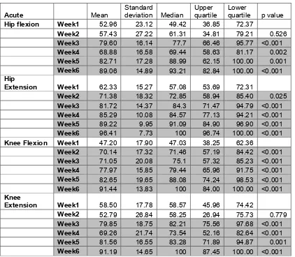
Locomotor function (measured using WISCI II) following RAGT showed a greater increase in acute subjects compared to subacute/chronic subjects (Figure 1).
Insert Figure 1 here.
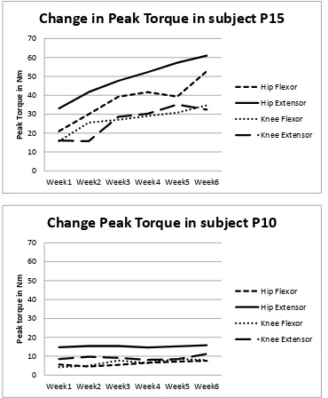
8 Figures 2a and 2b respectively. Both subjects (P15 and P10) were classified as AIS D on the ASIA scale. Subject P10 remained non-ambulant both prior to and after the RAGT and hence was given a score of 0 on the WISCI II scale. Subject P15 was unable able to walk prior to the RAGT but following six weeks of Lokomat training the subject was able to walk with the assistance of single elbow crutch and without any braces, hence progressing from a score of 0 to a score of 19 on the WISCI II scale. The change in ambulatory capacity in subject P15 was accompanied by an increase in peak torque generated as a result of isometric contraction of the leg muscles that were tested (Figure 2a). In contrast, P10 (Figure 2b) who remained non-ambulant showed little or no change in the peak torque generated by the lower limb muscles tested over the six weeks of RAGT.
Insert Figure 2 (2a& 2b) here.
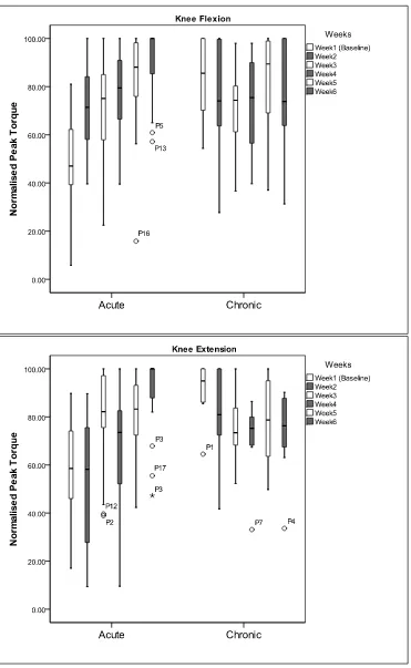
The normalized values of the peak torques generated in the hip flexor and extensors (Figure 3a and 3b) and knee flexors and extensors (Figure 4a and 4b) were analyzed separately for acute and subacute/chronic subjects. In the subacute/chronic subjects the pooled data showed no significant change in the peak torque generated throughout the six weeks of RAGT (Figure 3 and 4). This contrasts with data from acute subjects who show a gradual increase in their peak torque over the six weeks of RAGT (Figure 3 and 4).
Insert Figures 3(3a, 3b) & 4(4a,4b) here.
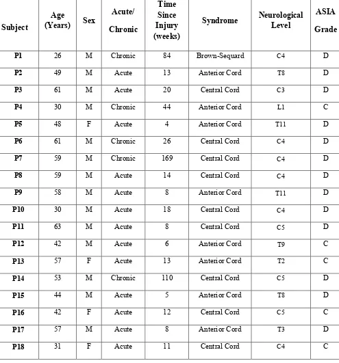
9 Wilcoxon signed rank test was used to compare the peak torques recorded between the training weeks in acute subjects. The results of these tests along with their p value are presented in Table 2.
Insert Table 2 here.
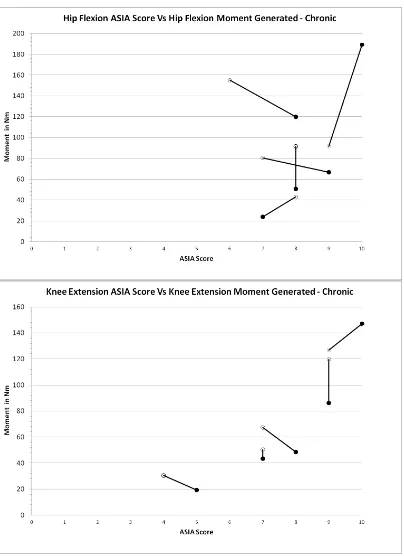
An increase in the lower limb motor score corresponded to an increase in the peak torque generated by the muscles that were tested. However this correlation between the motor score and peak torque was not a linear relationship (Figure 5a,5b,6a & 6b). When the subject had the ability to perform full ROM against gravity in both lower limbs i.e. having a lower limb motor score >6, the between subject variability of the peak torque measure increased. This observation was valid for both acute and sub-acute/chronic subjects who had participated in this study. In addition, subacute/chronic subjects had higher lower limb motor score in both hip and knee joints compared to the acute subjects. However, an increase in lower limb motor scores in subacute/chronic subjects was not necessarily accompanied by an increase in the peak torque generated by the muscle being assessed (Figure 6a & 6 b).
Insert Figures 5(5a,5b) & 6(6a,6b) here.
Discussion
10 score rising from 0 to 19). Improvements in gait function were observed in most acute subjects and this functional gain was accompanied by an increased peak isometric torque measurement. For the acute subjects, the functional gains in gait and muscle power are likely to reflect the combination of the natural recovery process post injury (Basso, 2000) supplemented by the impact of the intensive RAGT programme (Dietz, 2008). However, for some of the acute subjects, and the majority of chronic subjects, little or no change was observed in the recorded peak torques. Significantly, these subjects also failed to demonstrate significant improvements in walking ability as measured by WISCI II (refer to subject P10 illustrated in Figure 2b). The lack of significant changes in muscle strength in these subjects highlight that time post injury should be considered as an important factor to be taken into account in determining a gait rehabilitation programme for spinal cord injured subjects. In addition, the lack of evidence for RAGT promoting muscle strength changes in chronic subjects suggests that either the level of exercise provided by the training in this cohort was not sufficient to boost the force generating capacity of the muscle or that, they had reached a ceiling point in voluntary muscle performance post injury, thus re-emphasizing the criticality of the time window post injury for effective rehabilitation.
11 identified. Our observation also suggests that in acute subjects RAGT facilitates functional outcome above that produced by natural recovery processes on their own.
The observation that acute subjects who positively responded to RAGT had significant increase in peak torques within the first 3 weeks of training, suggests that this type of quantitative monitoring could also be considered as a useful marker in identifying subjects who are positively responding to RAGT using BWSTT. Accordingly, a change in peak torques generated by the lower limb muscles that exceeds the baseline measures, has the potential to be used as an indicator that helps the clinician to decide whether to continue training or to alter the rehabilitation strategy. This relationship between training time and muscle strength gain has not been previously reported in the literature. We have previously reported a similar pattern of improvement in gait function following Lokomat training.(Galen, Clarke, Allan, & Conway, 2011)
12 and further research will be required to explain these findings. It also needs to be noted that there were fewer chronic ISCI subjects in this study (n=5) than acute subjects (n=13). A larger group of subacute/chronic subjects with ISCI with more varied levels of mobility may need to be studied to fully understand the capacity of intensive RAGT to induce strength. Moreover the magnitude of the peak torque generated by the hip flexors and knee extensors were almost the same if not much greater in the subacute/chronic subjects compared to the acute subjects (Figures 5-6). Muscle atrophy secondary to SCI(Giangregorio & McCartney, 2006), has been reported previously, and has been considered a complication that has detrimental effects. However in the cohort of ISCI subjects who participated in this study, muscle strength seems to have been preserved. This is likely to be due to the fact that these subacute/chronic subjects were ambulant; therefore a better ambulatory capacity (Figure 2) following ISCI seems to stabilize muscle strength.
13 RAGT, in addition to having the potential to improve the force generating capacity of the lower limb muscles and gait function in responding ISCI subjects has also been reported to have an exercise impact on cardiovascular fitness and so contributes to improvements in the overall health and wellbeing of subjects irrespective of time post injury.(Jack, Purcell, Allan, & Hunt, 2011)
In recent years laboratory based studies on animal models of spinal cord injury have also demonstrated that intensive locomotor training when combined with other regenerative or pharmacological treatments produce better outcomes than when delivered on their own.(Edgerton, Kim, Ichiyama, Gerasimenko, & Roy, 2006) This suggests that when designing new pharmaceutical or regenerative treatments careful consideration should be given to the level and intensity of any concomitant rehabilitation programme. Use of interventions such as RAGT may offer advantages in such trials as both the dose and intensity of the rehabilitation programme can be easily monitored and standardized and that the integrated dynamometry provides for simple longitudinal collection of a set of important outcome measures.
Conclusion
14 of subjects. Similarly, regular monitoring for change in the force generating capacity of the lower limb muscles can provide a useful yet simple method for personalizing the rehabilitation programme and in identifying responding subjects.
Declaration of Interest
None declared.
Acknowledgement
15 References
Adams, M. M., Ditor, D. S., Tarnopolsky, M. A., Phillips, S. M., McCartney, N., & Hicks, A. L. (2006). The effect of body weight-supported treadmill training on muscle morphology in an individual with chronic, motor-complete spinal cord injury: A case study. J Spinal Cord Med, 29(2), 167-171.
Basso, D. M. (2000). Neuroanatomical substrates of functional recovery after experimental spinal cord injury: implications of basic science research for human spinal cord injury. Phys Ther, 80(8), 808-817.
Bolliger, M., Banz, R., Dietz, V., & Lunenburger, L. (2008). Standardized voluntary force measurement in a lower extremity rehabilitation robot. J Neuroeng Rehabil, 5, 23. doi: 1743-0003-5-23 [pii] 10.1186/1743-0003-5-23
Dietz, V. (2008). Body weight supported gait training: from laboratory to clinical setting. Brain Res Bull, 76(5), 459-463. doi: S0361-9230(08)00111-1 [pii]
10.1016/j.brainresbull.2008.02.034
Dietz, V., Colombo, G., Jensen, L., & Baumgartner, L. (1995). Locomotor capacity of spinal cord in paraplegic patients. Ann Neurol, 37(5), 574-582. doi: 10.1002/ana.410370506
Dittuno, P. L., & Dittuno Jr, J. F., Jr. (2001). Walking index for spinal cord injury (WISCI II): scale revision. Spinal Cord, 39(12), 654-656. doi: 10.1038/sj.sc.3101223 [doi]
Dudley-Javoroski, S., & Shields, R. K. (2008). Muscle and bone plasticity after spinal cord injury: review of adaptations to disuse and to electrical muscle stimulation. J Rehabil Res Dev, 45(2), 283-296.
Edgerton, V. R., Kim, S. J., Ichiyama, R. M., Gerasimenko, Y. P., & Roy, R. R. (2006). Rehabilitative therapies after spinal cord injury. J Neurotrauma, 23(3-4), 560-570. doi: 10.1089/neu.2006.23.560
Ellaway, P. H., Kuppuswamy, A., Balasubramaniam, A. V., Maksimovic, R., Gall, A., Craggs, M. D., . . . van Hedel, H. J. Development of quantitative and sensitive assessments of physiological and functional outcome during recovery from spinal cord injury: a clinical initiative. Brain Res Bull, 84(4-5), 343-357. doi: S0361-9230(10)00187-5 [pii]
10.1016/j.brainresbull.2010.08.007
Galen, S. S., Clarke, C. J., Allan, D. B., & Conway, B. A. (2011). A portable gait assessment tool to record temporal gait parameters in SCI. Med Eng Phys, 33(5), 626-632. doi: S1350-4533(11)00007-5 [pii]
10.1016/j.medengphy.2011.01.003
Giangregorio, L., & McCartney, N. (2006). Bone loss and muscle atrophy in spinal cord injury: epidemiology, fracture prediction, and rehabilitation strategies. J Spinal Cord Med, 29(5), 489-500.
Hicks, A. L., Adams, M. M., Ginis, K. M., Giangregorio, L., Latimer, A., Phillips, S. M., & McCartney, N. (2005). Long-term body-weight-supported treadmill training and subsequent follow-up in persons with chronic SCI: effects on functional walking ability and measures of subjective well-being. Spinal Cord, 43(5), 291-298. doi: DOI 10.1038/sj.sc.3101710
Hicks, A. L., & Martin Ginis, K. A. (2008). Treadmill training after spinal cord injury: It's not just about the walking. J Rehabil Res Dev, 45(2), 241-248.
16 Jack, L. P., Purcell, M., Allan, D. B., & Hunt, K. J. (2011). The metabolic cost of passive walking during robotics-assisted treadmill exercise. Technol Health Care, 19(1), 21-27. doi: 8765332286513V12 [pii]
10.3233/THC-2011-0608
Jayaraman, A., Shah, P., Gregory, C., Bowden, M., Stevens, J., Bishop, M., . . . Vandenborne, K. (2008). Locomotor training and muscle function after incomplete spinal cord injury: case series. J Spinal Cord Med, 31(2), 185-193.
Lunenburger, L., Colombo, G., Riener, R., & Dietz, V. (2004). Biofeedback in gait training with the robotic orthosis Lokomat. Conf Proc IEEE Eng Med Biol Soc, 7, 4888-4891. doi: 10.1109/IEMBS.2004.1404352
Marino, R. J., Barros, T., Biering-Sorensen, F., Burns, S. P., Donovan, W. H., Graves, D. E., . . . Priebe, M. M. (2003). International standards for neurological classification of spinal cord injury. J Spinal Cord Med, 26 Suppl 1, S50-56.
Nymark, J., DeForge, Z., Barbeau, H., Badour, M., Bercovitch, S., & Tomas, J. (1998). Body weight support treadmill gait training in the subacute recovery phase of incomplete spinal cord injury. Journal of Neurologic Rehabilitation, 12(3), 119-138.
Postans, N. J., Hasler, J. P., Granat, M. H., & Maxwell, D. J. (2004). Functional electric stimulation to augment partial weight-bearing supported treadmill training for patients with acute incomplete spinal cord injury: A pilot study. Arch Phys Med Rehabil, 85(4), 604-610. doi: S000399930301027X [pii]
Wirz, M., Zemon, D. H., Rupp, R., Scheel, A., Colombo, G., Dietz, V., & Hornby, T. G. (2005). Effectiveness of automated locomotor training in patients with chronic incomplete spinal cord injury: a multicenter trial. Arch Phys Med Rehabil, 86(4), 672-680. doi: S0003999304013279 [pii]
17 Table 1 : Details of Subjects who had participated in the study. Subjects who were <6months post injury at the time of recruitment were classified as acute and the rest were classified as Chronic. M=Male and F=Female.
Subject
Age
(Years) Sex
Acute/ Chronic Time Since Injury (weeks)
Syndrome Neurological Level
ASIA
Grade
P1 26 M Chronic 84 Brown-Sequard C4 D
P2 49 M Acute 13 Anterior Cord T8 D
P3 61 M Acute 20 Central Cord C3 D
P4 30 M Chronic 44 Anterior Cord L1 C
P5 48 F Acute 4 Anterior Cord T11 D
P6 61 M Chronic 26 Central Cord C4 D
P7 59 M Chronic 169 Central Cord C4 D
P8 59 M Acute 14 Central Cord C4 D
P9 58 M Acute 8 Anterior Cord T11 D
P10 30 M Acute 18 Central Cord C4 D
P11 63 M Acute 8 Central Cord C5 D
P12 42 M Acute 6 Anterior Cord T9 C
P13 57 F Acute 13 Anterior Cord T2 C
P14 53 M Chronic 110 Central Cord C5 D
P15 44 M Acute 5 Anterior Cord T8 D
P16 42 F Acute 12 Central Cord C5 C
P17 57 M Acute 8 Anterior Cord T3 D
18 Acute Mean Standard deviation Median Upper quartile Lower quartile p value
Hip flexion Week1 52.96 23.12 49.42 36.85 72.37
Week2 57.43 27.22 61.31 34.81 79.21 0.526
Week3 79.60 16.14 77.7 66.46 95.77 <0.001
Week4 68.88 16.58 69.44 58.63 81.17 0.002
Week5 82.71 17.28 88.99 62.15 100.00 0.001
Week6 89.06 14.89 93.21 82.84 100.00 <0.001
Hip
Extension Week1 62.33 15.27 57.08 53.69 72.31
Week2 71.38 18.32 72.85 58.94 85.40 0.025
Week3 81.72 14.37 84.3 71.47 94.79 <0.001
Week4 85.29 10.08 84.57 77.13 94.21 <0.001
Week5 89.22 9.95 91.09 84.90 96.90 <0.001
Week6 96.41 7.73 100 96.74 100.00 <0.001
Knee Flexion Week1 47.20 17.90 47.03 38.25 62.36
Week2 70.14 17.32 71.46 57.19 84.42 <0.001
Week3 71.05 20.08 75.1 57.32 85.23 <0.001
Week4 77.97 15.85 79.44 65.96 91.75 <0.001
Week5 82.65 19.65 88.08 74.24 98.53 <0.001
Week6 91.44 13.83 100 84.00 100.00 <0.001
Knee
Extension Week1 58.50 17.78 58.57 45.96 74.42
Week2 52.79 26.84 58.25 26.94 75.73 0.779
Week3 79.85 18.75 82.21 75.56 97.68 <0.001
Week4 69.26 21.74 73.54 52.16 82.64 <0.001
Week5 81.56 16.55 83.28 71.89 94.87 0.001
[image:18.595.71.493.68.436.2]Week6 91.19 14.65 100 87.45 100.00 <0.001
19 Figure1 : Change in ambulatory capacity (WISCI II score) before(Pre-intervention) and after(Post-intervention) lokomat training (RAGT) in acute and subacute/chronic subjects.
Figure2a and 2b: Peak Torques recorded in subjects P15 and P10 across key muscle groups in the lower limb during the six weeks of RAGT.
Figure 3a and 3b: Normalised peak torques presented as pooled data for chronic and acute subjects.
Figure 4a and 4b: Normalised peak torques presented as pooled data for chronic and acute subjects.
Figure 5a and 5 b: Peak Torque generatd as a result of isometric contraction of the hip flexor and knee extensor muscles, compared against the Lower limb motor score (ASIA Score), for each of the respective muscle in acute subjects. Results of ambulant subjects are shown as a solid line and results from non-ambulant subjects are shown using dashed line. Intial measures are denoted by open circles and final measures are denoted by filled circles.
21 Figure2a and 2b: Peak Torques recorded in subjects P15 and P10 across key muscle groups in the lower limb during the six weeks of RAGT.
0 10 20 30 40 50 60 70
Week1 Week2 Week3 Week4 Week5 Week6
Peak To rq u e in N m
Change in Peak Torque in subject P15
Hip Flexor Hip Extensor Knee Flexor Knee Extensor 0 10 20 30 40 50 60 70
Week1 Week2 Week3 Week4 Week5 Week6
Peak t o rq u e in N m
Change Peak Torque in subject P10
Hip Flexor
Hip Extensor
Knee Flexor
22
23