This is a repository copy of Rickets.
White Rose Research Online URL for this paper: http://eprints.whiterose.ac.uk/139788/
Version: Accepted Version
Article:
Elder, C.J. orcid.org/0000-0003-2390-5593 and Bishop, N.J.
orcid.org/0000-0001-7263-8546 (2014) Rickets. Lancet, 383 (9929). pp. 1665-1676. ISSN 0140-6736
https://doi.org/10.1016/S0140-6736(13)61650-5
Article available under the terms of the CC-BY-NC-ND licence (https://creativecommons.org/licenses/by-nc-nd/4.0/).
[email protected] Reuse
This article is distributed under the terms of the Creative Commons Attribution-NonCommercial-NoDerivs (CC BY-NC-ND) licence. This licence only allows you to download this work and share it with others as long as you credit the authors, but you can’t change the article in any way or use it commercially. More
information and the full terms of the licence here: https://creativecommons.org/licenses/
Takedown
If you consider content in White Rose Research Online to be in breach of UK law, please notify us by
Ελσεϖιερ Εδιτοριαλ Σψστεm(τm) φορ Τηε Λανχετ Μανυσχριπτ Dραφτ
Μανυσχριπτ Νυmβερ: ΤΗΕΛΑΝΧΕΤ−D−13−03383Ρ1
Τιτλε: Ριχκετσ
Αρτιχλε Τψπε: Ινϖιτεδ Σεmιναρ
Χορρεσπονδινγ Αυτηορ: Προφ. Νιχκ ϑ. Βισηοπ, ΜD
Χορρεσπονδινγ Αυτηορ∋σ Ινστιτυτιον: Τηε Υνιϖερσιτψ οφ Σηεφφιελδ
Φιρστ Αυτηορ: Χηαρλοττε ϑ Ελδερ, ΜΒ ΒΣ
Ορδερ οφ Αυτηορσ: Χηαρλοττε ϑ Ελδερ, ΜΒ ΒΣ; Νιχκ ϑ. Βισηοπ, ΜD
Title: Rickets
Authors (names, preferred degree, affiliations, full address): Charlotte Jane Elder MBBS
University of Sheffield
Academic Unit of Child Health, Sheffield Children’s Hospital, Western Bank Sheffield, S10 2TH, UK
Nicholas J Bishop University of Sheffield
Academic Unit of Child Health, Sheffield Children’s Hospital, Western Bank
Sheffield, S10 2TH, UK
Corresponding author: Nicholas J Bishop
University of Sheffield
Academic Unit of Child Health, Sheffield Children’s Hospital, Western Bank
Sheffield, S10 2TH, UK
Email: [email protected]
Abstract
Rickets, the English Disease, remains common worldwide. Lack of phosphate at the growth plate and mineralising bone surfaces due to inadequate vitamin
D supply either from sunlight exposure or diet remains the principal cause. Inherited disorders causing hypophosphataemia have cast light recently on
the intricacies of phosphate metabolism. Current advice regarding the provision of vitamin D to young infants needs to be clarified - the existing guidance is fragmentary and contradictory, and will not facilitate the
eradication of the disease.
Introduction
A century ago rickets affected over 25% of children in the UK. Today, rickets remains one of the commonest non-communicable diseases of children in the
developing world and has been thought to be on the rise again in the UK,1 although reliable recent data indicating the extent of the increase nationally is
lacking. Rickets is characterised by bony deformity and stunted growth. Lower limb deformities such as bow-legs, knock-knees, or windswept changes can
cause significant disability and pelvic deformity in girls can kill because of obstructed labour.2 There may be longer-term consequences for skeletal health with reduced bone size and mass predisposing to later osteoporotic
fracture.
The pathological definition of rickets, the failure to mineralise newly-formed
plate, with associated growth plate deformity.3 These features are the result of vitamin D deficiency in most cases, usually with an easily discernable clinical history and associated characteristic biochemical and radiological changes. What remains unclear is whether an absolute threshold for vitamin D exists
below which rickets is inevitable; rickets can also occur when vitamin D is within the range associated with maximal calcium absorption, but calcium
intake is low.4
In rare cases, abnormalities that primarily affect phosphate metabolism or bone tissue mineralisation may be the cause. Our understanding of the
intricacies of phosphate homeostasis is still evolving. This seminar will address these issues and other areas of controversy, such as the contribution
of low vitamin D to fractures in infancy.
Historical context
The original description of rickets is attributed variously to Whistler or Glisson, both practicing in England in the mid 1600s.3 The origin of the word itself is unclear, possibly relating to the German “wricken” meaning “twisted”. Glisson
clearly differentiated rickets from infantile scurvy based on post-mortem
observations although his suggested treatment of lamb’s wool ligatures to the extremities was wide of the mark. Trousseau in 1861-2 identified lack of sunlight and poor nutrition as likely causes of rickets and suggested
appropriate remedies, including cod-liver oil.5 Palm commented on the relationship of increasing latitude (and hence decreased sunlight exposure)
Columbia district of New York in 1916,7 predating the classic experiments of Mellanby who created and then cured rickets in sunlight-deprived dogs fed porridge.8 Hess and Unger also cured children’s rickets through exposure to sunlight.9 Infants fed cod liver oil in addition to their normal diet were observed by Daniels to grow more quickly than those receiving the same diet alone.10 The chemical characterisation of vitamin D2 and vitamin D3 by Windaus in 1932 was succeeded by the clinical studies of Jeans and Stearns in American orphanages in which wet-nursed infants received different doses of vitamin D2 or D3.11 Infants fed 8·5-10 g (340-400 units) of vitamin D daily were on average 2 cm longer at one year of age than infants fed 1·5-3·4 g (60-135 units). Infants on the lower dose exposed to sunlight grew more quickly.11 Infants who received doses of over 45 g/day (1800 units/day) in subsequent studies grew less quickly, and their growth rate improved when the dose was reduced to 10-15 g/day (400-600 units/day).12 More recent studies have not shown slower growth at these higher doses.13 The original dosing regimens are echoed in the current recommended daily intakes for the UK (table 1).
Vitamin D metabolism and actions; also see figure 1
Vitamin D2 (ergocalciferol) is obtained solely from diet whereas D3 (cholecalciferol) is found in cod liver oil and oily fish and is the form synthesised in skin. Sunlight, specifically Ultraviolet-B (UVB) in the 290-315
Stearns appeared equally effective at preventing rickets and improving linear
growth,11 and there is an equivalence of effect in raising serum 25-hydroxy vitamin D (calcidiol, 25OHD) concentrations.16
Vitamin D binds to vitamin D binding protein (VDBP) and is transported to the
liver for 25-hydroxylation (the main enzyme is CYP2R1)17 and then to the kidney. The vitamin D binding protein-25OHD complex is excreted and then
reabsorbed in the proximal tubule through the endocytic receptors megalin and cubilin,18 where it undergoes 1-hydroxylation by CYP27B1 resulting in the active metabolite 1,25-dihydroxyvitamin D (calcitriol, 1,25(OH)2D). Lack of the CYP27B1 enzyme results in vitamin D-dependent rickets type 1A19 and
treatment requires the use of calcitriol or 1-calcidol.
The 1,25(OH)2D binds to its cognate receptor (VDR), which heterodimerises with the retinoic acid receptor (RXR) to form a ligand-receptor complex that targets specific response elements on the genome. Mutations in the
ligand-binding domain of the VDR resulting in rickets can be overcome in some instances using high dose calcitriol therapy; mutations in the DNA-binding domain do not usually respond to this.20 Affected infants present with hypocalcaemia and severe rickets, and are typically alopecic. Such children require high dose intravenous calcium infusions daily until two years of age,
The primary action of 1,25(OH)2D is to increase gut calcium absorption by upregulating the calcium channel TRPV6, the intracellular transporter calbindin D, and the calcium pump PMCA1b needed to move calcium up the concentration gradient from enterocytes to serum.23 Calcium absorption reduces by 70-75% in animals lacking the vitamin D receptor,20, 24 however the extent to which there is a threshold of either 25OHD or 1,25(OH)2D for reduced calcium absorption is unclear. Fractional calcium absorption in children at lower 25OHD levels (25-50 nmol/l) is 0.34 compared to 0.28 at higher 25OHD concentrations (50-80 nmol/l),25 i.e. relatively more dietary calcium is being absorbed at these lower 25OHD concentrations. Need and colleagues reviewed the records of 319 adult subjects with comprehensive
calcium absorption and bone profile data and concluded that calcium absorption does not fall until 25OHD is below 10nmol/l;26 however, as similar work has not been performed in the paediatric population and the adult
calcium requirement is lower, it is unclear how this extrapolates to growing children. In addition 1,25(OH2)D acts in concert with osteoblast-derived factors to increase osteoclastic bone resorption. Thus vitamin D’s role in skeletal homeostasis seems primarily to keep serum calcium above the
threshold below which neuromuscular abnormalities occur.
Pathophysiology of rickets
It is lack of phosphate that results in the characteristic growth plate changes in rickets. Elegant experiments by Sabbagh and colleagues using different
hypertrophic chondrocyte to undergo apoptosis, a process dependent upon
phosphorylation of caspase-9 in those cells.27 In vitamin D deficiency, fasting phosphate is low with phosphate lost from the kidney as parathyroid hormone (PTH) rises in the face of the falling supply of calcium.
Some of the genetically determined forms of hypophosphataemic rickets (panel 1) lose phosphate as a result of inhibition of the renal
sodium-phosphate co-transporter. In these forms of rickets, there are elevated circulating levels of FGF23, a member of the “endocrine” fibroblast growth factors (FGFs) that lack heparin sulfate binding domains, and are hence not
restricted to the extracellular matrix.28 FGF23 binding to FGF receptors
requires the presence of klotho; the absence of either klotho or FGF23 in
humans results in hyperphosphataemia and ectopic calcification. FGF23
secretion is stimulated by increased phosphate intake, by 1,25(OH)2D and by PTH in some studies. In turn, FGF23 down-regulates the renal CYP27b1
enzyme that creates 1,25(OH)2D and upregulates the 24-hydroxylase that destroys 1,25(OH)2D.29 Whilst phosphate is required for the healing of the growth plate, the osteomalacia and bowing deformity of long bones in children
with hypophosphataemic rickets requires 1,25(OH)2D to resolve.30
At bone remodeling sites, where new bone replaces old bone, and at the
periosteal bone surface, lack of phosphate results in a failure of mineralisation of the fibrous component of bone, the osteoid. The balance of mineralisation inhibitors such as pyrophosphate and phosphate in the initiation and
propagation of mineral crystal deposition into bone matrix is thought to be under local control. Members of the SIBLING family of proteins, that includes
mineralisation through the balance of phosphate and mineralisation inhibitors
at the bone surface.31 DMP1 mutations result in autosomal recessive hypophosphataemic rickets type 1 (ARHR1). However, with the exception of DMP1, altered SIBLING proteins do not produce a rachitic phenotype in
humans. The SIBLING proteins all contain an acidic serine asparate-rich MEPE (ASARM) motif or cleavable peptide moiety.32 The ASARM peptide binds strongly to hydroxyapatite, can directly inhibit bone mineralisation, can provoke hypophosphataemia through inhibition of the renal sodium-phosphate co-transporter, and may be a substrate for PHEX.33
The similarity of the clinical and biochemical phenotypes in X-linked hypophosphataemic rickets (XLH) and ARHR1 suggest that PHEX and DMP1
act in the same pathway to regulate FGF23 expression.34 DMP1 and FGF23 are primarily expressed in osteocytes embedded deep within bone and PHEX in osteoblasts sitting on bone surfaces. The process of osteoid mineralisation
affects the envelopment of osteoblasts that will become osteocytes.35 The processes underpinning the interactions are thus spatially and temporally
complex and may affect other aspects of bone structure. It is notable that radiographs of bones from patients with inherited hypophosphataemic rickets
often show sclerosis rather than the osteopenia typical of the vitamin D pathway forms of the disease.
Infantile hypophosphatasia (HPP), in which lack of tissue non-specific alkaline
phosphatase results in failure to clear pyrophosphate and other mineralisation inhibitors, presents with a severe rachitic phenotype in the first days and
Recombinant bone-targeted enzyme replacement therapy is reported as
having some benefit.36
Rickets in infants born prematurely
Rickets has been described repeatedly in infants born prematurely. It is clear that inadequate supply of mineral substrates, rather than vitamin D deficiency,
is the root cause.37 Most of those affected are born at less than 28 weeks of gestation, have had many difficulties resulting in delay in the establishment of enteral feeding, and some may have chronic lung disease that necessitates
the use of steroids and diuretics causing hypercalciuria. Infants with such a history, and particularly those who develop conjugated hyperbilirubinaemia,
are at increased risk of fracture.38, 39 The risk of fracture is likely further increased in such infants by periods of immobilization associated with illness during the period of their hospitalization. Fractures of the ribs can be occult
and result from physiotherapy. The prevalence of such fractures at discharge is unclear as exit (from the neonatal unit) chest x-rays are not a routine part of
care; however recent data suggests that around 2% suffer rib fractures in the first year of life. 39
Rickets, low vitamin D and fractures in infants born at term
The extent to which reduced osteoid mineralisation diminishes bone strength in the early stages of rickets is unclear. Single but not multiple fractures have
risk during infancy and childhood. There is limited data regarding the
frequency of clinical rickets in infants who present with vitamin D deficiency-induced hypocalcaemia, and none regarding their subsequent fracture risk. Further research is needed to clarify these important issues. Such data are
needed both to advise parents regarding their handling of infants potentially at increased risk of fracture, and to better clarify the extent to which biochemical
parameters such as an isolated low 25OHD need to be taken account of in cases of suspected child abuse.41
Working definition of vitamin D deficiency
There is consensus that, as the circulating form of vitamin D with the longest
half-life, serum 25OHD is the most appropriate marker of vitamin D status.42 Controversy continues, however, about the definition of thresholds in 25OHD for vitamin D sufficiency and deficiency. What is “normal” depends on the
clinical endpoint of interest and thresholds from 25-100 nmol/l have been proposed. The value may be different when considering optimal general
health, good bone health43 or that needed to prevent rickets and osteomalacia. The majority of studies seeking the optimal vitamin D status for
good health have been done in adults and the childhood value is likely to be different and to vary with age.44 We do not know the absolute 25OHD level that unequivocally confers either good general or good bone health in
children. It is clear that as vitamin D falls, so the risk of ill-health rises, but there are numerous of examples of very low 25OHD levels with no evidence
Amongst UK paediatricians there seems emerging agreement that a serum
25OHD of <25 nmol/l represents deficiency with an increased likelihood of rickets and <50 nmol/l insufficiency.41, 45-49 Worldwide there is support for a higher cut off but different bodies advocate different definitions. The
Endocrine Society (USA) 2011 Clinical Practice Guideline defines >72·5 nmol/l as optimal, <50 nmol/l deficient and levels in-between as insufficient.50 The Institute of Medicine (USA) consider levels of 25OHD <30 nmol/l as deficiency51, cut offs supported by both the US and Canadian governments, and similarly the Lawson Wilkins Pediatric Endocrine Society define
deficiency as <37·5 nmol/l and insufficiency between 37·5-50-nmol/l.52 Some advocate levels of serum 25OHD that indicate an increase risk rather than
define deficiency.53 The lack of consensus reflects both the importance of considering covariates such as calcium intake and the lack of contemporary large scale controlled studies demonstrating benefit of administered vitamin D
either to bone or general health in children of differing ages.
Aetiology of vitamin D deficient rickets
Fetal vitamin D is acquired entirely from the mother and levels are dependent
on maternal vitamin D status which is frequently low in women of child-bearing age.54 Maternal 25OHD crosses the placenta and undergoes placental conversion to 1,25(OH)2D.55 At birth the 25OHD level in cord blood closely correlates to maternal levels and ranges from 68-108%.56 Vitamin D-replete mothers give birth to vitamin D D-replete infants and there are rare cases
mothers are taking near-pharmacological doses of 100 g/day (4000
units/day) of vitamin D.60 Babies born to vitamin D-replete mothers will have serum vitamin D levels consistent with deficiency after only eight weeks of exclusive breast feeding.55
Exposure to sunlight is reduced by living at latitude as negligible vitamin D synthesis occurs at latitudes greater than 35° in the northern hemisphere61 and greater than 32° in the southern hemisphere during the winter months.62 Covering up for religious or cultural custom and as a sun protective measure substantially reduces skin exposure to UVB light, as does the use of
sunscreen. Atmospheric pollution in rapidly industrialising nations63 may recapitulate the problems of the UK from a century ago. The same amount of
vitamin D synthesis requires more sunlight exposure in dark as opposed to light skinned individuals.64
The pathophysiology of rickets is such that it is most apparent, and therefore
clinically most frequently seen, at periods of peak growth, in particular in the first two years of life but also during the adolescent growth spurt. The most
typical presentation for a child with rickets is in the context of maternal insufficiency, due to darker skin colour and/or covering up without
supplementation during pregnancy, and prolonged breast-feeding without supplementation of the infant.
Hypocalcaemia and vitamin D deficiency
The earliest presentation is of neonatal hypocalcemia (“early” – less than 1 week of age; “late” 2-4 weeks), which may result in jitteriness, or progress to
secondary to hypocalcaemia and presumed vitamin D deficiency has been
reported.65 In an 11 year retrospective case study of 126 children presenting with vitamin D deficiency (<50 nmol/l) or rickets to paediatric centres in Sydney, Australia, hypocalcaemic seizures were the most common
presentation, seen in a third of cases.66 In the West Midlands, Callaghan and colleagues reported a quarter of presentations with symptomatic vitamin D
deficiency as due to hypocalcaemic convulsions although bowed legs were more common (46%).46
Measuring vitamin D status
Although 1,25(OH)2D is the active product of vitamin D synthesis its quantification is hampered by its presence in picomolar quantities (25OHD circulates in nanomols) and a considerably shorter half-life compared with 25OHD. Additionally as the rate-limiting step in vitamin D synthesis, the levels
of 25OHD need to be considerably reduced before there is any affect on serum 1,25(OH)2D levels, which may be misleadingly normal or even high due to secondary hyperparathyroidism.67
The measurement of 25OHD is analytically challenging. Until recently there
had been no reference methods or standard reference materials, resulting in considerable inter-laboratory variability.68-71
The current gold standard technique for measurement of vitamin D status,
Quality Assurance System) demonstrating both over- and under-estimations.
In addition current extraction techniques do not remove 3-epi-25OHD (not usually detected by immunoassays), initially only thought to be present in 22·7% of infant samples (contributing 8·7-61·1% of the total 25OHD),72 but more recently reported to be contributing approximately 5% of the total 25OHD (range 0 to 25·5%) in 99% of samples from patients of all ages.73 The biological activity of the 3-epimer is unclear.74
Incidence and Prevalence of rickets – a global perspective
Accurate incidence and prevalence data are undermined by the lack of a robust screening tool, with no global consensus for a vitamin D deficiency cut
off and confusion over the difference between vitamin D deficiency and rickets. There appears to be an increasing incidence of rickets worldwide, although good, up-to-date data are not available.57, 62, 75, 76 Previously published prevalence figures range from 70% in Mongolia, 42% in Ethiopia, 9% in Nigeria, 3·3% in The Gambia to 2·2% in Bangladesh.77 In NW England 1·6% of a predominantly Asian population were found to have rickets.78 In Hokkaido, Northern Japan, an estimate of rickets prevalence was 9/100,000
for under four year-olds.79 In Denmark the average incidence over a twenty year period was 2·9/100,000 per year, with 5·8/100,000 per year in the under three year-olds.80 In Eastern Turkey the incidence of rickets in children presenting to paediatric outpatient clinics was 0·1%.81
An Australian surveillance study has estimated the overall incidence of
National Diet and Nutrition Survey (2011) measured serum 25OHD in 160
young people aged 11-18 years and reported a mean of 44·6 nmol/l for boys and 42·2 nmol/l for girls indicating widespread insufficiency and probable deficiency.
The apparent increase in rickets has been seen worldwide. The occurrence of vitamin D-deficient rickets in areas of the tropics where cultural and religious
custom do not preclude adequate sunlight exposure has led to the suggestion of a significant aetiological role for a high phytate, low calcium diet.4, 83 A recent case-control study conducted in India has added further credence to
this by demonstrating no difference in 25OHD levels but significantly lower calcium and higher phytate dietary intakes in the group with rickets.84 Lower concentrations of calcium have been found in the breast milk of mothers of rachitic children.85 Radiological and biochemical rachitic changes observed in vitamin D sufficient (>25 nmol/l) hypocalcaemic children have been seen to
resolve rapidly with sole calcium supplementation as opposed to with only vitamin D, although the combination of both was most efficacious.4, 86
The increase in cases in industrialised countries is likely due to increased movement of darker skinned individuals to more temperate climes, as
individuals of Afro-Caribbean and Asian origin in Europe and African-Americans in North America are generally cited in published case series.62 Immigrant numbers continue to rise in the UK, with data from the 2011 census
showing that 13% of the UK population is now foreign born, the greatest numbers coming from India, Poland and Pakistan.87
The treatment of vitamin D deficiency-induced rickets is simple and cost
effective and usually entails an oral preparation of vitamin D with calcium supplementation in children with poor dietary intake or evidence of hypocalcaemia. Choice of vitamin D preparation, ergocalciferol or
cholecalciferol, and of dosing regimen, are contentious issues. There have been concerns about the efficacy of ergocalciferol, both in terms of its ability
to raise 25OHD and the precipitate fall in serum levels compared with cholecalciferol after completing treatment.88 Others have found the rise in 25OHD after administration of both forms to be equivalent89 including two studies in paediatric populations.90, 91 Most consensus statements and supplement/treatment guidance do not recommend one form over the other.
Other than the 80 year-old Jeans and Stearns studies, there is no data on functional outcome comparing the two forms.
The British National Formulary for Children (BNFc) recommends either form
of calciferol at treatment dose for 8-12 weeks after which supplemental doses should be employed, which we recommend continuing until completion of
linear growth (table 1).92 In practical terms, vitamin D deficiency will take longer to correct with lower vitamin D intake; consequently a sliding scale of
vitamin D treatment is suggested that takes some account of age-related changes in body size and rate of growth (table 1). Vitamin D insufficiency (<50 nmol/l but >25 nmol/l) is usually treated with supplement doses rather than
treatment doses. The BNFc recommends all patients receiving pharmacological doses to have serum calcium checked initially once or twice
to monitor asymptomatic patients and perform a bone profile and 25OHD
shortly after completion of treatment.
In the United States, dealing with an ethnically diverse population, the Endocrine Society Clinical Practice Guideline50 recommends the use of either vitamin D2 or D3 in a dose of 2,000 IU/day or 50,000 IU/week for 6 weeks in infants aged 0-1 years followed by maintenance intake of 400 IU/d; the same
schedule is recommended for children aged 1-18 years but with a maintenance dose of 600 IU/d.
We do not recommend administration of vitamin D as an intramuscular
injection as a routine measure in children. Stosstherapy (from the German “push”) with 600,000 units of vitamin D may result in hypercalcaemia and
nephrocalcinosis.93 There is no place for the routine use of 1 -hydroxylated preparations such as alfacalcidol or calcitriol in the treatment of rickets caused by vitamin D deficiency; their role is in the treatment of hypophosphatemic
rickets with raised FGF23 and the rare vitamin D pathway defects. They may have a place in the acute treatment of hypocalcaemic cardiomyopathy.65
Treating hypophosphataemic rickets
In the forms of hypophosphataemic rickets associated with raised serum FGF23, replacement of phosphate is required alongside the use of either
calcitriol or 1-calcidol. Regular review in a specialist paediatric metabolic
bone clinic is needed to monitor growth, bony deformity, and the
complications associated with these disorders and their treatment including root abscesses, craniosynostosis, nephrocalcinosis and parathyroid gland
particularly during periods of more rapid growth. Bowing deformity resulting in
genu varum with an intercondylar distance of more than 12 cm is likely to require surgical intervention; such intervention should only be undertaken when the bone disease is under control. A good clinical management
guideline was published in 2010.94
Anti-FGF23 antibody therapy has been assessed in the murine model of
X-linked hypophosphataemic rickets, the HYP mouse, and shown to correct both the hypophosphataemia and restore conversion of 25OHD to 1,25(OH)2D, as well as restoring longitudinal growth and improving osteomalacia.95 A phase 1 single dose escalation trial in adults has been completed but not yet reported (NCT00830674).
Prevention
Prevention of rickets can be summarised as adequate exposure to sunlight
and dietary intake. This is complicated by high profile public health campaigns advising sunlight avoidance, the need for different, culturally sensitive,
strategies for at risk groups and varying guidance internationally for the recommended daily intake of vitamin D. Population screening is currently not
a viable option due to the lack of a consensus over a diagnostic cut-off, lack of a test with appropriate levels of sensitivity and specificity and paucity of long term data regarding the sequelae of low serum 25OHD.
Adequate sunlight exposure
due to latitude/season. The strong epidemiological evidence linking sun
exposure and skin cancers has led the American Pediatric Association (APA) to support the guidance limiting exposure to sunlight in children and promotion of vitamin D supplementation throughout childhood.96 Studies on adults living in North West England (latitude of 53.5°N) have shown that sunlight exposure at recommended levels (15 minutes unshaded noontime exposure 3
times/week with 35% skin surface exposed), whilst improving 25OHD levels and being adequate for white skinned individuals, left all South Asian participants (N=15) vitamin D insufficient (<50 nmol/l).97 Increasing this exposure three-fold only achieved sufficiency in a quarter of the South Asian cohort demonstrating that sun exposure advice needs to be tailored to the
degree of skin pigmentation and may remain inadequate for a significant proportion of the population.98
Improvements in air quality enhance access of UVB to the skin. The Clean Air
Act of 1956 in Britain is thought to have contributed to a reduction in cases of rickets thereafter and similar government interventions in rapidly
industrialising nations may improve population 25OHD levels.
Vitamin D Supplementation (table 2)
Worldwide guidance and recommended vitamin D intakes during pregnancy vary (table 2). Advised supplementation doses are between 5-100 g/day
The upper limit considered safe for pregnant and lactating women was
increased in Europe following a large randomised control trial (RCT) in which 100 g/day (4000 IU/day) was found to be the dose most effective in achieving sufficiency in the absence of any adverse events.60, 100 The upper level of intake advised by the Institute of Medicine (IOM) in the US is similarly 100 g/day (4000 IU/day).51 An RCT is currently underway looking at
supplementation with higher doses (NCT01060735). The longer-term effects of such supplementation on the exposed fetal skeleton remain to be determined.
Although there remains concern about poor adherence to supplementation programmes, reductions in the prevalence of rickets101 and symptomatic vitamin D deficiency have been demonstrated following targeted and universal supplementation campaigns.102
Supplementation in breastfed infants
The WHO recommends exclusively breastfeeding infants until 6 months of
age. A 3.5 kg baby taking 150 ml/kg/day of breast milk would therefore receive little more than 0·75 g/day vitamin D (30 IU/day), below the intake
found to be inadequate to support normal linear growth in the Jeans and Stearns studies (inadequate 1·5-3·4 g/day (60-135 IU/day); adequate 8·5-15 (340-600 IU/day)).11 In order to increase the vitamin D concentration sufficiently in breast milk maternal supplementation would need to be in the range of 100-160 g/day (4000-6400 IU/day) but these doses have only been
The advice from the UK Department of Health (DH), based on the 2007
position statement by the Scientific Advisory Committee on Nutrition,42 is to commence supplementation of breastfed infants from age 6 months. A letter in 2012 from the UK Chief Medical Officers to GPs, Health Visitors,
Pharmacists and Practice Nurses stated “breast fed infants may need to receive drops containing vitamin D from one month of age if their mother has not taken vitamin D supplements throughout pregnancy”.104 It is clear that
adequate intake will not be achieved by breastfeeding without supplementation in the absence of adequate sun exposure and the advice is
therefore inconsistent with the original published data and confusing for health care practitioners.
Overdosing with vitamin D will cause hypercalcaemia and nephrocalcinosis. Serum calcium rises with increasing 25OHD; hypercalcaemia occurs when 25OHD exceeds 200 nmol/l. The UK tolerable upper intake level for vitamin D
for infants and children to age 10 is set at 25 mcg/day (1000 units/day). The European Food Safety Authority recently revised their limits to a tolerable
upper intake of 25 mcg/day (1000 IU/day) in infants, 50 mcg/day (2000 IU/day) for children under 10 years and 100 mcg/day (4000 U/day) for
children over 10 years.100 In North America the limits set by IOM are the same for infants under six months, 37·5 g/day (1500 IU/day) from six months to one year and 62·5 g/day (2500 IU/day) from one to three years, 75 g/day
(3000 IU/day) from four to eight years and 100 g/day (4000 IU/day), the tolerable upper limit, thereafter.51
In most countries infant formula milk is fortified to give a concentration of 10 g/l (400 IU/l). In the US milk and breakfast cereals are fortified and in
Canada milk and margarine.105 Following deaths from Idiopathic Infantile Hypercalcaemia in the 1950s in UK the Department of Health banned food
fortification with the exception of yellow spreads (margarines), cereals and infant formula milks. Fortification of chapatti flour to target low vitamin D status in the UK’s Asian community was shown to be effective but was not
universally introduced, although fortified flour is available.106
Summary/Take home messages
Rickets is a preventable disease and prevention should start in pregnancy.
The simplest measure for prevention is adequate sunlight exposure, however in populations where this is impracticable or implausible vitamin D supplementation should be instituted. There is no global consensus on the
amount of vitamin D offered in supplementation. The guidance in the UK from the Department of Health is fragmentary and confusing. Vitamin D 400IU/d is
sufficient to maintain vitamin D status in the range where adverse skeletal consequences are very unlikely; suggesting a daily supplement ensures that
irrespective of skin colour, latitude, sunlight exposure , pollution, and societal or cultural pressures to cover up, the growing skeleton will get what it needs. It is the view of the authors that supplementation until growth ceases with 10
mcg/day (400 IU/day) in all except those with a known contraindication (e.g. hypercalcaemia, sarcoidosis) should be recommended and that, without such
Acknowledgments
We thank Sarah Massey, Knowledge and Library Services Manager, Sheffield Children’s NHS Foundation Trust, for her help with literature searching and
sourcing papers.
Word Count
References
1. Ahmed SF, Franey C, McDevitt H, Somerville L, Butler S, Galloway P, et al. Recent trends and clinical features of childhood vitamin D deficiency presenting to a children's hospital in Glasgow. Arch Dis Child. 2011; 96(7): 694-6.
2. Loudon I. Deaths in childbed from the eighteenth century to 1935. Med Hist. 1986; 30(1): 1-41.
3. Pettifor JM. Nutritional Rickets. In: Glorieux FH, editor. Pediatric Bone; Biology and Diseases. San Diego: Academic Press; 2003. p. 541-65.
4. DeLucia MC, Mitnick ME, Carpenter TO. Nutritional rickets with normal circulating 25-hydroxyvitamin D: a call for reexamining the role of dietary calcium intake in North American infants. J Clin Endocrinol Metab. 2003; 88(8): 3539-45.
5. Trousseau A. Lectures on Clinical Medicine, delivered at the Hotel-Dieu, Paris. 3rd ed. London: New Sydenham Society; 1872.
6. Palm TA. The geographic distribution and etiology of rickets. Practitioner. 1890; 15: 321.
7. Hess AF, Unger LJ. Prophylactic therapy for rickets in a Negro community. JAMA. 1917; 19: 1583-6.
9. Hess AF, Unger LJ. The cure of infantile rickets by sunlight -
Preliminary note. Journal of the American Medical Association. 1921; 77: 39-. 10. Daniels AL, Hutton MK, Stearns G, Hejinian LM. The relation of rate of growth in infants to diet. American Journal of Diseases of Children. 1929; 37(6): 1177-86.
11. Jeans PC, Stearns G. Effectiveness of vitamin D in infancy in relation to the vitamin source. Proceedings of the Society for Experimental Biology and Medicine. 1934; 31(9): 1159-61.
12. Jeans PC, Stearns G. The effect of vitamin D on linear growth in infancy II The effect of intakes above 1,800 USPunits daily. Journal of Pediatrics. 1938; 13: 730-40.
13. Hypponen E, Fararouei M, Sovio U, Hartikainen AL, Pouta A,
Robertson C, et al. High-dose vitamin D supplements are not associated with linear growth in a large Finnish cohort. J Nutr. 2011; 141(5): 843-8.
14. Holick MF. Photosynthesis, metabolism and biologic actions of vitamin D. Rickets. 1991; 21: 1-22.
15. Lanham-New SA, Buttriss JL, Miles LM, Ashwell M, Berry JL, Boucher BJ, et al. Proceedings of the Rank Forum on Vitamin D. Br J Nutr. 2011; 105(1): 144-56.
16. Holick MF, Biancuzzo RM, Chen TC, Klein EK, Young A, Bibuld D, et al. Vitamin D2 is as effective as vitamin D3 in maintaining circulating
concentrations of 25-hydroxyvitamin D. J Clin Endocrinol Metab. 2008; 93(3): 677-81.
17. Cheng JB, Levine MA, Bell NH, Mangelsdorf DJ, Russell DW. Genetic evidence that the human CYP2R1 enzyme is a key vitamin D 25-hydroxylase. Proceedings of the National Academy of Sciences of the United States of America. 2004; 101(20): 7711-5.
18. Kaseda R, Hosojima M, Sato H, Saito A. Role of megalin and cubilin in the metabolism of vitamin D(3). Ther Apher Dial. 2011; 15 Suppl 1: 14-7. 19. Glorieux FH. Calcitriol treatment in vitamin dependent and vitamin D-resistant rickets. Metabolism. 1990; 39(4 Suppl 1): 10-2.
21. Chen H, Hewison M, Hu B, Adams JS. Heterogeneous nuclear
ribonucleoprotein (hnRNP) binding to hormone response elements: a cause of vitamin D resistance. Proc Natl Acad Sci U S A. 2003; 100(10): 6109-14. 22. Schlingmann KP, Kaufmann M, Weber S, Irwin A, Goos C, John U, et al. Mutations in CYP24A1 and idiopathic infantile hypercalcemia. N Engl J Med. 2011; 365(5): 410-21.
23. Christakos S, Dhawan P, Ajibade D, Benn BS, Feng J, Joshi SS. Mechanisms involved in vitamin D mediated intestinal calcium absorption and in non-classical actions of vitamin D. J Steroid Biochem Mol Biol. 2010; 121(1-2): 183-7.
24. Demay MB. Physiological insights from the vitamin D receptor knockout mouse. Calcif Tissue Int. 2013; 92(2): 99-105.
25. Abrams SA, Hicks PD, Hawthorne KM. Higher serum
25-hydroxyvitamin D levels in school-age children are inconsistently associated with increased calcium absorption. J Clin Endocrinol Metab. 2009; 94(7): 2421-7.
26. Need AG, O'Loughlin PD, Morris HA, Coates PS, Horowitz M, Nordin BE. Vitamin D metabolites and calcium absorption in severe vitamin D deficiency. J Bone Miner Res. 2008; 23(11): 1859-63.
27. Sabbagh Y, Carpenter TO, Demay MB. Hypophosphatemia leads to rickets by impairing caspase-mediated apoptosis of hypertrophic
chondrocytes. Proc Natl Acad Sci U S A. 2005; 102(27): 9637-42.
28. Masuyama H, Brownfield CM, St-Arnaud R, MacDonald PN. Evidence for ligand-dependent intramolecular folding of the AF-2 domain in vitamin D receptor-activated transcription and coactivator interaction. Mol Endocrinol. 1997; 11(10): 1507-17.
29. Martin A, David V, Quarles LD. Regulation and function of the FGF23/klotho endocrine pathways. Physiol Rev. 2012; 92(1): 131-55. 30. Harrell RM, Lyles KW, Harrelson JM, Friedman NE, Drezner MK. Healing of bone disease in X-linked hypophosphatemic rickets/osteomalacia. Induction and maintenance with phosphorus and calcitriol. J Clin Invest. 1985; 75(6): 1858-68.
32. Rowe PS. Regulation of bone-renal mineral and energy metabolism: the PHEX, FGF23, DMP1, MEPE ASARM pathway. Crit Rev Eukaryot Gene Expr. 2012; 22(1): 61-86.
33. Staines KA, MacRae VE, Farquharson C. The importance of the SIBLING family of proteins on skeletal mineralisation and bone remodelling. Journal of Endocrinology. 2012; 214(3): 241-55.
34. Rowe PSN. Regulation of Bone-Renal Mineral and Energy Metabolism: The PHEX, FGF23, DMP1, MEPE ASARM Pathway. Critical Reviews in Eukaryotic Gene Expression. 2012; 22(1): 61-86.
35. Prideaux M, Loveridge N, Pitsillides AA, Farquharson C. Extracellular matrix mineralization promotes E11/gp38 glycoprotein expression and drives osteocytic differentiation. PLoS One. 2012; 7(5): e36786.
36. Whyte MP, Greenberg CR, Salman NJ, Bober MB, McAlister WH, Wenkert D, et al. Enzyme-replacement therapy in life-threatening
hypophosphatasia. N Engl J Med. 2012; 366(10): 904-13.
37. Bishop N. Bone disease in preterm infants. Arch Dis Child. 1989; 64(10 Spec No): 1403-9.
38. Bishop N, Sprigg A, Dalton A. Unexplained fractures in infancy: looking for fragile bones. Arch Dis Child. 2007; 92(3): 251-6.
39. Lucas-Herald A, Butler S, Mactier H, McDevitt H, Young D, Ahmed SF. Prevalence and characteristics of rib fractures in ex-preterm infants.
Pediatrics. 2012; 130(6): 1116-9.
40. Chapman T, Sugar N, Done S, Marasigan J, Wambold N, Feldman K. Fractures in infants and toddlers with rickets. Pediatr Radiol. 2010; 40(7): 1184-9.
41. Arundel P, Ahmed SF, Allgrove J, Bishop NJ, Burren CP, Jacobs B, et al. British Paediatric and Adolescent Bone Group's position statement on vitamin D deficiency. BMJ. 2012; 345: e8182.
42. Scientific Advisory Committee on Nutrition. Update on Vitamin D. Position statement by the Scientific Advisory Committee on Nutrition 2007. p. ηττπ://ωωω.σαχν.γοϖ.υκ/πδφσ/σαχν_ποσιτιον_ϖιταmιν_δ_2007_05_07.πδφ
[Accessed: 10th May 13].
Dawson-for multiple health outcomes. The American Journal of Clinical Nutrition. 2006; 84(1): 18-28.
44. Valcour A, Blocki F, Hawkins DM, Rao SD. Effects of Age and Serum 25-OH-Vitamin D on Serum Parathyroid Hormone Levels. Journal of Clinical Endocrinology & Metabolism. 2012; 97(11): 3989-95.
45. Shaw NJ, Mughal MZ. Vitamin D and child health: part 2 (extraskeletal and other aspects). Archives of Disease in Childhood; 2013. p. [In press: Online] 2013. Available from: doi:10.1136/archdischild-2012-302585 [Accessed: 10th May 2013].
46. Callaghan AL, Moy RJ, Booth IW, Debelle GD, Shaw NJ. Incidence of symptomatic vitamin D deficiency. Archives of Disease in Childhood. 2006; 91(7): 606-7.
47. Pearce SHS, Cheetham TD. Diagnosis and management of vitamin D deficiency. BMJ: British Medical Journal 2010; 340: b5664.
48. Davies JH, Shaw NJ. Preventable but no strategy: vitamin D deficiency in the UK. Archives of Disease in Childhood. 2011; 96(7): 614-5.
49. Cancer Research UK. Consensus vitamin D position statement. 2010. p.
ηττπ://ωωω.χανχερρεσεαρχηυκ.οργ/προδ_χονσυmπ/γρουπσ/χρ_χοmmον/≅νρε/
≅συν/δοχυmεντσ/γενεραλχοντεντ/χρ_052628.πδφ [Accessed: 10th May 2013]. 50. Holick MF, Binkley NC, Bischoff-Ferrari HA, Gordon CM, Hanley DA, Heaney RP, et al. Evaluation, Treatment, and Prevention of Vitamin D Deficiency: an Endocrine Society Clinical Practice Guideline. Journal of Clinical Endocrinology & Metabolism. 2011; 96: 1911-30.
51. Institute of Medicine. Dietary reference intakes for calcium and vitamin D. Institute of Medicine 2010.
52. Misra M, Pacaud D, Petryk A, Collett-Solberg PF, Kappy M. Vitamin D deficiency in children and its management: review of current knowledge and recommendations. Pediatrics. 2008; 122(2): 398-417.
53. Aloia JF. The 2011 Report on Dietary Reference Intake for Vitamin D: Where Do We Go From Here? Journal of Clinical Endocrinology &
Metabolism. 2011; 96(10): 2987-96.
a comparative longitudinal investigation with UK Caucasian women. Osteoporosis International. 2013; 24(2): 477-88.
55. Salle BL, Delvin EE, Lapillonne A, Bishop NJ, Glorieux FH. Perinatal metabolism of vitamin D. The American Journal of Clinical Nutrition. 2000; 71(5): 1317s-24s.
56. Greer FR. 25-Hydroxyvitamin D: functional outcomes in infants and young children. The American Journal of Clinical Nutrition. 2008; 88(2): 529S-33S.
57. Shaw NJ, Mughal MZ. Vitamin D and child health Part 1 (skeletal aspects). Archives of Disease in Childhood. 2013; 98(5): 368-72.
58. Reeve LE, Chesney RW, DeLuca HF. Vitamin D of human milk: identification of biologically active forms. The American Journal of Clinical Nutrition. 1982; 36(1): 122-6.
59. Amukele TK, Soko D, Katundu P, Kamanga M, Sun J, Kumwenda NI, et al. Vitamin D levels in Malawian infants from birth to 24 months. Archives of Disease in Childhood. 2013; 98(3): 180-3.
60. Hollis BW, Johnson D, Hulsey TC, Ebeling M, Wagner CL. Vitamin D supplementation during pregnancy: Double-blind, randomized clinical trial of safety and effectiveness. Journal of Bone and Mineral Research. 2011; 26(10): 2341-57.
61. Holick MF. Resurrection of vitamin D deficiency and rickets. J Clin Invest. 2006; 116(8): 2062-72.
62. Pettifor JM. Vitamin D &/or calcium deficiency rickets in infants & children: a global perspective. Indian J Med Res. 2008; 127(3): 245-9. 63. Agarwal KS, Mughal MZ, Upadhyay P, Berry JL, Mawer EB, Puliyel JM. The impact of atmospheric pollution on vitamin D status of infants and toddlers in Delhi, India. Archives of Disease in Childhood. 2002; 87(2): 111-3. 64. Lo CW, Paris PW, Holick MF. Indian and Pakistani immigrants have the same capacity as Caucasians to produce vitamin D in response to
ultraviolet irradiation. The American Journal of Clinical Nutrition. 1986; 44(5): 683-5.
66. Robinson PD, Hogler W, Craig ME, Verge CF, Walker JL, Piper AC, et al. The re-emerging burden of rickets: a decade of experience from Sydney. Arch Dis Child. 2006; 91(7): 564-8.
67. Fraser W, Milan A. Vitamin D Assays: Past and Present Debates, Difficulties, and Developments. Calcified Tissue International. 2013; 92(2): 118-27.
68. Stepman HCM, Vanderroost A, Van Uytfanghe K, Thienpont LM.
Candidate Reference Measurement Procedures for Serum 25-Hydroxyvitamin D3 and 25-Hydroxyvitamin D2 by Using Isotope-Dilution Liquid
Chromatography-Tandem Mass Spectrometry. Clinical Chemistry. 2011; 57(3): 441-8.
69. Tai SSC, Bedner M, Phinney KW. Development of a Candidate Reference Measurement Procedure for the Determination of
25-Hydroxyvitamin D3 and 25-25-Hydroxyvitamin D2 in Human Serum Using Isotope-Dilution Liquid Chromatography-Tandem Mass Spectrometry. Analytical Chemistry. 2010; 82(5): 1942-8.
70. Roth HJr, Schmidt-Gayk H, Weber H, Niederau C. Accuracy and clinical implications of seven 25-hydroxyvitamin D methods compared with liquid chromatography-tandem mass spectrometry as a reference. Annals of Clinical Biochemistry. 2008; 45(2): 153-9.
71. Phinney KW, Bedner M, Tai SSC, Vamathevan VV, Sander LC, Sharpless KE, et al. Development and Certification of a Standard Reference Material for Vitamin D Metabolites in Human Serum. Analytical Chemistry. 2011; 84(2): 956-62.
72. Singh RJ, Taylor RL, Reddy GS, Grebe SKG. C-3 Epimers Can Account for a Significant Proportion of Total Circulating 25-Hydroxyvitamin D in Infants, Complicating Accurate Measurement and Interpretation of Vitamin D Status. Journal of Clinical Endocrinology & Metabolism. 2006; 91(8): 3055-61.
73. Lensmeyer G, Poquette M, Wiebe D, Binkley N. The C-3 Epimer of 25-Hydroxyvitamin D3 Is Present in Adult Serum. Journal of Clinical
Endocrinology & Metabolism. 2012; 97(1): 163-8.
vitamin D3: its synthesis, biological activity and crystal structure with its receptor. PLoS One. 2011; 6(3): e18124.
75. Prentice A. Vitamin D deficiency: a global perspective. Nutrition Reviews. 2008; 66: S153-S64.
76. Thacher TD, Fischer PR, Strand MA, Pettifor JM. Nutritional rickets around the world: causes and future directions. Annals of Tropical Paediatrics: International Child Health. 2006; 26(1): 1-16.
77. Prentice A. Nutritional rickets around the world. J Steroid Biochem Mol Biol. 2012/12/12 ed; 2012.
78. Ashraf S, Mughal MZ. The prevalence of rickets among non-Caucasian children [2]. Archives of Disease in Childhood. 2002; 87(3): 263-4.
79. Matsuo K, Mukai T, Suzuki S, Fujieda K. Prevalence and risk factors of vitamin D deficiency rickets in Hokkaido, Japan. Pediatr Int. 2009; 51(4): 559-62.
80. Beck-Nielsen SS, Brock-Jacobsen B, Gram J, Brixen K, Jensen TK. Incidence and prevalence of nutritional and hereditary rickets in southern Denmark. Eur J Endocrinol. 2009; 160(3): 491-7.
81. Ozkan B, Doneray H, Karacan M, Vancelik S, Yildirim ZK, Ozkan A, et al. Prevalence of vitamin D deficiency rickets in the eastern part of Turkey. Eur J Pediatr. 2009; 168(1): 95-100.
82. Munns CF, Simm PJ, Rodda CP, Garnett SP, Zacharin MR, Ward LM, et al. Incidence of vitamin D deficiency rickets among Australian children: an Australian Paediatric Surveillance Unit study. Med J Aust. 2012; 196(7): 466-8.
83. Pettifor JM. Nutritional rickets: deficiency of vitamin D, calcium, or both? Am J Clin Nutr. 2004; 80(6 Suppl): 1725S-9S.
84. Aggarwal V, Seth A, Aneja S, Sharma B, Sonkar P, Singh S, et al. Role of calcium deficiency in development of nutritional rickets in Indian children: a case control study. J Clin Endocrinol Metab. 2012; 97(10): 3461-6.
86. Thacher TD, Fischer PR, Pettifor JM, Lawson JO, Isichei CO, Reading JC, et al. A comparison of calcium, vitamin D, or both for nutritional rickets in Nigerian children. New England Journal of Medicine. 1999; 341(8): 563-8. 87. Office for National Statistics. 2011 Census.
ηττπ://ωωωονσγοϖυκ/ονσ/ρελ/χενσυσ/2011−χενσυσ/κεψ−στατιστιχσ−φορ−λοχαλ−
αυτηοριτιεσ−ιν−ενγλανδ−ανδ−ωαλεσ/ρπτ−ιντερνατιοναλ−mιγραντσητmλ. 2011.
88. Armas LAG, Hollis BW, Heaney RP. Vitamin D2 Is Much Less Effective than Vitamin D3 in Humans. Journal of Clinical Endocrinology & Metabolism. 2004; 89(11): 5387-91.
89. Holick MF, Biancuzzo RM, Chen TC, Klein EK, Young A, Bibuld D, et al. Vitamin D2 Is as Effective as Vitamin D3 in Maintaining Circulating
Concentrations of 25-Hydroxyvitamin D. Journal of Clinical Endocrinology & Metabolism. 2008; 93(3): 677-81.
90. Gallo S, Phan A, Vanstone CA, Rodd C, Weiler HA. The Change in Plasma 25-Hydroxyvitamin D Did Not Differ between Breast-Fed Infants That Received a Daily Supplement of Ergocalciferol or Cholecalciferol for 3
Months. The Journal of Nutrition. 2013; 143(2): 148-53.
91. Gordon CM, Williams AL, Feldman HA, May J, Sinclair L, Vasquez A, et al. Treatment of hypovitaminosis D in infants and toddlers. Journal of Clinical Endocrinology & Metabolism. 2008; 93(7): 2716-21.
92. Paediatric Formulary Committee. British National Formulary for
Children 2013. In: British Medical Association tRPSoGB, the Royal College of Paediatrics and Child Health, and the Neonatal and Paediatric Pharmacists Group, editor. 65 ed. London: BMJ Group and Pharmaceutical Press; 2013. 93. Hoppe B, Gnehm HE, Wopmann M, Neuhaus T, Willi U, Leumann E. [Vitamin D poisoning in infants: a preventable cause of hypercalciuria and nephrocalcinosis]. Schweiz Med Wochenschr. 1992; 122(8): 257-62. 94. Carpenter TO, Imel EA, Holm IA, Jan de Beur SM, Insogna KL. A clinician's guide to X-linked hypophosphatemia. J Bone Miner Res. 2011; 26(7): 1381-8.
96. Wagner CL, Greer FR. Prevention of rickets and vitamin D deficiency in infants, children, and adolescents. Pediatrics. 2008; 122(5): 1142-52.
97. Farrar MD, Kift R, Felton SJ, Berry JL, Durkin MT, Allan D, et al. Recommended summer sunlight exposure amounts fail to produce sufficient vitamin D status in UK adults of South Asian origin. The American Journal of Clinical Nutrition. 2011; 94(5): 1219-24.
98. Farrar MD, Webb AR, Kift R, Durkin MT, Allan D, Herbert A, et al. Efficacy of a dose range of simulated sunlight exposures in raising vitamin D status in South Asian adults: implications for targeted guidance on sun exposure. The American Journal of Clinical Nutrition. 2013; 97(6): 1210-6. 99. Hollis BW, Wagner CL. Assessment of dietary vitamin D requirements during pregnancy and lactation. The American Journal of Clinical Nutrition. 2004; 79(5): 717-26.
100. EFSA. Scientific Opinion on the Tolerable Upper Intake Level of vitamin D. EFSA Jounral. 2012; 10(7): 2813.
101. Dunnigan MG, Glekin BM, Henderson JB, McIntosh WB, Sumner D, Sutherland GR. Prevention of rickets in Asian children: assessment of the Glasgow campaign. BMJ. 1985; 291(6490): 239-42.
102. Moy RJ, McGee E, Debelle GD, Mather I, Shaw NJ. Successful public health action to reduce the incidence of symptomatic vitamin D deficiency. Archives of Disease in Childhood. 2012.
103. Taylor SN, Wagner CL, Hollis BW. Vitamin D Supplementation during Lactation to Support Infant and Mother. Journal of the American College of Nutrition. 2008; 27(6): 690-701.
104. UK Chief Medical Officers. Vitamin D – advice on supplements for at risk groups. letter; 2012. p.
https://ηττπ://ωωω.γοϖ.υκ/γοϖερνmεντ/πυβλιχατιονσ/ϖιταmιν−δ−αδϖιχε−ον− συππλεmεντσ−φορ−ατ−ρισκ−γρουπσ [Accessed 10th May 2013].
105. Calvo MS, Whiting SJ, Barton CN. Vitamin D fortification in the United States and Canada: current status and data needs. The American Journal of Clinical Nutrition. 2004; 80(6): 1710S-6S.
107. Wharton B, Bishop N. Rickets. The Lancet. 2003; 362(9393): 1389-400.
108. Mazhar SB. Vitamin D supplementation for women during pregnancy: RHL commentary. The WHO Reproductive Health Library; Geneva: World Health Organization. 2012.
109. Godel J. Vitamin D supplentation: Recommendations for Canadian mothers and infants. Paediatr Child Health. 2007; 12(7): 583-9.
110. De-Regil Luz M, Palacios C, Ansary A, Kulier R, Peña-Rosas Juan P. : Vitamin D supplementation for women during pregnancy. : Cochrane
Inserts and Panels
Insert 1: Search strategy
The overriding aim was to provide an update summarising the advances and
controversies that have arisen in the decade since the previous seminar in
2003 107. Particular attention has been paid to the defining vitamin D
deficiency, the difficulties measuring vitamin D, rachitic fractures, the role of
phosphate and public health policy with regard to supplementation. Literature
searches were performed on Medline, Embase, CINAHL, AMED, Health
Business Elite and HMIC with the keywords “rickets”, “hypophosphatemic”, “vitamin d”, “vitamin d dependent rickets” and “vitamin D resistant rickets” with multiple sub-searches performed on smaller, related topics. The selection of
quoted papers was based on our opinion of their scientific or historic
importance. Research published since 2008 was given particular attention as
they may be less well known to the reader. To reduce the number of
references up-to-date review papers were used. Book chapters and abstracts
were avoided where possible.
Insert 2: SI unit conversion
40
νγ/mλ
ΙΥ
νmολ/λ
mχγ
ΣΙ υνιτσ
Νον−ΣΙ υνιτσ
Panels
Panel 1: Genetic origin, clinical history and examination in inherited forms of rickets
Genetic origin Clinical history Features
Vitamin D pathway defects
CYP2R1 – vitamin D 25-hydroxylase deficiency; autosomal recessive
Consanguinity; later onset, washed out bones
Bowing deformities
CYP27B1 – 25-hydroxy vitamin D 1–hydroxylase deficiency; (PDDR) autosomal recessive
Consanguinity; early onset hypocalcaemia
Severe rickets, signs as for vitamin D deficiency
VDR – vitamin D receptor; (HVDRR) - autosomal recessive
Consanguinity; early onset hypocalcaemia
Severe rickets, signs as for vitamin D deficiency; alopecia in defects where DNA-binding domain affected
Hypophosphataemic rickets with raised FGF23
PHEX – X-linked dominant hypophosphataemic rickets (XLH)
Bowing deformity appearing around or just before walking; boys worse than girls
Usual rickets features plus craniosynostosis and tooth root abscesses
FGF23 – autosomal dominant hypophosphataemic rickets (ADHR)
Bone pain, fatigue, weakness, fractures and pseudofractures, tooth abscesses
Variable; rachitic features prominent in those
presenting at a younger age
DMP1 - autosomal recessive hypophosphataemic rickets type 1 (ARHR1)
Lower limb bowing deformity, dental caries, back pain, joint stiffness in later life
Skull base osteosclerosis Sensorineural hearing loss
ENPP1 - autosomal recessive hypophosphataemic rickets type 2 (ARHR2)
May present as infantile arterial calcification – treat with bisphosphonate
Rachitic features appear at usual time if arterial calcification survived
Hypophosphataemic rickets without raised FGF23
CLCN5 - X-linked recessive hypophosphataemic rickets
Nephrocalcinosis may result in renal failure in adulthood
Usual rickets features, small kidneys reported in one family
SLC34A3-
Hypophosphataemic rickets with hypercalciuria (HHRH) - autosomal recessive
Early onset bowing deformity and rachitic features
Treatment with phosphate alone effective; giving vitamin D analogues can cause nephrolithiasis
Other inherited rachitic disorders
TNSALP – hypophosphatasia
– autosomal dominant (mild); autosomal recessive (severe)
Age at presentation reflects severity.
Neonates and infants may die.
Perinatal/infant forms – severe rickets, respiratory failure, failure to thrive, craniosynostosis, motor delay.
Panel 2: Other hypophosphataemic disorders with rickets
Disorder History Features
Oncogenic osteomalacia Weakness, bone pain, fractures
Very high FGF23 leads to phosphaturia, suppression of vitamin D metabolism Metabolic bone disease of
prematurity
Usually less than 28 weeks gestation, with reduced mineral substrate intake plus illness-induced immobility
Rachitic features typically appear from 10 weeks; increased risk of fracture with prolonged intravenous feeding, conjugated hyperbilirubinaemia and prolonged oxygen requirement GNAS1 activating mutation -
McCune Albright syndrome
Polyostotic fibrous dysplasia Features of fibrous dysplasia plus phosphaturia
Fanconi renal tubular syndrome
Polydipsia and polyuria. Some develop rachitic features
Phosphaturia, glycosuria, and aminoaciduria
Panel 3: Clinical history and examination in vitamin D deficiency-induced rickets
History Skeletal features Non-skeletal features
Darker skin colour Reduced skin exposure No vitamin D
supplementation during pregnancy
Prolonged exclusive breast feeding
No vitamin D
supplementation of infant Use of foods high in phytates Iron deficiency
Slowing linear growth Metaphyseal swelling at long bone ends
Rickety rosary
Bowing deformity of long bones
Frontal bossing Craniotabes
Persistent anterior fontanelle
Harrison’s sulci
Hypocalcaemic convulsions Hypocalcaemic cardiac failure
Hypotonia
Delayed motor milestones Carpopedal spasm Enamel hypoplasia Delayed dentition Failure to thrive
Panel 4. Biochemical changes in rickets
Type of rickets Serum Biochemistry Urine biochemistry Other features
Phosphate Calcium PTH 25OHD 1,25OH2D FGF23 Alk Phos Phosphate Calcium
Hypocalcaemic vitamin D pathway defects
Vitamin D deficiency
Low Variable High Low May be increased
N/A increased increased low Variable aminoaciduria
VDRR1B Low Low High Very low Variable N/A increased increased low 25OHD does not increase after vitamin D dosing
VDRR1A Low Low High Normal/
high
Very low/ Not detected
N/A increased increased low 25OHD does increase after vitamin D dosing
VDRR2A Low Low High Normal/
high
High N/A increased increased low
VDRR2B Low Low High Normal/
high
High N/A increased increased low
Hypophosphataemic rickets with raised FGF23
PHEX Low Normal Normal or slightly high
Normal Low High increased increased Variable Urine calcium:creatinine used in monitoring therapy
FGF23 Low Normal Normal Normal Low High increased increased Variable DMP1 Low Normal Normal Normal Low High increased increased Variable ENPP1 Low Normal Normal Normal Low High increased increased Variable
Hypophosphataemic rickets without raised FGF23
CLCN5 Low Normal Normal Normal Normal Normal increased increased High Low molecular weight proteinuria
SLC34A3 Low Normal Normal Normal Normal Normal increased increased High No loss of low molecular weight protein
Klotho Low Normal Normal Normal Normal Normal increased increased Variable
Other inherited rachitic disorders
HPP - severe High High Low normal normal normal Very low Normal or high
High Raised levels of
TABLES
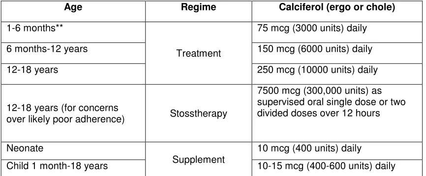
Table 1: Treatment and supplementation doses of vitamin D deficient rickets* as per BNFc 2013 Age Regime Calciferol (ergo or chole)
1-6 months**
Treatment
75 mcg (3000 units) daily
6 months-12 years 150 mcg (6000 units) daily
12-18 years 250 mcg (10000 units) daily
12-18 years (for concerns
over likely poor adherence) Stosstherapy
7500 mcg (300,000 units) as supervised oral single dose or two divided doses over 12 hours
Neonate
Supplement
10 mcg (400 units) daily
Child 1 month-18 years 10-15 mcg (400-600 units) daily
* Following completion of 8-12 weeks treatment children should receive a supplementation dose (table 2) until completion of linear growth.
**Babies receiving 500 mls or more of formula milk per day do not require supplementation following treatment.
Table 2: Summary of global supplementation advice
Supplemented group Advising body Advice
Pregnant and
DoH 2012 104 All pregnant and breastfeeding women to take
[image:41.842.65.507.127.304.2](CG62)
APA 2008 96 Measure maternal vitamin D status and
supplement if found to be “insufficient”
WHO 2012* 108 Call for rigorous RCTs to evaluate the benefit and safety of routine vitamin D
supplementation in pregnancy Canadian
Paediatric Society 2007 109
50 g/day for pregnant and lactating women,
especially during winter months
Institute of Medicine 2010 51
Recommended dietary allowance - 15 mcg/day. Upper level intake 100 mcg/day
Breastfed infants
DoH 2012 104 If mother has not taken supplements throughout pregnancy baby may need to receive drops from one month of age
Canadian
Paediatric Society 2007 109
5 g/day (200 IU/day) for premature infants, 10
g/day (400 IU/day) until the first birthday but 20 g/day (800 IU/day) during winter and for
those living at more northern latitudes than the 55th parallel or between 40-55th parallel with additional risk factors.
Institute of Medicine 2010 51
10 g/day (400 IU/day) during first year of life
Children and APA 2008
96
6 months until the age of five years
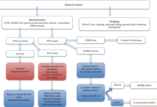
Figure 2: Rickets management algorithm
Clinical rickets
Imaging
(Wrist X‐ray: cupping, splaying, fraying, growth plate widening, osteopenia)
Biochemistry
(PTH, 25OHD, ALP, paired serum and urine calcium + phosphate, LMW protein)
PTH raised PTH not raised
ALP low
25OHD low Vitamin D deficiency
25OHD not low
Retake history: Any recent vitamin D/sunlight exposure?
Consider vitamin D pathway defect:
Check 1,25‐OH2‐D
HVDRR defect Raised
Consider disorders of
phosphate homeostasis e.g. XLH, HHRH etc
(see panel)
Measure fasting TMP/GFR, urine
ALP raised
Consider hypophosphatasia
Measure vitamin B6 and urine