0022-538X/95/$04.0010
Copyrightq1995, American Society for Microbiology
Vesicular Stomatitis Virus Glycoprotein Mutations That Affect
Membrane Fusion Activity and Abolish Virus Infectivity
BRENDA L. FREDERICKSENANDMICHAEL A. WHITT*
Department of Microbiology and Immunology, University of Tennessee, Memphis, Memphis, Tennessee 38163
Received 3 October 1994/Accepted 16 November 1994
We have introduced amino acid substitutions into two regions of the extracellular domain of the vesicular stomatitis virus (VSV) glycoprotein (G protein) and examined the effect of these mutations on protein transport, low-pH-induced stability of G protein oligomers, and membrane fusion activity. We suggested previously that the region between amino acids 118 and 139 may be important for the membrane fusion activity of G protein, on the basis of the characterization of a fusion-defective G protein mutant (M. A. Whitt, P. Zagouras, B. Crise, and J. K. Rose, J. Virol. 64:4907–4913, 1990). It has also been postulated by others that this region as well as the region between amino acids 181 and 212 may constitute putative internal fusion domains of VSV G protein. In this report, we show that three different amino acids substitutions between residues 118 and 139 (G-1243E, P-1273D, and A-1333K) either altered or abolished low-pH-dependent membrane fusion activity. In contrast, substitutions between residues 192 and 212 resulted either in G proteins that had wild-type fusion activity or in mutant proteins in which the mutation prevented transport of G protein to the cell surface. Two of the substitutions between residues 118 and 139 (G-1243E and P-1273D) resulted in G proteins that were fusion defective at pH 5.7, although syncytia were observed after cells were treated with fusion buffer at pH 5.5, albeit at levels significantly less than that induced by wild-type G protein. Interestingly, when either G-1243E or P-1273D was incorporated into tsO45 virions, the resulting particles were not infectious, presumably because the viral envelope was not able to fuse with the proper intracellular membrane. These results support the hypothesis that the region between amino acids 118 and 139 is important for the membrane fusion activity of VSV G protein and may constitute an internal fusion domain.
Vesicular stomatitis virus (VSV) is an enveloped, negative-strand RNA virus which enters the host cell through receptor-mediated endocytosis (23). The glycoprotein (G protein) of VSV, which is located in the viral envelope, is responsible both for binding of virus to the cell surface and for fusion of the viral envelope with the endosomal membrane after endocytosis (9, 10, 26, 31). This fusion event releases the viral nucleocapsid into the cytoplasm of the cell and is required for VSV to initiate an infection. It is thought that G protein undergoes a pH-dependent conformational change (6), and this conforma-tional change is believed to be required for G protein to cat-alyze fusion of the viral envelope with the endosomal mem-brane (2, 22). However, the regions of G protein involved in the conformational changes and in the actual membrane fusion event have not been definitively identified.
Although membrane fusion is required for all enveloped virus to enter cells and cause infection, only in the case of influenza virus hemagglutinin (HA) are the processes leading up to the fusion event understood at the molecular level. Mem-brane fusion is believed to result from the interaction of a specific region of HA, called the fusion peptide or fusion do-main, with the target membrane (14). During transport, HA is
proteolytically cleaved to generate two subunits, HA1and HA2
(15, 17). The fusion peptide is located at the amino terminus of
the HA2subunit and at neutral pH is sequestered within the
HA trimer (40). Recently, it has been shown that HA under-goes a massive structural rearrangement after exposure to
acidic pH and that this conformational change moves the fu-sion peptide over 100 Å (10 nm), positioning it so that it may interact with the target membrane (4).
For noncleaved fusion proteins, such as VSV G, the identi-fication of a fusion domain has proven more difficult. In gen-eral, the amino termini of noncleaved fusogenic proteins are not particularly hydrophobic, and it is thought that these pro-teins have internal fusion domains. The current evidence for the existence of internal fusion domains is based on the char-acterization of mutant proteins that have altered fusion activ-ities (19, 20, 38, 41). Internal fusion peptides are currently defined as regions of 16 to 17 apolar amino acids which are bounded by a positively charged amino acid on the N-terminal side and a negative or positively charged residue on the C-terminal side (33). These regions are relatively hydrophobic and are often rich in alanine and glycine residues. Lastly, internal fusion domains are thought to contain helix-breaking residues at their centers (1, 33).
We previously described the characterization of a G protein mutant that contained a single amino acid substitution which results in the addition of an extra glycosylation site at amino acid 117. This mutant (QN-1) was transported to the cell sur-face normally but did not induce membrane fusion (38). We suggested that the extra oligosaccharide side chain is located near a region important for the membrane fusion activity of G protein and that the additional oligosaccharide may block the interaction of this region with the host cell membrane. Because the region between amino acids 118 and 139 is conserved between VSV serotypes, we and others suggested that this region may be a potential internal fusion domain for VSV G protein (25, 38). Recent evidence using site-directed mutagen-esis of this region supports this suggestion (41).
To further investigate the importance of the region between
* Corresponding author. Mailing address: 858 Madison Ave., De-partment of Microbiology and Immunology, University of Tennessee, Memphis, Memphis, TN 38163. Phone: (901) 448-4634. Fax: (901) 448-8462. Electronic mail address: [email protected]. EDU.
1435
on November 9, 2019 by guest
http://jvi.asm.org/
residues 118 and 139 in the membrane fusion activity of G protein, we constructed several new G protein mutants that have single amino acid substitutions within this region and then examined the effects of these mutations on the membrane fusion activity of G protein. We also constructed single amino acid substitutions in the region between amino acids 192 and 212, which others have postulated may also be important for the membrane fusion activity of VSV G protein (25).
The G protein mutants that are described in this report were grouped into four classes based on their fusion phenotypes. The first group of mutant G proteins had fusion activities similar to that of wild-type G protein. This class included all amino acid substitutions made in the region from amino acids 192 to 212 and many of the mutations in the region between amino acids 118 and 139. The second class of mutants had slightly lower pH thresholds for fusion but still fused like wild-type G protein at a pH of 5.7. The third class of mutants, which
included G-1243E and P-1273D, had a significantly reduced
pH threshold for fusion. Even at pH 5.2, these proteins had fusion activities significantly less than that of wild-type G
pro-tein. Despite the observation that G-1243E and P-1273D
were able to induce membrane fusion to a limited extent, these mutant G proteins were unable to initiate infection of cells when incorporated into tsO45 particles. The last class, which
consisted of a single mutant G protein (A-1333K), was
com-pletely defective for fusion activity at all pHs tested. Our re-sults and those of Zhang and Ghosh (41) support our earlier hypothesis that the region between amino acids 118 and 139 is important for the membrane fusion activity of VSV G protein and may constitute an internal fusion domain.
MATERIALS AND METHODS
Cell culture, viruses, transfections, and rescue assay.HeLa cells were grown in high-glucose Dulbecco’s modified Eagle medium (DMEM) containing 10% iron-supplemented calf serum, 100 U of penicillin per ml, and 100mg of strep-tomycin per ml. Baby hamster kidney (BHK-21) cells were grown in DMEM containing 5% fetal bovine serum, 100 U of penicillin per ml, and 100mg of streptomycin per ml. Stocks of tsO45 and the recombinant vaccinia virus vTF7-3 (13) were prepared as described previously (36). Cell transfections were per-formed as described previously (35), using a suspension of liposomes composed of dimethyldioctadecyl ammonium bromide (DDAB) andL-a
-dioleoylphosphati-dylethanolamine (DOPE) at a weight ratio of 1:2.5 (29). Rescue assays were performed as described previously (36) except that all transfections were per-formed with DDAB-DOPE liposomes.
Plasmids and oligonucleotide-directed mutagenesis.The cDNA for the G protein of the Indiana serotype (San Juan strain) of VSV was recovered from pAR-G (36) and cloned into pBluescript-SK1(pBS-SK1; Stratagene) between the XhoI and BamHI sites such that G protein expression could be driven by the bacteriophage T7 promoter. This construct is called BS-G and contains EcoRI sites immediately flanking both ends of the G protein cDNA and an additional XhoI site adjacent to the EcoRI site at the 39end of the cDNA. These additional restriction sites were present in pSVGL1 (28), which was the parent plasmid of pAR-G. Therefore, the restriction sites in BS-G beginning at the 59XhoI site in the pBS-SK1polylinker are in the following order: XhoI-EcoRI-G protein cDNA-EcoRI-XhoI-BamHI. All of the mutated G protein cDNAs described below were constructed by using either pBS-SK1or pBS-KS1as the vector. In addition, the mutant G protein cDNAs that were used in the rescue assay were also inserted into pAR-X (36), which is derived from the T7 expression vector pET-3 (30). All mutations that resulted in amino acid substitutions were intro-duced into the G protein cDNA by subcloning a PCR-amplified fragment into the wild-type G protein cDNA backbone. Sequences of the oligonucleotides and the conditions used for PCR amplification for the individual clones are available by request.
For substitutions at amino acids 119 to 127, PCR-amplified fragments were generated by using (i) antisense, mutagenic oligonucleotides which contained the appropriate nucleotide changes for the indicated amino acid substitutions and that spanned the unique SfaNI site in the G protein cDNA and (ii) an oligonu-cleotide complementary to the T7 promoter. The template for these PCR am-plifications was pAR-G. The amplified fragments were extracted with phenol-chloroform, precipitated with ethanol, and resuspended in TE buffer (10 mM Tris, 1 mM EDTA [pH 8.0]) prior to digestion with SfaNI and BamHI. The digested fragments were gel purified and then used to replace the corresponding fragment from the wild-type G protein cDNA clone in a three-way ligation. The
components for each ligation were BS-KS1digested with BamHI and XhoI, the PCR-amplified fragment digested with BamHI and SfaNI, and a fragment from the wild-type G protein cDNA clone containing sequences from the SfaNI site to the XhoI site at the 39 end of pAR-G. Mutations were confirmed by dideoxynucleotide sequencing of several individual clones for each of the mutant constructs, using Sequenase (United States Biochemical Corp.). The sequence of the entire region that was amplified and used in the ligation was determined for all of the mutants to confirm that the PCR-generated fragment contained the desired mutation only and that no other mutations were introduced during the amplification. Variations of the strategy described above were used to obtain cDNAs encoding the other amino acid substitutions listed in Table 1. The following descriptions detail these variations. For substitutions at amino acids 128 to 133, PCR-amplified fragments consisting of the 59portion of the G protein cDNA were used to replace the corresponding region from the wild-type G protein cDNA in a three-way ligation which consisted of pBS-SK1digested with EcoRI and BamHI, the PCR fragment digested with EcoRI and SfaNI, and the remaining 39region of the wild-type G protein cDNA from BS-G digested with SfaNI and BamHI. The mutation at amino acid 137 was introduced into the wild-type G protein cDNA backbone by amplifying a fragment by using BS-G as the template, a sense-strand mutagenic oligonucleotide that contained a new AvaI site at nucleotide 396, and an antisense, nonmutagenic oligonucleotide that overlapped the unique NcoI site in the G protein cDNA. The amplified fragment was digested with AvaI and NcoI and then used in a three-way ligation to regenerate the intact G protein cDNA. The 59portion of this clone was derived from the mutant G-1313A, which contained, in addition to the nucleotide changes at 421 (G to C) for the substitution of glycine 131 to alanine, nucleotide changes at positions 398 (A to C) and 401 (C to G) that resulted in the intro-duction of the new AvaI site. These nucleotide changes were silent and did not result in any additional amino acid changes. This strategy was necessary because the codon specifying amino acid 137 is part of the recognition/cleavage site for SfaNI. For mutations at amino acids 139 and 147, the region between the SfaNI and NcoI sites was amplified by using sense-strand, mutagenic oligonucleotides that overlapped the SfaNI site and an antisense, nonmutagenic oligonucleotide that contained sequences overlapping the NcoI site. The PCR-generated frag-ments containing the mutations were digested with SfaNI and NcoI and then used in ligations with BS-G digested with ApaI and NcoI plus an ApaI-to-SfaNI fragment corresponding to the 59end of the wild-type G protein cDNA. The substitutions at amino acids 192 and 197 were generated by using an antisense, mutagenic oligonucleotide that overlapped the NcoI site and a nonmutagenic, sense-strand oligonucleotide that overlapped the SfaNI site. The amplified frag-ments were used to replace the corresponding fragment in a three-way ligation containing BS-G digested with ApaI and NcoI, the ApaI-to-SfaNI fragment also from BS-G, and the PCR-amplified region digested with SfaNI and NcoI. Sub-stitutions at amino acids 203 to 212 were introduced on a fragment generated by using a sense-strand, mutagenic oligonucleotide that spanned the NcoI site and an antisense, nonmutagenic oligonucleotide that overlapped the unique PstI site in the G protein cDNA. The corresponding wild-type G fragment was replaced by using a 59ApaI-to-NcoI fragment from BS-G, the PCR-amplified NcoI-to-PstI fragments that contained the appropriate mutations, and a vector derived from BS-G that was digested with ApaI and PstI.
Surface expression and syncytium formation assays.Duplicate plates of HeLa cells were rinsed two times in serum-free DMEM (SF-DMEM) and infected with vTF7-3 at a multiplicity of infection of 10 for 30 min at 378C. The inoculum was removed, and the cells were transfected with plasmids encoding either wild-type or mutant G proteins as described previously (35) or were incubated in SF-DMEM without DDAB-DOPE liposomes (mock transfected). At 7 h posttrans-fection, cells from one set of duplicate plates were processed for flow cytometry as described previously (38). To examine low-pH-induced membrane fusion activity, cells from the other set of duplicate plates were rinsed once with fusion medium [10 mM Na2HPO4, 10 mM NaH2PO4, 150 mM NaCl, 10 mM
2-(N-morpholino)ethanesulfonic acid (MES), 10 mM N-2-hydroxyethylpiperazine-N9 -2-ethanesulfonic acid (HEPES); titrated to the indicated pH with HCl] and then bathed for 1 min in fresh fusion medium at room temperature. After 1 min of incubation, the fusion medium was replaced with DMEM containing 10% calf serum and the plates were returned to a 378C, 5% CO2incubator for 20 min.
Following the 20-min incubation, the medium was removed and ice-cold phos-phate-buffered saline containing 0.9 mM CaCl2and 0.5 mM MgCl2(PBS1) was
added. The cultures were maintained at 48C to prevent further cell-cell fusion until individual cultures could be examined for syncytium formation by using an inverted light microscope and phase-contrast optics. After the addition of ice-cold PBS1, the number of syncytia and the number of single (unfused) cells were counted in three to five separate fields containing approximately 300 cells per field for each of the mutants and for parallel cultures of cells that were either mock transfected or were expressing wild-type G protein. Fusion activities were quantitated by determining the percentage of fused cells per field for each of the mutants. The percentage of fused cells was determined by first subtracting the number of syncytia containing three or more nuclei present in the mock-trans-fected cultures from the number of syncytia seen in the cells expressing either wild-type G protein or the G protein mutants. This value represents the cor-rected number of fused cells (fusedcorr) and excludes any background fusion
resulting from spontaneously fused cells or from cells that may have fused as a result of the activity of the vaccinia virus fusion protein (5, 27). The percentage
1436 FREDERICKSEN AND WHITT J. VIROL.
on November 9, 2019 by guest
http://jvi.asm.org/
of fused to unfused cells was then determined by calculating the ratio of fusedcorr
to unfused cells in each field. The percent cells fused represents the average value obtained from each of the fields.
Indirect immunofluorescence was performed essentially as described previ-ously (28) except that the primary antibody was the G protein-specific monoclo-nal antibody I1 (18) and the secondary antibody was either fluorescein isothio-cyanate- or lissamine rhodamine sulfonyl chloride-labeled, affinity-purified donkey anti-mouse antibody (Jackson ImmunoResearch Laboratories, Inc.).
Metabolic labeling, transport kinetics, and trimer stability assays.HeLa cells expressing either wild-type or mutant G proteins were rinsed once with methi-onine-free SF-DMEM (Met-free DMEM) at 3 to 4 h posttransfection, incubated for 10 min in Met-free DMEM, and then radioactively labeled for various amounts of time in 1 ml of Met-free DMEM containing sufficient Express protein labeling mix (DuPont/New England Nuclear) to give 50mCi of [35
S]me-thionine per ml. Following the pulse, the radioactive labeling medium was re-moved, and either the cells were lysed immediately by adding 1 ml of detergent lysis buffer (10 mM Tris [pH 7.4], 66 mM EDTA, 1% Triton X-100, 0.4% deoxycholic acid, 0.02% sodium azide) or the labeling medium was replaced with DMEM containing 10% calf serum and 2 mM additional methionine (chase medium). Immunoprecipitation was performed essentially as described previ-ously (28) except that the postnuclear cell lysates were made to 0.3% sodium dodecyl sulfate (SDS) and the antigen-antibody complexes were formed for 30 min at 378C. Digestion of immunoprecipitates with endoglycosidase (endo) H was performed as instructed by the manufacturer (New England Biolabs).
Trimer stability was examined by lysing radioactively labeled cells with 1% Triton X-100 in 23MNT buffer [40 mM 2-(N-morpholino)ethanesulfonic acid (MES), 60 mM Tris, 200 mM NaCl, 2.5 mM EDTA] at the pH indicated as described previously (34). Cell lysates were centrifuged on 5 to 20% sucrose gradients as described previously (34). The fractions were immunoprecipitated with polyclonal anti-VSV serum and electrophoresed through 10% polyacryl-amide gels containing SDS (16), and the resolved proteins were visualized fol-lowing fluorography (3).
RESULTS
To identify regions that are important for the membrane fusion activity of G protein, we introduced single amino acid substitutions into the extracellular domain of G protein, using site-directed mutagenesis of the G protein cDNA. We chose two regions in the extracellular domain that have been sug-gested previously to be important for the membrane fusion activity of G protein to begin our mutagenesis study (25, 38). The first region includes amino acids 118 to 139, which we had proposed might constitute a putative internal fusion domain on the basis of the characterization of a fusion-defective mutant of G protein called QN-1 (38). The second region, which spans amino acids 181 to 212, was suggested by others because this region has properties expected for putative internal fusion domains (25). The rationale for the types of amino acid sub-stitutions that were made was based on the premise that in-troduction of charged residues into a domain that is involved in membrane fusion should affect the ability of that domain to interact with a target membrane and subsequently alter or abrogate the fusion activity of the mutant G protein. Because several putative internal fusion domains of other fusion pro-teins have helix-breaking residues in the middle of the domain (1, 33), we also replaced proline residues with either alanine, glycine, or various charged amino acids. Table 1 lists the po-sitions of the amino acids in G protein that were changed and the substitutions that were made.
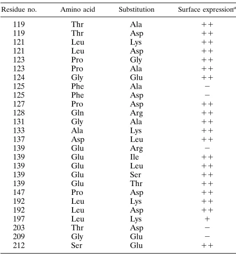
Surface expression of mutant G proteins.Before we could examine whether the mutations listed in Table 1 alter the membrane fusion activity of G protein, we needed to first determine if the mutant proteins were transported to the cell surface. To assay for surface expression, we examined cells expressing each of the mutant proteins by indirect immunoflu-orescence. Most of the mutant G proteins were expressed on the cell surface, and the majority of these had signal intensities similar to that of cells expressing wild-type G protein (Table 1).
One mutant (L-1973K) was found on the surface at levels
lower than that of wild-type G protein, while five of the mu-tants were not detected on the cell surface by
immunofluores-cence. Because our assays for membrane fusion activity require transport to the plasma membrane, mutant proteins that were not detected on the cell surface were not examined further.
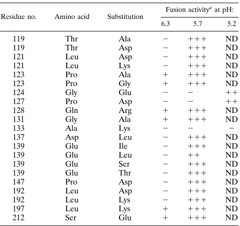
Membrane fusion activity of mutant G proteins.We initially examined the effects of the amino acid substitutions on the membrane fusion activity of G protein by using a syncytium formation assay. Cell fusion occurs when cells expressing wild-type G protein are incubated briefly in acidic pH buffer. The pH threshold for fusion activity of wild-type G protein is ap-proximately 6.3 (11, 24, 31), and cells expressing G protein form extensive syncytia after treatment with pH 5.7. Cells ex-pressing either wild-type G protein or individual G proteins mutants were treated with fusion buffer at pH 6.3, 5.7, or 5.2, and the cultures were examined for fused cells (Table 2). For some experiments, the amount of plasmid encoding wild-type G protein that was transfected was varied so that wild-type G protein was expressed on the cell surface at levels similar to those of the mutant G proteins. Fusion activities were deter-mined by counting the number of syncytia formed after incu-bating cells expressing equivalent levels of either the mutant or wild-type G proteins at a given pH.
We found that the mutants could be grouped into four classes based on their ability to induce syncytia after incubation in fusion buffer at pH 6.3, 5.7, or 5.2. The first class are mutants which had fusion activities similar to that of wild-type G pro-tein. All of the mutants in the region between amino acids 192 and 212 belonged to this class, as did most of the mutants having amino acid changes between residues 118 and 139. The second class of mutants consists of those that had slightly
reduced fusion activities at pH 5.7. The mutant E-1393L
[image:3.612.315.554.90.348.2]represents this class of G protein, which appeared to have a
TABLE 1. Amino acid substitutions and surface expression of G protein mutants
Residue no. Amino acid Substitution Surface expressiona
119 Thr Ala 11
119 Thr Asp 11
121 Leu Lys 11
121 Leu Asp 11
123 Pro Gly 11
123 Pro Ala 11
124 Gly Glu 11
125 Phe Ala 2
125 Phe Asp 2
127 Pro Asp 11
128 Gln Arg 11
131 Gly Ala 11
133 Ala Lys 11
137 Asp Leu 11
139 Glu Arg 2
139 Glu Ile 11
139 Glu Leu 11
139 Glu Ser 11
139 Glu Thr 11
147 Pro Asp 11
192 Leu Lys 11
192 Leu Asp 11
197 Leu Lys 1
203 Thr Asp 2
209 Gly Glu 2
212 Ser Glu 11
a
Determined by indirect immunofluorescence on HeLa cells that had been transfected with 5mg of the indicated plasmids and fixed at 7 h posttransfection.
11, fluorescence intensity similar to that of cells expressing wild-type G protein;
1, detectable surface expression, but signal is less than that for cells expressing wild-type G protein;2, no surface expression.
on November 9, 2019 by guest
http://jvi.asm.org/
slightly reduced pH threshold for fusion. The third class of
mutants is represented by G-1243E and P-1273D, which
were fusion defective at pH 5.7. However, cells expressing
G-1243E or P-1273D did form syncytia after treatment at
pH 5.2, albeit at levels significantly less than that of wild-type G protein. These mutants were classified as pH-shift fusion mutants, indicating that they have a greatly reduced pH thresh-old for fusion activity. The fourth class consisted of the mutant
A-1333K, which was fusion defective at all pHs tested.
To examine the pH dependence for fusion activity of the
mutants G-1243E, P-1273D, and A-1333K in greater detail,
we compared the fusion activities of these mutants with that of wild-type G protein at pHs between 6.3 and 5.2. Because the extent of syncytium formation in these cultures is dependent on both the number of cells expressing G protein and the amount of G protein on the cell surface, we used flow cytom-etry to determine the relative level of surface expression and the number of cells expressing each of the G protein mutants. Relative fusion activities are presented as the percentage of cells fused in the culture for each of the proteins at the pH indicated (Fig. 1). In all of these experiments, the cells were expressing equivalent levels of either wild-type or mutant G
proteins. The results confirm that neither G-1243E nor
P-1273D induces syncytium formation until the pH is below
5.7, which indicates that the pH threshold for both of the
mutants is between 5.7 and 5.5. In addition, neither G-1243E
nor P-1273D induces cell-cell fusion to the extent that G
protein does. Cultures of cells expressing the mutant
A-1333K had less than 5% fused cells after treatment with
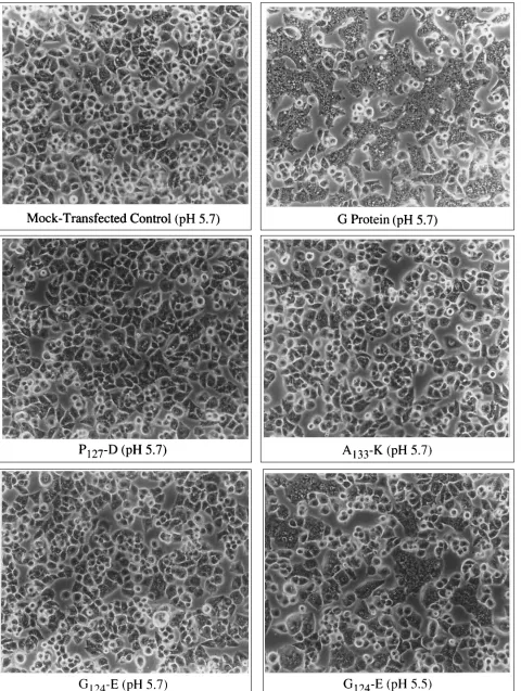
fusion medium at all pHs examined. This level of fusion was equivalent to the background fusion seen in cells that were infected with the recombinant vaccinia virus vTF7-3 and mock transfected only. Figure 2 shows examples of a typical cell fusion assay for the three fusion-defective mutants, for
wild-type G protein and a nontransfected control culture at pH 5.7,
and for G-1243E at pH 5.5.
Folding and transport of G protein fusion mutants. To determine the basis for the altered fusion activities exhibited
by G-1243E, P-1273D, and A-1333K, we examined several
different parameters that might affect the fusion activity of these mutant G proteins. These parameters include oligomer-ization, trimer stability, and transport kinetics. To examine
whether G-1243E, P-1273D, and A-1333K were
trans-ported out of the endoplasmic reticulum (ER) properly, we determined the rate at which these proteins acquired endo H-resistant oligosaccharides. Acquisition of endo H-resistant sugars indicates that G protein has been transported to the medial compartment of the Golgi apparatus (8), and this assay allows us to measure the rate and extent of transport of G protein from the ER to the Golgi apparatus.
Both G-1243E and P-1273D were transported with
kinet-ics similar to that of wild-type G protein (Fig. 3). These results indicated that neither of the substitutions adversely affected
the folding or transport of the mutants. In contrast, A-1333K
was transported to the Golgi apparatus more slowly than wild-type G protein, and only 75% of the pulse-labeled material became endo H resistant after 2 h. This result suggested that
A-1333K may have difficulty attaining the proper
conforma-tion for transport; however, results from the flow cytometric
analysis showed that A-1333K did accumulate on the cell
surface, but to levels less than that of wild-type G protein when cells were transfected with equal amounts of plasmid DNA (data not shown).
pH-dependent trimer stability of G protein fusion mutants.
Wild-type G protein trimers dissociate during centrifugation in sucrose gradients buffered to a pH of 6.3 or higher (6). Because the pH at which trimers dissociate correlates very closely with the pH threshold for the membrane fusion activity of G pro-tein, trimer stability is thought to be a measure of the confor-mational change required for induction of the membrane fu-sion activity of G protein (6).
To examine the pH-dependent stability of the mutant G
protein trimers, cells expressing either G-1243E, P-1273D,
A-1333K, or wild-type G protein were detergent solubilized at
[image:4.612.316.556.71.243.2]the pH indicated, and the lysates were centrifuged through sucrose gradients having the same pH as the lysis buffer. The
FIG. 1. pH dependence of G protein membrane fusion activity. HeLa cells expressing equivalent amounts of either wild-type G protein (wt-G) or mutant G proteins, or that were infected with vTF7-3 only (mock), were treated with fusion medium at the pH indicated, and the percentage of fused cells in the culture was determined as described in Materials and Methods.
TABLE 2. Fusion activities of G protein mutants at pHs 6.3, 5.7, and 5.2
Residue no. Amino acid Substitution
Fusion activityaat pH:
6.3 5.7 5.2
119 Thr Ala 2 111 ND
119 Thr Asp 2 111 ND
121 Leu Asp 2 111 ND
121 Leu Lys 2 111 ND
123 Pro Ala 1 111 ND
123 Pro Gly 1 111 ND
124 Gly Glu 2 2 11
127 Pro Asp 2 2 11
128 Gln Arg 1 111 ND
131 Gly Ala 1 111 ND
133 Ala Lys 2 2 2
137 Asp Leu 2 111 ND
139 Glu Ile 2 111 ND
139 Glu Leu 2 11 ND
139 Glu Ser 2 111 ND
139 Glu Thr 2 111 ND
147 Pro Asp 2 111 ND
192 Leu Asp 2 111 ND
192 Leu Lys 2 111 ND
197 Leu Lys 1 111 ND
212 Ser Glu 1 111 ND
a
Calculated by determining the ratio of fused to unfused cells in three to five separate fields containing at least 300 cells per field for each experiment. The percentage of fused cells for each mutant is represented as follows:2,,5% fused cells per field;1, 5 to 25% fused cells per field;11, 25 to 70% fused cells per field;111,.70% fused cells per field; ND, not determined.
1438 FREDERICKSEN AND WHITT J. VIROL.
on November 9, 2019 by guest
http://jvi.asm.org/
[image:4.612.58.299.90.317.2]FIG. 2. Syncytium formation in cells expressing wild-type or mutant G proteins. HeLa cells from the experiment presented in Fig. 1 were photographed by using phase-contrast optics. The pH of the fusion medium is indicated in parentheses, and the G protein that was expressed is indicated below each photomicrograph. The mock-transfected control shows the amount of background fusion resulting from treatment of vTF7-3-infected cells with fusion medium at pH 5.7.
1439
on November 9, 2019 by guest
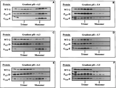
dissociation profiles for both P-1273D (Fig. 4) and A-1333K (not shown) were identical to that of wild-type G protein. This
result indicated that P-1273D and A-1333K were properly
folded and presumably undergo conformational changes sim-ilar to that of wild-type G protein. In contrast, trimers of
G-1243E were significantly less stable to centrifugation in
sucrose gradients at pH 6.3. Monomers were the predominate
form that was recovered from gradients containing G-1243E
at pH 6.3, whereas the majority of wild-type G and P-1273D
were trimers at this pH (Fig. 4C). Even at pH 6.1, at which both
wild-type G and P-1273D were predominantly trimers,
G-1243E was found in the monomer fraction (Fig. 4E). These
results suggested that G-1243E undergoes the conformational
change required for trimer stability at a significantly lower pH
that either wild-type G protein or P-1273D. Alternatively, the
difference in trimer stability may simply reflect an increased requirement for protonation of the acidic residue at position 124 in order to prevent trimer dissociation during centrifuga-tion.
Assembly and function of fusion-defective G proteins in virions.The second assay that we used to examine the fusion activity of mutant G proteins relies on the ability of the mutant proteins to assemble into virus particles. To accomplish this, we utilized an assay in which expression of either wild-type or transport-competent mutant G proteins is used to rescue the temperature-sensitive mutant tsO45 (34, 36–38). Because in-fectivity of the rescued particles depends on the fusion activity of the incorporated G protein, this assay provides a sensitive means for determining whether a mutant G protein is biolog-ically active.
To examine whether G-1243E, P-1273D, or A-1333K
could function in the viral envelope and bring about cell in-fection after complementation of tsO45, we first needed to determine if these proteins were transported to the cell surface at the nonpermissive temperature for tsO45. Flow cytometry was used to compare the relative levels of surface expression for each of the mutants with that of wild-type G protein at the
restrictive temperature (40.58C). We found that both
G-1243E and P-1273D were expressed at levels equivalent to
that of wild-type G protein at 40.58C; however, A-1333K was
not expressed on the cell surface at this temperature (data not shown). Because incorporation into virions requires expression
of G protein on the cell surface, A-1333K could not be
ex-amined by this assay. We also exex-amined whether the two mu-tants retained their low-level cell-cell fusion activities when expressed at the restrictive temperature. Although both
G-1243E and P-1273D were expressed on the cell surface at
40.58C, no syncytia were detected after treatment with fusion
medium at either pH 5.5 or pH 5.2. This lack of fusion activity may be due slight differences in the conformation of the pro-teins when expressed at the restrictive temperature, although these differences were not significant enough to affect trans-port to the cell surface.
To determine whether G-1243E or P-1273D could
assem-ble and function in virus particles, we used the tsO45 rescue assay described previously (36). We found that neither
G-1243E nor P-1273D could rescue tsO45 infectivity when
assayed by the ability to form plaques at the permissive
tem-perature of 328C (Table 3). The lack of infectivity was not due
to the inability of either of the mutants to assemble into the
viral envelope, since both G-1243E and P-1273D were
in-corporated into virions as efficiently as wild-type G protein (Fig. 5). These results demonstrated that both mutants were fusion defective when expressed and assembled into tsO45 virions at the restrictive temperature and showed that neither
G-1243E nor P-1273D could induce fusion of the viral
en-velope with the proper intracellular membrane and initiate a productive infection.
DISCUSSION
The region between residues 118 and 139 as well as the region between amino acids 181 and 212 have been postulated to be involved in the membrane fusion activity of VSV G protein (20, 25, 38). Both of these regions have properties that are thought to characterize internal fusion domains (32), and mutations in both of these regions have previously been shown to alter the membrane fusion activity of G protein (20, 38, 41). To investigate the role of these regions in the membrane fusion activity of VSV protein further, we constructed several cDNAs for mutant G proteins that have single amino acid substitutions within both regions and then examined the effects of these substitutions on membrane fusion activity. The majority of the substitutions introduced charged residues into these regions. We postulated that if either of these regions interact directly with the host cell membrane as a prerequisite to or during the fusion event, the introduction of a charged residue may inhibit the ability of G protein to induce membrane fusion. Internal fusion domains have been postulated to contain helix-breaking residues near their centers (1, 33). The region between amino acids 118 and 136 contains prolines at amino acid 123, 126, and 127. We therefore replaced all three of these residues with either aspartic acid, glycine, or alanine. We also made several conservative substitutions in which residues were replaced with amino acids that have similar properties. These conservative substitutions were made to examine the possibility that a spe-cific amino acid at a spespe-cific position is essential for membrane fusion activity.
All of the mutant G proteins were first examined for trans-port to the cell surface. Not surprisingly, some of the mutations resulted in G proteins which were not transported to the cell surface; therefore, these mutants could not be analyzed for defects in membrane fusion activity. The majority of mutants were transport competent and were grouped into four classes
FIG. 3. Transport kinetics of wild-type and mutant G proteins. Cells express-ing either wild-type G protein (wt G), G-1243E, P-1273D, or A-1333K were radioactively labeled with [35S]methionine for 15 min at 3 h posttransfection and
then chased for the times indicated in medium containing excess methionine. The G proteins were immunoprecipitated from cell lysates, and one-half of the immunoprecipitate was digested with endo H. Endo H-resistant and -sensitive forms of G protein were resolved by SDS-polyacrylamide gel electrophoresis and visualized following fluorography. The amount of G protein that had acquired endo H-resistant oligosaccharides was determined by image analysis of fluoro-grams, using a Millipore BioImage analyzer.
1440 FREDERICKSEN AND WHITT J. VIROL.
on November 9, 2019 by guest
http://jvi.asm.org/
based on the effect that the amino acid substitution had on fusion activity of the mutant G protein. Class 1 mutants were defined as those substitutions which did not significantly alter the ability of G protein to induce syncytium formation com-pared with wild-type G protein. The majority of the mutants fell into this category. Importantly, all of the substitutions made between amino acids 192 and 212 that were expressed on the cell surface had wild-type G fusion activity. Although it was reported previously that a mutant G protein having an amino acid insertion between residues 194 and 195 appeared to have reduced fusion activity (20), it is unlikely that this region is directly involved in membrane fusion because one of the two conserved N-linked glycosylation sites in G protein is located near this region at amino acid 179. The addition of a hydro-philic oligosaccharide at N-179 would likely block this region of G protein from interacting with the host cell membrane. Our results showing that several amino acid substitutions between
residues 192 and 212 did not alter the fusion activity of G protein support the conclusion that this region is probably not involved directly with the membrane fusion activity of G pro-tein.
Members of the second class of mutant G proteins appeared to have a slightly reduced pH threshold for fusion but were still
[image:7.612.113.502.71.368.2]FIG. 4. Stability of G protein trimers to centrifugation through sucrose gradients as a function of pH. HeLa cells expressing either wild-type G protein (wt G), G-1243E, or P-1273D were radioactively labeled for 15 min and then chased for 1 h in the presence of excess methionine. Cells were lysed at the pH indicated, and the lysates were centrifuged through 5 to 20% sucrose gradients buffered to the same pH as the lysis solution. G proteins were recovered from fractions by immunoprecipitation, and each fraction was analyzed by SDS-polyacrylamide gel electrophoresis and fluorography. Only the relevant portion of the fluorogram is shown. The positions of G protein trimers and monomers are indicated for each set of gradients.
FIG. 5. Incorporation of G protein mutants into tsO45 virions. Labeling conditions for rescued tsO45 particles have been described elsewhere (36). Ra-dioactively labeled virions were centrifuged through a 20% sucrose cushion, and the pellet fraction was resuspended and then immunoprecipitated with poly-clonal anti-VSV serum. Viral proteins were resolved on 10% polyacrylamide gels containing SDS, and the gel was subjected to fluorography. Lanes from the left: VSV markers, particles from cells expressing wild-type G protein (wt G), parti-cles from cells expressing G-1243E, partiparti-cles from cells expressing P-1273D, and particles from mock-transfected cells.
TABLE 3. Titers of virus particles containing fusion-defective G protein mutants
Expressed protein Titer (PFU/ml)a
Wild-type G...2.93104–9.03104
G-1243E... 0–101
P-1273D ... 0–43101
None (mock) ... 0–101
a
Titers from four separate rescue experiments are given. Virus titers were determined on mouse L cells.
on November 9, 2019 by guest
http://jvi.asm.org/
[image:7.612.373.499.526.652.2]capable of inducing syncytium formation equivalent to that of
wild-type G protein at pH 5.7. The E-1393L substitution
typ-ifies this class of mutant G protein. The basis for the reduced fusion activity observed at a pH slightly below the threshold pH for wild-type G protein fusion activity was not fully explored. Protonation of acidic residues in internal fusion domains is thought either to contribute to the conformational change re-quired for fusion activity or to act by increasing the hydropho-bicity of the region. By replacing E-139 with L, we may have affected either one of these properties. A more likely explana-tion is that this mutaexplana-tion has altered the conformaexplana-tion of G protein slightly such that maximal fusion activity is not ob-served near the pH threshold.
The third class of mutant G proteins had significantly altered fusion activities compared with wild-type G protein. The
pro-line-to-aspartic acid substitution at amino acid 127 (P-1273D)
and glycine-to-glutamic acid substitution at amino acid 124
(G-1243E) belong to this class. Both of these G proteins did
not induce syncytium formation at pH 5.7; however, syncytia were observed when cells expressing the mutants were treated with fusion buffer at pH 5.2. To determine whether substitu-tion of other prolines also altered the fusion activity of G protein, we exchanged the proline at amino acid 147 with an aspartic acid and the proline at amino acid 123 with either
alanine or glycine. In contrast to the P-1273D mutation,
sub-stituting the prolines at amino acids 123 and 147 had no effect on the fusion activity of G protein. These results indicate that
the altered fusion activity of P-1273D is specific for the
pro-line residue at amino acid 127. Our results for G-1243E and
P-1273D support the results of Zhang and Ghosh, who also
showed that substitutions at amino acids 124 and 127 altered the fusion activity of G protein (41). In addition, we obtained one mutant, an alanine-to-lysine substitution at amino acid 133
(A-1333K), that was completely fusion defective at all pHs
tested. This protein constituted the fourth class of mutants in our study.
One explanation for the altered fusion activities G-1243E,
P-1273D, and A-1333K is that the substitutions caused
sub-tle changes in the folding or oligomerization the mutant G proteins. To indirectly examine the extent of misfolding of the mutant G proteins, we determined the rate at which the pro-teins were transported out of the ER. It has been previously shown that misfolded G proteins are retained in the ER until they achieve a transport-competent form (7). Therefore, by comparing the rate of transport of the mutant G proteins with that of wild-type G protein, we can indirectly determine the extent of misfolding of the mutant G proteins within the ER.
We found that both P-1273D and G-1243E were transported
at rates similar to that of wild-type G protein. These results
suggest that neither the P-1273D nor the G-1243E
substitu-tion has a detrimental effect on the initial folding of the mutant
proteins. In contrast, A-1333K was transported more slowly
than wild-type G protein. This result indicated that A-1333K
requires longer to achieve a transport-competent
conforma-tion and suggests that A-1333K may be slightly misfolded.
We were also interested in examining whether the mutant G proteins could undergo the pH-dependent conformational change that is thought to be required for induction of G pro-tein’s membrane fusion activity. Because there are no assays currently available to examine low-pH-induced conformational changes in G protein directly, we examined whether there was any difference in the stability of the mutant G protein trimers at different pHs. It has been shown previously that trimers of G protein dissociate to monomers when centrifuged through neu-tral pH sucrose gradients containing Triton X-100 unless cells lysates are maintained and centrifuged at acidic pHs (6, 21).
There is also a strong correlation between the pH at which G trimers become stable to centrifugation on sucrose gradients and the pH required to induce the fusion activity of G protein (6). An explanation for this observation is that G protein un-dergoes a conformational change at low pH which stabilizes the trimers during centrifugation and this conformational change is required for activation of G protein’s fusion potential (6). Because of this correlation, we used trimer stability to measure potential differences in the pH-dependent
conforma-tional changes of the mutant G proteins. Both A-1333K and
P-1273D had trimer stability profiles identical to that of
wild-type G protein. These results showed that the introduction of either acidic or basic amino acids at these positions did not alter the quaternary structure of G protein significantly. These
results also suggested that trimers of P-1273D and A-1333K
appear to undergo the conformational changes required to stabilize trimers. On the basis of these results, we suggest that the altered fusion activities of both of these mutants are due to the introduction of a charge into a domain directly involved in
membrane fusion. In the case of P-1273D, it appears that this
effect can be partially reversed by lowering the pH below 5.5, although decreasing the pH to 5.2 is not sufficient to restore wild-type fusion activity. The lower pH may partially neutralize the charge on the side chain of aspartic acid, allowing interac-tion between G protein and the host cell membrane.
The G-1243E mutant had a pattern of trimer stability much
different from that of wild-type G protein. The majority of
G-1243E trimers were not stable until pH 5.9, suggesting that
G-1243E does not undergo the conformational change
re-quired for stabilization of trimers and possibly for membrane fusion activity until pH 5.9 or lower. However, our observation
that G-1243E is still not capable of inducing syncytium
for-mation until pH 5.5 indicates that this substitution has an additional effect on fusion activity. As we postulated for
P-1273D, the addition of a charged amino acid at this position
may interfere with the interaction of this region with target
membranes, and as with P-1273D, when the charge is partially
neutralized at lower pHs, syncytium formation does occur to a limited extent. An alternative explanation is that both G-124 and P-127 are essential for fusion activity, possibly by providing a necessary structural motif important for interaction of the fusion domain with target membranes, and that no other amino acids are allowable at these positions. As we had shown previously (12) and as reported by Zhang and Ghosh (41), both G-124 and P-127 are amino acids that are conserved between the putative internal fusion domains of Semliki Forest virus and many of the vesiculoviruses. Therefore, replacing either of these amino acids may affect the fusion activity of G protein. This interpretation appears plausible in light of the results obtained by Zhang and Ghosh, who showed that substitution of G-124 with alanine and P-127 with glycine resulted in G proteins that had altered fusion activities (41).
The initial assay that was used to characterize the mutants described in this study relied on the ability of the mutant G proteins to induce cell-cell fusion. However, the inability to induce syncytium formation does not necessarily correspond to a complete lack of fusion activity (32). In other virus systems, it was shown that some mutant viral fusion proteins which were not capable of inducing syncytium formation were still able to mediate infection of a host cell (39). To examine whether mutant G proteins that do not efficiently induce syncytium formation can function in viral entry, we assembled mutant G proteins into tsO45 particles and examined whether the result-ing virions were infectious. When transiently expressed in
tsO45-infected cells, both P-1273D and G-1243E were
in-corporated into tsO45 particles, but neither was capable of
1442 FREDERICKSEN AND WHITT J. VIROL.
on November 9, 2019 by guest
http://jvi.asm.org/
rescuing tsO45 infectivity. The lack of biological activity, with respect to infectivity, is most likely due to an inability of the mutant G proteins to induce fusion of the viral envelope with an intracellular membrane, since neither of the mutant pro-teins induced cell-cell fusion after incubation at pH 5.5 or
lower when expressed at 40.58C. Alternatively, the lack of
infectivity could result from the inability of the mutant G proteins to bind to the cell surface. Although this is formally possible, our previous results showing that an additional N-linked oligosaccharide at amino acid 117 had no effect on binding to cells (38) suggest that this region can tolerate sig-nificant changes without affecting the binding properties of G protein.
The identification of single amino acid substitutions within the domain defined by amino acids 118 to 136 that alter the low-pH-induced fusion activity of VSV G protein and that prevent virus infectivity strongly suggests that this region is important for the membrane fusion activity of G protein. Al-though we have shown that two of the substitutions appear to be folded properly, we cannot exclude the possibility that these mutations simply alter the conformation of another region that is directly responsible for the fusion event. Until there is a direct demonstration that a single domain in G protein medi-ates membrane fusion, the mechanism responsible for this event will remain uncertain.
ACKNOWLEDGMENTS
We thank Yulia Kovari and Kimberly Prince for synthesizing oligo-nucleotides and Felicia Waller for performing the flow cytometric analysis. Oligonucleotides and flow cytometry facilities were provided by the Molecular Resource Center at the University of Tennessee, Memphis. We also thank Elizabeth Stillman for suggestions after read-ing the manuscript.
This work was supported by grant VM-43 from the American Can-cer Society.
REFERENCES
1. Blobel, C. P., T. G. Wolfsberg, C. W. Turck, D. G. Myles, P. Primakoff, and
J. M. White.1992. A potential fusion peptide and an integrin ligand domain in a protein active in sperm-egg fusion. Nature (London) 356:248–252. 2. Blumenthal, R., A. Bali-Puri, A. Walter, D. Covell, and O. Eidelman. 1987.
pH-dependent fusion of vesicular stomatitis virus with Vero cells. J. Biol. Chem. 262:13614–13419.
3. Bonner, W. M., and R. A. Laskey. 1974. A film detection method for tritium-labelled proteins and nucleic acids in polyacrylamide gels. Eur. J. Biochem.
46:83–88.
4. Bullough, P. A., F. M. Hughson, J. J. Skehel, and D. C. Wiley. 1994. Structure of influenza haemagglutinin at the pH of membrane fusion. Nature (Lon-don) 371:37–43.
5. Doms, R. W., R. Blumenthal, and B. Moss. 1990. Fusion of intra- and extracellular forms of vaccinia virus with the cell membrane. J. Virol. 64: 4884–4892.
6. Doms, R. W., D. S. Keller, A. Helenius, and W. E. Balch. 1987. Role for adenosine triphosphate in regulating the assembly and transport of vesicular stomatitis virus G protein trimers. J. Cell Biol. 105:1957–1969.
7. Doms, R. W., A. Ruusala, C. Machamer, J. Helenius, A. Helenius, and J. K.
Rose.1988. Differential effects of mutations in three domains on folding, quaternary structure, and intracellular transport of vesicular stomatitis virus G protein. J. Cell Biol. 107:89–99.
8. Dunphy, W. G., and J. E. Rothman. 1985. Compartmental organization of the Golgi stack. Cell 42:13–21.
9. Fan, D. P., and B. M. Sefton. 1978. The entry into host cells of Sindbis virus, vesicular stomatitis virus, and Sendai virus. Cell 15:985–992.
10. Florkiewicz, R. Z., and J. K. Rose. 1984. A cell line expressing vesicular stomatitis virus glycoprotein fuses at low pH. Science 225:721–723. 11. Florkiewicz, R. Z., A. Smith, J. E. Bergmann, and J. K. Rose. 1983. Isolation
of stable mouse cell lines that express cell surface and secreted forms of the vesicular stomatitis virus glycoprotein. J. Cell Biol. 97:1381–1388. 12. Fredericksen, B., and M. A. Whitt. 1993. Characterization of a putative
fusion domain in the glycoprotein of VSV, abstr. 29-5, p. A67. In Abstracts of the 12th Annual Meeting of the American Society of Virology. 13. Fuerst, T. R., E. G. Niles, R. W. Studier, and B. Moss. 1986. Eukaryotic
transient-expression system based on recombinant vaccinia virus that syn-thesizes bacteriophage T7 RNA polymerase. Proc. Natl. Acad. Sci. USA
83:8122–8126.
14. Harter, C., P. James, T. Bachi, G. Semenza, and J. Brunner. 1989. Hydro-phobic binding of the ectodomain of influenza hemagglutinin to membranes occurs through the ‘‘fusion peptide.’’ J. Biol. Chem. 264:6459–6464. 15. Klenk, H.-D., R. Rott, M. Orlich, and J. Blodorn. 1975. Activation of
influ-enza A viruses by trypsin treatment. Virology 68:426–439.
16. Laemmli, U. K. 1970. Cleavage of structural proteins during the assembly of the head of bacteriophage T4. Nature (London) 227:680–685.
17. Lazarowitz, S. G., and P. W. Choppin. 1975. Enhancement of the infectivity of influenza A and B viruses by proteolytic cleavage of the hemagglutinin polypeptide. Virology 68:440–454.
18. Lefrancois, L., and D. S. Lyles. 1982. The interaction of antibody with the major surface glycoprotein of vesicular stomatitis virus. I. Analysis of neu-tralizing epitopes with monoclonal antibodies. Virology 121:157–167. 19. Levy-Mintz, P., and M. Kielian. 1991. Mutagenesis of the putative fusion
domain of the Semliki Forest virus spike protein. J. Virol. 65:4292–4300. 20. Li, Y., C. Drone, E. Sat, and H. P. Ghosh. 1993. Mutational analysis of the
vesicular stomatitis virus glycoprotein G for membrane fusion domains. J. Virol. 67:4070–4077.
21. Lyles, D. S., V. A. Varela, and J. W. Parce. 1990. Dynamic nature of the quaternary structure of the vesicular stomatitis virus envelope glycoprotein. Biochemistry 29:2442–2449.
22. Marsh, M., and A. Helenius. 1989. Virus entry into animal cells. Adv. Virus Res. 36:107–151.
23. Matlin, K., H. Reggio, A. Helenius, and K. Simons. 1982. The pathway of vesicular stomatitis entry leading to infection. J. Mol. Biol. 156:609–631. 24. Mifune, K., M. Ohuchi, and K. Mannen. 1982. Hemolysis and cell fusion by
rhabdoviruses. FEBS Lett. 137:293–297.
25. Ohnishi, S.-I. 1988. Fusion of viral envelopes with cellular membranes. Curr. Top. Membr. Transport 32:257–298.
26. Riedel, H., C. Kondor-Koch, and H. Garoff. 1984. Cell surface expression of fusogenic vesicular stomatitis virus G protein from cloned cDNA. EMBO J.
3:1477–1483.
27. Rodriguez, J. F., E. Paez, and M. Estaban. 1987. A 14,000-Mrenvelope
protein of vaccinia virus is involved in cell fusion and forms covalently linked trimers. J. Virol. 61:395–404.
28. Rose, J. K., and J. E. Bergmann. 1982. Expression from cloned cDNA of cell-surface and secreted forms of the glycoprotein of vesicular stomatitis virus in eucaryotic cells. Cell 30:753–762.
29. Rose, J. K., L. Buonocore, and M. A. Whitt. 1991. A new cationic liposome reagent mediating nearly quantitative transfection of animal cells. BioTech-niques 10:520–525.
30. Rosenberg, A. H., B. N. Lade, D.-C. Chui, S.-W. Lin, J. J. Dunn, and F. W.
Studier.1987. Vectors for selective expression of cloned DNAs by T7 RNA polymerase. Gene 56:125–135.
31. White, J., K. Matlin, and A. Helenius. 1981. Cell fusion by Semliki Forest, influenza and vesicular stomatitis virus. J. Cell Biol. 89:674–679. 32. White, J. M. 1990. Viral and cellular membrane fusion proteins. Annu. Rev.
Physiol. 52:675–697.
33. White, J. M. 1992. Membrane fusion. Science 258:917–924.
34. Whitt, M. A., L. Buonocore, C. Prehaud, and J. K. Rose. 1991. Membrane fusion activity, oligomerization, and assembly of the rabies virus glycopro-tein. Virology 185:681–688.
35. Whitt, M. A., L. Buonocore, J. K. Rose, V. Ciccarone, A. Chytil, and G.
Gebeyehu.1991. TransfectACE reagent: transient transfection frequencies greater than 90%. Focus 13:8–12.
36. Whitt, M. A., L. Chong, and J. K. Rose. 1989. Glycoprotein cytoplasmic domain sequences required for rescue of a vesicular stomatitis virus glyco-protein mutant. J. Virol. 63:3569–3578.
37. Whitt, M. A., and J. K. Rose. 1991. Fatty acid acylation is not required for membrane fusion activity or assembly of the VSV glycoprotein in virions. Virology 185:875–878.
38. Whitt, M. A., P. Zagouras, B. Crise, and J. K. Rose. 1990. A fusion-defective mutant of vesicular stomatitis virus glycoprotein. J. Virol. 64:4907–4913. 39. Wilson, C. A., J. W. Marsh, and M. V. Eiden. 1992. The requirements for
viral entry differ from those for virally induced syncytium formation in NIH 3T3/DTras cells exposed to Moloney murine leukemia virus. J. Virol. 66: 7262–7269.
40. Wilson, I., J. Skehel, and D. Wiley. 1981. Structure of the hemagglutinin membrane glycoprotein of Influenza virus at 3 Å resolution. Nature (Lon-don) 289:366–373.
41. Zhang, L., and H. P. Ghosh. 1994. Characterization of the putative fusogenic domain in vesicular stomatitis virus glycoprotein G. J. Virol. 68:2186–2193.