0022-538X/95/$04.0010
Copyrightq1995, American Society for Microbiology
Interaction of Polypyrimidine Tract-Binding Protein with the
5
9
Noncoding Region of the Hepatitis C Virus RNA
Genome and Its Functional Requirement in
Internal Initiation of Translation
NAUSHAD ALIANDALEEM SIDDIQUI*
Department of Microbiology and Program in Molecular Biology, University of Colorado Health Sciences Center, Denver, Colorado 80262
Received 9 May 1995/Accepted 12 July 1995
Initiation of translation of the human hepatitis C virus (HCV) RNA genome occurs by internal ribosome entry into the 5*noncoding region (5*NCR) in a cap-independent manner. The internal ribosome entry site of the HCV 5*NCR has been previously defined to encompass almost the entire 5*NCR. Here we report the interaction of polypyrimidine tract-binding protein (PTB) at three distinct regions within the 5*NCR by UV cross-linking assays. All three regions contain a consensus polypyrimidine tract motif. The evidence for the interaction of recombinant PTB at multiple sites within the 5*NCR is based on the use of 5*NCR mutants as competitors and by direct UV cross-linking of the mutant RNAs. Furthermore, the PTB isomers from HeLa nuclear extracts interact with the HCV 5*NCR, as shown by immunoprecipitation of a UV cross-linked complex with anti-PTB serum. Immunodepletion of PTB from translation lysates suggested the functional requirement for PTB during translation initiation of the HCV RNA. Addition of purified PTB to immunodepleted lysates did not restore translation mediated by the HCV 5*NCR, indicating the requirement of PTB-associated factors that were removed during immunodepletion.
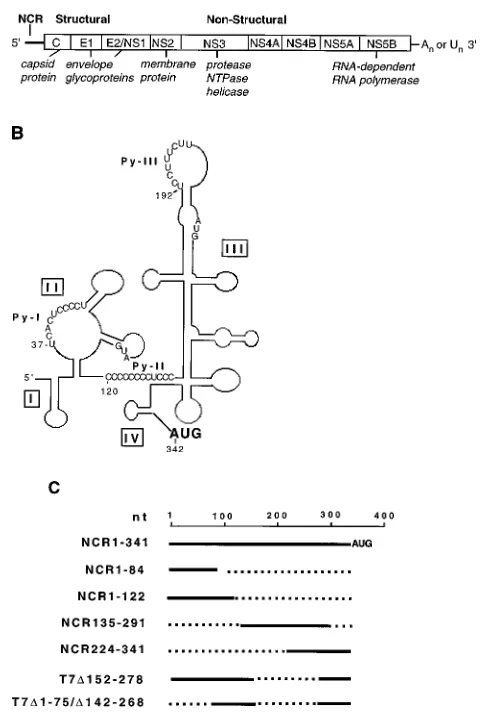
Human hepatitis C virus (HCV) is recognized as the prin-cipal causative agent of parenteral non-A, non-B hepatitis (for reviews, see references 20 and 37). HCV infection leads to chronic hepatitis in the majority of infected individuals (29). A strong link between HCV infection and development of hep-atocellular carcinoma has been documented (43). On the basis of several criteria, including genome organization, molecular features, and biochemical properties, HCV has been classified as one of the genera of the Flaviviridae family (9, 23, 32, 41). The HCV genome is 9.4 kb long and has positive polarity, consisting of various lengths of 59noncoding region (59NCR) (331 to 341 nucleotides [nt]), a long open reading frame en-coding a polyprotein consisting of approximately 3,000 amino acids, and a short 39NCR (20). The polyprotein is further processed into at least two structural and several nonstructural functionally active proteins (Fig. 1A).
While clinical isolates of the HCV genomes display consid-erable heterogeneity within the coding region, the 59NCR is highly conserved (over 98% homology) (7, 17). A complex secondary structure has been proposed for the 59NCR from phylogenetic comparative sequence analysis (6). There are multiple cryptic AUG codons within the 59NCR. These fea-tures, coupled with experimental evidence derived from suc-cessful expression of the second cistron in the context of dicis-tronic expression vectors, strongly argue for the presence of an internal ribosome entry site (IRES) within the HCV 59NCR (24, 42, 46). Kettinen et al. (24) further demonstrated a func-tional role of the coding region as a part of the IRES element located immediately 39distal to the initiator AUG. A detailed examination of the HCV IRES by extensive mutational
anal-ysis revealed the functional role of a helical structure associ-ated with the pyrimidine tract II (previously defined as Py-I [47]) (Fig. 1B) and a probable pseudoknot structure located immediately upstream of the initiator AUG (45, 48). These secondary-tertiary interactions are essential cis elements of the HCV IRES (45, 48). Deletion and insertional mutagenesis studies suggest that almost the entire 59NCR with the excep-tion of 29 nt at the extreme 59 end may constitute the HCV IRES element (48). Thus, the translational strategy used by HCV is similar to that employed by members of the family
Picornaviridae (for reviews, see references 1, 31, and 38). On
the basis of several features, the HCV IRES element appears to be similar to the type II IRES elements, in which IRES is located either immediately upstream of the initiator AUG or includes the AUG triplet (2, 21, 48). While the majority of the eukaryotic mRNAs are translated by a ribosome-scanning mechanism (25), IRES-mediated translation has been ob-served for a few cellular mRNAs (27, 31). These observations further suggest that eukaryotic translation machinery can sup-port cap-independent translation.
Among the various cellular trans-acting factors, the polypy-rimidine tract-binding protein (PTB), also known as heteroge-neous nuclear ribonucleoprotein I or p57 (13, 14, 22), princi-pally involved in splicesome assembly (5, 15, 33), has been shown to be functionally required for internal initiation of translation by picornaviruses (12, 22, 26, 36). PTB binds to picornavirus IRES elements at one or more sites (18, 26, 49), and its removal from cellular extracts abrogates translation initiation of picornavirus RNA but notb-globin mRNA (3, 19, 34).
In this study, we investigated the possibility of PTB binding within the HCV 59NCR. UV cross-linking assays carried out with recombinant PTB clearly demonstrated the interaction of PTB with 59NCR at at least three distinct sites. Our studies performed with PTB derived from HeLa nuclear lysates also * Corresponding author. Mailing address: Department of
Microbi-ology, B172, University of Colorado Health Sciences Center, 4200 E. 9th Ave., Denver, CO 80262. Phone: (303) 7016. Fax: (303) 270-8330.
6367
on November 9, 2019 by guest
http://jvi.asm.org/
show PTB’s interaction with the HCV 59NCR, as evidenced by immunoprecipitation with a monoclonal antiserum directed against PTB. Furthermore, the immunodepletion of PTB from translation lysates completely inhibited the translational capac-ity of 59NCR. These studies suggest that PTB plays a functional role in the internal initiation of the HCV RNA genome.
MATERIALS AND METHODS
Construction of recombinant plasmids. The construction of plasmid pGEM59NC, which contains the full-length 59NCR of HCV-I, has been de-scribed previously (46). NCR135-291, which represents domain III of the 59NCR, was PCR amplified from pGEM59NC with sense (59CGGGAATTCAGAGC CATAGTG39) and antisense (NC/AS [59AGGCGGATCCAGTACCACAAG G39]) primers. The amplified product was cloned at EcoRI-BamHI sites in pGEM-3. Nucleotides 224 to 341 of 59NCR were amplified from plasmid pGEM59NC with sense (59TGGAAGCTTAGATTTGGGCGTG39) and anti-sense (NC/AS) primers and cloned at HindIII-SacI sites in pGEM-4 to construct NCR224-341. Plasmid T7C75-332 represents the 59NCR of HCV-II, in which the upstream 75 nt have been deleted (46). The AgeI-StuI fragment was deleted from this plasmid to construct the mutant T7CD1–75/D142–268 plasmid. All of the plasmids generated for this study are listed in Fig. 1C. Construction of the
plasmids pHC5NC/126, pHC5NC/121/126/319, and T7CD152–278 has been de-scribed previously (47).
In vitro transcription.Plasmid DNAs were purified by the CsCl density gra-dient centrifugation method. pGEM59NC was linearized with EcoRI and tran-scribed in vitro with T7 RNA polymerase to produce full-length HCV 59NCR RNA (NCR1-341). NCR1-84 and NCR1-122 RNAs were prepared similarly by digestion of pGEM59NC with NcoI and SmaI, respectively. Plasmids pHC5NC/ 126 and pHC5NC/121/126/319 were linearized with XbaI, and RNA was synthe-sized with SP6 RNA polymerase. T7CD152–278 and T7CD1–75/D142–268 were digested with XbaI to linearize them before transcription with T7 RNA poly-merase. Plasmids NCR135-291 and NCR224-341 were linearized with BamHI and NcoI, respectively, and transcribed with T7 RNA polymerase. Plasmid pT7-GEM-Polio(N) containing the full-length poliovirus (Mahony) cDNA was di-gested with Msc 1, which cleaves the DNA at nt 627, for use in UV cross-linking studies (see Fig. 5A). The same plasmid was also digested with BamHI (nt 220) to generate poliovirus NCR1-220 RNA for the competition studies described in the legend to Fig. 2C. The poliovirus RNAs were synthesized by T7 RNA polymerase. Plasmid T7C1-341 was linearized with HpaI and transcribed with T7 RNA polymerase. All of the DNA templates, after digestion with the appropriate restriction enzyme, were purified by elution of the desired fragment from the agarose gels before transcription. The radioactive RNA probes were synthesized under similar reaction conditions with 4-thio-UTP and [32
P]CTP.
Expression of recombinant PTB.The plasmid GST-2TK/PTB, which encodes the glutathione S-transferase (GST)–PTB fusion protein, was a generous gift of M. A. Garcia-Blanco. The expression of GST-PTB was induced with 1 mM IPTG (isopropyl-b-D-thiogalactopyranoside) in Escherichia coli (JM101) cells and affinity purified on glutathione-Sepharose beads (Pharmacia). The final prepa-ration was dialyzed against 5 mM HEPES (N-2-hydroxyethylpiperazine-N9 -2-ethanesulfonic acid [pH 7.6])–1 mM EDTA–1 mM dithiothreitol–0.2 mM–phen-ylmethylsulfonyl fluoride–10% (vol/vol) glycerol. GST was also purified by a similar procedure and used as a control.
UV cross-linking of RNA with proteins.4-Thio-UDP (Sigma) was phosphor-ylated with nucleoside 59-diphosphate kinase to prepare 4-thio-UTP according to the method of Stade et al. (39). The 4-thio-UTP and [32
P]CTP were included in an in vitro transcription reaction mixture to produce thio-U-containing 32 P-labeled RNA. Protein samples were incubated with RNA probe for 30 min at 308C in an RNA binding buffer (5 mM HEPES [pH 7.6], 25 mM KCl, 2 mM MgCl2, 0.1 mM EDTA, 10 mM dithiothreitol, 15% [wt/vol] glycerol). Yeast tRNA (10 mg) and 40 U of RNasin (Promega) were also included in each reaction mixture (15 to 20ml). Samples were irradiated with UV light (312 nm) for 30 min with a UV Stratalinker model 1800 (Stratagene) at 48C. Unbound RNA was digested with 10 U of RNase A (U.S. Biochemical Corp.) for 15 min. Samples were analyzed by sodium dodecyl sulfate-polyacrylamide gel electro-phoresis (SDS-PAGE) [10% polyacrylamide]) followed by autoradiography. In all of the competition assays, appropriate competitor RNAs were added along with other components of the reaction mixture prior to UV cross-linking.
Purification of PTB from HeLa nuclear extracts.Nuclear extracts from cul-tured cells were prepared essentially as described by Dignam (11). The final preparation was dialyzed against buffer D (20 mM HEPES-NaOH [pH 7.9], 20% [vol/vol] glycerol, 100 mM KCl, 0.2 mM EDTA, 0.5 mM dithiothreitol, 0.5 mM phenylmethylsulfonyl fluoride). Purification of p57/PTB was carried out accord-ing to the modified method of Gil et al. (15). One milliliter of slurried poly(U)-Sepharose 4B (Pharmacia), preequilibrated with buffer E (buffer D plus 0.05% Nonidet P-40 and 2.0 mM MgCl2) was mixed with 1.5 mg of nuclear extracts and incubated at 48C on a rotary shaker for 1 h. The mixture was loaded on a minicolumn, and the unbound materials were washed with buffer E containing 0.5 M KCl after the slurry had completely settled. The bound proteins were eluted with 0.8 M KCl added to the same buffer. Fractions were collected, and the protein concentration was determined. Fractions were dialyzed against buffer D, and PTB binding activity was determined by a UV cross-linking method.
Immunoprecipitation of the PTB-HCV 5*NCR complex.The ribonucleopro-tein complexes formed between HCV 59NCR RNA and partially purified HeLa PTB were prepared as described above. After UV cross-linking and RNase treatment, samples were diluted to 500ml in buffer containing 50 mM Tris-HCl (pH 7.4), 5 mM EDTA, 1 mM dithiothreitol, 100 mM NaCl, 0.05% Nonidet P-40). Monoclonal anti-PTB antibody 7G12 (a generous gift of Gideon Dreyfuss) was added, and the mixture was incubated for 1 h at 48C. Ten micrograms of anti-mouse immunoglobulin G (IgG) antibody (Sigma) was added, and immu-nocomplexes were immobilized on protein A-Sepharose beads (Pharmacia) and washed four times with the same buffer. The bound proteins were analyzed by SDS-PAGE followed by autoradiography.
Immunodepletion of PTB from cell lysates.For immunodepletion of PTB from the cell-free translation system, 8ml of 7G12 antibody was added to 50ml of rabbit reticulocyte lysate (RRL [Promega]) or HeLa S10 extract and incubated for 1 h at 48C. Protein G-Sepharose (Pharmacia) was added, and the mixture was incubated further for an additional hour at 48C on a rotary shaker. The mixture was centrifuged, and the supernatant was used for translation.
In vitro translation.The translation reactions were performed with RRLs. Proteins were radiolabeled with Trans-35
S label (ICN Biomedicals) and analyzed by SDS-PAGE. Luciferase activity in the RRLs was measured according to the method of de Wet et al. (10). Immunodepleted RRL and HeLa S10 fractions FIG. 1. (A) Genomic organization of the HCV RNA and polyprotein
pro-cessing scheme. (B) Schematic representation of computer-generated RNA sec-ondary structure of the 59NCR (adapted from Brown et al. [6]). The locations of the pyrimidine tracts (Py-I, Py-II, and Py-III) and the four structural domains within the open box are shown. (C) Maps of HCV 59NCR deletion mutants. Dotted lines represent the extent of deletion. The nucleotide position (nt) indi-cates the first nucleotide of the mutants.
on November 9, 2019 by guest
http://jvi.asm.org/
[image:2.612.54.295.89.447.2]were supplemented with various amounts of recombinant PTB. The reconsti-tuted lysates were also used for translation of T7C1-341 RNA.
RESULTS
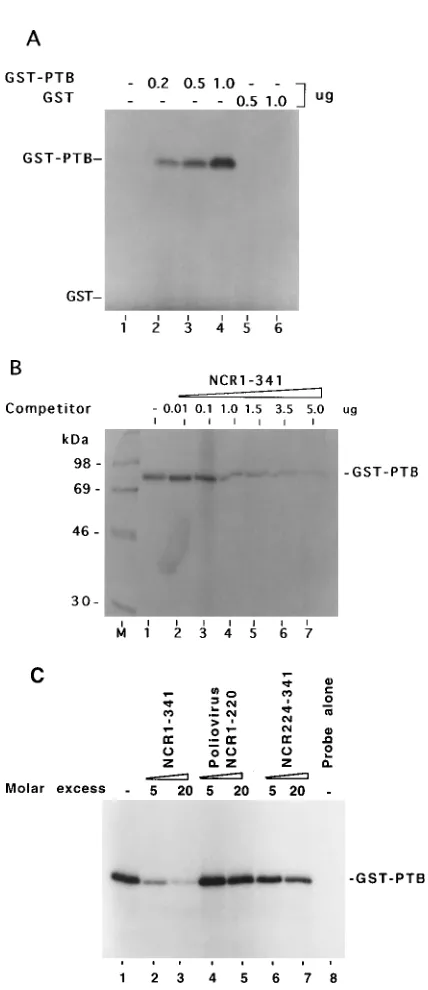
Binding of PTB to the HCV 5*NCR.There are at least three potential pyrimidine-rich tract motifs located within the HCV 59NCR. They are located at nt 37 to 46 (Py-I), 120 to 131 (Py-II), and 192 to 199 (Py-III) as shown in Fig. 1B. Py-I and Py-III sequences are part of single-stranded regions, but Py-II sequences are partially base paired with nt 319 to 324 (Fig. 1B) (6). The putative Py sequences of the HCV 59NCR may serve as potential binding sites for PTB. We investigated the inter-action of recombinant PTB with the HCV 59NCR by UV cross-linking assays. The RNA probe was enzymatically syn-thesized in the presence of [32P]CTP and 4-thio-UTP. The
radiolabeled thio-U-containing 59NCR probe was incubated with purified GST-PTB or GST at various protein concentra-tions. The reaction mixtures were subsequently UV irradiated and digested with RNase A prior to fractionation by SDS-PAGE. Appearance of a radioactive protein band in this anal-ysis is due to the covalent interaction between 32P-labeled
probe and PTB. A radiolabeled PTB-RNA complex was formed between GST-PTB and the HCV 59NCR, and the intensity of this complex increased as a function of protein concentration (Fig. 2A, lanes 2 to 4). However, under similar binding conditions, GST alone was not cross-linked to the probe (lanes 5 and 6). These results show interaction of 59NCR with the PTB moiety in the GST-PTB fusion protein. The binding specificity of PTB to 59NCR was confirmed by a com-petition assay (Fig. 2B). Various concentrations of unlabeled homologous RNA (NCR1-341) were added to the reaction mixture before UV cross-linking. The binding of PTB to the probe was considerably inhibited in the presence of unlabeled 59NCR RNA (lanes 4 to 7). These results indicate the speci-ficity of PTB’s interaction with HCV 59NCR. The interaction of the wild-type HCV 59NCR with PTB was then compared with that of a heterologous RNA derived from nt 1 to 220 of poliovirus 59NCR and a homologous RNA representing nt 224 to 341 (NCR224-341) of HCV 59NCR in a competition assay. The results of this experiment are presented in Fig. 2C. With similar molar excess of the competitors, while unlabeled NCR1-341 RNA completely inhibited the PTB interactions with the RNA probe (lanes 2 and 3), poliovirus-derived RNA (lanes 4 and 5) failed to compete. Similarly, the HCV mutant RNA (NCR224-341), devoid of all of the Py motifs (Py-I, -II, -III), was unable to inhibit PTB interaction with the full-length HCV 59NCR RNA (lanes 6 and 7). These observations con-firm the specificity of PTB binding with HCV 59NCR.
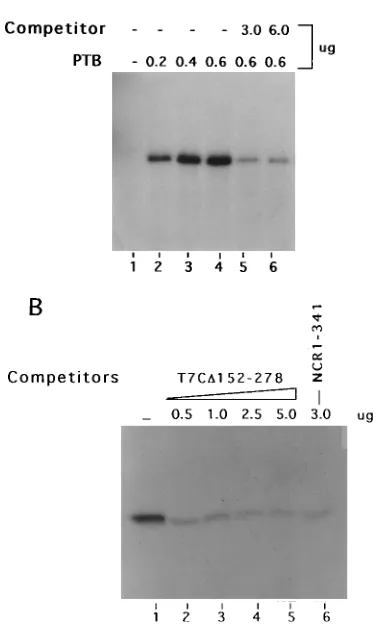
Mapping of PTB binding sites in HCV 5*NCR.Our mapping strategy involved the use of several mutant 59NCR RNAs both in direct binding to PTB and as unlabeled competitors in the UV cross-linking of the wild-type 59NCR. In the first analysis, the T7CD152–278 thio-U-containing deletion mutant RNA was used as a probe. This deletion essentially eliminates the upper portion of domain III, including the apical loop contain-ing the Py-III motif. The enhancement of the probe transfer signal as a result of PTB binding to the labeled RNA was evident with increasing concentrations of the protein (Fig. 3A, lanes 2 to 4). Notably, the intensity of the binding signal was reduced considerably when unlabeled homologous competitor RNA was added to the reaction mixture (lanes 5 and 6). The binding specificity of PTB was further confirmed in a compe-tition assay in which wild-type NCR1-341 RNA probe was used for UV cross-linking with PTB in the presence of unlabeled T7CD152–278 RNA (Fig. 3B). The competitor RNA drasti-FIG. 2. UV cross-linking of the GST-PTB fusion protein with 59NCR of
HCV RNA (NCR1-341) followed by SDS-PAGE (10% polyacrylamide). (A) Interaction of GST-PTB with the full-length wild-type HCV 59NCR RNA probe. Lanes: 1, probe alone; 2 to 4, increasing amounts of GST-PTB; 5 and 6, UV cross-linking of GST with the probe. (B) Interaction of PTB with HCV 59NCR with increasing amounts of unlabeled homologous RNA used as competitor (lanes 2 to 7). GST-PTB (0.5mg) was UV cross-linked to;105cpm of the NCR1-341 RNA probe. The 59NCR RNA probe was synthesized in vitro with 4-thio-UTP and [32P]CTP under standard conditions. Lane 1 contained no com-petitor. (C) Effects of competitors on PTB binding to the HCV 59NCR RNA in UV cross-linking reactions. Lanes: M, molecular mass markers; 1, without com-petitor; 2 and 3, NCR1-341; 4 and 5, poliovirus 59NCR representing nt 1 to 220; 6 and 7 NCR224-341; 8, probe alone. UV cross-linking of wild-type HCV 59NCR thio-U-containing32P-labeled RNA probe with GST-PTB (80 ng) was carried out as described above. RNA competitors in 5 and 20 molar excess of the probe concentration were included in the reaction mixtures in the competition assay.
on November 9, 2019 by guest
http://jvi.asm.org/
[image:3.612.71.285.81.576.2]cally inhibited the interaction between PTB and wild-type 59NCR1-341 (lanes 2 to 5). The unlabeled competitor wild-type 59NCR RNA is included as a positive control, which as expected abolished the PTB-RNA complex (lane 6). These results together suggest that both Py-I and Py-II motifs along with other structural elements may be potential sites of PTB interaction. The mutant T7CD152–278 HCV 59NCR is devoid of the Py-III motif but retains both Py-I and Py-II, including the helical element (Fig. 1B).
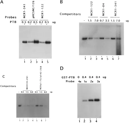
To further identify these regions as PTB binding sites, we used two candidate RNA probes along with the wild-type 59NCR. First, a truncated RNA representing nt 1 to 122 from the extreme 59 end, which includes a pyrimidine-rich tract (Py-I; nt 37 to 46) was used. PTB binding to this sequence is efficient (Fig. 4A, lanes 4 and 5) and is comparable to that of the wild-type 59NCR (lane 1). Next, we selected a mutant pHC5NC/126 RNA in which four pyrimidines of the Py-II tract (nt 126 to 129) were altered to purines (47). These nucleotide changes essentially disrupt the helical interaction between nt 126 to 134 and nt 315 to 323. Although this mutation dramat-ically affects translation initiation (47), it has no effect on PTB binding (Fig. 4A, lanes 2 and 3). The specific binding of PTB
to the nt 1 to 122 sequence of 59NCR was further substantiated by the use of several unlabeled RNAs encompassing that se-quence. These include homologous NCR1-122, NCR1-84, and the wild-type 59NCR RNAs. In all cases, the level of interac-tion between PTB and the NCR1-122 probe was reduced (Fig. 4B). Binding of these RNAs to PTB unambiguously defines the presence of a PTB binding site within nt 1 to 84, which is designated as PTB site I.
NCR135-291 RNA, which represents the largest stem-loop structure of domain III, was used as a competitor in UV cross-linking of PTB with the full-length wild-type 59NCR. The re-sults of competitive inhibition are shown in Fig. 4C (lanes 2 to 4). Effective competition by the unlabeled NCR135-291 RNA was obtained at all concentrations, suggesting the presence of a PTB binding site within that sequence. Located within this sequence at nt 192 to 200 is a characteristic pyrimidine-rich sequence followed by a noninitiator AUG, designated here as PTB site II.
A double-deletion mutant RNA, NCRD1–75/D142–268, was constructed in which the pyrimidine-rich motif (Py-II; nt 120 to 131) was linked to the primary sequences required to form the helical structural elements at the 39 border of wild-type 59NCR. This RNA was found to compete with wild-type 59NCR for PTB binding (Fig. 4C, lanes 5 to 7). The competi-tive binding achieved by this mutant further established that PTB site III was within the HCV 59NCR located in the 39 border region of the IRES. We cannot rule out the possibility that this mutant may not have formed the native helical struc-ture because of a large deletion. It is evident that a larger amount of competitor was needed to achieve the diminution of PTB binding (compare lanes 2 and 3 with lanes 5 and 6 in Fig. 4C).
In another approach, a double compensatory mutant RNA, pHC5NC/121/126/319 (47), was used as a probe. This mutant is similar to pHC5NC/126, except that the helical structural re-gion at the Py-II tract was restored by the introduction of mutations in the complementary sequences and two more py-rimidine residues at nt 122 and 123 were mutated to purines. With these rearrangements of nucleotide sequences, the mu-tant contains the same structure as the wild type, but 60% of the pyrimidine residues in Py-II are altered to purines (47). This mutant translates efficiently both in vitro and in vivo. In this analysis, a constant amount of GST-PTB was used while increasing amounts of the 4-thio-U-containing32P-labeled
mu-tant probe were added. The intensity of the radiolabeled PTB signal correlates with the amount of probe added to the reac-tion mixture (Fig. 4D). Thus, a funcreac-tionally active mutant HCV 59NCR retains its capacity to bind PTB despite the fact that only 40% of the pyrimidine residues were present in the Py-II motif.
In summary, the results of this detailed analysis identify three noncontiguous binding regions of PTB within the HCV 59NCR, designated as sites I, II, and III. Interestingly, all of these regions contain characteristic stretches of pyrimidine-rich tracts (Fig. 1B).
[image:4.612.82.271.98.410.2]Interaction of cellular PTB with the HCV 5*NCR.The PTB binding studies described above were carried out with a bac-terially expressed GST fusion protein. To demonstrate the interaction of PTB from cell lysates with the HCV 59NCR, partially purified HeLa nuclear proteins were used. The puri-fied protein fraction was subjected to a UV cross-linking assay with the RNA probes transcribed from wild-type HCV 59NCR. The poliovirus 59NCR RNA probe, known to efficiently bind HeLa PTB (19, 34), was included as a positive control. Two closely migrating ribonucleoprotein complexes with an average molecular mass of 57 kDa (p57) were resolved as major bands FIG. 3. Binding of GST-PTB with HCV 59NCR RNA probe lacking domain
III sequences. (A) UV cross-linking of T7CD152–278 RNA probe with increasing amounts of PTB (lanes 2 to 4). Lane 1 contained probe alone, and lanes 5 and 6 show specific inhibition of PTB binding with the probe by unlabeled homolo-gous RNA. The UV cross-linked fractions were subjected to SDS-PAGE (10% polyacrylamide). (B) Inhibitory effect of heterologous competitor T7CD152–278 on the interaction of GST-PTB with the NCR1-341 RNA probe. The reaction mixtures were fractionated by SDS-PAGE. Lanes: 1, no competitor; 2 to 5, increasing amounts of competitor RNA; 6, homologous competitor.
on November 9, 2019 by guest
http://jvi.asm.org/
(Fig. 5A, lane 1). These complexes also correspond to the cross-linked products of the poliovirus RNA probe (lane 3). The cross-linking specificity of the p57 complex was confirmed by addition of unlabeled homologous competitor RNA under the same reaction conditions. The interaction of p57 doublets with HCV probe was considerably abolished by the unlabeled homologous competitor (lane 2), clearly indicating the specific interaction of these proteins with the HCV 59NCR. In this analysis, two additional protein bands, 72 and 110 kDa, also show a relatively weaker but specific interaction with the RNA probes. A few low-molecular-mass species were cross-linked with the poliovirus 59NCR but not with the HCV 59NCR. This
[image:5.612.83.532.72.499.2]is consistent with previous observations in which smaller poly-peptides selectively interact with the 59NCRs of picornaviruses (8, 12). With a similar approach, the purified p57 proteins were UV cross-linked with the HCV 59NCR and subsequently im-munoprecipitated with a monoclonal anti-PTB antibody (7G12). The results of this analysis, shown in Fig. 5B, lane 2, identify the p57 doublet UV cross-linked with HCV 59NCR as PTB. In contrast, immunoprecipitation was not observed with unrelated serum (lane 1). These observations unambiguously demonstrate that the cellular p57 complex of PTB proteins interacts directly with the HCV 59NCR. This complex of pro-tein bands ranging from 57 to 62 kDa in size represents isomers FIG. 4. Mapping of PTB binding sites in the HCV 59NCR. All of the NCR RNA probes contained 4-thio-U and were32
P labeled. The UV cross-linked RNA-protein complexes were subjected to SDS-PAGE (10% polyacrylamide). (A) Direct binding of HCV 59NCR mutant RNA probes. UV cross-linking was carried out with the labeled probes at the indicated amounts of GST-PTB. Lanes: 1, NCR1-341 RNA; 2 and 3, pHC5NC/126 RNA probe. In this mutant (pHC5NC/126), Py-II sequences are mutated to purines, leading to disruption of the helical structure (47). Lanes 4 and 5 contained the NCR1-122 RNA probe, which contains the putative Py-I motif and represents the 59end sequences of the NCR. (B) Binding of PTB with the NCR1-122 RNA probe in the presence of unlabeled competitor NCR mutants. Lanes: 1, no competitor; 2 and 3, NCR1-122 RNA; 4 and 5, NCR1-84 RNA; 6 and 7, NCR1-341 RNA. (C) Competitive inhibition of PTB binding to the wild-type NCR1-341 RNA probe by deletion mutant 59NCR RNAs. Lanes: 1, no competitor; 2 to 4, increasing amounts of NCR135-291 (contains domain III of the HCV 59NCR); 5 to 7, increasing amounts of NCRD1–75/D142–268 (contains Py-II and downstream sequences involved in a probable pseudoknot structure) (45). (D) Direct binding of a double compensatory mutant, pHC5NC/121/126/319, RNA probe to PTB. Increasing amounts of probe (13 51.63104cpm) were used for the UV cross-linking, while the amount of GST-PTB was kept constant.
on November 9, 2019 by guest
http://jvi.asm.org/
of PTB (5, 13, 33). These results are consistent with those from experiments conducted with picornavirus 59NCRs (18, 49).
Effect of PTB on HCV IRES-mediated translation initiation. In this study, the functional role of PTB interaction with the HCV 59NCR was examined. The plasmid T7C1-341, in which a full-length HCV 59NCR was cloned in front of the luciferase gene such that the translation of the reporter gene is dictated by the HCV IRES, was used (46). The transcripts were trans-lated in RRLs in the presence of various amounts of a mono-clonal anti-PTB antibody, 7G12. The translational efficiency of the luciferase was progressively inhibited by the addition of increasing amounts of antibody (Fig. 6A, lanes 2 to 4). In
contrast, addition of unrelated antibodies such as anti-mouse IgG (lanes 8 and 9) or a normal rabbit serum (lanes 6 and 7) did not show any inhibitory effect on the cap-independent translation. These results provide evidence that the role of PTB is functionally indispensable in the HCV IRES-mediated translation. The role of PTB in the HCV IRES-mediated translation was further determined by PTB immunodepletion-reconstitution experiments. HeLa S10 fraction and RRLs ca-pable of efficient translation of T7C1-341 RNA were immu-nodepleted with anti-PTB serum. These lysates were then used for translation of T7C1-341 LUC transcripts. The results show that PTB-depleted lysates were incapable of translating lucif-erase RNA (Fig. 6B, lanes 1 and 5). The IRES-mediated trans-lation could not be restored after addition of exogenous PTB to the immunodepleted lysates (lanes 2, 3, 6, and 7), suggesting the requirement of cellular factors associated with PTB.
DISCUSSION
PTB has been shown to play an important role in spliceo-some assembly of the eukaryotic pre-mRNAs and in cap-inde-FIG. 5. Interaction of cellular PTB with the 59NCR of HCV. (A) A 0.5-mg
aliquot of protein fraction purified with poly(U)-Sepharose 4B matrix from HeLa nuclear extracts was UV cross-linked with the HCV 59NCR (NCR1-341) 32P-labeled RNA probe (lane 1). Unlabeled homologous RNA (5.0mg) was included as a competitor (lane 2) in the reaction mixture during UV cross-linking. Poliovirus (PV) 59NCR RNA (lane 3) was used as an RNA probe to UV cross-link similar protein fractions. M, molecular mass standard. Two RNA-protein bands located at 72 and 110 kDa are indicated by arrowheads. (B) Immunoprecipitation of HeLa PTB UV cross-linked to the wild-type 59NCR. An affinity-purified fraction was used in UV cross-linking as described above. A monoclonal anti-PTB antibody, 7G12 (lane 2), was used to immunoprecipitate the UV cross-linked fractions (normal serum [lane 1]).
FIG. 6. (A) Inhibition of HCV IRES-mediated translation of the luciferase gene (LUC) by monoclonal anti-PTB serum. Increasing amounts of anti-PTB serum (lanes 2 to 4, 0.1, 1.0, and 2.0ml, respectively) were added to the trans-lation mixture. Lane 1 contained no anti-PTB serum. In another set of experi-ments, 1 and 2ml of normal rabbit serum (lanes 6 and 7, respectively) or 2 and 4mg of anti-mouse IgG (lanes 8 and 9, respectively) were added to the transla-tion mixture. Lane 5 contained no serum, and in lane 10, 2ml of anti-PTB serum served as a control. The in vitro-transcribed T7C1-341 RNA was translated in RRLs in the presence of Trans-35S label and analyzed by SDS-PAGE (10% polyacrylamide) followed by autoradiography. The percentage of luciferase ac-tivity in the antibody-containing samples was normalized against that of the control without antibody (lane 1). (B) Translation of HCV IRES-mediated luciferase in the PTB-immunodepleted RRL and HeLa lysates. The indicated amounts of GST-PTB were added to the lysates. Translation of T7C1-341 RNA in immunodepleted RRLs and HeLa S10 cell fractions is shown in lanes 1 to 3 and 5 to 7, respectively. The immunodepleted lysates were supplemented with recombinant PTB (lanes 2, 3, 6, and 7). Lanes 1 and 5 were without exogenous PTB. Lanes 4 and 8 show translation in native (not antibody treated) RRLs and HeLa S10 cell fractions, respectively, under similar assay conditions. Luciferase is indicated by an arrowhead.
on November 9, 2019 by guest
http://jvi.asm.org/
[image:6.612.100.258.65.491.2] [image:6.612.317.552.69.353.2]pendent translation initiation of picornaviruses (3, 13–15, 18, 19, 26). Translation of HCV RNA is also initiated by cap-independent internal ribosome binding to the 59NCR. Impor-tant features of the HCV 59NCR include a complex secondary structure (6) containing potential pyrimidine-rich tracts at more than one location, the presence of multiple noninitiator AUG triplets, a probable pseudoknot structure at the 39border of the IRES element, and the requirement of nearly the entire 59NCR for IRES function. Experimental results accumulated in several laboratories which identified these structural fea-tures provide conclusive evidence for the IRES-mediated translation of the HCV RNA genome (24, 42, 45–48).
Among picornaviruses, there is ample evidence that internal initiation of translation is facilitated by interactions of cellular and or viral proteins (3, 16, 22, 28). However, one of the unsolved questions is whether IRES function requires factors other than those involved in translation initiation of eukaryotic mRNAs. Identification of additional factors required for inter-nal initiation of translation has been the topic of intense in-vestigation. Of the several trans-acting cellular factors, two factors, p57/PTB and La autoantigen (40), have gained prom-inence in the recent past. In this study, we have investigated the binding of PTB to the 59NCR of the HCV. The evidence presented here shows that PTB, which binds picornavirus 59NCRs at multiple sites (18), also binds the HCV 59NCR and is functionally required for internal initiation of translation. Although PTB interacts with three noncontiguous regions of the HCV 59NCR, exact sequences including the Py motif or the structural elements with which PTB makes contact could not be determined. Of interest in this respect, is a double compen-satory mutant, pHC5NC/121/126/319, in which about 60% of the pyrimidines (Py-II) were altered to purines, which main-tained the PTB interaction, suggesting that PTB binding may be focused more on structural motifs than on the pyrimidine-rich sequence per se. This is not without precedent, because PTB binding has been shown to occur in a pre-mRNA element that does not contain a pyrimidine tract (30). Furthermore, in another study, a pyrimidine homopolymer was unable to dis-place PTB binding (19). On the basis of their detailed exami-nation of the sites of PTB binding in picornavirus 59NCRs, Witherall et al. (49) suggested that common features of PTB binding include such structural features as helix–single-stranded RNA–helix. Further studies will be needed to define the exact sites and the primary and structural components of PTB binding sites in the HCV 59NCR.
The HCV 59NCR contains three independent binding sites for PTB comprising all or part of nt 1 to 84 (site I), nt 135 to 291 (site II), and site III, which includes a helical structure encompassing nt 126 to 134 (base paired with nt 315 and 323) located in the vicinity of initiator AUG. Among the three PTB binding sites in the poliovirus 59NCR, one is located within the pyrimidine tract while the others are identified as non-pyrim-idine stem-loop or helical structures (18). In the UV cross-linking and competition assay, PTB binding site I (1 to 75 nt) in the HCV 59NCR appears to interact with a relatively higher affinity (Fig. 4B). These sequences contain a putative pyrimi-dine-rich motif (Py-I, beginning at nt 37 to 46) with a nonini-tiator AUG located at nt 85 farther downstream, but a long stem may bring the triplet in close structural proximity to the Py sequences and thus may constitute a characteristic Yn-Xm-AUG motif. Additional features of site I include a stem and single-stranded region followed by a second stem. Such struc-tural features have been previously defined for picornavirus encephalomyocarditis virus 59NCR (49, 50) (Fig. 1B). Earlier studies from this laboratory showed that deletion of this region markedly reduced the translational efficiency of the HCV
IRES (46). Interestingly, an antisense oligonucleotide directed against sequences between nt 38 and 65, which overlaps the putative Py-I sequence, blocked HCV RNA translation (44). These observations support the functional importance of this 59upstream region.
The region in which PTB binding site II is located contains the largest stem-loop structure, or domain III as defined by Brown et al. (6). A characteristic pyrimidine-rich sequence motif, Yn-Xm-AUG, is located in the apical loop of the do-main, followed by a noninitiator AUG triplet 17 nt down-stream. Previous studies from this laboratory in which pyrim-idine residues and the AUG codon were mutated did not lead to any appreciable decrease in the translational efficiency of the 59NCR (47). However, a major deletion of 127 nt, which essentially eliminates the upper portion of this domain includ-ing the Py-III tract, had detrimental effects on IRES-mediated translation initiation (47). In the present analysis, PTB binding to an RNA derived from this region (NCR135-291) was main-tained.
The third cis element (site III) that is identified as a PTB binding site represents a higher-order structure consisting of secondary and tertiary interactions. First, a highly conserved sequence maintains a helical structure in which nt 126 to 131 are base paired with nt 311 to 319. Disruption of this helical interaction resulted in a dramatic reduction in translation ini-tiation (47). This mutant RNA did compete for PTB binding to the wild-type 59NCR (data not shown). A compensatory mu-tation in which pyrimidine residues in nt 121 to 131 were altered to purines (about 60%), but with a complementary strand which contained a mutation restoring base pairing in these sequences, recovered translational efficiency of the 59NCR. UV cross-linking of this RNA molecule with PTB was also observed in the present analysis (Fig. 4D). Further biochemical and genetic analysis of this region supports the existence of a pseudoknot structure in this region (45). These structural ele-ments may serve as recognition sites for RNA-protein interac-tions including PTB. Thus, the HCV IRES resembles the IRES elements of picornaviruses in terms of the presence of the multiple cis elements involved in PTB interaction (4, 18, 26, 34).
UV cross-linking of the HCV 59NCR with the PTB partially purified from HeLa cellular lysates revealed two closely mi-grating species with molecular masses ranging from 57 to 62 kDa. These isomers of PTB cross-linked to 59NCR, were con-firmed by the use of monoclonal anti-PTB serum (Fig. 5B). This pattern is consistent with previous studies of PTB complex interactions with cellular pre-mRNA and picornavirus 59NCR (3, 14, 30).
Indirect evidence suggests that p57/PTB is an essential fac-tor in translation of picornavirus mRNA (19, 22, 34). This conclusion is based on observations that specific anti-PTB se-rum inhibited cap-independent translation but not cap-depen-dent translation (19). Using a similar approach, we investi-gated the functional role of PTB in HCV IRES-dependent translation. Addition of monoclonal anti-PTB serum abolished translation initiation (Fig. 6A), while normal serum or nonspe-cific antibodies had no such effect. This observation lends sup-port to the notion that PTB plays a key role in cap-independent translation. Immunodepletion of PTB from cell lysates did not restore translational efficiency after reconstitution with recom-binant PTB, suggesting the removal of PTB and PTB-associ-ated factors, which may constitute important protein com-plexes that are essential components of internal initiation of translation. This observation is consistent with previous results with picornavirus 59NCRs (19, 36). Furthermore, the observa-tion that the splicing activities of poly(U)-depleted nuclear
on November 9, 2019 by guest
http://jvi.asm.org/
extracts were restored only by supplementing PTB with a 100-kDa protein factor supports the results presented here (33).
The mechanism by which the HCV IRES element or those of picornaviruses recruits the 40S ribosomal subunit to form an initiation complex remains elusive. On the basis of previous studies of picornavirus IRES elements, the following have been predicted. (i) Ribosomes directly land at the IRES ele-ment by interaction of ribosomal proteins with 59NCRs. (ii) Ribosomal subunits are recruited by trans-acting cellular fac-tors that interact with 59NCR. (iii) A characteristic, Yn-Xm-AUG motif may provide a Shine-Dalgarno sequence-like motif for the interaction of IRES with complementary sequence of 18S rRNA. A considerable level of sequence complementarity has been noted between the 39 border of the picornavirus IRES elements and the 18S rRNA (35). A potential 18S rRNA binding site has also been localized between nt 192 and 203 of HCV 59NCR. These sequences are complementary to bases 461 to 471 of human 18S RNA (6). Mutations introduced in these sequences of HCV exhibit no inhibitory effect on HCV IRES activity (47). While molecular mechanisms underlying PTB interaction are not clearly understood, PTB’s absolute requirement in translation initiation for picornaviruses and HCV invokes an essential functional role. Possible roles of PTB could include serving as a nucleation site for general initiation factors, mediating ribosome binding, and facilitating direct binding of ribosomes and other factors by modulating the structure of the IRES (19, 22, 36). These appear to be the essential molecular interactions for the assembly of the trans-lation initiation complex.
In summary, the present analysis unambiguously demon-strates a functional role of PTB and PTB-associated factors via physical interactions at multiple sites within the 59NCR in the internal initiation of translation of the HCV RNA genome.
ACKNOWLEDGMENTS
We thank M. A. Garcia-Blanco for GST-2TK/PTB plasmid and polyclonal anti-PTB serum and G. Dreyfuss for monoclonal anti-PTB antibody (7G12). We also thank Peter Sarnow for constant help throughout the course of this study.
A.S. received a grant from the Lucille P. Markey Charitable Trust. N.A. received support from the Colorado Advanced Technology In-stitute.
REFERENCES
1. Agol, V. I. 1991. The 59-untranslated region of picornaviral genomes. Adv. Virus Res. 40:103–180.
2. Ali, N., C. Wang, and A. Siddiqui. Translation of HCV RNA genome. In Proceedings of the 25th Tokyo Princess Takamatsu Symposium on HCV and Hepatocellular Carcinoma, in press.
3. Borman, A., M. T. Howell, J. G. Patton, and R. J. Jackson. 1993. The involvement of a spliceosome component in internal initiation of human rhinovirus RNA translation. J. Gen. Virol. 74:1775–1788.
4. Borovjagin, A. V., M. V. Ezrokhi, V. M. Rostapshov, T. Y. Ugarova, T. F. Bystrova, and I. N. Shatsky. 1991. RNA-protein interactions within the internal translation initiation region of encephalomyocarditis virus RNA. Nucleic Acids Res. 19:4999–5005.
5. Bothwell, A. M., D. W. Ballard, W. M. Philbrick, G. Lindwall, S. E. Maher, M. M. Bridgett, S. F. Jamison, and M. A. Garcia-Blanco.1991. Murine polypyrimidine tract binding protein: purification, cloning, and mapping of the RNA binding domain. J. Biol. Chem. 266:24657–24663.
6. Brown, E. A., H. Zhang, L.-H. Ping, and S. M. Lemon. 1992. Secondary structure of the 59nontranslated regions of hepatitis C virus and pestivirus genomic RNAs. Nucleic Acids Res. 20:5041–5045.
7. Bukh, J., R. H. Purcell, and R. H. Miller. 1992. Sequence analysis of the 59 noncoding region of hepatitis C virus. Proc. Natl. Acad. Sci. USA 89:4942– 4946.
8. Chang, K. H., E. A. Brown, and S. M. Lemon. 1993. Cell type-specific proteins which interact with the 59nontranslated region of hepatitis A virus RNA. J. Virol. 67:6716–6725.
9. Choo, Q.-L., L. K. Richman, J. H. Han, K. Berger, C. Lee, C. Dong, C. Gallegos, D. Coit, A. Medina-Selby, P. J. Barr, A. J. Weiner, D. W. Bradly,
G. Kuo, and M. Houghton.1991. Genetic organization and diversity of the hepatitis C virus. Proc. Natl. Acad. Sci. USA 88:2451–2455.
10. de Wet, J. R., K. V. Wood, M. DeLuca, D. R. Helinski, and S. Subramani. 1987. Firefly luciferase gene: structure and expression in mammalian cells. Mol. Cell. Biol. 7:725–737.
11. Dignam, J. D. 1990. Preparation of extracts from higher eukaryotes, p. 194–203. In M. P. Deutscher (ed.), Guide to protein purification. Academic Press, Inc., New York.
12. Ehrenfeld, E., and J. G. Gebhard. 1994. Interaction of cellular proteins with the poliovirus 59noncoding region. Arch. Virol. Suppl. 9:269–277. 13. Garcia-Blanco, M. A., S. F. Jamison, and P. A. Sharp. 1989. Identification
and purification of a 62,000-dalton protein that binds specifically to the polypyrimidine tract of introns. Genes Dev. 3:1874–1886.
14. Ghetti, A., S. Pinol-Roma, W. M. Michael, C. Morandi, and G. Dreyfuss. 1992. hnRNP I, the polypyrimidine tract-binding protein: distinct nuclear localization and association with hnRNAs. Nucleic Acids Res. 20:3671–3678. 15. Gil, A., P. A. Sharp, S. F. Jamison, and M. A. Garcia-Blanco. 1991. Char-acterization of cDNAs encoding the polypyrimidine tract-binding protein. Genes Dev. 5:1224–1236.
16. Hambidge, S. J., and P. Sarnow. 1992. Translational enhancement of the poliovirus 59noncoding region mediated by virus-encoded polypeptide 2A. Proc. Natl. Acad. Sci. USA 89:10272–10276.
17. Han, J. H., V. Shyamala, K. H. Richman, M. J. Brauer, B. Irvine, M. S. Urdea, P. Tekamp-Olson, G. Kuo, Q.-L. Choo, and M. Houghton.1991. Characterization of the terminal regions of hepatitis C viral RNA: identifi-cation of conserved sequences in the 59untranslated region and poly(A) tails at the 39end. Proc. Natl. Acad. Sci. USA 88:1711–1715.
18. Hellen, C. U. T., T. V. Pestova, M. Litterst, and E. Wimmer. 1994. The cellular polypeptide p57 (pyrimidine tract-binding protein) binds to multiple sites in the poliovirus 59nontranslated region. J. Virol. 68:941–950. 19. Hellen, C. U. T., G. W. Witherell, M. Schmid, S. H. Shin, T. V. Pestova, A.
Gil, and E. Wimmer.1993. A cytoplasmic 57-kDa protein that is required for translation of picornavirus RNA by internal ribosomal entry is identical to the nuclear pyrimidine tract-binding protein. Proc. Natl. Acad. Sci. USA 90:7642–7646.
20. Houghton, M., A. Weiner, J. Han, G. Kuo, and Q.-L. Choo. 1991. Molecular biology of the hepatitis C viruses: implication for diagnosis, development and control of viral disease. Hepatology 14:381–388.
21. Jackson, R. J., M. T. Howell, and A. Kaminiski. 1990. The novel mechanism of initiation of picornavirus RNA translation. Trends Biochem. Sci. 15:477–483. 22. Jang, S. K., and E. Wimmer. 1990. Cap-independent translation of encepha-lomyocarditis virus RNA: structural elements of the internal ribosomal entry site and involvement of a cellular 57-kDa RNA-binding protein. Genes Dev. 4:1560–1572.
23. Kato, N., M. Hijikata, Y. Ootsuyama, M. Nakagawa, S. Ohkoshi, T. Sug-imura, and K. Shimotohno.1990. Molecular cloning of the human hepatitis C virus genome from Japanese patients with non-A non-B hepatitis. Proc. Natl. Acad. Sci. USA 87:9524–9528.
24. Kettinen, H., K. Grace, S. Grunert, B. Klarke, D. Rowlands, and R. Jackson. 1994. Mapping of the internal ribosome entry site at the 59end of the hepatitis C virus genome, p. 125–131. In K. Nishioka, H. Suziki, S. Mishiro, and T. Oda. (ed.), Proceedings of the International Symposium on Viral Hepatitis and Liver Disease, Tokyo.
25. Kozak, M. 1989. The scanning model for translation: an update. J. Cell Biol. 108:229–241.
26. Luz, N., and E. Beck. 1991. Interaction of a cellular 57-kilodalton protein with the internal translation initiation site of foot-and-mouth disease virus. J. Virol. 65:6486–6494.
27. Macejak, D. G., and P. Sarnow. 1991. Internal initiation of translation me-diated by 59leader of a cellular mRNA. Nature (London) 353:90–94. 28. Meerovitch, K., Y. V. Svitkin, H. S. Lee, F. Lejbkowicz, D. J. Kenan, E. K. L.
Chan, V. I. Agol, J. D. Keene, and N. Sonenberg.1993. La autoantigen enhances and corrects aberrant translation of poliovirus RNA in reticulocyte lysate. J. Virol. 67:3798–3807.
29. Miller, R. H., and R. H. Purcell. 1990. Hepatitis C virus shares amino acid sequence similarity with pestiviruses and flaviviruses as well as members of two plant virus supergroups. Proc. Natl. Acad. Sci. USA 87:2057–2061. 30. Mulligan, G. J., W. Guo, S. Wormsley, and D. M. Helfman. 1992.
Polypy-rimidine tract binding protein interacts with sequences involved in alterna-tive splicing ofb-tropomyosine pre-mRNA. J. Biol. Chem. 267:25480–25487. 31. Oh, S.-K., M. P. Scott, and P. Sarnow. 1993. Homeotic gene antinnapedia mRNA contains 59-noncoding sequences that confer translational initiation by internal ribosome binding. Genes Dev. 6:1643–1653.
32. Okamoto, H., S. Okada, Y. Sugiyama, K. Kurai, H. Iizuka, A. Machida, Y. Miyakawa, and M. Mayumi.1991. Nucleotide sequence of the genomic RNA of hepatitis C virus isolated from a human carrier: comparison with reported isolates for conserved and divergent regions. J. Gen. Virol. 72: 2697–2704.
33. Patton, J. G., S. A. Mayer, P. Tempst, and B. Nadal-Ginard. 1991. Charac-terization and molecular cloning of polypyrimidine tract-binding protein: a component of a complex necessary for pre-mRNA splicing. Genes Dev. 5:1237–1251.
on November 9, 2019 by guest
http://jvi.asm.org/
34. Pestova, T. V., C. U. T. Hellen, and E. Wimmer. 1991. Translation of polio-virus RNA: role of an essential cis-acting oligopyrimidine element within the 59nontranslated region and involvement of a cellular 57-kilodalton protein. J. Virol. 65:6194–6204.
35. Pilipenko, E. V., A. P. Gmyl, S. V. Maslova, Y. V. Svitkin, A. N. Sinyakov, and V. I. Agol.1992. Prokaryotic-like cis elements in the cap-independent inter-nal initiation of translation on picornavirus RNA. Cell 68:119–131. 36. Schmid, M., and E. Wimmer. 1994. IRES-controlled protein synthesis and
genome replication of poliovirus. Arch. Virol. Suppl. 9:279–289. 37. Sherlock, D. S. 1993. Viral hepatitis C. Curr. Opin. Gasteroenterol. 9:341–
348.
38. Sonenberg, N. 1990. Poliovirus translation. Curr. Top. Microbiol. Immunol. 161:23–47.
39. Stade, K., J. Rinke-Appel, and R. Brimacombe. 1989. Site-directed cross-linking of mRNA analogues to the Escherichia coli ribosome; identification of 30S ribosomal components that can be cross-linked to the mRNA at various points 59 with respect to the decoding site. Nucleic Acids Res. 17:9889–9908.
40. Svitkin, Y. V., K. Meerovitch, H. S. Lee, J. N. Dholakia, D. J. Kenan, V. I. Agol, and N. Sonenberg.1994. Internal translation initiation on poliovirus RNA: further characterization of La function in poliovirus translation in vitro. J. Virol. 68:1544–1550.
41. Takamizawa, A., C. Mori, I. Fuke, S. Manabe, S. Murakami, J. Fujita, E. Onishi, T. Andoh, I. Yoshida, and H. Okayama.1991. Structure and orga-nization of the hepatitis C virus genome isolated from human carriers. J. Virol. 65:1105–1113.
42. Tsukiyama-Kohara, K., N. Iizuka, M. Kohara, and A. Nomoto. 1992. Inter-nal ribosome entry site within hepatitis C virus RNA. J. Virol. 66:1476–1483. 43. Tsukuma, H., T. Hiyama, S. Tanaka, M. Nakao, T. Yabuuchi, T. Kitamura, K. Nakanishi, I. Fujimoto, A. Inoue, H. Yamazaki, and T. Kawashima.1993. Risk factors for hepatocellular carcinoma among patients with chronic liver disease. N. Engl. J. Med. 328:1797–1801.
44. Wakita, T., and J. R. Wands. 1994. Specific inhibition of hepatitis C virus expression by antisense oligodeoxynucleotides: in vitro model for selection of target sequence. J. Biol. Chem. 269:14205–14210.
45. Wang, C., S.-Y. Le, N. Ali, and A. Siddiqui. 1995. An RNA pseudoknot is essential for internal initiation of translation of the hepatitis C virus RNA genome. RNA 1:526–537.
46. Wang, C., P. Sarnow, and A. Siddiqui. 1993. Translation of human hepatitis C virus RNA in cultured cells is mediated by an internal ribosome-binding mechanism. J. Virol. 67:3338–3344.
47. Wang, C., P. Sarnow, and A. Siddiqui. 1994. A conserved helical element is essential for internal initiation of translation of hepatitis C virus RNA. J. Virol. 68:7301–7307.
48. Wang, C., and A. Siddiqui. 1995. Structure and function of the hepatitis C virus internal ribosome entry site. Curr. Top. Microbiol. 203:99–115. 49. Witherell, G. W., A. Gill, and E. Wimmer. 1993. Interaction of
polypyrimi-dine tract binding protein with the encephalomyocarditis virus mRNA in-ternal ribosome entry site. Biochemistry 32:8268–8275.
50. Witherell, G. W., and E. Wimmer. 1994. Encephalomyocarditis virus internal ribosomal entry site RNA-protein interactions. J. Virol. 68:3183–3193.