BUPIVACAINE IN SUPRACLAVICULAR BRACHIAL
PLEXUS BLOCK
A STUDY OF 60 CASES
DISSERTATION SUBMITTED FOR
DOCTOR OF MEDICINE
BRANCH X (ANESTHESIOLOGY)
THE TAMILNADU DR.M.G.R. MEDICAL
UNIVERSITY
CHENNAI,
TAMILNADU
CERTIFICATE
This is to certify that the Dissertation “ COMPARISON BETWEEN EFFECTS OF BUPRENORPHINE AND CLONIDINE ADDED TO BUPIVACAINE IN SUPRACLAVICULAR BRACHIAL PLEXUS BLOCK “
presented herein by Dr. J. NISHA SARAL is an original work
done in the Department of Anaesthesiology, Tirunelveli Medical
College Hospital, Tirunelveli for the award of Degree of M.D.
(Branch X) Anesthesiology under my guidance and supervision
during the academic period of 2010 - 2013.
DEAN
CERTIFICATE
This is to certify that this dissertation entitled “COMPARISON BETWEEN EFFECTS OF BUPRENORPHINE AND CLONIDINE ADDED TO BUPIVACAINE IN SUPRACLAVICULAR BRACHIAL PLEXUS BLOCK ”
submitted by DR. J.NISHA SARAL to the faculty of ANAESTHESIOLOGY, The TamilNadu Dr. M.G.R. Medical University, Chennai, in partial fulfillment of the requirement in the award of degree of M.D. Degree, Branch -X (ANAESTHESIOLOGY), for the April 2013 examination is a bonafide research work carried out by her under our direct supervision and guidance.
PROF. DR. A.THAVAMANI, M.D, D.A,
I, Dr.J.NISHA SARAL, declare that the dissertation titled
“COMPARISON BETWEEN EFFECTS OF BUPRENORPHINE AND CLONIDINE ADDED TO BUPIVACAINE IN SUPRACLAVICULAR BRACHIAL PLEXUS BLOCK” has been prepared by me.
This is submitted to The Tamil Nadu Dr. M.G.R. Medical University, Chennai, in partial fulfilment of the requirement for the award of M.D.Degree,Branch X (ANAESTHESIOLOGY) degree Examination to be held in April 2013.
Place : TIRUNELVELI
DR.A.THAVAMANI M.D., D.A., Professor and Head of the Dept. of
Anaesthesiology, Government Tirunelveli Hospital and Tirunelveli Medical College, Tirunelveli for his kind encouragement and valuable guidance during the period of this study, without which this dissertation would not have been materialized. I would like to place on record my indebtedness to my Prof. Dr.A.BALAKRISHNAN M.D., D.A., Additional Professor of
Anaesthesiology, Tirunelveli Medical College, Tirunelveli for his whole hearted help and support in doing this study.I would like to convey my gratitude to Dr.R.AMUTHA RANI M.D., senior assistant professor of Anaesthesiology,
Tirunelveli Medical College, Tirunelveli for her whole hearted help and guidance in doing this study.I would like to thank all my Asst. Professors for their valuable advice and kind co-operation in doing this study. I express my sincere thanks to The DEAN, Tirunelveli Medical College and Tirunelveli Medical College Hospital for permitting me to utilize clinical material of this hospital.
I am also thankful to the Department of Ortho Paedic Surgery for their cooperation in this study.
3 BRACHIAL PLEXUS 5
4 TECHNIQUES OF BRACHIAL PLEXUS BLOCK 6
5 REVIEW OF LITERATURE 11
6 LOCAL ANAESTHETICS 23
7 PHARMACOLOGY OF BUPRENORPHINE 36
8 PHARMACOLOGY OF CLONIDINE 38
9 MATERIALS & METHODS 42
10 STATISTICAL ANALYSIS 48
11 OBSERVATION AND RESULTS 49
12 DISCUSSION 71
13 SUMMARY 75
14 CONCLUSION 76
15 REFERENCES 77
16 PROFORMA 81
There are various techniques to anaesthetise a patient. It is really a divine
venture to alleviate the pain. Regional techniques are now a days becoming
more and more popular because it has its own advantages of providing
anaesthesia and pain relief and relaxation required for the surgery
pertaining to the area required only without unnecessarily blocking the
other sites and avoiding unnecessary complications which can be better
avoided just by reducing the poly pharmacy as in general anaesthesia
which is not always needed or better to be said it is unnecessary for all
the cases.
The generalized thought of general anaesthesia for any surgery has
been totally out dated now a days. Thanks to the regional anaesthesia as
such because it is feasible many a time and has won very good patient
compliance also.
Among the regional techniques we are going to focus mainly on
the regional anaesthesia for the upper limb that is the brachial plexus
blockade.
There are various available approaches and techniques in brachial
plexus blockade. Among them this study concentrates on the supra
anaesthetics and its varying concentrations used and their respective
effects upon the patients.
ADJUVANTS:
THESE ARE NOTHING BUT THE DRUGS WHICH ARE ADDED
TO LOCAL ANAESTHETICS IN ORDER TO
• HASTEN THE ONSET OF BLOCKADE OR
• TO PROLONG THE DURATION OF BLOCK OR
• TO IMPROVE THE QUALITY OF BLOCKADE.
Humpty number of studies have been conducted upon using
various and different classes of drugs as adjuvants in brachial plexus
block. To enumerate upon the drugs :
• Epinephrine
• Sodium bicarbonate
• Clonidine
• Dexamethasone
• Tramadol
• Dexmedetomidine
• Fentanyl
• Alfentanyl
And so on . Each and every drug
behaved in a different fashion.
[1,2,3,4]
But among all those studies there have not been any study focused on
comparison between buprenorphine and clonidine.
Hence this special study is focused to contrast the effects of
buprenorphine and clonidine as analgesic adjuvants to bupivacaine in
AIM OF THIS PARTICULAR STUDY:
To compare the effectiveness of adding buprenorphine and clonidine
as adjuvants to 0.3% bupivacaine in supraclavicular brachial plexus blockade.
OBJECTIVES:
To evaluate
a) The onset time for sensory and motor blockade,
b) The time for complete motor and sensory blockade
c) The total duration of sensory and motor blockade
REALITY BEHIND BRACHIAL PLEXUS: [40]
Brachial plexus - one which supplies nerves to the upper limb. This
is formed by the ventral divisions in the 5 TH to the 8 TH Cervical
plus the 1 ST thoracic nerves, with its contributions from the 4 th
cervical and the 2 nd thoracic roots.
Then roots join to form the
• Superior trunk
• Middle trunk
• Inferior trunk
These trunks now divide into divisions namely
• Anterior division
• Posterior division
The contained three posterior divisions unite to form posterior
cord. The major branches of this posterior cord : --- AXILLARY,
also the RADIAL NERVE.
cord. major branches of the lateral cord are including :
MUSCULO CUTANEOUS N., also LATERAL ROOT
contribution to MEDIAN NERVE.
Lowest anterior division is forming the medial cord.
Medial cord then provides:
MEDIAL ROOT contribution to MEDIAN NERVE,
terminating as -- ULNAR NERVE.
sympathetic contributions to this plexus are being
derived of so called MIDDLE CERVICAL GANGLION, also
STELLATE GANGLION. [5]
VARIOUS TECHNIQUES OF BRACHIAL PLEXUS BLOCK: [6-10]
Four classic routes had been described for blocking the brachial plexus
namely:
• Paravertebral – kappis method
• Axillary – Hirschel method
• Infraclavicular – Louis,Bazi method
The first two techniques have been virtually abandoned now days. But the
INFRACLAVICULAR AND SUPRACLAVICULAR TECHNIQUES are
seen to be achieving a replenishing surge in their use now a days that too
with the aid of ultrasound imaging.
The use of the ultrasound imaging is directing the anaesthesiologist
towards the targeted nerve providing a live guidance with concurrent
visualization of the important vascular plus other vital structures situated
there.
This is being used in this era with increasing frequency in aiding
the performance of various established regional blocks -- brachial plexus,
femoral and sciatic nerves.
The perivascular techniques of brachial plexus block elaborated by
WINNIE & COLLINS by axillary, inter scalene or the subclavian approach
are found to be the most commonly used techniques. Each is known for its
advantages and disadvantages.
When there is an advantage or significant benefit in a Technique, it
is a well aware fact also that that kind of advantage may also be
overshaded or else set beside that of the disadvantages also which is to
ADVANTAGES:
• Most compactly arranged form of nerve fibres
• Most intensive blockade achieved
• Smaller volume being required
• Quicker onset achieved
• All of the nerves are being reliably blocked
• Can be performed also if arm is immobilized
DISADVANTAGES:
• Demonstrable PARAESTHESIAS REQUIRED WHICH IS
UNPLEASANT FOR THE PATIENT
• 0.5 – 6% of pneumothorax incidence seen
• 40-60% phrenic nerve blockade seen
• 70-90% stellate ganglion blockade recorded
• Possibility of neuritis also seen.
INTERSCALENE BLOCK :
ADVANTAGES:
• This is Ideal for shoulder surgery
• Clear landmarks appreciated
• Lower volume sufficient for block
• Lesser chance of pneumothorax than in supraclavicular block
DISADVANTAGES:
• Lower trunk anaesthesia may be missed here
• May also block phrenic nerve, vagus, recurrent laryngeal nerve, also
cervical sympathetic nerves.
• Inadvertent entry of drug to other sites and epidural, subdural, spinal
anaesthesia have been seen
• Intra vascular injection into vertebral artery may also occur.
AXILLARY BLOCK:
ADVANTAGES:
• Provides Excellent anaesthesia for forearm and hand
• Performance wise Easy technique
• Easily demonstrable landmark
• Safest of all the techniques
• Easiest to do in paediatric group
• Insufficient for the shoulder and the upper arm surgery
• To perform this technique , abduction of the arm is required
• Intra vascular injection may be seen
• Haematoma formation
PERIVASCULAR SUBCLAVIAN BLOCKADE:
ADVANTAGES:
• EASY LAND MARKS TO IDENTIFY this.
• Can be done even when the patient’s arm is immobilized
• Smaller volume is being needed compared to axillary block
• Lesser chances of leaving the lower trunk unblocked
DISADVANTAGES:
• TO ENSURE THE SUCCESS of this block, paraesthesia needed
• Pneumothorax may be seen
• With large volumes of local anaesthetic this method may also block
Review of Literature
Various Biochemical studies performed to purify the opioid receptor
protein but not successful. Since the early ages of 1990s, many molecular
biologic studies were elucidating the molecular structures, signal transduction
mechanisms in the opioid receptors.
Four very different cDNAs were isolated as the members of this opioid
receptor family. Three receptors of them corresponding to the
pharmcacologically elaborated µ-, δ-, and κ-opioid receptors. And the fourth receptor was not binding with the opioid ligands with higher affinity. Then, a
newer peptide namely nociceptin / orphanin FQ then identified as the
endogenous agonist of this fourth member of this opioid receptor family. The
µ-receptors are also located in both the brain and the spinal cord and is
mediating a various number of pharmacologic actions of opioids.
Further the pharmacological classification of this µ-receptor is as µ1, µ2,
and µ3 which has been proposed. The post-translational modification of this
µ-receptor was the molecular basis of the µ-µ-receptor subtypes. [11-14]
The Opioids also produce analgesia through a peripheral mechanism.
The Immune cells infiltrating the inflammatory site also release endogenous
opioid-like substances which act on opioid receptors which is located on the
Buprenorphine is found to produced variable affection of the
respiratory adequacy at a dose higher than 3.0 µg/kg to 50% of baseline, in
opposition to fentanyl, in which there is dose-dependent respiratory depression
and resulting in apnea at doses higher than 2.9 µg/kg. [21]
Sarkar D et al ,[22] compared the effects of adding buprenorphine
and fentanyl in supra clavicular block in a controlled study of 75
comparable adults conducted over a period of one year allocated into 3
equal groups with group A as 2 % lignocaine 10 ml with adrenaline + 0.5 5
bupivacaine 20 ml + distilled water 1o ml and group B has added 0.3 mg
of Buprenorphine and group C with added 50 mcg fentanyl to the same
drug mixture as group A .
He has concluded that the addition of fentanyl had no significant
benefits on the duration of analgesia but the addition of buprenorphine
has significantly increasing time of devoid of the pain that is analgesia
to nearly 1.5 times that of the control group and has observed no
significant side effects of adding buprenorphine. [22]
Ashok jadan et al [23] did a study with addition of buprenorphine as
adjuvant to 30 ml of 0.3 % bupivacaine and he observed that the
anaesthesia for the ortho surgery -- the time to obtain complete level
of sensory blockade is lengthened but hastens onset to motor block
time. It improves the quality in block, lengthens the pain free time that is
analgesia but without affecting the duration of motor blockade.
Popping et al. [24] in his meta analysis of the randomized trials had
showed that there is good pleasing actions in clonidine in the time of pain
free period -. They noticed that there is prolongation of motor block which
was higher while the same drug then mixed with the bupivacaine as
compared to ropivacaine. The least effect is being noted with prilocaine.
There are no published studies which compare the effects of clonidine
and buprenorphine as adjuvants in supraclavicular block, that too with
0.3 % bupivacaine. Hence we have chosen this study of comparing the
effects of clonidine and buprenorphine as adjuvants in the 30 ml of 0.3%
bupivacaine in supraclavicular block.
TECHNIQUES NEEDED TO LOCALISE THE NERVES:
Various tactics are differently said to catch hold of identifying
target, that includes “pops,” also elicitation of one or more paresthesias,
also field infiltration. Now days, direct imaging by ultrasonography, the
fluoroscopy, the computed tomography (CT), and the magnetic resonance
imaging (MRI) has also been used. Although there is no definitive study which
has identified the best method guiding for needle placement, generalities are
possible. The elicitation of a paresthesia is appearing to be equivalent to the
electrical stimulation.
The Success rates and the onset times of paresthesia and nerve
stimulation techniques are now further improved if the multiple injections are
performed. The Transarterial injection is variably successful and a
two-injection transarterial technique is also comparable to the single-two-injection
paresthesia or nerve stimulator techniques.
The success rate using a fascial pop or click is variable, it also may be
much more reliable in pediatric patients than in the adults. [40]
Now a days the peripheral techniques are being performed under
ultrasound guidance moreover with or without confirmation of needle
placement by the electrical stimulation. Both the approaches involve the
The Ultrasound images of the distribution of the local anesthetic
provided the clinicians with the visualization of successful blocks and also
have documented why are the single-injection techniques less reliable.
[42]
The risk of intravascular injection of large-volume local anesthetic may
be reduced by experienced ultra-sonographers’ hands.
Supraclavicular Block
CLINICAL USES:
Indications for supraclavicular block are the operations on the elbow,
forearm, and hand. The Blockade occurs at the LEVEL OF distal trunk–
proximal division level. At this AREA, the brachial plexus is very compact and
thus small volume of solution produces a rapid onset of reliable blockade in the
brachial plexus. Another additional advantage is that this block can also be
performed with the patient's arm in any position.
Ensured supraclavicular blockade requires elicitation of a paresthesia or a
motor response. The classic block may be appearing somewhat difficult to
describe and to demonstrate. A proposed modification of the technique, the so
named plumb-bob approach, decrease the complications and is simplifying
TECHNIQUE OF BLOCK:
Several anatomic points are important for performance of the
supraclavicular approach. The plexus - its three trunks are being clustered
vertically overlying the rib number one cephalad and behind the artery of
subclavian, that can often be palpated in a slender, relaxed patient.
This neurovascular bundle lying inferior to the clavicle at about the
midpoint. The first rib acting as a medial barrier to needle reaching the pleural
dome and it is short, broad, and flat, having an anteroposterior orientation at the
site of the plexus.
Now ask and instruct the patient to lie in the couch in the normal
supine posture and instruct him to look to the opposite side of block
intended side by turning his head there. The arm that ought be
anesthetized should be adducted, then the hand should be extended toward the
ipsilateral knee as far as possible.
In this classic technique, the midpoint of clavicle should be identified
and marked first. The posterior border of the sternocleidomastoid may be
palpated easily when the patient is asked to raise the head slightly. Then
palpating fingers can roll over the belly of the anterior scalene muscle dipping
2.0 cm behind the clavicle ‘s middle point. Palpation of the subclavian
artery now at this site confirms the landmark.
Then after appropriate preparation and also development of a skin
wheal, now the anesthesiologist stands at the side of the patient facing patient's
head. The needle to do block is pricked in this marked site in the
downward , inward and behind direction till we are getting the motor
response or paresthesia or the rib number one is reached.
While a syringe is attached, this exact orientation causes the needle
shaft and syringe to lie almost parallel in a line joining the skin entry site and
also the patient's ear. If the first rib is being encountered without elicitation of a
paresthesia, the needle then can be systematically walked anteriorly and
posteriorly over the rib until the plexus or the subclavian artery is located .The
Location of the artery provides a useful landmark and then the needle can be
withdrawn and reinserted now in a more posterolateral direction, that
generally results in a paresthesia or motor response. After localization of the
brachial plexus, aspiration for blood should be done before incremental
injections of a total volume local anaesthetic solution of 20 to 30 ml.
The rib is usually contacted at a needle depth of 3 to 4 cm and in an obese
solution, exact depth may exceed the length of the needle. But, before the
needle is advanced farther, fine probing in the anterior and posterior directions
must be done at the 2- to 3-cm depth if at all paresthesias are not obtained.
Multiple injections will improve the quality or shorten the onset of blockade.
The modified plumb-bob approach uses same arrangement and patient
positioning, though the needle entry is at the point where the lateral margin of
neck muscle sterno cleido mastoid is inserting into clavicle.
After aseptic preparation and raising of a skin wheal, a 22-gauge, 4-cm
needle now is inserted while mimicking a plumb-bob as if suspended over the
needle entry site. Usually, a paresthesia or motor response is elicited before
contacting WITH THE NEEDLE the first rib or artery.
If no paresthesia or motor response is being elicited, the needle is
reinserted when angling the tip of the needle upward towards head and then
COMPLICATIONS :
Although the block is more difficult to perform in obese patients, there
did not appear to be an increased number of complications. The prevalence of
pneumothorax after performing a supraclavicular block is about 0.5% to 6% and
this diminishes with experience.
The onset of symptoms is sometimes delayed and may take even up to 24
hours. Routine chest x ray after the block is not justified. The supraclavicular
technique is best avoided when the patient is not cooperative or he cannot
tolerate any degree of respiratory compromise due to the underlying diseases.
Other noted complications include frequent phrenic nerve block (40% to
60%), also Horner's syndrome, and neuropathy. The scene of phrenic or
cervical sympathetic nerve blockade usually requires only reassurance.
And though the nerve damage can occur, it is uncommon and normally
self-limited.
NERVE IMAGING STUDY WITH ULTRASOUND: [27-32]
The Fascicles of peripheral nerves can be detected with a high-resolution
ultrasound imaging. The fascicular echotexture is often the most distinguishing
as the cervical ventral rami, with fewer fascicles, therefore can appear as
monofascicular on ultrasound scans.
One of the most powerful techniques to clinch the nerve fascicles is to
slide a broad linear transducer on the area of peripheral target N. being
transverse cross section view.
Nerves can appear round, oval, or triangular. Although nerve shape can
be changing on course, cross-sectional area is same and constant in the
absence of major branching. The Peripheral nerves are pathologically enlarged
also by entrapment or in certain other neuromuscular disorders such as
Charcot-Marie-Tooth disease of type IA. There is also some evidence to suggest that the
patients with diabetic neuropathy are also having enlarged peripheral nerves.
It is true that direct nerve imaging has led to a phenomenal good
increase in ultrasound-guided regional anesthesia, but still the identification of
other nearby structures like the fascia and other connective tissue is
critical in this endeavor.
These significant structures permit favorable distribution of local
anaesthetic that the nerve contact with the block needle is not mandated.
ULTRASOUND AND ITS ARTIFACTS IN REGIONAL ANAESTHESIA:
There are several common assumptions in the ultrasound imaging. First
of all, the velocity of sound is assumed to be around 1540 msec. This estimate
was achieved from measurements on soft tissue at physiological body
temperature.
When the local heterogeneities exist, then artifactual bending of the
block needle can be seen with sonography, the so-called bayonet artifact. The
Speed of sound artifacts relate to the time-of-flight considerations and to the
refraction at the interface of tissues with the different speeds of sound.
[33,34,35]
Second thing, sound waves are assumed to take a straight linear path to
and from the tissue. When this does not occur, the reverberation artifacts occur
from the multipath echoes.
Then comet tail artifact is a type of reverberation artifact. At the low
receiver gain, the comet tail is seen as a typical tapering series of discrete and
clear echo bands just deep to a strongly reflecting structure.
Then spacing between the bands represents the distance seen between
the anterior and posterior side walls of the object. Internal clear
Moreover the pleura is a strong reflector that causes the comet tail
artifact. Reverberation echoes are usually seen while strong specular
reflections are being received.
During supraclavicular block, the mirror-image artifacts can be observed
from the reverberation. While the pleura is adjacent to the subclavian artery,
the mirror-image artifacts can occur with gray-scale type sonographic
imaging.
Third to say, all reflectors are assumed to be on one central ray of the
transducer beam. When this is not occurring true, out-of-plane artifacts are also
observed that are slice thickness artifacts.
Definitive proof of the out-of-plane artifacts requires multiple views
that are recommended when ambiguities arise.
Not like the adjacent tissue, biologic fluids is not significantly
attenuating the sound beam and therefore will cause acoustic enhancement
The Acoustic enhancement artifacts deep to vessels may be erroneously
For example, acoustic enhancement lying deep to the axillary artery
that is in the axilla can mislead .[36-39]
LOCAL ANAESTHETICS: [25]
Local anaesthetics are those drugs which on application topically
or as a local injection causing reversible loss of the sensory perception,
specially to quote, the pain, in a restricted area where applied of the
body.
They block the origination and also conduction of impulses
pertaining to nerve mainly at all parts of specific neuron where they
have come in the contact without causing any much structural damage to
the neuron
Thus to say, not only sensory impulses but motor impulses are also
interrupted when this drug is being applied to a mixed nerve, leading on
to muscular paralysis and also loss of the autonomic control too.
MAIN DIFFERENCES BETWEEN THE GENERAL ANAESTHESIA AND
LOCAL ANAESTHESIA:
• Acting site - CNS for GA,
• Consciousness level – lost in GA
Unaltered in LA
• Care of vital functions – needed in GA
Not in LA
• Physiological alterations – much in GA
Less in; LA
• Ill health patient - high risk for GA
Low risk for LA
• Use in ill cooperating patients – easy for GA, not in LA
• Major surgery - GA –preferred technique
LA – with some difficulty
The local anaesthetics are actually classified as
• Amide group
• Ester group
The clinically useful type local
anesthetic drugs are the weak bases with
tertiary type of amine nature on one
side and a lipophilic nature of
aromatic residue on the other side are
joined by an alkyl chain together
Through an ester type or amide type
linkage.
ESTER LINKED TYPE LOCAL ANAESTHETICS:
• COCAINE
• CHLOROPROCAINE
• PROCAINE
• TETRACAINE
• BENZOCAINE
AMIDE LINKED LOCAL ANAESTHETICS:
• LIGNOCAINE
• BUPIVACAINE
• DIBUCAINE
• PRILOCAINE
THE AMIDES :
Bupivacaine AND
8.1 Ropivacaine
Lignocaine and
7.8 Prilocaine
Etidocaine has 7.7
Mepivacaine has 7.6
ESTERS
Chloroprocaine has 9.0
Procaine has 8.9
Cocaine has 8.7
Tetracaine has 8.2
FEATURES OF AMIDE LOCAL ANAESTHETICS ARE FOUND TO BE
FOLLOWING SIGNIFICANT APPRECIATING FEATURES:
• Produce much intense and a longer lasting anaesthesia
• Bind to the protein alpha 1 acid glycoprotein in plasma
• Not at all hydrolysed by the enzyme plasma esterases
• Very rarely may cause hypersensitivity reactions, no reported cross
sensitivity has been seen with ester type local anaesthetics.
THE MECHANISM OF LOCAL ANAESTHETIC DRUG ACTION:
These drugs are found to block the conduction of nerve impulses
by decreasing the penetration of sodium ions in the period of upstroke
of the action potential.
And going to the increased concentration level the rise rate of
action potential and the top most occurring depolarization reduces leading
on to the slowness of conduction.
Thus to say here is THE FAILING OF LOCAL
DEPOLARISATION to reach to threshold potential leading to the
drug that is able to go near the receptor only if the channel remains in
the open conformation at the inner aspect side, and too it binds at
significant avid nature to the inactive conformation of the channel.
Hence to say here is, a resting conformation nerve is actually
resistant to the action of blocking and the block action is developing
rapidly in repeatedly being stimulated nerve.
The frequency of this multiple stimulation is also a tribute factor
on which the blockade degree is relying. So the degree of blockade relies
mainly on many vital factors.
Greater level of blockade quality has been seen with higher
stimulation frequency. And moreover the exposure to a huge
concentration of calcium is actually seen to decrease the inactivation of
sodium channels and thereby it is lessening the degree of the block.
This process of interruption of impulses travel by these local anaesthetic
drugs are found to be not mainly due to the hyperpolarisation BUT
INDEED THE RESTING POTENTIAL OF THE MEMBRANE IS NOT
CHANGED because the channels of potassium will be getting blocked
only at a very high local anaesthetic level.
of p -ka. Those drugs with lesser p -ka, to say like
• Lignocaine
• Mepivacaine
are found to be quicker initiation acting, the reason for it is near to thirty
to forty percent of that drug is in the base form without dissociation at
the normal body level p-H of 7.4 and this exact form is going to
penetrate the axon.
The drugs which are actually found to be having high status of
p-ka namely:
• Procaine
• Tetracaine
• Bupivacaine
are really having slower initiation of action and its good reason is the
lesser percent of only fifteen is in base form without ionization.
The one drug which is really not falling in this trap categorical
scene is chloroprocaine which has quicker initiation action even though it
These drugs clinically have no or to say minimal irritant local action.
They decrease the release of acetyl choline also from the endings of
motor nerve.
When it is being injected around the motor or mixed nerve it will
produce anaesthesia of skin and also paralysis of the voluntary muscular
structure supplied by that nerve. The sensory and motor fibres are equally
having sensitivity profile.
The sensitivity is determined by the fibre’s diameter as well so as
what variety of fibre it is. usually the fibres that are thinner are much
sensitive than which are in larger compared size. Another thing to notice
is that the fibres that are covered by myelin or sheathed are having
lesser than expected propensity to get blocked than when compared in
the unsheathed fibres.
Fibres also vary by the significant length of axon which must be
getting revealed to this drug to deliver an effective interruption of
impulses in the travelling propagation.
The fibres which are small are also having a short significant
length of axons because in these fibres the alterations in the voltage
Another thing is more thin axons are found to be having shorter
distances of inter nodes and the drugs of local anaesthetic variety enter
through the nodes of ranvier only in to the axons. The sodium channel
density is very higher in these specific sites, the so called ranvier nodes.
It has been noted already that the block degree also significantly
rely on the frequency stimulation. That fact applies well here that is
SENSORY FIBRES THAT TOO THE SMALLER TYPES ARE VERY
VULNERABLE as they are generating frequency of higher level with
longer sustaining action potentials when contrasted with the motor fibres.
When contrasted with the somatic fibres, the autonomic type fibres
are much susceptible. While being applied to the tongue, the taste sense
of bitter is found to be lost first, next being followed there by the
sweetness taste and sour one and the last to all is the salty.
SYSTEMIC ACTION PERTAINED TO THIS DRUG:
ANY OF LOCAL ANAESTHETIC DRUG when it is injected or
if it is locally applied, anyway ultimately it is being absorbed entering
circulation and naturally producing the effects on the vital systems which
• Neural system
• Cardiac system
• Vascular territorial system
NEURAL SYSTEM:
All these local acting drugs are producing continuous events of
stimulation that is sequently followed by depression. The powerful
STIMULATING drug on CNS, the so called cocaine drug - in sequence
it is causing.
• Euphoria
• Excitedness
• Confusion of mentation
• Restless nature
• Tremor
• Muscle targeted twitches
• Convulsive events
• Unconscious state
• Depression of respiratory adequacy
• Finally - death
Procaine and also other synthetic locally acting drugs are really
having much lesser potency as far as this regard. At the safe doses of
clinical usage, they are seen to produce vague apparent CNS effects.
But on the counter level scale to be noticeable
thing is that at a higher level doses or while it has accidentally reached
in to the vascular space, it is found to produce stimulation of the neural
system followed by the phenomenon - depression. On to move to other
drugs that is to say on lignocaine which causes drowsy state, lethargy
state -- higher doses seen to be producing stimulation of neural system---
then depression.
CARDIAC SYSTEM ACTION:
These are actually the depressing type drugs on the cardiac state.
But to say is that conventionally administered dose levels - no notable
effects seen. Still at high dose levels, or probable reach inside vascular
Channel, then in cardiac tissue:
• Automaticity depressed
• Excitability reduced
• Contractility blunted
• Conductivity slowed
notorious for:
• Ventricular level tachycardia
• Fibrillation of the ventricles
VASCULAR SYSTEM ACTION OF THE DRUG:
They are in a profile to produce the decline in the blood pressure
which is mainly because of the block of the sympathetic tract but
at the high level dosages, they are seen to be causing direct stimulated
relaxation of the smooth muscular component of the arteriolar area. But
it is to be remembering that at doses of toxic reaching levels , it will
cause the collapse in the cardio vascular profile .
Procaine and also the relating drugs seen to possess:
• Weaker anti cholinergic
• Also antihistaminic
• Blocking action at ganglion
• Blocking act at neuromuscular area
• Relaxing effect on smooth muscle
This is a potent and also long acting amide linked local
anaesthetic drug which can be used for infiltration, nerve block,epidural
and spinal anaesthesia of a long duration. A 0.25 to 0.5 % solution of drug
injected epidurally resulted adequate analgesia without that much motor
blockade. And hence this has become very popular in obstetrics ( mother
can actively participate in vaginal delivery ) and thus used for post surgery
relief of pain by using continuous infusions epidurally.
This possesses a high lipid solubility, distributes high in the
tissues than in the blood after giving the subarachnoid / epidural injections
and thereby these possesses a lesser chance of reaching the foetus (when
being used in labour) to produce neonatal depression.
This drug is more liable to prolong the QTc interval and will
induce ventricular level tachycardia or cardiac depression as already
mentioned. For this reason, this should not be used for intravenous
regional analgesia. Bupivacaine is metabolized in the liver. It provides
duration of nerve block to 180 – 360 minutes without any added
adjuvants.
The R+ enantiomer Levobupivacaine is equally potent to
racemic bupivacaine but less cardiotoxic and also less prone to cause
This is actually a synthetic type of drug. This is seen to possess the
drug thebaine as its congener. This is having a higher scale of lipid – fat –
dissolving nature. It is a mu receptor level action drug. It is possessing the
analgesic action. It has twenty five times high potency than morphine
drug .
It has slower initiating action but the total acting time is longer.
After giving one dosage, the time free of pain is seen to sustain for six
to eight hours. If it is used again and multiple occasions it will provide
the time free of pain to twenty four hours.
The various seen side effects of this drug buprenorphine are mainly:
• Side effect of sedation
• Side effect of nausea
• Side effect of vomiting
• Side effect of miosis
• Subjective illness
• Side effects in cardiac system
• Less marked constipation
• Side effect of postural hypotension
This drug is seen to be having tolerance at a degree of lower status.
Also physical type of and also the so called psychological type of the
status called dependence is also seen with persons using the drug
buprenorphine on a chronic scale use.
The syndrome showing the different symptoms because of withdrawing
from its use pertaining to buprenorphine drug is somehow mimicking like
the drug called morphine but the clarifying, differentiating things from
that are:
• This may take certain days to develop itself
• This is milder
• This is sustaining symptom for longer
• Liability of nature towards abuse rated less than morphine
Naloxone is having ability to reverse its effects just
partially only because the drug buprenorphine is tightly
appearing bound to its receptor .
This is mainly excreted in the same unchanged form in the bile
secretion, so it naturally finds its way of excretion through the faecal
residue.
• Intra muscular level administered as range of from
0.3 to 0.6 mg
• Subcutaneous dose same as the IM delivery
• Slow administering intravenous dose same as above
• Sublingually also dose is as 0.2-0.4 mg
USES IN THE CLINICAL ASPECT:
•CHRONIC LASTING PAIN AS WITH THE
ONCOLOGICALLY AFFECTED PATIENTS
•As in premedication
•For providing pain freeness after surgery
•Also in myocardial infarction
CLONIDINE DRUG: [25,26]
This is actually a derivative of the imidazoline nucleus. This drug is
related to the drug called naphazoline, still this has complex profile of
actions. This is a partial level agonist with a high level affinity and also
high level intrinsic activity at the alpha two receptors, that too in a
of brain. This produces various and major range of effects on
haemodynamics found to be resulting from the stimulation of the alpha
two A receptors seen mainly post junctionally in the vasomotor site
centre in the medulla. This leads to the fall in the outflow of sympathetic
throws and thereby fall in the pressure exerted on the vessel lateral
walls.
This may lead to bradycardia also because of the raise in the
tone of the vagal territory , thus the plasma holding level of nor
adrenaline also is declining . This has the vital potential to PREVENT
the release of NA directly from the peripheral sites still this effect is
not so proving in the doses of clinical use .
UNWANTED EFFECTS OR HARM EFFECTS:
These are of disturbing and of harming nature when given for
clinical use, to say are:
• Side effect of depressed mentation
• Side effect of so called sedation
• Disturbed nature of sleep
• Dryness of the area of mouth and also nose , eyes
• Bradycardia
• Hypotension when erect posture
• If dose missed in 2 days - threatened rise of pressure on lateral
wall of vessel
• A syndrome may occur that is more like pheochromocytoma
• Tachycardia if dose missed
• Anxiety if dose missed
• Headache also occurs if dose missed
INTERACTIONS WITH VARIOUS OTHER PHARMACOLOGICAL
PREPARATIONS:
This drug may also like other ones are seen to be reacting with
other different drugs namely:
• Tricyclic anti-depressant drug
• Chlorpromazine
Because of these specific interactions, the intended anti-hypertensive
USES CLINICALLY:
• In the treatment of moderate level hypertension especially in
a combined dose with diuretic.
• In order to treat the syndrome associated with the effect of
withdrawing from using opioids.
• Also having the analgesic activity which is made use of to
remove the pain of surgery.
• Also as a premedication
• To treat the symptoms of vasomotor effect of menopausal
syndrome.
STUDY DESIGN:
Prospective randomized controlled observer blinded study.
POPULATION:
60 patients
INCLUSION CRITERIA:
• ASA I, II
• age 20 to 50
• unilateral upper limb orthopaedic surgeries
• both sexes
BLINDING:
It is mainly an observer blinded study.
SAMPLE SIZE-
GROUP ALLOCATION :
• Group A (n = 20) – patients receiving 30 ml of 0.3% bupivacaine with
1 ml of normal saline in supraclavicular block as CONTROL GROUP
• Group B (n = 20) – patients receiving 30 ml of 0.3% bupivacaine with
300 mcg buprenorphine [ 1 ml ] in supraclavicular block as
• Group C ( n= 20) - patients receiving 30 ml of 0.3% bupivacaine with
150 mcg clonidine [ 1 ml ] in supraclavicular block as CLONIDINE
GROUP
EXCLUSION CRITERIA:
• Suspected coagulopathy
• Infection at the site of block
• Age less than 20 and more than 50
• Coexisting diseases like hypertension, diabetes, seizure disorder, liver
disease, renal disease, CAHD
• Patient refusal
• Allergy to local anaesthetics and study drugs
• Pregnant women
• ASA III , IV
• Bilateral upper limb procedures
• Pneumothorax even on any one side
In all the patients,
• age,
• I.P. No.,
• body weight, and
• baseline vital parameters
were recorded.
History regarding
• previous anaesthesia, surgery,
• any significant medical illness,
• medications and
• allergy were recorded.
Complete physical examination and airway assessment were done.
Following laboratory investigations were done: like
• haemoglobin %,
• blood sugar & urea,
• serum creatinine and
RANDOMISATION:
The population is allocated into these defined groups
according to computer generated random numbers.
STUDY METHOD:
After getting institutional level ethical authorized committee level
permission, written level informed, explained consent from the patients, they
have been allocated randomly into 3 as:
• Group A taken as control,
• B as Buprenorphine group and
• Group C as Clonidine group as mentioned earlier.
All the adults were given Inj.Atropine 0.02mg per kilo weight i.m.
45 min prior to anaesthesia. Standard monitoring was used during
anaesthesia and surgery. HR, MAP and SpO2 were recorded before surgery and
at regular intervals during and after the surgery.
TECHNIQUE:
After sterile preparation of the neck region, 22 G hypodermic needle
of 4 cm was used for supraclavicular block by paraesthesia elicitation
aspiration for blood if at all to prevent intravascular flow of the drug. While
giving the drug was finished, the sensory block was assessed by
sensations to pinprick and to evaluate the motor blockade, movements
in thumb:
• adduction for ulnar N,
• abduction for radial N,
• opposition for median N,
• flexion of elbow, the supination and the pronation of forearm for
musculocutaneous N were assessed.
Hollmen scale made use of to examine sensory as well as motor block.
Examination was conducted every one minute after giving the drug and the
time taken for initiation of the motor and sensory block was noted. Time
for onset is explained as level of at the least grade 2 in measuring
hollmen’s scale.
Time for complete block is defined as while motor, sensory scores
amount to grade 3 according to hollmen scale. Total time of motor
block is explained to be the time from the injection of the drug
and recovery of muscle power.
Total duration of sensory block is defined -- the time from giving
When intubation and GA needed for unsuccess of block or not an
adequate block that was excluded from the study.
Moreover the intraoperative and post-operative complications were
noted which is also to be adequately seen and scored for the necessity of
grading the drugs.
HOLLMEN SCALE:
FOR MOTOR BLOCK:
Normal level of function of muscle = 1
Mild level function weakness = 2
Moderate level function weakness =3
total action of muscle lost = 4
Sensory block assessment:
Good level pinprick sensation = 1
perceived -- pointed sharp weak sensation than other hand = 2
Perceived sense- blunt touch with needle=3
Nil sensation to pinpriock =4
STATISTICAL ANALYSIS:
The Randomised three groups were matched by
their demographic factors like
• age
• weight
baseline Physiological factors –
• pulse rate,
• MAP and
• SPO2
By the technique of ANOVA (Analysis of Variance). The differences
between them were interpreted by the Post hoc test of Bonferroni. Similarly,
the time for sensory block, time for motor block analysed in the anova
technique.
Intra and post-operative pulse rate, MAP and SPO2 at different intervals
BEING CLEARLY ANALYSED in the anova technique and
interpreted difference by Post hoc test of Bonferroni. The Type of
The above statistical procedures were performed by the statistical
package IBM SPSS statistics- 20. The P – values less than 0.05 (P<0.05)
were treated as significant in two tail condition.
RESULTS:
group matching:
The three groups namely A, B and C were selected with each group
encorporating 20 subjects. For randomization the three groups were matched
according to their selected and related demographic characteristics such as age
and weight.
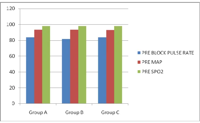
The baseline Physiological characteristics such as pre pulse rate, MAP
and SPO2 and the type of surgery was also matched. There were no much
marked differences among these haemodynamic variables prior to the
Table -1. Matching of three groups according to their demographic
characteristics
Group
n Mean S D ANOVA
’F’
df Significance
AGE
A 20 34.5 7.4
2.461 2, 57 P>0.05
B 20 40.1 8.7
C 20 36.6 8.1
WEIGHT
A 20 65.6 4.3
2.994 2, 57. P>0.05
B 20 64.3 5.4
Figure - 1. Comparison of age and weight between the three groups
The demographic variables of three groups were
matched in the above table -1. The age and weight of three analysed -
statistically significant difference --nil (P>0.05).
Table number 2. Matching of three groups according to their
physiological characteristics
Variables
Group
n Mean S D ANOVA
’F’
df Significance
Pre pulse
rate
A 20 83.6 4.6 1.136
2, 57 P>0.05
B 20 81.6 5.5
C 20 83.4 3.8
Pre MAP
A 20 93.4 1.6
0.325 2, 57. P>0.05
B 20 93.1 2.5
C 20 92.9 1.7
A 20 97.8 0.6
1.715
Pre SPO2 B 20 98.0 0.7 2, 57 P>0.05
Figure - 2. Comparison of baseline haemodynamic variables.
Haemodynamic variables of three groups before surgery were matched in
the table-2.
In this systematic process of the matching analysis,the
• Pulse rate,
• MAP and
Table - 3. Matching of the type of Surgery
Type of surgery A B C Total χ2 df Sig
Bone grafting, distal
locking 0 0 1 1
40.871 34 .194 Buttress plating with k
wire fixation
1 0 0 1
Deroofing & curettage 1 0 0 1
Implant exit, lrs
fixation
0 0 1 1
Implant exit & im
nailing
0 0 1 1
K wire fixation 1 0 0 1
Oblique osteotomy & k
wire fixation
1 0 0 1
ORIF with berbert
screw
0 1 0 1
ORIF with kwire
fixation
1 0 0 1
ORIF with nailing 0 0 1 1
ORIF with narrow
DCP
2 1 2 5
ORIF with plate
osteosynthesis
7 2 1 10
ORIF with plate
osteotomy 0 1 0 1
Radial head excision 0 1 0 1
Refixation of k wire 0 0 1 1
Total
Number of cases 20 20 20 60
The type of surgery was compared between the groups in table-3. There
Comparison of PR, MAP, & SPO2 at different time
intervals between groups:
Table - 4 Comparison of pulse rate between groups at
different time interval.
Pulse
Rate at
Group n Mea
n
S D F df Sig (P)
5
Minutes
A 20 84.4 5.3 1.346 2, 57 .248
B 20 82.8 4.4
C 20 85.2 3.6
10
Minutes
A 20 84.4 6.0 .494 2, 57 .613
B 20 83.4 4.6
C 20 84.8 3.9
20
Minutes
A 20 84.8 4.1 .974 2, 57 .384
B 20 83.6 4.4
C 20 85.3 3.6
30
Minutes
A 20 85.1 3.4 1.236 2, 57 .298
B 20 83.8 4.2
[image:64.612.93.477.273.714.2]1 Hour B 20 83.5 3.8
C 20 85.6 3.0
2 Hour
A 20 84.5 4.6 2.792 2, 57 .070
B 20 83.2 4.3
C 20 86.2 3.0
3 Hour A 20 84.9 3.1
1.391 2, 57 .257 B 20 84.0 4.4
C 20 85.9 3.5
4 Hour
A 20 85.7 2.6 1.634 2, 57 .204
B 20 84.1 4.3
C 20 86.0 3.6
5 Hour
A 20 85.5 3.7 1.892 2, 57 .160
B 20 83.2 5.0
C 20 85.4 3.5
6 Hour
A 20 86.0 3.4 .926 2, 57 .402
B 20 84.4 4.6
C 20 85.6 3.8
8 Hour
A 20 86.0 4.4 .953 2, 57 .392
B 20 84.2 4.3
C 20 85.7 3.6
16 Hour
A 20 85.9 4.9 .731 2, 57 .486
B 20 84.3 3.9
C 20 85.2 4.0
24 Hour
A 20 85.5 4.5 .658 2, 57 .522
B 20 84.4 3.8
C 20 85.8 3.9
The above table-4 shows the pulse rate between three groups at
different time interval. The pulse rates at different interval not significantly
altering among these three (P>0.05).
Table - 5. Comparison of MAP between groups at different time interval.
MAP at Group n Mean S D F df Sig (P)
5
Minutes
A 20 93.6 1.8
1.539 2, 57 .223 B 20 92.5 2.3
10
Minutes
B 20 93.4 1.6
C 20 93.1 1.4
20
Minutes
A 20 92.6 4.4
.582 2, 57 .562 B 20 93.4 1.8
C 20 93.6 1.9
30
Minutes
A 20 93.8 0.9
.487 2, 57 .617 B 20 93.4 1.7
C 20 93.8 2.2
1 Hour
A 20 93.9 1.2
.885 2, 57 .419 B 20 93.2 1.8
C 20 93.5 1.6
Second
hour
A 20 93.9 1.6
.673 2, 57 .514 B 20 93.2 1.9
C 20 93.6 1.9
Third
hour
A 20 93.8 1.6
.143 2, 57 .867 B 20 93.5 2.9
C 20 93.8 1.9
Fourth
hour
A 20 93.4 1.6
1.884 2, 57 .161 B 20 94.0 1.6
hour
C 20 93.9 1.6
6thhour
A 20 93.1 2.6
1.764 2, 57 .181 B 20 93.4 1.9
C 20 94.3 1.7
Eighth
hour
A 20 92.9 2.3
1.234 2, 57 .299 B 20 93.2 1.7
C 20 93.9 2.2
12 Hour
A 20 93.4 2.6
1.007 2, 57 .372 B 20 92.8 1.8
C 20 93.7 1.6
16 Hour
A 20 93.4 2.9
.044 2, 57 .957 B 20 93.6 1.6
C 20 93.6 1.7
24 Hour
A 20 93.4 2.6
.622 2, 57 .540 B 20 93.8 1.6
The above table-5 states the MAP at different intervals between the three
groups. The MAP of three groups were not significantly differing at all intervals
[image:69.612.88.481.314.724.2]between them (P>0.05).
Table - 6. Comparison of spo2 between groups at different
time interval.
SPO2 at Group n Mean S D F df Sig (P)
5
Minutes
A 20 97.4 0.7
.763 2, 57 .471 B 20 97.6 0.5
C 20 97.6 0.7
10
Minutes
A 20 97.4 0.7
.808 2, 57 .451 B 20 97.5 0.6
C 20 97.6 0.6
20
Minutes
A 20 97.2 0.7
2.323 2, 57 .107 B 20 97.5 0.6
C 20 97.7 0.6
30
Minutes
A 20 97.6 0.6
.676 2, 57 .513 B 20 97.6 0.7
C 20 97.6 0.7
2 Hour
A 20 97.5 0.5
1.267 2, 57 .290 B 20 97.6 0.7
C 20 97.8 0.6
3 Hour
A 20 97.4 0.5
2.192 2, 57 .121 B 20 97.8 0.6
C 20 97.8 0.8
4 Hour
A 20 97.4 0.5
2.369 2, 57 .103 B 20 97.6 0.6
C 20 97.7 0.7
5 Hour
A 20 97.2 0.4
2.019 2, 57 .142 B 20 97.5 0.5
C 20 97.4 0.7
6 Hour
A 20 97.3 0.6
1.054 2, 57 .355 B 20 97.5 0.5
C 20 97.6 0.9
8 Hour
A 20 97.5 0.5
1.144 2, 57 .326 B 20 97.8 0.7
12 Hour B 20 97.7 0.6
C 20 98.0 0.8
16 Hour
A 20 97.4 0.8
1.421 2, 57 .250 B 20 97.7 0.5
C 20 97.7 0.6
24 Hour
A 20 97.5 0.6
.868 2, 57 .425 B 20 97.8 0.6
C 20 97.8 0.7
[image:71.612.89.480.64.344.2]
The SPO2 of three groups at different intervals was shown in the above
table-6. The observed differences between the three groups at different intervals
were not found to be statistically significant (P>0.05).
Comparison of Motor Blocks and Sensory blocks:
The onset, completion, total sensory, motor block time are analysed
between three. The initiation times for sensory, motor blocks of these
different sets of population were recorded after the procedure and
between groups.
Blocks Groups n Mean SD ANOVA
‘F’
df Sig
(P)
Significantly
differed
groups
OTSB
A 20
6.4 0.5
81.419
2,
57
.000
B differed
with A&C.
A&C not
differed.
B 20
4.8 0.4
C 20
6.1 0.3
OTMB
A 20
4.3 0.5
609.548
2,
57
.000
A&B not
differed. C
differed with
A&B
B 20
4.0 0.6
C 20
9.5
0.
6
[image:72.612.91.553.185.574.2]
Figure - 3. Comparison of onset time for sensory & motor
block
.Onset time in min
The onset time of Sensory block and Motor blocks were shown in the
above table -7. The group B significantly differed with A&C in respect of their
sensory block on set time and it was comparatively quicker (P<0.001).
But, the onset time difference was not significant between the groups
groups was not significant (P>0.05). The C group significantly differed with
[image:74.612.91.552.326.603.2]A&B groups and it is longer (P<0.001).
Table - 8. Comparison of Time for Complete Sensory and Motor blocks
between groups.
Blocks Groups n Mean SD ANOVA
‘F’ df Sig (P) Significantly differed groups TCSB
A 20 20.6 1.9 0.364
2,
57
.697
All groups did not
differ
significantly B 20 20.8 3.0
C 20 21.4 3.6
TCMB
A 20 17.8 1.7
19.866 2, 57 .000 A&B not differed. C differed with A&B B 20 19.3 2.2
C 20 22.9 3.5
The above table shows time for complete Sensory block and Motor
blocks. In respect of their time for complete sensory block all groups did not
Figure - 4. Comparison of TCSB & TCMB.
Regarding the time for complete motor block, the difference between A&B
groups was not significant (P>0.05). The C group significantly differed with A
group (P<0.001) and also differed with B group (P<0.001) and it is
Table - 9. Comparison of Total Duration of Sensory and Motor blocks
between the groups.
Blocks Groups n Mean SD ANOV A ‘F’
df Sig (P)
Significantly differed groups
TDSB
A 2
0 330.8 14.5
208.49 2, 57 .000
All groups differed
significantly B 2
0 638.5 19.4
C 2
0 464.5 79.1
TDMB
A 2
0 313.2 11.9
2.019 2, 57 .142
No significant differences
between the groups B 2
0 330.2 11.8
C 2
Figure - 5. Comparison of TDMB & TDSB
Total duration of Sensory and Motor blocks were compared in table -9. In
respect of TDSB, the three groups significantly differed between them
(P<0.001). It is longer for B than A and C.
It is longer for C than A but less than B.
Regarding the TDMB,
no significant differences were observed between the groups
[image:77.612.94.436.222.429.2]The durations of surgery between the three groups were compared.
Table - 10. Comparison of surgery duration between the groups.
Groups n
Mean SD
ANOVA
‘F’
df Significance
A 20
113.5
16.0
2.029
2, 57
p>0.05
B 20
108.3
13.7
C 20
117.5
13.9
[image:78.612.130.470.475.676.2]
The above table-10 compares the duration of surgery between three
groups. The durations were not significantly different between the three groups
(P>0.05).
DISCUSSION:
In our study 5 cases had failed block and they were eliminated
from the study. The mean ages of A, B, and C groups were 34.5±7.4, 40.1± 8.7
and 36.6±8.1 years respectively. They did not differ between them
significantly(P>0.05). The mean weights of three groups were 65.6±4.3,
64.3±5.4 and 61.4±6.9 Kgs respectively. The weights of the three groups were
not significantly different (P>0.05).
The pre pulse rates of three groups namely A, B, and C were 83.6 ± 4.6,
81.6±5.5 and83.4±3.8 respectively. Even fortunately the sex ratio of each
group were also similar making the matching unnecessary. The pre MAP
were 93.4 ± 1.6, 93.1±2.5and 92.9±1.7 respectively.
The Pre SPO2 was 97.8±0.6, 98.0±0.7 and 98.1±0.4 respectively. The pre
Physiological variables between the three groups were not significantly
time interval like 5 Minutes, 10 Minutes, 20 Minutes, 30 Minutes, 1 Hour, 2
Hours, 3 Hours, 4 Hours, 5 Hours, 6 Hours, 8 Hours, 12 Hours, 16 Hours
and 24 Hours were analysed statistically. The pulse, MAP and SPO2 recorded
in the above time were not significantly different (P>0.05).
The onset time of sensory blocks of A group was 6.4 ±0.5 minutes, B
group was 4.8±0.4 and C group was 6.1±0.3 minutes. The OTSB of B group
was significantly lower than A & C groups (4.8±0.4 < 6.4 ±0.5& 6.1±0.3 and
P<0.05). The onset time of motor blocks of A group was 4.3± 0.5, B group
was 4.0 ±0.6 and C group was 9.5±0.6 minutes.
The OTMB of C group was significantly higher than A&B groups
(9.5±0.6 > 4.3± 0.5 &4.0 ±0.6 and P<0.05).The above findings have differed
from the observations of the study conducted by Jadon et al in which the
onset time of motor block alone is hastened by adding buprenorphine
but the onset time for sensory block was prolonged. [23]
The Time for Complete Sensory Block of A group was 20.6 ±1.9, B
group was 20.8±3.0 and C group was 21.4±3.6 minutes. The TCSB of three
groups were not significantly different (20.6 ±1.9=20.8±3.0=21.4±3.6 and
P>0.05). The Time for Complete Motor Block of A group was 17.8 ± 1.7, B
The TCMB of C group was significantly higher than A&B groups (22.9
± 3.5> 17.8 ± 1.7&19.3± 2.2 and P<0.05
The Total Duration of Sensory Block of A group was 330.8 ±14.5, B
group was 638.5 ± 19.4 and C group was 464.5± 79.1minutes. The TDSB of B
group was significantly longer than the C and C longer than A groups (638.5 ±
19.4> 464.5± 79.1>330.8 ±14.5 and P<0.001).
The Total Duration of Motor Block of A group was 313.2±11.9, B group
was 330.2 ± 11.8 and C group was 331.8± 13.9 Minutes and thus did not differ
significantly.
The Durations of surgery between the three groups were not significantly
different and hence they were equal (113.5±16.0 ≈ 108.3±13.7 ≈ 117.5±13.9 and P>0.05).
Adverse effects were also watched for in all the