MICROSPHERES LOADED WITH LISINOPRIL
DIHYDRATE
DISSERTATION
Submitted to
The Tamilnadu Dr. M.G.R. Medical University
Chennai – 32
In partial fulfillment for the award of the Degree of
MASTER OF PHARMACY
In the Department of Pharmaceutics
MARCH 2012
DEPARTMENT OF PHARMACEUTICS
PADMAVATHI COLLEGE OF PHARMACY AND RESEARCH
INSTITUTE
CERTIFICATE
This is to certify that this dissertation entitled
FORMULATION AND EVALUATION OF GELATIN
MICROSPHERES LOADED WITH LISINOPRIL
DIHYDRATE
Constitutes the original work carried out by
Reg. No-26106809
For the partial fulfillment of the requirements for the award of Degree of
Master of Pharmacy in Pharmaceutics, carried out in Department of
Pharmaceutics, Padmavathi College of Pharmacy and Research Institute
under my guidance and supervision.
GUIDE
Place: Dharmapuri Dr. K.L.SENTHIL KUMAR, M.Pharm., Ph.D.,
Date:
Padmavathi College of Pharmacy & Research
Institute,
This is to certify that this dissertation entitled
FORMULATION AND EVALUATION OF GELATIN
MICROSPHERES LOADED WITH LISINOPRIL
DIHYDRATE
Constitutes the original work carried out by
Reg. No-26106809
Under the guidance and supervision of
DR. K.L.SENTHIL KUMAR, M.Pharm., Ph.D.,
PRINCIPAL
Padmavathi College of Pharmacy and Research Institute,
Dharmapuri -635205.
PRINCIPAL
Place: Dharmapuri Dr. K.L.SENTHIL KUMAR, M.Pharm., Ph.D.,
Date:
Padmavathi College of Pharmacy & Research
Institute,
CERTIFICATE
This is to certify that this dissertation entitled
FORMULATION AND EVALUATION OF GELATIN
MICROSPHERES LOADED WITH LISINOPRIL
DIHYDRATE
Constitutes the original work carried out by
Reg. No-26106809
Under the guidance and supervision of
Dr. K.L.SENTHIL KUMAR, M.Pharm., Ph. D.,
PRINCIPAL
Padmavathi College of Pharmacy and Research Institute,
Dharmapuri-635205
Head of the Department
Place: Dharmapuri Dr. R.P. EZHIL MUTHU, M.Pharm., Ph.D.,
Date: Department of Pharmaceutics,
Padmavathi College of Pharmacy & Research
Institute,
“With the blessings of Lord Ganesh”
I humbly owe the completion this dissertation work to the almighty whose love and blessing will be with me very moments of life.
It is a delightful moment for me to put in towards all my deep sense gratitude to my esteemed guide Dr.K.L.SENTHIL KUMAR M.Pharm., Ph.D., the department of Pharmaceutics of Padmavathi College of Pharmacy and Research Institute, for his unstinted guidance, innovative ideas, constructive criticism constant encouragements and continues supervision and also for making the requisite arrangement to enable to me to complete me dissertation work at Padmavathi College of Pharmacy and Research Institute in Dharmapuri.
I am heartily thankful to Dr.K.L.SENTHIL KUMAR M.Pharm., Ph.D., Principal of Padmavathi College of Pharmacy and Research Institute, Dharmapuri, for his help and motivation to carry out dissertation work in College.
It is with great pleasure and humble thanks to my eminently esteemed teacher
Dr.R.P.EzhilMuthu M.Pharm., Ph.D., Vice Principal & HOD of Pharmaceutics, Padmavathi College of Pharmacy and Research institute, Dharmapuri for his valuable guidance, keen interest, inspiration, unflinching encouragement and moral support throughout my dissertation work.
I would like to express our sincere thanks to Kalvi Kodai Vallal M.G.Sekar B.A.B.L., Ex.M.P & M.L.A., Chairman of Saptagiri Padmavathi & Pee Gee Group of institutions.
I wish my thanks to Words are insufficient to express my gratefulness and indebtedness to my Pharmaceutis department staffs Mr.M.Muthukumar M.Pharm., Mrs.P.Sumathi M.Pharm., Mr.A.Vasanthan M.Pharm., MBA., for providing relevant information and discussing the bottlenecks with my project superior, valuable suggestions and inspirations.
Mr.L.Samuel M.Pharm., Dr.V.Maithili M.Pharm., ph.D., Mrs.Gnanambigai M.Pharm.,
Mr.Vimalan M.Pharm., Mrs.Valarmani M.pharm., Mr.P.Siva Kumar M.Pharm.,
Mrs.Kartikayani, M.Pharm., Mr.Raj kumar M.Pharm., Head, Department of Pharmacognosy,
Mr.M.Saravanan, M.Pharm., Mr.Ravi, M.sc., Librarian and all of our college teaching and non teaching staff for their valuable suggestion.
Words are not sufficient to express my depressed love and appreciation to my affectionate bellowed parents and my sister who always inspired and cherished me and fill my heart with their love strength which makes my project completion successful.
Friendship is a treasured gift and true friends are few. I am lucky to have enough of them like Yamini, Navya, Sateesh, Pavan, Madhubabu, Sandeep for giving me constant encouragement, moral support and dynamic cooperation throughout my dissertation work.
Finally I consider this is an opportunity to express my gratitude to all the dignitaries, who have been involved directly and or indirectly with the successful completion of this dissertation.
At last but not least, I would like to thank almighty with whose blessings this work has been completed
Thanks to one & all
CHAPTER CONTENTS Page No
1 INTRODUCTION 1-18
1.1 Microspheres As Controlled Drug Delivery System 1-2
1.2 Lisinopril Dihydrate 2-4
1.3 Microencapsulation 4-11
1.4 Polymers Used For Microspheres 12-14 1.5 Methods of Preparation of Gelatin Microspheres 14-16 1.6 Characterization of Microspheres 17-18
2 LITERATURE REVIEW 19-22
3 AIM AND OBJECTIVE 23-24
4 PLAN OF WORK 25-26
5 EXPERIMENTAL INVESTIGATION 27-44
5.1 Material And Equipment Used 27-28
5.2 Drug Profile 29-32
5.3 Excipient Profile 33-41
5.4 Preformulation Studies 42-43
5.5 Standard Plot For Lisinopril Dihydrate 43-44
6 FORMULATION AND DEVELOPMENT 45-46
7 EVALUATION STUDIES 47-73
7.1 Particle Size Determination 47-49 7.2 Determination of Angle of Repose 49-50 7.3 Determination of Bulk Density 50 7.4 Drug Entrapment Efficiency 51 7.5 Determination of Percentage Yield of Microspheres 52 7.6 Scanning Electron Microscopy 53-54 7.7 FT-IR Compatibility Studies of Lisinopril Dihydrate With
Polymers 54-59
7.9 Kinetics of Drug Release 70-73
8 ACCELERATED STABILITY STUDIES 74
9 RESULTS AND DISCUSSION 75-77
10 SUMMARY AND CONCLUSION 78
TABLE
NO LIST OF TABLES PAGE NO.
1 Microencapsulation Processes and Their Applications 7 2 Materials Used For Formulations 27
3 Equipments Used 28
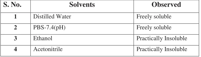
4 Solubility Studies of Lisinopril dihydrate 41 5 Standard Calibration Curve of Lisinopril dihydrate 42 6 Formulation Design of Microparticles 46 7 Common Techniques of Measuring Particle Size 47
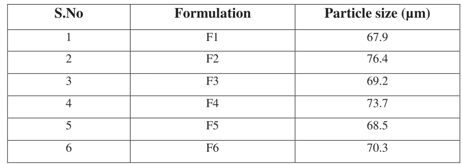
8 Mean Particle size of microsphere formulations 48
9 Relation Between Angle of Repose And Flow Properties 49 10 Angle of Repose of Microparticles 50 11 Bulk Density of Formulations 50 12 Drug Entrapment Efficiency of Microparticles 51 13 Percentage Yield of Formulations 52 14 Comparision of IR Spectra of Lisinopril Dihydrate And In
Combination With Polymers 55
15 In-vitro Dissolution Profile For Formulation F1 61
16 In-vitro Dissolution Profile For Formulation F2 62
17 In-vitro Dissolution Profile For Formulation F3 63
18 In-vitro Dissolution Profile For Formulation F4 64
19 In-vitro Dissolution Profile For Formulation F5 65
20 In-vitro Dissolution Profile For Formulation F6 66
21 Comparative Cumulative Percentage Release Profile of
Formulations F1-F6 67
22 Kinetic Release Data of Formulations 73 23 Stability Studies of Percentage Drug Content of Formulation F1& F5 74
[image:9.612.68.572.92.678.2]
LIST OF GRAPHS
Graph
NO. LIST OF GRAPHS PAGE.NO.
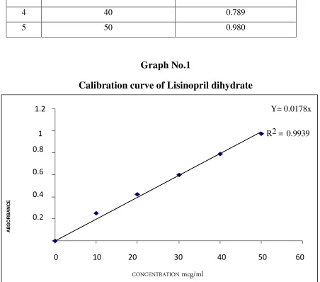
1 Standard Calibration Curve 44
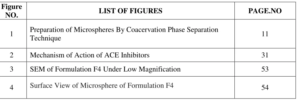
LIST OF FIGURES
Figure
NO. LIST OF FIGURES PAGE.NO
1 Preparation of Microspheres By Coacervation Phase Separation
Technique 11
2 Mechanism of Action of ACE Inhibitors 31 3 SEM of Formulation F4 Under Low Magnification 53
4 Surface View of Microsphere of Formulation F4 54
LIST OF ABBREVIATIONS
ACE Angiotensin Converting Enzyme
ATI Angiotensin I
ATII Angiotensin II
mEq/L Milli equivalents per litre
FDA Food And Drug Administration
SEM Scanning Electron Microscopy
PBS Phosphate Buffer Saline
NF National Formulary
Mg Milligram
MS Microspheres
FTIR Fourier Transform Infrared Spectroscopy
RH Relative Humidity
GI Gastro Intestinal
PVP Poly Vinyl Pyrrolidone
PVA Poly Vinyl Alcohol
PMMA Poly Methyl Methacrylate
KV Kilo Volts
gm Gram
GRAS Generally Regarded As Safe
USP United States Pharmacopoeia
IP Indian Pharmacopoeia
W/O Oil in Water
°C Degree celsius
ml Milli litre
min Minutes
KCl Potassium chloride
Mw Molecular weight
DMA Dexamethasone Acetate
PLGA Polylactic-co-glycolic acid
BSA Bovine Serum Albumin
INTRODUCTION
Padmavathi College of Pharmacy And Research Institute 1
INTRODUCTION
1.1.
MICROSPHERES AS
CONTROLLED DRUG DELIVERY SYSTEM
For many decades, medications of an acute disease or a chronic illness has been accomplished by delivering drugs to the patients via various pharmaceutical dosage forms like tablets, capsules, pills, creams, ointments, liquids, aerosols, injectables and suppositories as carriers. To achieve and then to maintain the concentration of drug administered within the therapeutically effective range needed for medication, it is often necessary to take this type of drug delivery systems several times a day. This results in a fluctuated drug level and consequently undesirable toxicity and poor efficiency.
This factor as well as other factors such as repetitive dosing and predictable absorption leads to the concept of controlled drug delivery systems.1, 2.
The objectives of controlled release drug delivery include two important aspects namely spatial arrangement and temporal delivery of drug.
Spatial placement relates to targeting a drug to a specific organ or tissue, while temporal delivery refers to controlling the rate of drug delivery to the target tissue.
An approximately designed controlled release drug delivery system can be a major advance towards solving these two problems. It is this reason that the science and technology responsible for development of controlled release pharmaceuticals have been and continue to be the focus of a great deal of attention both industrial and academic laboratories.
A controlled release system includes any drug delivery system that “achieves slow release of the drug over an extended period of time”. If the system can provide some control weather this of a temporal or a spatial nature, in other words, if the system is successful in maintaining predictable and reproducible kinetics in the target tissue or cell, it is considered as a controlled release system.
Padmavathi College of Pharmacy And Research Institute 2
The objectives in designing a controlled release system are to deliver the drug at a rate necessary to achieve and maintain a constant drug blood level. This rate should be analogous to that achieved by continuous intravenous infusion where a drug is provided to the patient at a rate just equal to its rate of elimination. This complies that the rate of delivery must be independent of the amount of drug remaining in the dosage form and constant over time. That is release from the dosage form should follow zero-order kinetics3.
The several advantages of a controlled drug delivery system over a conventional dosage forms:
• Improved patient convenience and compliance due to less frequent drug administration. • Reduction in fluctuation in steady state levels and therefore better control of disease
condition and reduced intensity of local or systemic side effects.
• Increased safety margin of high potency drugs due to better control of plasma levels. • Maximum utilization of drug enabling reduction in total amount of dose administered. • Reduction in health care costs through improved therapy, shorter treatment period, less
frequency of dosing and reduction in personnel time to dispense, administer and monitor patients4.
1.2. LISINOPRIL DI HYDRATE
Lisinopril is a potent, competitive inhibitor of angiotensin-converting enzyme (ACE) and is used to treat high blood pressure (hypertension), heart failure and to improve survival after heart attack. Lisinopril, one of the few ACE inhibitors that is not a pro-drug, compete with ATI for binding to ACE and inhibits and enzymatic proteolysis of AT-I to AT-II. Lisinopril, a synthetic peptide derivative, is chemically described as (S)-1-[N2-(1-carboxy-3- phenylpropyl)-L-lysyl]-L-proline dihydrate5. Lisinopril inhibits angiotensin-converting enzyme (ACE) in human subjects and animals. ACE is a peptidyl dipeptidase that catalyzes the conversion of angiotensin I to the vasoconstrictor substance, angiotensin II. Angiotensin II also stimulates aldosterone secretion by the adrenal cortex.
INTRODUCTION
Padmavathi College of Pharmacy And Research Institute 3
results in decreased plasma angiotensin II which leads to decreased vasopressor activity and to decrease aldosterone secretion. The latter decrease may result in a small increase of serum potassium. In hypertensive patients with normal renal function treated with Lisinopril alone for up to 24 weeks, the mean increase in serum potassium was approximately 0.1mEq/L however, approximately 15% of patients had increases greater than 0.5mEq/L and approximately 6% had a decrease greater than 0.5mEq/L. In the same study, patients treated with Lisinopril and hydrochlorothiazide for up to 24 weeks had a mean decrease in serum potassium of 0.1mEq/L; approximately 4% of patients had increases greater than 0.5 mEq/L and approximately 12% had a decrease greater than 0.5mEq/L.
Concomitant administration of Lisinopril and hydrochlorothiazide further reduced blood pressure in Black and non-Black patients and any racial differences in blood pressure response were no longer evident. Administration of Lisinopril to patients with hypertension results in a reduction of both supine and standing blood pressure to about the same extent with no compensatory tachycardia. Symptomatic postural hypotension is usually not observed although it can occur and should be anticipated in volume and/or salt-depleted patients. In most patients studied, onset of antihypertensive activity was seen at one hour after oral administration of an individual dose of Lisinopril, with peak reduction of blood pressure achieved by 6 hours. Although an antihypertensive effect was observed 24 hours after dosing with recommended single daily doses, the effect was more consistent and the mean effect was considerably larger in some studies with doses of 20 mg or more than with lower doses; however, at all doses studied, the mean antihypertensive effect was substantially smaller 24 hours after dosing than it was 6 hours after dosing. In some patients achievement of optimal blood pressure reduction may require two to four weeks of therapy.
Upon multiple dosing, Lisinopril exhibits an effective half-life of accumulation of 12 hours. Impaired renal function decreases elimination of Lisinopril, which is excreted principally through the kidneys, but this decrease becomes clinically important only when the glomerular filtration rate is below 30mL/min.
Padmavathi College of Pharmacy And Research Institute 4
superior to hydrochlorothiazide in effects on systolic and diastolic pressure in a population that was 3/4 Caucasian. Lisinopril was approximately equivalent to Atenolol and Metoprolol in effects on diastolic blood pressure, and had somewhat greater effects on systolic blood pressure. In patients with renovascular hypertension Lisinopril has been shown to be well tolerated and effective in controlling blood pressure.
1.3 Microencapsulation
Microencapsulation is a process by which very tiny droplets or particles of liquid or solid material are surrounded or coated with a continuous film of polymeric material. In microencapsulation particle size is ranging from several tenths to 5000 microns6.
Microencapsulation provides the means of converting liquids to solids, of altering
colloidal and surface properties, of providing environmental protection and of controlling the release characteristics or availability of coated materials. Several of these properties can be attained by macro packaging techniques; however, the uniqueness of microencapsulation is the smallness of the coated particles and their subsequent use and adaptation to a wide variety of dosage forms7.
REASONS FOR MICROENCAPSULATION
8The primary reason for microencapsulation is found to be either for sustained or prolonged drug release.
This technique has been widely used for masking taste and odor of many drugs to improve patient compliance.
This technique can be used for converting liquid drugs in a free flowing powder.
The drugs, which are sensitive to oxygen, moisture or light, can be stabilized by microencapsulation.
Incompatibility among the drugs can be prevented by microencapsulation.
Vaporization of many volatile drugs e.g. methyl salicylate and peppermint oil can be prevented by microencapsulation.
INTRODUCTION
Padmavathi College of Pharmacy And Research Institute 5
Alteration in site of absorption can also be achieved by microencapsulation.
Toxic chemicals such as insecticides may be microencapsulated to reduce the possibility of sensitization of factorial person.
Bakan and Anderson reported that microencapsulated vitamin A palmitate had enhanced stability8.
RELEASE MECHANISMS
8Mechanisms of drug release from microspheres are
1. Degradation controlled monolithic system:
-The drug is dissolved in matrix and is distributed uniformly throughout. The drug is strongly attached to the matrix and is released on degradation of the matrix. The diffusion of the drug is slow as compared with degradation of the matrix.
2. Diffusion controlled monolithic system:
-Here the active agent is released by diffusion prior to or concurrent with the degradation of the polymer matrix. Rate of release also depend upon where the polymer degrades by homogeneous or heterogeneous mechanism.
3. Diffusion controlled reservoir system: -
Here the active agent is encapsulated by a rate controlling membrane through which the agent diffuses and the membrane erodes only after its delivery is completed. In this case, drug release is unaffected by the degradation of the matrix.
4. Erosion: -
Erosion of the coat due to pH and enzymatic hydrolysis causes drug release with certain coat material like glyceryl mono stearate, beeswax and steryl alcohol etc.
METHODS TO MANUFACTURE MICROSPHERES
9Basic Microencapsulation processes can be divided into
Chemical methods
Padmavathi College of Pharmacy And Research Institute 6
Chemical methods
• Solvent Evaporation
• Interfacial polymerization/cross linking • In-situ polymerization
• Matrix polymerization
• Polymer-polymer compatibility • In-liquid drying
• Thermal and ionic gelation in liquid media
Physico-chemical methods
• Ionotropic gelation
• Coacervation phase separation
Mechanical methods
• Pan coating
• Air suspension coating • Centrifugal extrusion • Spray drying
• Spray coating
• Spinning disk or rotational suspension separation • Electrostatic deposition
• Pressure extractionor spraying into solvent extraction bath11.
INTRODUCTION
Padmavathi College of Pharmacy And Research Institute 7
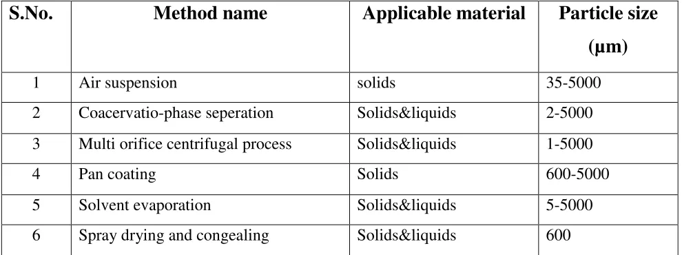
Table No.1
Microencapsulation Processes and Their Applicabilities
S.No.
Method name
Applicable material
Particle size
(µm)
1 Air suspension solids 35-5000 2 Coacervatio-phase seperation Solids&liquids 2-5000 3 Multi orifice centrifugal process Solids&liquids 1-50004 Pan coating Solids 600-5000
5 Solvent evaporation Solids&liquids 5-5000 6 Spray drying and congealing Solids&liquids 600
Ideal characteristics of drug for microencapsulation
5Particle size requirement
The lower the molecular weight, faster and complete is the absorption of the drug. The drugs having size 150-600 Daltons they can easily diffuse through the membrane but diffusivity (the ability of drug to diffuse through the membrane) is inversely related to molecular size.
The drug or protein should not be adversely affected by the process.
Reproducibility of release profile and the method
No stability problem
Drugs unstable in GI environment cannot be administered as oral controlled release formulation because of bioavailability problems. E.g.Nitroglycerine.
[image:20.612.85.580.154.339.2]Padmavathi College of Pharmacy And Research Institute 8
Therapeutic range
A candidate drug for controlled delivery system should have a therapeutic range wide enough such that variations in the release rate do not result in a concentration beyond this level.
Therapeutic index
The ratio of maximum safe concentration to the minimum effective concentration of drug is called as therapeutic index. The release rate of drug with narrow therapeutic index should be such that the plasma concentration is attained between the therapeutically safe and effective range. It is necessary because such drugs have toxic concentration nearer to their therapeutic range.
Elimination half life
Smaller the half life larger the amount of drug to be incorporated in the controlled release dosage form. Drugs with t1\2 in the range of 2-4 hours make a good candidates for such a system. E.g.propronalol.
Plasma Concentration-Response relationship
Drugs whose pharmacological activity is independent of its concentration are poor candidates for controlled release systems.
Microencapsulation of pharmaceuticals is undertaken for the following
applications
9, 10:
INTRODUCTION
Padmavathi College of Pharmacy And Research Institute 9
Microencapsulation by Coacervation Phase Separation Technique
8Preparation
The core material will be added to the solution. The core material should not react or dissolve in water (maximum solubility 2%) The core material is dispersed in the solution. The particle size will be defined by dispersion parameter, as stirring speed, stirrer shape, surface tension and viscosity. Size range 2µm - 1200µm
Coacervation
• Coacervation starts with a change of the pH value of the dispersion, e.g. by adding organic acids. The result is a reduction of the solubility of the dispersed phases (shell material).
• The shell material (coacervate) starts to precipitate from the solution. • The shell material forms a continuous coating around the core droplets.
Cooling and hardening phase
• The shell material is cooled down to harden and forms the final capsule. • Hardening agents like formaldehyde can be added to the process.
• The microcapsules are now stable in the suspension and ready to be dried.
Drying phase
• The suspension is dried in a spray dryer or in a fluidized bed drier. • Spray Drying is a suitable method for heat sensitive Products.
• The atomized particles assume a spherical shape. The rapid the coating material keeps the core material below 100°C, even if the temperature in the drying chamber is much greater.
• Microencapsulation makes the spray drying process easier for sticky products like fruit pulp or juice, with a high content of invert sugar.
Coacervation-Phase Separation
The general outline of the processes consists of three steps carried out under continuous agitation:
Padmavathi College of Pharmacy And Research Institute 10
material phase, an immiscible polymer in a liquid state, is formed by utilizing one of the methods of the methods of phase separation-coacervation, i.e., by changing the temperature of the polymer solution; or by adding a salt, nonsolvent, or incompatible polymer to the polymer solution; or by inducing a polymerpolymer interaction.
2. Deposition of the coating – It consists of depositing the liquid polymer coating upon the core material. This is accomplished by controlled, physical mixing of the material in the manufacturing vehicle. Deposition if the liquid polymer coating around the core material occurs if the polymer is adsorbed at the interface formed between the core material and the liquid vehicle phase, and this adsorption phenomenon is a prerequisite to effective coating. The continued deposition of the coating material is promoted by a reduction in the total free interfacial energy of the system, brought about by the decrease of the coating material surface area during coleasance of the liquid polymer droplets.
3. Rigidization of the coating – It involves rigidizing the coating, usually by thermal, cross-linking, or desolvation techniques, to form a self-sustaining microcapsules.
Padmavathi College of Pharmacy
Preparation of Microspheres
Aqueous
Drug dispersed or dissolved in the polymer solution
Phase sepration
Hardening by
Microspheres
INTRODUCTION
Pharmacy And Research Institute
Figure No.1
Microspheres by Coacervation Phase Separation
Technique
Aqueous solution of polymer
Drug dispersed or dissolved in the polymer solution
sepration by
Temperature change
Polymer rich globules
Hardening by
adding cross linking agent
Microspheres in aqueous/organic phase
Separation and Drying
MICROSPHERES
INTRODUCTION
11
Phase Separation
Padmavathi College of Pharmacy And Research Institute 12
1.4. POLYMERS USED FOR MICROSPHERES
12Synthetic Polymers
Non-biodegradable
PMMA Acrolein
Epoxy polymers
Biodegradable
Lactides and Glycolides copolymers Polyalkyl cyanoacrylates
Polyanhydrides
Natural polymers
Proteins
Albumins Gelatin Collagen
Carbohydrates
Starch agarose Carrageenan Chitosan
Chemically modified carbohydrates
Poly (acryl) dextranPoly (acryl) starch
CLASSIFICATION OF POLYMERS
13Water-Soluble Synthetic Polymers
• Poly (acrylic acid) cosmetic, pharmaceuticals, immobilization of Cationic drugs, base for
Carbopol polymers.
• Poly (ethylene oxide) Coagulant, flocculent, very high molecular-weight up to a few
millions, swelling agent.
• Poly (ethylene glycol) Mw <10,000; liquid (Mw <1000) and wax (Mw >1000),
Plasticizer, base for suppositories.
• Poly (vinyl pyrrolidone) used to make betadine (iodine complex of PVP) with less
toxicity than iodine, plasma replacement and tablet granulation.
• Poly (vinyl alcohol) Water-soluble packaging, tablet binder, tablet coating.
• Polyacrylamide Gel electrophoresis to separate proteins based on their molecular
INTRODUCTION
Padmavathi College of Pharmacy And Research Institute 13
• Poly (isopropyl acrylamide) and poly (cyclopropyl methacrylamide) are thermogelling
acrylamide derivatives, its balance of hydrogen bonding, and hydrophobic association changes with temperature.
Cellulose-Based Polymers
13• Ethyl cellulose is insoluble but dispersible in water, used in aqueous coating system for
sustained release applications.
• Carboxymethyl cellulose is super disintegrant and emulsion stabilizer.
• Hydroxyethyl and hydroxypropyl celluloses are soluble in water and in alcohol, used in
tablet coating.
• Hydroxypropyl methyl cellulose is Binder for tablet matrix and tablet coating, gelatin
alternative as capsule material.
• Cellulose acetate phthalate generally used in Enteric coating.
Hydrocolloids
• Alginic acid is used in oral and topical pharmaceutical products; thickening and
suspending agent in a variety of pastes, creams, and gels, as well as a stabilizing agent for oil-in-water emulsions; binder and disintegrant.
• Gelatin a protein derivative and natural polymer used for controlled release of drug by using suitable cross linking agent.
• Chitosan Cosmetics and controlled drug delivery applications, mucoadhesive dosage
forms, rapid release dosage forms.
Water-Insoluble Biodegradable Polymers
13• (Lactide-co-glycolide) polymers Microparticle–nanoparticle for protein delivery.
Starch-Based Polymers
• Starch is used as Glidant, a diluent in tablets and capsules, a disintegrant in tablets and
capsules, a tablet binder.
Padmavathi College of Pharmacy And Research Institute 14
1.5.
METHODS
TO
MANUFACTURE
OF
GELATIN
LOADED
MICROSPHERES
8, 10Gelatin loaded microspheres can be prepared by using any one of the different following techniques
Single Emulsion Polymerization Technique
Single Emulsion Technique the micro particulate carriers of natural polymers, i.e. those of proteins (e.g. gelatin) and carbohydrates (sodium alginate, chitosan) are prepared by single emulsion technique. The natural polymers are dissolved or dispersed in aqueous medium followed by dispersion in the non-aqueous medium e.g., oil. In the second step of preparation, cross linking of the dispersed globule is carried out. The cross-linking can be achieved either by means of heat or by using the chemical cross linkers (gluteraldehyde, formaldehyde, calcium chloride, genipin etc.)
Double Emulsion Technique
Double emulsion technique Double emulsion method of microspheres preparation involves the formation of the multiple emulsions or the double emulsion of type w/o/w and is best suited to the water-soluble drugs, peptides, proteins and the vaccines. This method can be used with both the natural as well as the synthetic polymers. The aqueous protein solution is dispersed in a lipophilic organic continuous phase. This protein solution may contain the active constituents.
Spray Drying
INTRODUCTION
Padmavathi College of Pharmacy And Research Institute 15
Phase Separation Coacervation Technique
Phase Separation Coacervation Technique Phase separation method is specially designed for preparing the reservoir type of the system to encapsulate water soluble drugs e.g. peptides, proteins. Some of the preparations are of matrix type particularly, when the drug is hydrophobic in nature e.g. steroids. In matrix type device, the drug or the protein is soluble in the polymer phase. The process is based on the principle of decreasing the solubility of the polymer in the organic phase to affect the formation of the polymer rich phase called the ‘coacervates’. In this technique the polymer is first dissolved in a suitable solvent and then drug is dispersed by making its aqueous solution, if hydrophilic or dissolved in the polymer solution itself, if hydrophobic. Phase separation is then accomplished by changing the solution conditions by using any of the method mentioned. Salt addition Non-solvent addition addition of in-compatible polymer Change in pH the process is carried out under continuous stirring to control the size of the microparticles8.
Interfacial Cross Linking
Interfacial cross-linking is derived from interfacial polycondensation, and was developed to avoid the use of toxic diamines, for pharmaceutical or cosmetic applications. In this method, the small bifunctional monomer containing active hydrogen atoms is replaced by a biosourced polymer, like a protein. When the reaction is performed at the interface of an emulsion, the acid chloride reacts with the various functional groups of the protein, leading to the formation of a membrane. The cross-linked protein microcapsules are biocompatible and biodegradable, and the presence of the protein backbone renders the membrane more resistant and elastic than those obtained by interfacial polycondensation10. The method is very versatile, and the properties of the microcapsules (size, porosity, degradability, mechanical resistance) can be easily tuned by varying the preparation parameters. A carbohydrate can be added to the protein, for the modulation of particle biodegradability.
Complex Coacervation
Padmavathi College of Pharmacy And Research Institute 16
polymer-polymer incompatibility mentioned earlier in that one phase contains most of the two polymers whereas the second phase is a dilute polymer solution. Polymer-polymer incompatibility yields two phases with each containing predominately one of the two polymers. For proper encapsulation, it is preferred to have liquid coacervates which can flow around or completely wrap the internal phase particles or droplets.
Solvent Evaporation
INTRODUCTION
Padmavathi College of Pharmacy And Research Institute 17
1.6. CHARACTERIZATION OF MICROSPHERES
Particle size, Shape and Morphology
All the microspheres were evaluated with respect to their size and shape using optical microscope fitted with an ocular micrometer and a stage micrometer. The particle diameters of more than 100 microspheres were measured randomly by optical microscope. Scanning Electron photomicrographs of drug‐loaded microspheres were taken. A small amount
of microspheres was spread on gold stub. Afterwards, the stub containing the sample was placed in the Scanning electron microscopy (SEM). A Scanning electron photomicrograph was taken at an acceleration voltage of 20KV.
Entrapment Efficiency
The capture efficiency of the microspheres or the percent entrapment can be determined by allowing washed microspheres to lyse. The lysate is then subjected to the determination of active constituents as per monograph requirement. The percent encapsulation efficiency is calculated using following equation
% Entrapment = Actual content/Theoretical content x 100.
Scanning Electron Microscopy (SEM)
The surface morphology of blank microspheres, drug loaded microspheres and microspheres collected after dissolution studies were examined by a Scanning electron
microscope (Hitachi, S-3600N, Japan). The samples were fixed on brass sub using double-sided tape and then gold- coated in vacuum by a sputter coater. The SEM pictures were then taken at an excitation voltage of 15 KV14.
Swelling Index
15Padmavathi College of Pharmacy And Research Institute 18
dried mass of sample. The swelling index of the microsphere was calculated by using the formula
Swelling index= (mass of swollen microspheres - mass of dry microspheres/mass of dried microspheres) 100.
In Vitro
Drug Release
To carry out In Vitro drug release, accurately weighed 50 mg of loaded microspheres were dispersed in dissolution fluid in a beaker and maintained at 37±2°C under
continuous stirring at 100 rpm. At selected time intervals 5 ml samples were withdrawn through a hypodermic syringe fitted with a 0.4 µm millipore filter and replaced with the same volume of pre-warmed fresh buffer solution to maintain a constant volume of the receptor compartment. The samples were analyzed spectrophotometrically. The released drug content was determined from the standard calibration curve of given drug.
In Vitro
Diffusion Studies
In Vitro diffusion studies were performed using in vitro nasal diffusion cell. The receptor chamber was filled with buffer maintained at 37±2°C. Accurately weighed microspheres
equivalent to 10 mg were spread on sheep nasal mucosa. At selected time intervals 0.5 ml of diffusion samples were withdrawn through a hypodermic syringe and replaced with the same volume of prewarmed fresh buffer solution to maintain a constant volume of the receptor compartment. The samples were analyzed spectrophotometrically.
Accelerated Stability Studies
The preparation was divided into 3 sets and stored at 4°C (refrigerator), room temperature and 40°C (thermostatic oven). After 15, 30 and 60 days drug content of all the formulation was determined Spectrophotometrically.
LITERATURE REVIEW
Padmavathi College of Pharmacy and Research Institute 19
LITERATURE REVIEW
Santanu Chakraborty et al., (2010) prepared microparticles of aceclofenac by gelation technique using a blend of sodium alginate and carbopol 934 as release retardant. The entrapment efficiency was found within the range of 73.6 ± 3.21% to 90.7 ± 2.48%. All the formulations were investigated for various evaluation parameters like particle size, flow behavior, percentage swelling, surface morphology study and in vitro drug release etc. All the formulation showed good flow behavior as compared to the pure drug. SEM study revealed that the spheres were almost spherical in shape with rough outer surface. In-vitro drug release study showed that by increasing the polymer concentration the drug release of all the formulation were gradually decreased and the optimized formulation (F7) was able to sustain the drug release for 12 hours15.
Ying Lu, Guoqing Zhang et al., (2007)prepared Flubiprofen gelatin microspheres (FP-GMS) by an emulsion-congealing method. The particle size of FP-GMS with optimized formulation was 2.5, 12.3µm with a mean size of gelatin microspheres of 7.53µ m. The drug loading efficiency was 5.02% (w/w). A total of 96% original FP was remained in the microspheres after being stored under 75% humidity and 37°C for 3 months. These data indicate that the simple emulsion-congealing method can be used to encapsulate water soluble drugs16.
Anand kumar et al., (2011)prepared controlled release of Lamivudine microspheres by W/O/W multiple emulsion solvent evaporation technique using osmogen like sodium chloride, polymers like ethyl cellulose, cellulose acetate and polyvinyl alcohol as a continuous phase. The prepared microspheres were characterized for the percent drug content, encapsulation efficiency, FTIR, scanning electron microscopy (SEM), In vitro dissolution studies, in vitro kinetic studies and accelerated stability studies17. The release kinetics data and characterization studies indicate that drug release from microspheres was diffusion-controlled and that the microspheres were stable.
Padmavathi College of Pharmacy and Research Institute 20 conventional microcapsules to all other small particulate systems such as self assembling structures that involve preparative manipulation. The review covers encapsulation materials, physics of release through the capsule wall and desorption from carrier, techniques of preparation, many uses to which microcapsules are put.
P.V. Kozlov et al., (1983) studied Structural diversity of gelatin chain units determines the specific features of gelatin properties. Most synthetic polymers show no such features that are typical of most biopolymers. The first peculiarity of gelatin common to all biopolymers arises from the presence of both acidic and basic functional groups in the gelatin macromolecules. The second peculiarity of gelatin is its capacity to form a specific triple-stranded helical structure not observed in synthetic polymers. The third peculiarity of gelatin as a biopolymer is its specific interaction with water which is different to that observed with synthetic hydrophilic polymers18.
Vikas Parashar et al., (2010)prepared Bovine Serum Albumin microspheres in four batches. The emulsion cross-linking method was used for the preparation. The quantity of BSA varies for each formulation. The microspheres were spherical, discrete and compact and size distribution was between 33.28 to 36.25 µm. In vitro studies were carried out at different pH for a period of 18 h and compared with marketed formulation. From all the batches it is concluded that when concentration of polymer increases microspheres shows more controlled and prolonged release19.
Satish Balakrishna Bhise et al., (2005) developed RIF’s loaded porous microspheres as a controlled release dosage form. Eudragit based porous microspheres of RIF were prepared by emulsion solvent diffusion method. Prepared porous microspheres were evaluated for its entrapment efficacy, morphology, thermal behavior, crystalline nature, in-vitro drug release and stability in simulated gastric fluid20. In vitro drug release studies indicated that drug to polymer ratio of 2:1 showed more than 85% drug release over the period of 3hrs.
LITERATURE REVIEW
Padmavathi College of Pharmacy and Research Institute 21
Sunil K. Jain et al., (2004) studied microsphere dosage forms have gained increasing importance as oral controlled drug delivery systems. These systems present several advantages in comparison to unit dosage forms such as more predictable gastric emptying and less local irritation. Microsphere systems also minimize the possible intestinal retention of undigested polymer materials in chronic dosing. The Eudragits are a family of polymers based on acrylic and methacrylic acids suitable for use in orally administered drug delivery systems22.
Kishore Narra et al., (2009) prepared the rifampicin loaded sodium alginate beads by ionic gelation method. In this method, various formulation variables such as the addition of PVA to the drug-alginate mixture and addition of PVP to cross linking solution were studied. PVA and PVP can be used in the formulation to improve in-vitro characteristics of rifampicin loaded alginate beads23.
Sanat Kumar Basu et al., (2009) prepared Microspheres (MS) of Ketorolac Tromethamine (KT) for oral delivery were prepared by complex coacervation (method-1) and simple coacervation (method-2) methods without the use of chemical cross–linking agent (glutaraldehyde) to avoid the toxic reactions and other undesirable effects of the chemical cross-linking agents. Alternatively, ionotropic gelation was employed by using sodium-tripolyphosphate (Na-TPP) as cross linking agent. Chitosan and gelatin B were used as polymer and copolymer respectively24.
Bhaskar Mazumder et al., (2009) studied the micro particles mean particle size, percentage yield were decreased significantly with decrease in drug-polymer ratio, surfactant concentration, stirring speed and volume of continuous phase. It was found that in vitro release were decreased significantly with decrease in drug-polymer ratio and stirring speed but increased significantly with increase in surfactant concentration and volume of continuous phase25.
Chowdary K.P.R et al., (1989) carried out microencapsulation of aspirin, diazepam and nitrofurantoin by calcium alginate. The microparticles were found to be discrete, spherical and free flowing. The release mechanism was found to be of diffusion type and release depended on solubility of the core material in the dissolution fluid43.
Padmavathi College of Pharmacy and Research Institute 22 surfaced alginate microcapsules of ibuprofen were obtained by this method. The preparation was based on dispersion of sodium alginate-ibuprofen matrix in liquid paraffin followed by coating process by calcium chloride44.
Prasant K Rout et al., (2009) describes preparation of microspheres by solvent evaporation and W/O emulsion solvent evaporation methods followed by in vitro characterization of microspheres to evaluate the effect of method of preparation on physical properties and drug release profile of microspheres. The mean geometric particle size of microspheres prepared by solvent evaporation method was found in the ranges of 40-50 µm and the microspheres prepared by W/O emulsion solvent evaporation method was found in a ranges of 126-150µm, respectively26.
Alf Lamprecht, Ulrich Schafer et al., (2000) demonstrated the potential of confocal laser scanning microscopy as a characterization tool for different types of microparticles which are prepared by various methods including complex coacervation ,spray drying, double emulsion solvent evaporation technique and ionotropic gelation technique45.
LakshmanaPrabu S et al., (2009) studied that drug:polymer ratio had a considerable effect on the entrapment efficiency, however particle size distribution of microspheres was more dependent on the volume of dichloromethane and polyvinyl alcohol concentration rather than on the drug: polymer ratio. Drug, polymer concentrations were varied to obtain optimum release profile for sustaining the action of the drug27.
AIM AND OBJECTIVE
Padmavathi college of pharmacy and Research Institute 23
AIM AND OBJECTIVE
AIM
The main aim of present work is to develop Lisinopril dihydrate loaded Gelatin microspheres by using Coacervation phase separation technique. Oral bioavailability of Lisinopril dihydrate is 25-28%. Dose of Lisinopril is 10-80mg per day, thus conventional dosage form has several draw backs, so the main aim behind this work is to retard the release of drug at controlled rates throughout the day and to improve bioavailability as well as patient compliance by using gelatin and other polymer mixtures and to reduce dosage frequency.
OBJECTIVES
Lisinopril dihydrate is an angiotensin-converting enzyme used in the treatment of hypertension, myocardial infarction, heart failure. It is used in dose of 10-80 mg twice daily. The present study “Lisinopril microparticles by coacervation phase separation technique” was meticulously designed to improve the therapeutic efficacy and to prolong its release.
The following experimental protocol was therefore designed to allow systemic approach to the study:
1) Compatibility study- compatibility with various polymers by using IR
2) Preparation of standard curve for Lisinopril dihydrate in phosphate buffer (pH 7.4)
3) To prepare microparticles of Lisinopril dihydrate using polymers gelatin, carbopol and sodium alginate by using coacervation phase separation technique.
4) The following evaluation parameters were carried out based on laboratory experiments: a) Drug entrapment efficiency
b) Flow property of prepared microparicles
c) In-vitro dissolution studies by using dissolution tester USP (XXIII) basket method release.
Padmavathi college of pharmacy and Research Institute 24 • To overcome the rapid elimination of drug and to develop the oral controlled drug
delivery system.
• To increase the biological half life of the drug or to maintain the constant drug
concentration in the body.
PLAN OF WORK
Padmavathi College of Pharmacy and Research Institute 25
PLAN OF WORK
Present work was carried out to design and evaluate the Lisinopril dihydrate loaded gelatin microspheres.
1) Selection of drug 2) Literature review 3) Preformulation studies
a) Identification of drug
physical appearance
melting point
FT-IR spectra
b) Solubility studies
c) Partition coefficient
d) FT-IR study for drug excipient interaction
e) Determination of absorption maxima (λ max)
4) Preparation of standard curve in phosphate buffer pH 7.4 5) Preparation of gelatin loaded microspheres
6) Characterization of prepared gelatin microspheres
• Yield of production
• percent entrapment efficiency • particle size analysis
• surface morphology by scanning electron microscopy (SEM)
7) Characterization of prepared gelatin microspheres of Lisinopril dihydrate
a) Angle of repose b) Bulk density
Padmavathi College of Pharmacy and Research Institute 26 d) Interpretation of drug release mechanism by kinetic models
• Zero-order • First order • Higuchi model
• Korsmeyer-peppas model.
EXPERIMENTAL INVESTIGATION
Padmavathi College of Pharmacy and Research Institute 27
MATERIALS AND METHODS
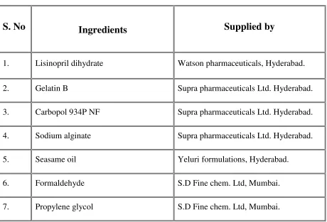
[image:40.612.78.538.219.532.2]The following materials were used as supplied by the manufacturers without further purification:
Table No.2
5.1. Materials Used For Formulations
S. No
Ingredients
Supplied by
1. Lisinopril dihydrate Watson pharmaceuticals, Hyderabad. 2. Gelatin B Supra pharmaceuticals Ltd. Hyderabad. 3. Carbopol 934P NF Supra pharmaceuticals Ltd. Hyderabad. 4. Sodium alginate Supra pharmaceuticals Ltd. Hyderabad. 5. Seasame oil Yeluri formulations, Hyderabad.
6. Formaldehyde S.D Fine chem. Ltd, Mumbai. 7. Propylene glycol S.D Fine chem. Ltd, Mumbai.
Padmavathi College of Pharmacy and Research Institute 28
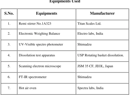
Table No.3
Equipments Used
S.No.
Equipments
Manufacturer
1. Remi stirrer No.1A323 Titan Scales Ltd. 2. Electronic Weighing Balance Electro labs, India 3. UV-Visible spectro photometer Shimadzu
4. Dissolution test apparatus USP Rotating basket dissolution. 5. Scanning electron microscope JSM 35 CF, JEOL, Japan
6. FT-IR spectrometer Shimadzu
EXPERIMENTAL INVESTIGATION
Padmavathi College of Pharmacy and Research Institute 29
5.2 DRUG PROFILE
Lisinopril Dihydrate
Lisinopril dihydrate is a lysine derivative, an oral angiotensin-converting enzyme (ACE) inhibitor.
Chemical Name
(S)-1-(N2-(1-Carboxy-3-phenylpropyl)-L-lysyl)-L-proline dihydrateMolecular Weight
441.52Structural Formula
Category
AntihypertensiveAppearance
white to off-white, crystalline powderSolubility
It is soluble in water, sparingly soluble in methanol, and practically insoluble in ethanol.Melting Point
Around 160̊CPadmavathi College of Pharmacy and Research Institute 30
Pharmacokinetics
Following oral administration of Lisinopril, peak serum concentrations of Lisinopril occur within about 7 hours. Lisinopril does not appear to be bound to other serum proteins. Lisinopril does not undergo metabolism and is excreted unchanged entirely in the urine. Declining serum concentrations exhibit a prolonged terminal phase which does not contribute to drug accumulation. This terminal phase probably represents saturable binding to ACE and is not proportional to dose. Systemic bioavailability of Lisinopril dihydrate is approximately 25%28.
• DOSE : 5-80mg daily • ONSET OF ACTION : 1Hour
• PEAK PLASMA CONCENTRATION : 6 Hours • BIOAVAILABILITY : 25%-28%
• EXCRETION : Unchanged in urine
• HALF LIFE ELIMINATION : 12 HOURS on multiple dosing. • PROTEIN BINDING : Doesn’t bound to serum proteins • VOLUME OF DISTRIBUTION : 28ML/MIN
•
Mechanism of Action
EXPERIMENTAL INVESTIGATION
Padmavathi College of Pharmacy and Research Institute 31
renal function treated with Lisinopril alone for up to 24 weeks, the mean increase in serum potassium was approximately 0.1mEq/L29.
Fig.No.2
Padmavathi College of Pharmacy and Research Institute 32
SIDE EFFECTS
First doses of Lisinopril can cause dizziness due to a drop in blood pressure.
Lisinopril can cause nausea, headaches, anxiety, insomnia, drowsiness, nasal congestion, and sexual dysfunction.
Like all ACE inhibitors, Lisinopril may cause a nonproductive cough that resolves when the drug is discontinued.
Lisinopril should be stopped if there are symptoms or signs of an allergic reaction including feelings of swelling of the face, lips, tongue or throat. Severe allergic reactions (anaphylaxis) and hives occasionally occur. (Ref FDA).
Rarely, Lisinopril may cause a decrease in red blood cells (anemia), white blood cells (leukopenia), and platelets (thrombocytopenia).
CONTRAINDICATIONS
Pregnancy
: Lisinopril should not be taken during pregnancy because fetuses and neonates have died when Lisinopril was administered during pregnancy29.Nursing Mothers
: It is not known whether Lisinopril is excreted in breast milk.EXPERIMENTAL INVESTIGATION
Padmavathi College of Pharmacy and Research Institute 33
5.3
EXCIPIENT PROFILE
5.3.1. Gelatin Type B
Gelatin type B, with isotonic point of 4.8 to 5.2, is the result of an alkaline pretreatment of the collagen. Gelatin is a heterogeneous mixture of single or multi-stranded polypeptides, each with extended left-handed proline helix conformations and containing between 50 - 1000 amino acids. Gelatin is produced by partial hydrolysis of collagen extracted from the boiled bones, connective tissues, organs and some intestines of animals such as domesticated cattle, and pigs32. Type A gelatin (dry and ash free) contains 18.5 % nitrogen, but due to the loss of amide groups, Type B gelatin contains only about 18% of nitrogen.
Gelatin melts to a liquid when heated and solidifies when cooled again. Together with water, it forms a semi-solid colloid gel. Gelatin forms a solution of high viscosity in water, which sets to a gel on cooling, and its chemical composition is, in many respects, closely similar to that of its parent collagen.
Gelatin is an amphoteric protein with isotonic point between 5 and 9 depending on raw material and method of manufacture. Like its parent protein, collagen, it is unique in that it contains 14% hydroxyproline, 16 % proline and 26 % glycine.
The cross-linking of gelatin with aldehydes is being used to extend the uses of gelatin. In particular, treatment of gelatin films with glutaraldehyde is receiving considerable study in order to improve their thermal resistance, decrease their solubility in water as well as to improve their mechanical properties.
Solution pH (1%) is usually about pH 5 but can vary considerably. At this pH the viscosity of Type B gelatin is minimal and the gel strength is maximal, hence from the manufacturers’ point of view it is advantageous to manufacture gelatin at this pH30.
Padmavathi College of Pharmacy and Research Institute 34
penetrometer type instruments have been adapted to determine Bloom Strength. In the present work the bloom strength of gelatin B used was 250.
Structure of gelatin
Empirical formula of gelatin
Gelatin contains many glycine (almost 1 in 3 residues, arranged every third residue), proline and 4-hydroxyproline residues. A typical structure is -Ala-Gly-Pro-Arg-Gly-Glu-4Hyp-Gly-Pro-.
Synonyms
Gelatine, Jelly, gel.
Solubility
EXPERIMENTAL INVESTIGATION
Padmavathi College of Pharmacy and Research Institute 35
Functional categories
USP: Suspending agent, viscosity increasing agent, gelling agent, stabilizer30.
IP: gelling agent , thickener , film former , protective colloid , adhesive agent , stabilizer , emulsifier.
Others: Foaming/whipping agent, beverage fining agent, gelling agent in food and cosmetic manufacturing.
Method of manufacture
Gelatin is made from by-products of the meat and leather industry. Gelatin is derived mainly from pork skins, pork and cattle bones, or split cattle hides. The raw materials are prepared by different curing, acid, and alkali processes which are employed to extract the dried collagen hydrolysate30. These processes may take up to several weeks, and differences in such processes have great effects on the properties of the final gelatin products.
The manufacturing processes of gelatin consist of three main stages:
1. Pretreatments to make the raw materials ready for the main extraction step and to remove impurities which may have negative effects on physiochemical properties of the final gelatin product,
2. The main extraction step, which is usually done with hot water or dilute acid solutions as a multi-stage extraction to hydrolyze collagen into gelatin, and finally,
3. The refining and recovering treatments including filtration, clarification, evaporation, sterilization, drying, rutting, grinding, and sifting to remove the water from the gelatin solution, to blend the gelatin extracted, and to obtain dried, blended and ground final product.
Stability and storage conditions
Padmavathi College of Pharmacy and Research Institute 36
The stability of gelatin in solution depends on temperature and pH. Generally, to minimize loss of gel strength and viscosity with time, the pH of the solution should be in the range 5 to 7 and the temperature should be kept as low as possible, consistent with the avoidance of gelation and the suitability of the solution viscosity to the particular application.
Safety
Gelatin is regarded as a food ingredient rather than an additive and it is Generally Regarded as Safe (GRAS). In 1993 the FDA reiterated the GRAS status of gelatin and stated that there was no objection to the use of gelatin from any source and any country provided that the hide from animals showing signs of neurological disease were excluded and also Specified Raw Materials were excluded from the manufacturing process.
Application in pharmaceuticals
Microencapsulation
: Gelatin is amphoteric, i.e. it has both positive and negative charges on the molecule (and no net charge at the isoionic point). Hence, at a pH where the basic side chains do not carry a charge, acid groups for example from gum arabic can react with the basic groups of gelatin to form an insoluble gelatin-arabate complex which can be precipitated around emulsified oil droplets, forming micro-ecapsulated oil. The microcapsules are hardened with formaldehyde or gluteraldehyde before harvesting and drying31.Milk – Food stabilizing industry
: Gelatin is used as a stabilizer particularly in yoghurt, where the addition of 0.3 - 0.5 % acts to prevent syneresis thus allowing the production of stirred and fruit containing products.Emulsifying properties
: Gelatin gives limited emulsifying and emulsion stabilising properties used in the manufacture of toffees and water in oil emulsions like low fat margarine31.Texturising properties:
Gelatin is used in dried soups to provide the appropriate mouth feel (viscosity) to the final product.EXPERIMENTAL INVESTIGATION
Padmavathi College of Pharmacy and Research Institute 37
Adhesive properties:
An example of this use of gelatin is in pharmaceutical or confectionery tableting and in liquorices’ all-sorts where it can be used to join the layers.Gelling properties
: In the manufacture of these products gelatin is combined with sugar and glucose syrups.Review of past work on gelatin
BSA-loaded gelatin microspheres were prepared they are with a high loading efficacy and a particle size from 1 to 10µm.In vitro degradation and drug release studies were performed in order to evaluate the potential of gelatin microspheres as regulated and sustained release systems. During 4 days, between 80 and 91 % of gelatin microspheres were degraded. The diffusion exponents were superior to 0.5 indicating the anomalous Fickian diffusion, thus suggesting the influence of a polymer relaxation and diffusion through the matrix in BSA release33.
Padmavathi College of Pharmacy and Research Institute 38
5.3.2. Carbopol 934P NF
(
“carboxy polymethylene
”)
Structural formula
Empirical formula
(C3H4O2) n
Description
It is fluffy, white, dry powder (100% effective). The carboxyl groups provided by the acrylic acid backbone of the polymer are responsible for many of the product benefits. Carbopol 934P NF is cross-linked with allyl sucrose and is polymerized in solvent benzene.
Method of manufacture
Carbomers are synthetic, high-molecular-weight, cross linked polymers of acrylic acid. These acrylic acid polymers are cross linked with allyl sucrose or allyl pentaerythritol. The polymerization solvent used previously was benzene; however, Carbopol 934P NF is cross-linked with allyl sucrose and is polymerized in solvent benzene35.
Grades
EXPERIMENTAL INVESTIGATION
Padmavathi College of Pharmacy and Research Institute 39
Solubility
Swellable in water, glycerin and after neutralization in ethanol (95%). Carbomers do not dissolve but merely swell to a remarkable extent, since they are three-dimensionally cross linked micro gels. In addition to its hydrophilic nature, its cross-linked structure and it’s essentially insolubility in water makes Carbopol a potential candidate for use in controlled release drug delivery systems.
Stability and storage conditions
Carbomers are stable, hygroscopic materials that may be heated at temperatures below 104̊̊C for up to 2 hours without affecting their thickening efficiency. However, exposure to excessive temperatures can result in discoloration and reduced stability. Dry powder forms of
carbomer do not support the growth of molds and fungi. At room temperature, carbomer
dispersions maintain their viscosity during storage for prolonged periods.
Safety
Carbomers are generally regarded as essentially nontoxic and nonirritant materials. There is no evidence of systemic absorption of carbomer polymers following oral administration. Acute oral toxicity studies in animals indicate that carbomer 934P NF has a low oral toxicity.
Applications
The readily water swellable Carbopol polymers are used in a diverse range of pharmaceutical applications to provide:
• Controlled release in tablets.
• Bioadhesion in buccal, ophthalmic, intestinal, nasal, vaginal and rectal applications. • Thickening agent in topical, lotions, creams and gels, oral suspensions and transdermal
gel reservoirs.
• Permanent suspensions of insoluble ingredients in oral suspensions and topicals.
• Emulsifying topical oil-in-water systems permanently, even at elevated
Padmavathi College of Pharmacy
5.3.3. Sodium Alginate
Structural formula
Empirical formula
(CH7O6Na)nDescription
Sodium alginate occurs as a
alginate produces aqueous solutions that forms gel on the addition of small amount of soluble calcium salt.
Grades
Various grades of sodium alginate are available yielding aqueous solutions of varying viscosities within a range of 20-400 centipoises.
Solubility
Sodium alginate is very slowly soluble in water forming a viscous colloidal solution practically it is insoluble in alcohol, chloroform and ether and in hydro alcoholic solutions in which alcohol content is greater than 30% by weight.
Stability and storage conditions
Sodium alginate is hygroscopic;
relative humidity. Dry storage stability is excellent when the powder is stored in a well closed container at temperatures of 25̊C or less
harmacy and Research Institute
Sodium alginate occurs as awhite or buff powder which is odorless and tasteless. Sodium
solutions that forms gel on the addition of small amount of soluble
Various grades of sodium alginate are available yielding aqueous solutions of varying 400 centipoises.
Sodium alginate is very slowly soluble in water forming a viscous colloidal solution practically it is insoluble in alcohol, chloroform and ether and in hydro alcoholic solutions in which alcohol content is greater than 30% by weight.
tability and storage conditions
Sodium alginate is hygroscopic; the moisture content at equilibrium is a function of
relative humidity. Dry storage stability is excellent when the powder is stored in a well closed C or less36.
40
white or buff powder which is odorless and tasteless. Sodium solutions that forms gel on the addition of small amount of soluble
Various grades of sodium alginate are available yielding aqueous solutions of varying
Sodium alginate is very slowly soluble in water forming a viscous colloidal solution practically it is insoluble in alcohol, chloroform and ether and in hydro alcoholic solutions in
EXPERIMENTAL INVESTIGATION
Padmavathi College of Pharmacy and Research Institute 41
Incompatibility
Depending on the concentrations sodium alginate is incompatible with phenols and
parabens37.
Applications of Sodium Alginate
• Food Application
• Pharmaceutical Application • Dental application
• Cosmetic Application
Food Application
Used as an emulsifier and thickener in areas like food industry. It is al