1
A STUDY ON MICROBIAL KERATITIS
DISSERTATION
SUBMITTED
FOR
BRANCH
–
IV
–
M.D
.
DEGREE
(MICROBIOLOGY
)
APRIL
2012
THE
TAMILNADU
DR.
M.G.R
MEDICAL
UNIVERSITY
CERTIFICATE
This
is
to
certify
that
the
dissertation
entitled
“MICROBIAL
KERATITIS”
submitted
by
Dr.D.SARADHA
to
the
Tamilnadu
Dr.
M.G.R
Medical
University,
Chennai,
in
partial
fulfillment
of
the
requirement
for
the
award
of
M.D.
Degree
Branch
–
IV
(Microbiology)
is
a
bonafide
research
work
carried
out
by
her
under
direct
supervision
&
guidance.
Director,
Institute of Microbiology, Madurai Medical College,
Madurai.
3
DECLARATION
I, Dr.D. SARADHA declare that, I carried out this work on
“MICROBIAL KERATITIS” at the institute of Microbiology, Madurai Medical
College, I also declare that this bonafide work or a part of this work was not
submitted by me or any others for any award, degree, or diploma to any
other University, Board, either in India or abroad.
This is submitted to the Tamilnadu Dr. M.G.R. Medical University,
Chennai in partial fulfillment of the rules and regulations for the
M.D Degree examination in Microbiology.
Place : Madurai Dr. D. SARADHA
Date :
ACKNOWLEDGEMENT
I humbly submit this work to the ALMIGHTY who has given the
health and ability to pass through all the difficulties in the compilation
and proclamation of this blue print.
I wish to express my sincere thanks to our DEAN Dr.A. Edwin Joe,
M.D. for permitting me to use the resources of this institution for my
study.
I feel indebted to Prof. Dr. M. Mohamed Meeran, M.D.,D.V.,
M.D., PROFESSOR & DIRECTOR, INSTITUTE OF MICROBIOLOGY, Madurai
Medical College, Madurai for his constant encouragement, innovative
ideas, and timely suggestion during my work.
I owe special thanks to my Professors Dr. P.A.T. Jagadeeswari,
M.D., Dr.Jhansi Charles, M.D., Dr. R.Vibushanan, M.D., Dr.
V.Dhanalakshmi, M.D. Institute of Microbiology, Madurai Medical
College, Madurai for their constant support, invaluable suggestion,
erudite guidance in my study and for being a source of inspiration in my
5
My sincere thanks to Dr.T.Thiyagarajan, M.S., PROFESSOR
AND H.O.D., DEPARTMENT OF OPHTHALMOLOGY, Govt. Rajaji Hospital,
Madurai for permitting to carry out my study.
I would like to thank Dr. Lalitha, M.D., CHIEF MICROBIOLOGIST,
ARAVIND EYE HOSPITAL, MADURAI for her ideas and guidance to my
study.
I express my sincere thanks to our Assistant Professors Dr. S.
Ganesan, M.D., Dr. S. Lallitha, M.D., Dr. C. Sugamari, M.D., Dr. N.
Ram Murugan, M.D., Dr. N. Anuradha, M.D., Dr. M.R. Vasantha priyan,
M.D., Dr. D.S. Kavitha, M.D. for their valuable guidance and technical
support for my study.
I wish to thank Dr.John Victor, M.Sc. Ph.D., SENIOR
ENTOMOLOGIST, INSTITUTE OF MICROBIOLOGY, Madurai Medical
College, Madurai.
I would like to thank all my colleagues, technical staff and
Posts Graduates students Dr. D.Therese Mary, Dr.S.Ramalatha, Dr.T.
R.Lavanya, Institute of Microbiology, Madurai Medical College, Madurai
for their help.
I would like to thank the Institutional Ethical Committee for
approving my study.
I also extend my thanks to all the patients who participated in my
study.
Finally I am indebted to my husband Mr.V.Pandiya Rajan.
M.Pharm., and family members for their constant support and
encouragement.
7
CONTENTS
Sl.No. Title Page No.
1 INTRODUCTION 1
2 REVIEW OF LITERATURE 11
3 AIMS OF THE STUDY 29
4 MATERIALS AND METHODS 30
5 RESULTS 44
6 DISCUSSION 57
7 CONCLUSION 65
8 SUMMARY 67
9
ANNEXURES
PROFORMA
APPENDIX
BIBLOGRAPHY
INTRODUCTION
Keratitis is an inflammation of the cornea caused by infectious
organisms or non infectious agents. Microbial keratitis is potentially a
vision threatening condition that can be caused by bacteria, viruses, fungi or
parasites. Infectious keratitis is a significant public health problem. The
reported incidence range from 11 per 1,00,000 person years in the United
States to 799 per 1,00,000 person years in the developing nations like Nepal.
In India the annual incidence is reported to be 11.3 per 10,000. Infectious
keratitis requires prompt diagnosis and treatment to prevent blindness or
even enucleation.
Few clinical signs distinguish infectious keratitis from corneal
inflammation associated with trauma, hypersensitivity and immune
mediated conditions. Diagnosis is assisted by the patient’s history and
ocular examination, focusing on the presence or absence of an epithelial
defect and stromal inflammation. Microbiological tests are needed to
establish aetiological agents and antimicrobial susceptibility.Presumptive
treatment of the keratitis is often begun immediately after specimens are
obtained for isolation.The regimen may be changed based on reports of
9
Given the rapid progression and virulent nature of many infectious
agents, any corneal inflammation should be considered a threat to vision,
requiring prompt evaluation and treatment.
Subsequent endophthalmitis (inflammatory process involving the
ocular cavity and adjacent structures), leading to loss of vision or even loss
of the eye is an ever present danger in such settings.
Cornea is a transparent avascular structure which consists of 5 layers.
1. Corneal epithelium with its basement membrane
2. Bowman’s membrane
3. Substantia propria (stroma)
4. Descemet’s membrane
5. Endothelium
Normal mechanisms which prevent corneal ulcerations include
* Eyelid – is a physical barrier providing protection against
mechanical injuries.
* Smooth corneal surface with intact epithelium
* Tear film containing enzymes combined with the mechanical
and survival on the corneal surface.
Generally microbial agents do not cause keratitis in immuno competent
hosts or hosts without prior epithelial injury. There are exceptions however
in which organisms such as Neisseria gonorrhoea, Listeria monocytogens,
shigella and corynebacterium spp, may invade an intact epithelial surface.
Corneal Ulcer :
Is an inflammatory or more seriously infective condition of the cornea
involving disruption of its epithelial layer with involvement of corneal
stroma.
Predisposing risk factors associated with microbial keratitis usually
involve disruption of the corneal epithelium such as wearing of contact
lenses, trauma (Iatrogenic and traumatic), contaminated ocular medications,
and altered structure of the corneal surface.
Contributing risk factors include diabetes mellitus, immunodeficiency,
exposure keratoplasty (eg. Grave’s exophthalmopathy, Bell’s palsy).
Surface alterations from or with dysfunctional tear states (eg. Sjogren’s
syndrome, neurotrophic cornea, chemical burn, Steven Johnson syndrome,
medication related) and anatomical abnormalities (eg. Neoplasia, cicatrical
11
Ocular trauma other than corneal surgery repeatedly account for 48% to
65% of all corneal ulcers in some developing countries. But such trauma
was responsible for only 27% of corneal ulcer in U.S., whereas in India
trauma accounts for 60% of the corneal ulceration.
Contact lenses are the most common risk factor for microbial keratitis
diagnosed in the US. The annual incidence of contact lens associated
keratitis is estimated at 0.04% for individual with daily wear soft lenses and
0.21% for individuals with extended wear lenses.
Several studies have reported that bacterial pathogens are responsible
for most of the cases of microbial keratitis.
Most of the bacterial keratitis are caused by 5 major groups.
Staphylococcus spp, streptococcus spp, (streptococcus pneumoniae, Group
A through G. Streptococci) other Gram positive organisms (Bacillus and
Propionobacterium spp) Gram negative organisms (eg Pseudomonas,
Hemophilus and Moroxella) and the Enterobacteriacae, (Proteus,
Klebsiella, Enterobacter and Citrobacter)
With the advent of refractive surgery, especially Laser Assisted Insitu
Kerato Mileusis (LASIK), more unusual organisms such as Nocardia and
The apparent change in the causal organisms could be the result of
numerous factors such as improved isolation techniques, increased use of
topical corticosteroid (ie. Refractive and cataract surgery) increased
population of immuno deficient patients and an expansion in the use of soft
contact lenses, especially extended wear and cosmetic lenses.
Fungi are generally responsible for less than 10% of corneal infections
in most clinical cases reported in the United States whereas in India,fungal
keratitis accounts for more than 60% of the cases. Keratitis due to moulds
occur more commonly in areas with a warmer and more humid environment.
The fungi are usually inoculated into the cornea by trauma involving
plant or vegetable matter.
Topical cortico steroids for medical or surgical ocular conditions
(LASIK) and the use of soft contact lenses as a bandage for post operative or
damaged corneas may increase the likelihood of fungal keratitis.
The incidence of fungal keratitis varies according to geographic
location and ranges from 2% in NewYork to 35% in Florida. Fusarium spp
are the most common cause of fungal corneal infection in the Southern US
whereas candida and Aspergillus spp are more common in the Northern
13
by Aspergillus species.
Patients with fungal keratitis generally have fewer inflammatory signs
and symptoms than patients with bacterial keratitis.
In 2006, the CDC began to receive reports of an increased incidence of
contact lens associated Keratitis.
Major predisposing risk factors for keratitis resulting from Candida spp
are prolonged epithelial ulceration, topical cortico steroid use, recent
keratoplasty and current use of a bandage soft contact lens (ie. Recurrent
erosion, persistent epithelial defect).
Fungal keratitis remains a diagnostic and therapeutic challenge.
Difficulties are related to establishing a clinical diagnosis, isolating the
causative agent in the laboratory and treating the keratitis effectively with
topical antifungal agents.
Even if the diagnosis is made accurately, management remains a
challenge because of the poor corneal penetration and limited commercial
availability of antifungal agents
The small area of active infection and the need to avoid excessive
corneal thinning by unnecessary scraping needs ocular akinesia and patient
This may be accomplished through use of topical anaesthetics in
patients old enough to cooperate, with general anaesthesia potentially
needed in children.
Specimens are collected by using sterile surgical blades, blunt platinum
spatulas or calcium alginate swab (often dipped in trypticase soybroth).
Materials from the scraping is transferred directly to glass slides and
appropriate culture media. The slides should be clean to avoid artifacts and
sterile to avoid contaminating the instrument. Multiple slides are desirable
to permit Gram stain, calcoflour and KOH wet mount and acid fast stain.
If the patient had been treated before evaluation, and there is
uncertainty regarding the diagnosis, it may be wise to consider stopping the
medication for 12 to 24 hrs and then proceeding with culture.
Antimicrobials should not be stopped in cases of severe or rapidly
progressive destruction.
As a clinical routine for microbiologic evaluation of the patient with
suspected keratitis, direct inoculation of material from corneal scrapings into
blood, chocolate and Sabouraud’s agar plates with ‘C’ Streaks provide the
support for growth of majority of bacterial and fungal pathogens.
15
material from corneal scrapings from the spatula or surgical blade to a cotton
tipped applicator or calcium alginate swab. The swab is then inserted into
the bottom of the tube to enhance the growth of possible anaerobic
pathogens.
Aerobic and anaerobic cultures of the corneal scraping should be
incubated for 7 days before being reported as no growth. Mycobacterial
and fungal cultures should be incubated for 4 to 6 weeks before being
reported as no growth.
The results of corneal cultures should be interpreted with regard to the
clinical situation, the adequacy of sampling and the possibility of
contamination by organisms present on the skin, eyelids and conjunctiva.
Supportive evidence for a pathogenic role of species are growth on
two or more media, heavy growth of the organism and a Gram stain directly
smeared from the lesion containing organisms compatible with those
isolated from culture.
Antibiotic sensitivity testing was performed by Kirby- Bauer disc
diffusion technique, using 0.5 Mac Farland’s turbidity as the standard
inoculum’s density on Mueller Hinton agar plates.
number of newer antifungal agents have multiplied the demand and interest
for invitro antifungal susceptibility testing.
WHO Treatment Guidelines for the treatment of corneal ulcers:
No fungal hyphae seen on smear Fungal hyphae seen on smear
Cefazolin 5% and
Gentamycin 1.4% drops hourly
Natamycin 5% drops hourly alone
(no antibiotics)
Ciprofloxacin may be used instead
of gentamycin.
- if hourly drops is not possible
- then a sub-conjunctival inj. can
be considered.
Or Amphotericin B 0.15% drops
17
Treatment frequency, duration and followup:
- Daily examination until the ulcer
starts improving
- Examination every 2 days until
the ulcer starts improving
- Then gradually reduce the
frequency of drops and follow up
over 2 weeks
- Then continue drops at least 3
hourly for at least 2 weeks after
healing of the ulcer.
Refer to tertiary ophthalmic centre if:
Not improving after 3 days
treatment
Not improving after 7 days
treatment
Adjunctive therapy:
- Includes cycloplegics; analgesics; anti-glaucoma medication if indicated.
- Do not use any preparation containing steroids.
Investigate for diabetes mellitus as a possible risk factor for corneal
REVIEW OF LITERATURE
According to the National programme for control of blindness in 1992
Ministry of Health and Family Welfare, New Delhi, the number of blind
people in the world is 45 million. Out of which 5.4 million blind people are
in our country. corneal ulcer is a major cause of blindness through out the
world. About 10% cases of blindness are due to corneal ulceration68.
Bharathi MJ et al from South India in 2003 reported that microbial keratitis
is a major cause of corneal opacity and loss of vision world wide8.
Boucier T et al in 2003 from US has reported that the most common
causative organisms are bacteria although fungi and protists are also
pathogens 10.
M.J. Bharathi et al in 2003 from South india has reported that the
epidemiology and etiology of bacterial keratitis is specific to the region.
Screening patients for predisposing factors, treating the co-existing ocular
diseases, and educating them about proper lens care and risk of infection
may reduce the occurrence of bacterial keratitis8.
Green M et al in 2008 from US has reported that several specific risk
19
Cesar et al in 2008 from UK says that trauma is the leading predisposing
factor13.
Dr. Rajan K. Anand in 2010 from Bihar has reported that corneal
ulcer is a common vision threatening condition among the rural population,
next only to cataract. The annual incidence in India is reported to be 11.3 per
10,00085.
Geetha Kumari et al from Kerala in 2011 has reported that the
regional information of aetiological agent is very important as this will help
us to have a high degree of clinical suspicion in starting the appropriate
initial treatment before getting the microbiological confirmation28.
This information will also help primary and secondary care
ophthalmologists in initiating therapy as many of these centers lack adequate
microbiology facilities.
Singh SK et al from Nepal in 2011 has reported that the incidence of
corneal ulceration in Nepal is one of the highest reported in the world. The
Bhaktapur Eye study revealed it to be 799 per 100,000 population per year.
(Upadhyay et al, 2001) which is seven times higher than in South India
(Gonzales et al, 1996) and seventy times greater that reported in the USA
B.H. Jeng et al in 2003 in US has reported that the highest rate of
corneal ulceration was found in females (63%)46,47.
Youhanna HW Ibrahim et al in 2009 from UK has reported
predominance of corneal ulcer in female (54%)119.
Sadia Sethi et al in 2010 from Peshawar (India) has reported that the
incidence of microbial keratitis was high in males (67%)89.
M.Srinivasan et al in 1997 from Madurai has reported increased
incidence of corneal ulceration in males (65%)97.
B.H. Jeng et al in 2003 from UK has reported that risk factors for
corneal ulceration included contact lens use (55%), ocular surface disease
(16.6%), trauma (11.9%), and bullous keratopathy (1.3%)46.
Youhanna HW Ibrahim et al in 2009 from UK has reported that the
contact lens wear was the main predisposing factor in (31%)119.
M.Srinivasan et al in 1997 from Madurai has reported that corneal
injury (65.4%) was the major predisposing factor in the aetiology of corneal
ulcer97.
Sadia Sethi et al from Peshawar in 2010 has reported that ocular
trauma was the most common cause found in 39% of patients89.
21
with vegetative matter is the most common risk factor86.
Youhanna et al in 2009 from UK has reported that among the bacterial
isolates, Staphylococcus aureus is the predominant organism (71.1%) 119.
Sadia Sethi et al in 2010 from Peshawar has reported that
Pseudomonas spp was the most common organism cultured in 50% of
cases89.
M. Jayahar Bharathi et al from South India Tamil Nadu in 2007 has
reported that S.Pneumoniae (63%) was the predominant organism isolated
from corneal ulcers43.
Feilmeir et al in 2010 from Nepal has said that fungal organisms are
the most common cause of infectious keratitis in patient population.
Aspergillus (35%) among fungus and S.Pneumoniae among bacteria were
the most common organisms responsible for keratitis24.
M.Srinivasan et al from Madurai in 1997 has reported that
S.pneumoniae (44.3%) was the predominant organism followed by
Pseudomonas spp and the most common fungal pathogen isolated was
Fusarium spp(47.1%) followed by Aspergillus spp. (16.1%)97
Jayahar Bharathi et al in 2007 from South India has reported that the
incidence of fungal keratitis (66%) was more with agricultural workers and
Streptococcus Pneumoniae with alpha hemolysis
Streptococcus Pneumoniae (Microscopic)
Streptococcus Pneumoniae
(Optochin sensitive) & Streptococcus Viridans
(Optochin resistant)
Streptococcus Pneumoniae ( Bile solubility)
23
Streptococcus Viridans (Microscopic)
Nocardia ( Chalky white colonies in BAP)
Nocardia (Microscopic) Pseudomonas (Antibiogram)
where as the bacterial keratitis (57%) was more common in non agricultural workers 43.
Noda AL yousuf et al in 2009 from Bahrain has reported that contact
lens wear was the major risk factor for microbial keratitis in Bahrain.
Pseudomonas aeruginosa was the most common bacteria isolated, sleeping
with contact lens is the major risk factor among contact lens wearer70.
Gogi et al in 1983 has reported that the most important cause of corneal
ulceration was due to indiscriminate use of corticosteroids (or) due to
lowering of host resistance as a result of acute (or) chronic ailments (or)
systemic steroid therapy31.
Ferrec C et al in 2011 from US reported that LASIK treatment is a
predisposing factor for bacterial keratitis even years after surgery26.
Prashant Garg et al from Vadavalli in 2010 reported that the incidence
of this complication is estimated to be 1 in 5000 procedures83.
Jorma B. Mueller et al in 2008 reported that prolonged exposure to
UV light (or) brief exposure to intense UV light flashes can produce
photokeratitis of non infectious origin49.
According to Jagadish Chander et al from Chandigarh in 2008, the
prevalent organisms involved in microbial keratitis were Aspergillus spp.
25
(5.88%) and Bipolaris species (5.88%)42.
Samar K Basak et al from West Benghal in 2005 reported that fungal
ulcers are more common than bacterial ulcers. Aspergillus and
Staphylococcus aureus were the most common fungus and bacteria
respectively90.
MJ. Bharathi et al in 2003 from South India has reported that a high
index of suspicion of Nocardia infection should exist in patients with history
of trauma to the eye by soil (or) sand9.
Usha Arora et al in 2009 from Amristar has said that Aspergillus spp
was the most common isolate followed by Fusarium, Penicillium and
Curvularia108.
Lisa J. Keay et al in 2011 from US has reported that trauma, contact
lens wear and ocular surface disease predispose patients to developing
fungal keratitis58.
Laila Aktar et al in 2009 from Bangladesh has reported that
Pseudomonas spp (24%), S.Pneumoniae (17%), Aspergillus spp (13%),
Fusarium spp (7 %) and Curvularia spp (6%) were found as pathogens
causing suppurative corneal ulcer55.
Kursiah M.R.et al from Malaysia in 2008 reported that most of the
contact lens induced corneal ulcer were caused by Pseudomonas aeruginosa
and this finding will help in determining the empirical treatment to be
initiated54.
Philip Thomas from Trichy in 2002 has reported that the fungal
infection of the cornea continues to be an important cause of ocular
morbidity, particularly in the agricultural communities of the developing
world. A proper understanding of agent and host factors involved in these
infections will improve the outcome of this condition103.
In 2008, in an eye camp conducted at Perambalur by a private
hospital,66 patients underwent surgery for cataract.Postoperatively,the
patients developed pain and irritation of eyes followed by loss of vision.The
reason for loss of vision was attributed to the use of contaminated fluid
during surgery.(The Times of India,Madurai/Trichy Wednesday,September
27
Pathogenesis :
Adherence of microbes to cornea
Invasion into corneal stroma
Inflammation and neovascularisation
Interruption of the host immune response
Stromal degradative process
Penetration of exogenous organisms into the corneal epithelium typically
requires a defect in the surface of squamous epithelial layer.
By virtue of specialized enzymes and virulence factors a few bacteria
such as N. gonorrhoea, N. meningitidis, C. diphtheriae, Shigella and Listeria
may directly penetrate the corneal epithelium.
Reichert R et al in 1984 reported that the adherence of S. aureus,
S.pneumoniae and Pseudomonas to ulcerated corneal epithelium is
significantly higher than other bacteria and may account in part for their
Haepelman AIM et al in 1992 reported that receptor recognition is
only the first step in the pathogenesis of infection directed by microbial
adhesion molecules39.
Hyndiuk RA et al (in 1981) reported that in addition to adhesions, the
adherence of P. aeruginosa and N.gonorrhoea to adhere to susceptible cells
producing slime aggregates that are resistant to phagocytosis40.
Koch JM et al in 1990 reported that similar coatings may form on
contact lenses to facilitate adherence of bacteria to the lens material52.
Clinical manifestations :
Patients generally present with complaints of pain, redness, reflex
watering, photophobia and diminished vision.
On examination there may be conjunctival chemosis, congestion,
purulent discharge, hypopyon and stromal infiltration.
Feilmeier, Michael R et al from Nepal in 2010 has reported that smear
microscopy is reliable in determining the etiology of the corneal infection
and can be used to help guide initial therapy in this setting24.
Wilhelmus KR et al reported that (in 1994) laboratory diagnosis of
ocular infection by culture is the gold standard for clinical management.
29
by stain or culture115.
If the patient had been treated before evaluation and there is
uncertainity regarding the diagnosis it may be wise to consider stopping the
medication for 12 to 24 hrs and then proceeding with culture.
Antimicrobials should not be stopped in cases of severe or rapidly
progressive ulceration.
Agarwal V et al in 1994 reported that corneal scraping are collected
under strict aseptic precautions by an ophthalmologist, using sterile No.15
Bard Parker blade after instillation of local anaesthetics like 2% lignocaine
hydrochloride from the leading edge of the ulcer1.
Gram Staining :
Noopur Gupta et al in 2008 reported that smears prepared by corneal
scraping and Gram staining done to observe the bacteria and yeast cells71.
Bharathi et al in 2006 reported 100% sensitivity of Gram stain
procedure in the diagnosis7.
Gomez et al in 1988 and Groden et al in 1990 reported that the
acridine orange stain accurately predicts culture results in 71%, 84% of
Vajpayee et al in 1993 reported that 10% KOH mount demonstrate
fungal elements in 94.3% of total culture positive cases of keratomycoses111.
Chowdhry et al in 2005 reported that direct microscopic examination
of KOH mount is a rapid, reliable and inexpensive diagnostic modality,
which would facilitate the institution of early antifungal therapy before
culture reports become available thus providing to be sight saving14.
In 1998, Silverberg et al reported that 10% KOH mount positive in
100% total culture proven cases94.
Usha Gopinathan et al in 2008 from Hyderabad stated that simple
KOH preparation of corneal scraping alone is highly beneficial in
confirming the diagnosis109.
Lactophenol cotton mount :
Thomas et al in 1991 documented the correlation of macroscopic
morphology with microscopic findings in LPCB mount104.
Culture :
Wihelmus et al in 1994 reported that the culture media recommended
for evaluation of suspected microbial keratitis have the potential to support
31
‘O’Brien et al in 1994 said that the SDA agar should not contain
cycloheximide which may inhibit the saprophytic fungi commonly
responsible for ocular infection72.
Commonly used Culture media
Medium Organism that can be cultured
Standard media
Blood agar Aerobic, Facultative anaerobic bacteria and fungi
Chocolate agar Aerobic, Facultative anaerobic bacteria , fungi +
Neisseria and Hemophilus
SDA with Gentamycin Fungi
Thioglycollate broth Aerobic and Anaerobic bacteria
Additional media
BHI broth Fungi
LJ Agar slant, middle brook agar
slant Mycobacterium
Schaedler’s agar,
Brucella agar Anaerobic bacteria
Thayer martin agar Neisseria
Non nutrient agar with E.coli
overlay Acanthamoeba
S. viridans (antobiogram) Nocardia (Antibiogram)
33 Anti microbial susceptibility testing :
According to CLSI (Clinical & Laboratory Standard Institute) standard
disc diffusion or micro dilution are the preferred lab methods for anti
microbial susceptibility testing of ocular bacterial isolates.
Antibiotic sensitivity testing was performed by the Kirby-Bauer disc
diffusion technique, using 0.5 Mac Farland’s turbidity as the standard
inoculum’s density on Mueller Hinton agar plates.
Antifungal susceptibility testing :
The recent increased incidence of fungal infections and the growing
number of new antifungal agents have multiplied the demand and interest for
invitro antifungal susceptibility testing.
The CLSI sub committee on antifungal sensitivity has developed both
micro dilution and disc diffusion method for testing susceptibility of
filamentous fungi.
Recent methods :
Polymerase chain reaction amplification can be used to detect the
presence of as low as 10 organisms per 100 ml volume of clinical specimen.
Corneal scrapings are processed for DNA extraction which is amplified by
products are sequenced and analysed by single standard confirmation
polymorphism for species identification.
Motoki Hahashi et al in 2010 have stated that the real time PCR can
accurately and simultaneously detect bacterial and fungal pathogens in a
speedy fashion66.
Itahashi M et al has reported that the real time PCR can
simultaneously detect and quantitate bacterial and fungal pathogens in
patients with corneal ulcer. Real time PCR can be a test diagnostic tool and
may be useful as an adjunct to identify potential pathogens41.
ELMA KIM et al in 2008 has stated that yield and concordance with
culture are higher for fungal than bacterial ulcer20,21.
Ferrer et al in 2002 highlighted the benefit of time factor in
diagnosing fungal corneal ulcer. PCR assay produced results in 8 hrs,
culture confirmation took almost 10 days26.
Sujith venayil et al in 2009 has reported that although PCR has several
advantages due to its rapidly and wide spread applicability to bacteria, fungi
and viruses the technique has various reported complexities and drawbacks
as evidenced from their study also some of the limitations are logistic and
35
Among them is the difficulty in optimization especially in case of
fungi, apart from the difficulty in differentiating between active and latent
infections, viable and non viable cells and high chance of false positivity can
be caused by lab contaminants from reagents, intra sample contamination,
and processing of positive control specimens.
Elma Kim et al in 2008 in a study from Madurai has stated that PCR
detects microbial DNA in the majority of the bacterial and fungal corneal
ulcers and identifies potentially pathogenic organisms in a high proportion
of culture negative cases. Yield and concordance with culture are higher
for fungal than bacterial ulcers20,21.
Practical use of the technique is limited by artifactual amplification of
non pathogenic organisms, PCR may be used as an adjunct to culture to
identify potential pathogens in microbial keratitis
Management of bacterial keratitis :
Topical administration is the method of administration of choice.
Because it provides a rapid high level of drug in the cornea and anterior
chamber.
Baccum J et al in 1983 reported that subconjunctival injection is
risk of globe perforation while failing to provide enhanced corneal levels of
antibiotics compared with topical drops4.
Davis SD et al reported that oral or parenteral administration
establishes a relatively low level of antibiotic in the cornea and does not
appear to contribute to the effect of topically applied drug17.
Systemic antibiotics are advised only when keratitis is complicated by
scleritis or there is a risk of perforation or endophthalmitis.
Significantly higher corneal level of drugs can be established with more
frequent application of drops.
‘O’ Brien TP et al in 1995 and Panda A et al in 1991 reported that
initial regimens of fluroquinolone or aminoglycoside combined with a
cephalosporins is effective in approximately 95% of cases of bacterial
keratitis74,77.
All fluroquinolones demonstrate excellent activity against Gram
negative organisms with good to excellent activity against Gram positive
organisms but variable activity against anaerobes and S. Pneumoniae.
Ciprofloxacin remains the fluroquinolone of choice for pseudomonas.
Amikacin is a semi synthetic aminoglycoside that is useful in the
37
and tobramycin.
Fiscella RG et al in 1995 reported that to minimize the development of
resistance, empirical therapy is not encouraged. Nocardia infection is also
responsive to treatment with sulfonamides. A combination of
Trimethoprim and sulfamethoxazole to be administered both topically and
systemically27.
Management of fungal keratitis :
Stephen keye et al in 2010 from UK has said that topical application of
an antimicrobial to the cornea may achieve a very different tissue
concentration and bioavailability than in the serum98.
Sonali. S. Tuli from US in 2011 has reported that topical Natamycin is
the most commonly used medication for filamentous fungi while
Amphotericin B is the most commonly used for yeast. Voriconazole is
rapidly becoming the drug of choice for all fungal keratitis, because of
its wide spectrum of coverage and increased penetration into the
cornea96.
Thomas PA et al in 2003 from India says that Natamycin (5%) (or)
Amphotericin B (.15%) remain the drug of choice for superficial
Therese K.L. et al in 2006 from Malaysia has reported that among
filamentous fungi, Asp.niger followed by Asp.terreus exhibited higher
percentage of resistance to Amphotericin B102.
Usha Arora et al in 2006 from Amristar has reported that 81% of
Aspergillus species were resistant to Flucanazole107.
Pankaj K Agarwal et al in 2001 from Calcutta has reported that
Itraconazole is effective in treating mycotic corneal ulcers78.
Usha Gopinathan et al in 2009 from South India has reported that a
significantly large no of patients with fungal Keratitis required
surgical (50.8%) intervention compared to bacterial Keratitis thus
39
AIMS AND OBJECTIVES
To find out the etiological agents causing corneal ulcer.
To identify the predisposing factors causing corneal ulcers
To find out the role of cofactors like age, sex, occupation predisposing
to corneal ulcers.
To find out the anti microbial sensitivity pattern of bacterial and fungal
MATERIALS AND METHODS
The study group comprised of 120 patients attending the cornea
clinic at Department of Ophthalmology, Govt. Rajaji Hospital, Madurai
(tertiary care hospital) and Aravind Eye Hospital(a private sector hospital
dedicated to Ophthalmology), Madurai during the period from December
2010 to July 2011 The Institutional ethical committee clearance was
obtained for study .
INCLUSION CRITERIA:
Patients having proven corneal ulcer on clinical examination.
Both outpatient and inpatient were included in the study.
Postoperative patients of ocular surgery with suspicion of impending
corneal ulcer.
COLLECTION OF SPECIMENS:
Written consent from the participants (or) their guardians included in
the study was obtained after providing full explanation of the current study
in their local language. All the data collected were kept confidential.
Specimens were collected from patients with corneal ulcer and
41
the patients and data were collected as per proforma. Corneal scrapings were
collected for investigations by the Ophthalmologist.
6. Patient was made to lie down comfortably on a couch.
7. The affected eye was cleansed with sterile saline using sterile swabs.
8. Sterile 2% Xylocaine was applied to the eye taking care not to apply too
much of it as it may inhibit the growth of the organism.
9. Care was taken to see that the eyelids did not contaminate the specimens.
Eye speculum was used whenever necessary.
10.Patients were given relevant instructions regarding position and
restriction of eyeball movement during the scraping procedure.
11.No.15 and Bard Parker blades were used to scrap the ulcer. A new sterile
blade was used for each patient.
12.The corneal scraping was inoculated in a C. Streak pattern on culture
media (Blood agar, chocolate agar, potato dextrose agar, sabouraud’s
agar).
13.Direct Gram’s staining and 10% KOH wet mount were made on the
direct scraping.
14.Blood agar and chocolate agar plates were incubated at 370c in the
15.Sabouraud’s dextrose agar slant and potato dextrose agar slant were
incubated at 250C aerobically.
16.The culture plates and slants were looked for the growth of organisms.
17.If bacterial growth was observed, staining (Gram’s and modified acid
fast) was performed.
18.Biochemical tests were done to identify the pathogen.
19.Antibiotic sensitivity pattern was performed to identify the sensitivity
pattern of pathogens to the antibiotics.
If the fungal growth was observed, lactophenol cotton blue staining was
performed and fungus was identified based on the spore morphology.
SPECIMEN PROCESSING:
The following tests were performed on the specimens that were collected.
Gram staining
1. Thin smear of the specimen was prepared on a clean sterile glass slide.
2. Then the smear was fixed by heating over a bunsen burner flame.
3. The smear was flooded with 1% gentian violet for 1 minute & washed
with distilled water.
43
with distilled water.
5. and decolorized with acetone, washed with distilled water and counter
stained with dilute carbol fuschin for 30 seconds.
Modified acid fast staining
1. Thin smear of the specimen was prepared and dried in the air.
2. The smear was fixed by heating over a Bunsen burner flame.
3. The smear was flooded with strong carbol fuschin stain for 5 minutes.
4. Washed with distilled water and flooded with 1% sulphuric acid for 3
minutes.
5. Washed with distilled water and counter stained with 3%methylene
blue for 3 minutes.
6. Washed with distilled water, dried, and examined under oil immersion
microscope.
KOH wet mount
- A clean glass slide was taken.
- The specimen was placed in the centre of the slide.
- and observed under microscope.
Lactophenol cotton blue staining
- With the help of a sterile teasing needle a small fragment of the colony to
be identified was taken.
- A drop of lactophenol cotton blue stain was placed in the centre of the
slide.
- By using teasing needles, the growth was spread over the slide and the
coverslip was placed without trapping any air bubbles.
- Under low power and high power objective, the morphology of hyphae,
conidia were observed and was correlated with macroscopic features.
Slide culture method:
This was done to see the morphology of structures of fungi such as
spores, conidiophores and hyphae.
1. A round piece of filter paper was placed on the bottom of Sterile Petri
dish. A pair of thin glass rods was placed over the filter paper.
2. A 3 inch x 1 inch glass microscopic slide was placed over the glass rods.
45
dish with the help of sterile scalpel and the agar block was
transformed to the microscope slide.
4. The fungal colony was inoculated into four sides of the agar block by
using sterile needle.
5. The agar block was covered with sterile coverslip in the Petri dish.
6. Moistened filter paper was placed within the Petri dish.
7. The Petri dish was incubated at room temperature and examined for
growth periodically.
8. When a growth appeared visually, the coverslip was removed from the
surface of the agar block with forceps.
9. The coverslip was placed on a drop of lactophenol cotton blue stain on a
glass slide.
10.Like wise, the agar block was removed and the fungal growth adhering to
the surface of the microscopic slide was stained with lactophenol
cotton blue and new coverslip was placed over that.
11.The shape and arrangement of conidia were observed microscopically.
Microbial culture were considered significant,
plate.
b) If there was confluent growth at the site of inoculation in solid media.
c) Growth was consistent with microscopic findings (KOH mount, Gram
stain and modified acid fast stain).
d) If the same organism was grown from repeated scraping from the
patients.
Interpretation of Bacterial culture:
Bacterial culture plates were observed for growth at 24 hrs and 48 hrs.
Any growth seen outside the ‘C’ streak was considered as contaminant.
Bacterial isolates were identified by means of Gram’s staining, motility and
biochemical reactions by standard microbiological techniques as
recommended by Clinical and Laboratory Standard Institute (CLSI).
Interpretation of Fungal culture:
Inoculated SDA slants were incubated at 300C for minimum of 4 weeks
before discarding as negative. These slants were inspected daily during the
first week and twice weekly during the next three weeks. Growth on two
slants or growth on one medium with presence of hyphal elements in 10%
KOH preparations was regarded as significant fungal growth. Identification
47
mount and studying the morphology of hyphal and conidial arrangement.
SENSITIVITY TESTING OF ISOLATES
ANTIBACTERIAL SENSITIVITY TEST
Bacterial isolates were subjected to antibiotic sensitivity testing by the
Kirby-Bauer’s Disc Diffusion technique on Mueller Hinton agar plates as
recommended by CLSI. Peptone water culture of the bacterial isolates
corresponding to 0.5 McFarland’s turbidity was used as inoculum.
The surface of Mueller-Hinton agar plate (after ensuring drying) was
evenly swabbed in three different directions with a sterile cotton swab
dipped into the inoculum Maximum six antibiotic discs were used for each 9
cm diameter plate. These plates were incubated at 370C for 16-18 hours in
Ambient air. The diameters of zones of inhibition were interpreted according
to CLSI standards for each organism. Media and discs were tested for
quality control using standard strains.
The antibiotic discs used for bacterial isolates were: Gatifloxacin,
Tobramycin, Ceftazidime, Vancomycin and Cotrimoxazole.
ANTI FUNGAL SUSCEPTIBILITY TESTS
Disc diffusion method
Broth microdilution method
Agar dilution method
The Clinical and Laboratory Standards Institute (CLSI) subcommittee
on Antifungal Susceptibility Tests has developed a reproducible procedure
for antifungal susceptibility testing of filamentous fungi by a broth
microdilution. Recently, an agar diffusion method has been developed for
testing filamentous fungi.
INVITRO SUSCEPTIBILITY TESTING:
The Clinical Laboratory Standard Institute (CLSI) which describes the
standard parameters for testing MIC (Minimum Inhibitory Concentration) of
established agents against filamentous fungi.
Antifungal susceptibility testing is receiving attention with the advent
of newer anti fungal drugs. However susceptibility testing of filamentous
fungi is not as advised as susceptibility testing. In vitro susceptibility tests
should provide a reliable measure of relative activity of the antifungal agent,
correlate with in vivo activity and predict the likely outcome of the therapy,
provide a means with which to monitor the development of resistance and
49
Invitro susceptibility testing of fungi is influenced by a number of
technical variables such as inoculums size and preparation, medium
composition and pH, duration and temperature of incubation and MIC end
point determination. In addition there are problems unique to fungi like their
slow growth rates and the ability of some of them to grow either as yeasts
with blastoconidia or as moulds with variety of conidia depending on pH,
temperature and medium composition.
DISK DIFFUSION METHOD:
1. Inoculum preparation:
The fungal colony to be tested was grown in Potato dextrose agar slants
at 35c to induce the conidium and sporangiospore formation. After 7 to 10
days of incubation with well grown spores, the culture was taken for testing.
This method dilution method was performed on Nutrient agar or Muller
Hinton agar plates supplemented with 2% glucose.
The plate was allowed to dry for 10 minutes. Using a pair of flame
sterilized forceps the antifungal disks were applied onto the surface of the
inoculated plate. The plates were incubated at 35c for 48 hours. The plates
were read at 24 hrs and 48 hrs.
Amphotericin B 100units Itraconazole 10ug
Fluconazole 10 ug Nystatin 100 units
The following standard strains were tested each time to ensure quality
control. Aspergillus flavus ATCC 204304
Aspergillus fumigatus ATCC 204305
2. Interpretation:
Zone diameters were measured at the point where there was prominent
reduction of growth. The results were compared with broth microdilution
method for respective fungal isolates.
AGAR DILUTION METHOD:
Procedure & Interpretation:
1) 1.8 ml of molten Nutrient agar poured into sterile test tubes and allowed
to cool to 50oC.
2) 0.2 ml of drug dilutions from stock solution added in descending
concentration to NA slope.
3) 100ul of standardised inoculums added to all tubes except sterility control
tube.
4) Tubes incubated at 300C for 2 days.
51
6) Lowest concentration of the drug which permitted no macroscopically
visible growth after 2-3 days is taken as MIC.
BROTH MICRODILUTION METHOD
1. Growth Medium Preparation:
1. The completely synthetic medium Rosewell Park Memorial Institute –
1640 (RPMI-1640) supplemented with 0.3g of L-glutamate per liter
without sodium bicarbonate was used as a growth medium in
antifungal susceptibility testing. The medium should be buffered at
the pH of 7.0 - 7.2 at 35oC.
2. The buffer used was MOPS (3-N-morpholinopropane sulfonic acid) with
optimal concentration of 0.165 mol/L with pH of 7.0.
3. RPMI 1640 was dissolved in MOPS. The final solution was sterilised by
filtration through membrane filter and stored at 4oC.
4. The same medium was used for the preparation of the drug dilutions.
2. Drug Dilution Preparation:
1. The drug dilutions were prepared following the additive two fold drug
dilution scheme described in the NCCLS M38-A method.
2. Stock drug solutions were first diluted to 100x the final concentration in
medium to obtain the 2x drug concentration. The final drug
concentration was 0.125 to 32 for Amphotericin B and Itraconazole.
3. These volumes were adjusted according to the total number of tests
required. Because there will be 1:2 dilution of the drug when
combined with the inoculum, working antifungal solutions were 2 fold
more concentrated than the final concentration.
3. Inoculation in RPMI – 1640 Medium:
1. The inoculation was done in sterile 96 – well microlitre plate with flat
bottom.
2. Each well was inoculated with 100 ul of the conidial suspension.
3. 100ul of the diluted drugs were added correspondingly to each well.
4. The growth control well was inoculated only with the 200 ul of diluted
conidial suspension with the growth medium without any antifungal
agents.
5. The sterility control well was inoculated with 200 ul of the growth
medium alone without any conidium.
6. All microtitre plates were incubated at 35oC for 48 hours without
agitation and evaluation was done after four days of incubation.
53
1. The test was read when the growth control shows adequate growth,
which is typically 24-48 hours for most moulds, but it could be up to
96 hours.
2. Read MICs the first day that the growths controls showed the visible
growth and then 24 hours later.
3. Scores were given as follows, (1) 0 – optically clear (2) 1 + = slightly
hazy (3) 2+ prominent reduction in turbidity compared with that of the
drug-free growth control.
4. 3+ = slight reduction in turbidity compared with that of the drug-free
growth control.
5. 4+ = no reduction in turbidity compared with that of the drug-free
Aspergillus fumigatus (Macroscopic) Aspergillus fumigatus (Microscopic)
Aspergillus niger (Macroscopic) Aspergillus niger (Microscopic)
55
RESULT
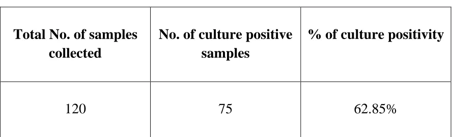
A total of 120 patients with infectious corneal ulcer were selected for
study. This study involves males and females of all age group. 75 cases were
culture positive.
TABLE 1
CULTURE POSITIVITY IN THE CORNEAL SCRAPING SAMPLES
N = 120
Total No. of samples collected
No. of culture positive samples
% of culture positivity
120 75 62.85%
TABLE 2
SMEAR POSITIVITY AMONG CORNEAL ULCER ISOLATES
Gender Total no of
specimens
10% KOH
positivity
Gram stain
positivity
Male 77 46 45
Female 43 25 24
[image:55.612.88.532.308.443.2] [image:55.612.89.535.560.673.2]FIGURE ‐ 1
CULTURE POSITIVITY IN THE CORNEAL SCRAPING SAMPLES
62.85
37.15 % of Culture Positive Sam ples
% of Culture Negative Sam ples
FIGURE ‐ 2
GENDER DISTRIBUTION OF INFECTIOUS CORNEAL ULCER
77
48 43
27
0 10 20 30 40 50 60 70 80
Total No. of Cases No. of Culture Positivity
Male
[image:56.612.139.503.155.320.2] [image:56.612.171.481.423.640.2]57
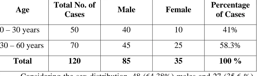
The cases were analysed under the following parameters. Out of 120
cases 77 patients were male and 43 patients were female. (50/120) 41%
cases were found to be in the age group between 30-60 years and 58.3 %
[image:57.612.94.532.299.429.2](70/120) of cases were in the age group of > 60 years.
TABLE – 3
AGE DISTRIBUTION OF INFECTIOUS CORNEAL ULCER
N = 120
Age Total No. of
Cases Male Female
Percentage of Cases
0 – 30 years 50 40 10 41%
30 – 60 years 70 45 25 58.3%
Total 120 85 35 100 %
Considering the sex distribution, 48 (64.38%) males and 27 (35.6 %)
female patients showed positive culture. A high prevalence of keratitis was
seen among males contributing to 64.38% of cases.
Table 2 and 3 shows the age and sex distribution of the patients along
with the positive culture for bacteria and fungi. This study shows that the
FIGURE – 3
AGE DISTRIBUTION OF INFECTIOUS CORNEAL ULCER
40 45 10 25 0 10 20 30 40 50 60 70 80 90 100 110 120
0-30 years 30-60 years
Male Female
FIGURE – 4
DISTRIBUTION OF PREDISPOSING FACTORS CAUSING CORNEAL ULCER
48 50 27 15 0 10 20 30 40 50 60 70 80 90 100 110 120
Culture Positive Traumatic Origin
[image:58.612.145.434.135.344.2] [image:58.612.137.425.443.641.2]59
The distribution of cases among rural and urban areas, showed
increased prevalence of keratitis in rural population accounting for 70%.
[image:59.612.90.507.465.669.2]TABLE – 4
DISTRIBUTION OF CASES AMONG RURAL AND URBAN AREAS
Total Rural Urban
120 84 36
Parentage % 70 % 30 %
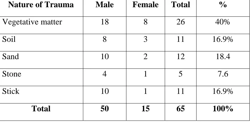
Numerous predisposing factors have been implicated, trauma alone
contributed to 54.16% of the cases in the development of keratitis.
TABLE – 5
DISTRIBUTION OF CORNEAL ULCER AMONG TRAUMATIC CASES N = 120
Nature of Trauma Male Female Total %
Vegetative matter 18 8 26 40%
Soil 8 3 11 16.9%
Sand 10 2 12 18.4
Stone 4 1 5 7.6
Stick 10 1 11 16.9%
FIGURE – 5
DISTRIBUTION OF CORNEAL ULCER AMONG TRAUMATIC CASES
10
4
10
2 1 1
18 8 8 3 0 10 20 30 40 50 60 70 Vegetative matter
Soil Sand Stone Stick Male Female
FIGURE – 6
DISTRIBUTION OF PREDISPOSING FACTORS OTHER THAN TRAUMA
2 6 0 3 6 4 4 3 0 10 20 30 Previous ocular surgery Steroid application Lid abnormalities
History of prior antifungal use (followup cases)
[image:60.612.158.454.140.359.2] [image:60.612.166.453.447.649.2]61
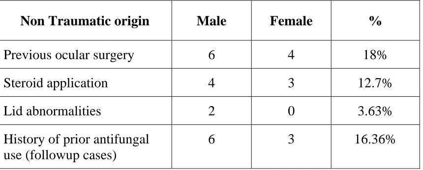
Co-existing ocular diseases such as lid abnormalities, previous ocular
surgery, steroid application and history of prior antifungal agents contribute
[image:61.612.90.520.282.455.2]to 3.63%, 18%, 12.7% and 16.36% respectively.
TABLE – 6
DISTRIBUTION OF PREDISPOSING FACTORS OTHER THAN TRAUMA
Non Traumatic origin Male Female %
Previous ocular surgery 6 4 18%
Steroid application 4 3 12.7%
Lid abnormalities 2 0 3.63%
History of prior antifungal use (followup cases)
6 3 16.36%
The relationships of influence of various predisposing factors on the
In analyzing the contribution of different trauma lesions in corneal
ulcer, trauma with vegetative matter like paddy, leaf and wood were
responsible for 40% of cases
TABLE – 7
DISTRIBUTION OF CORNEAL ULCER AMONG TRAUMATIC CASES
N = 120
Nature of Trauma Male Female Total %
Vegetative matter 18 8 26 40%
Soil 8 3 11 16.9%
Sand 10 2 12 18.4
Stone 4 1 5 7.6
Stick 10 1 11 16.9%
63
Among the bacterial isolates, S.Pneumoniae 9/22 (40%) was the
Predominant organism followed by Pseudomonas 7/22 (31%), Nocardia
[image:63.612.88.532.308.515.2]4/22 (18%) and S.viridans 2/22(9.09%).
TABLE – 8
DISTRIBUTION OF BACTERIAL AGENTS CAUSING CORNEAL ULCER
Total 22
Bacterial Isolate Total No. of Isolate %
Strep.pneumoniae 9 40%
Pseudomonas 7 31%
Nocardia 4 18%
Strep.viridans 2 9.09%
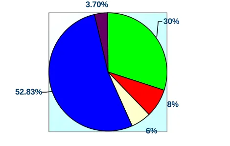
Among the fungal isolates, 28 out of 53 (52.83%) cases were due to
Fusarium species and next common agent isolated was Aspergillus flavus
16/53 (30%), Aspergillus fumigatus 4/53 (7.5%), Aspergillus niger 3/53
TABLE – 9
DISTRIBUTION OF FUNGAL AGENTS CAUSING CORNEAL ULCER
Total 53
Fungal Isolate Total No. of Isolates %
Aspergillus flavus 16 30%
Aspergillus fumigatus 4 7.5%
Aspergillus niger 3 5.6%
Fusarium 28 52.83%
Bipolaris 2 3.7%
Fusarium species were the most common fungal agent isolated 52.83%
Antibacterial sensitivity testing was performed by Kirby-Bauer
method with drugs such as Gatifloxacin, Tobramycin, Ceftazidime,
Vancomycin and Cotrimoxazole. 80% of S.Pneumoniae, 85.71% of
Pseudomonas, 75% of Nocardia and 50% of S.viridians were sensitive to
Gatifloxacin.
88% of S.Pneumoniae, 71% of Pseudomonas, 50% of Nocardia, and
50% of S.viridans were sensitive to Tobramycin. 55.55% of S.Pneumoniae,
65
FIGURE – 7
DISTRIBUTION OF BACTERIAL AGENTS CAUSING CORNEAL ULCER
31%
40% 19.09%
18%
Strep.pneumoniae Pseudomonas Nocardia Strep.viridans
FIGURE – 8
DISTRIBUTION OF FUNGAL AGENTS CAUSING CORNEAL ULCER
3.70%
8% 30%
52.83%
6%
[image:65.612.221.427.146.317.2] [image:65.612.187.422.470.619.2]sensitive to ceftazidime. All the 4 species were sensitive to
vancomycin and 22.2% of S.Pneumoniae, 42.8% of Pseudomonas, 50% of
[image:66.612.88.555.285.486.2]Nocardia, and 50% of S.viridans were sensitive to Cotrimoxazole.
TABLE – 10
ANTI BACTERIAL SUSCEPTIBILITY PATTERN OF BACTERIAL ISOLATES
Organism Gatiflox Tobramycin Ceftazidime Vancomycin Cotrimoxazole
Strep.pneumoniae 8(88%) 8(88%) 5(55.55%) 9(100%) 2(22.22%)
Pseudomonas 6(85.71%) 5(71%) 3(42.85%) 7(100%) 3(42.8%)
Nocardia 3(75%) 2(50%) 2(50%) 4(100%) 2(50%)
67
FIGURE –9
ANTI BACTERIAL SUSCEPTIBILITY PATTERN OF BACTERIAL ISOLATES
8888 55.55 100 22.22 85.71 71 42.85 5050 75 5050 100 50 505050 100100 0 10 20 30 40 50 60 70 80 90 100 110 120
Strep.pneumoniae Pseudomonas Nocardia Strep.v iridans
Gatiflox Tobramycin Ceftazidime Vancomycin Cotrimoxazole
[image:67.612.117.452.148.341.2]
FIGURE– 10
ANTI FUNGAL SUSCEPTIBILITY PATTERN OF FUNGAL ISOLATES
62% 50% 66% 71% 50% 75% 75% 66% 82% 50% 0% 10% 20% 30% 40% 50% 60% 70% 80% 90%
s>15m m Am pho B (20ug) S>23m m Itraconazole (10ug)
[image:67.612.113.485.440.676.2]Antifungal susceptibility pattern of fungal isolates by Disc diffusion
method shown 62% of A. flavus, 50% of A.fumigatus, 66.6% of A. niger,
[image:68.612.88.561.278.590.2]82% of Fusarium and 50% of Bipolaris were sensitive to Amphotericin B.
TABLE – 11
ANTI FUNGAL SUSCEPTIBILITY PATTERN OF FUNGAL ISOLATES (Disk Diffusion Method)
Organisms No. of
Isolates
S > 15mm Ampho B
(20ug)
S>23mm Itraconazole
(10ug)
Flucanazole
Aspergillus flavus 16 10(62%) 12(75%) 0
Aspergillus fumigatus
4 2(50%) 3(75%) 0
Aspergillus niger 3 2(66%) 2(66%) 0
Fusarium 28 20(71%) 23(82%) 0
Bipolaris 2 1(50%) 1(50%) 0
75% of A. flavus, 75% of A. fumigatus, 100% of A.niger, 82% of
Fusarium and 100% Bipolaris were sensitive to Itraconazole.All the fungal
isolates 100% of the (Aspergillus species, Fusarium and Bipolaris) were
69
MIC of Amphotericin B by agar dilution method showed MIC of less
than 2 micro gram/ml. for all the Aspergillus species. Fusarium species
showed MIC of less than 2 micro gram in 100% isolates. Bipolaris showed
[image:69.612.90.568.299.580.2]100% sensitive range for Amphotericin B.
TABLE – 12
MIC OF AMPHOTERICIN B BY AGAR DILUTION METHOD
Organisms .625ug 0.125ug .25ug .5ug 1ug 2ug 4ug 8ug
Aspergillus
flavus
6 4 2 4
Aspergillus
fumigatus
2 2
Aspergillus
niger
3
Fusarium 10 12 6
Bipolaris 1 1
MIC of Itraconazole by agar dilution method, all the isolates showed
Aspergillus, Fusarium and Bipolaris species showed MIC of less than 2
micro gram/ml for Itraconozole.
TABLE – 13
MIC OF ITRACONAZOLE BY AGAR DILUTION METHOD
Organisms .625ug 0.125ug .25ug .5ug 1ug 2ug 4ug 8ug
Aspergillus
flavus
3 5 4 3 1
Aspergillus
fumigatus
2 2
Aspergillus
niger
2
Fusarium 10 15 3
[image:70.612.86.570.254.533.2]71
MIC determination by broth microdilution method also showed that
the MIC range was comparable with agar dilution method. Good correlation
was observed between agar dilution and broth microdilution method.
TABLE – 14
MIC OF AMPHOTERICIN B BY MICRO DILUTION METHOD
Organisms .625ug 0.125ug .25ug .5ug 1ug 2ug 4ug 8ug
Aspergillus flavus
6 7 2 1
Aspergillus fumigatus
2 1 1
Aspergillus niger
3
Fusarium 8 10 8 2
Bipolaris 1 1
TABLE – 15
MIC OF ITRACONAZOLE BY MICRO DILUTION METHOD
Organisms .625ug 0.125ug .25ug .5ug 1ug 2ug 4ug 8ug
Aspergillus flavus
4 6 2 2 2
Aspergillus fumigatus
2 1 1
Aspergillus niger
3
Fusarium 4 10 8 2 4 4
TABLE – 16
COMPARISON OF MIC IN AGAR DILUTION AND BROTH MICRO DILUTION
Drug Conc Ampho B
MIC < 2ug
Itraconazole MIC < 2ug
Org Agar
Dilution Broth Micro Dilution Agar Dilution Broth Micro Dilution
Aspergillus flavus 16 15 14 14
Aspergillus fumigatus
4 3 3 3
Aspergillus niger 3 3 2 3
Fusarium 24 24 25 25
Bipolaris 1 1 2 2
A good correlation was observed between agar dilution method and
broth micro dilution method in the sensitivity pattern of fungal