COMPARATIVE EVALUATION OF THE EFFECTS OF TWO
CERAMIC FINISHING SYSTEMS AND DIAMOND POLISHING
PASTE ON THE SURFACE ROUGHNESS OF TWO CERAMIC
MATERIALS USED FOR CERAMO-METAL RESTORATIONS
-AN IN VITRO STUDY
Dissertation submitted to
THE TAMILNADU Dr. M. G. R. MEDICAL UNIVERSITY
In partial fulfillment for the Degree of
MASTER OF DENTAL SURGERY
BRANCH VI
PROSTHODONTICS
CERTIFICATE
This is to certify that this dissertation titled “COMPARATIVE EVALUATION
OF THE EFFECTS OF TWO CERAMIC FINISHING SYSTEMS AND DIAMOND
POLISHING PASTE ON THE SURFACE ROUGHNESS OF TWO CERAMIC
MATERIALS USED FOR CERAMO-METAL RESTORATIONS - AN IN VITRO
STUDY” a bonafide record of work done by Dr. CHERRY ANMOL under our
guidance and to our satisfaction during her postgraduate study period 2006-2009.
This Dissertation is submitted to THE TAMILNADU Dr. M.G.R. MEDICAL
UNIVERSITY, in partial fulfillment for the award of the Degree of MASTER OF
DENTAL SURGERY– PROSTHODONTICS, BRANCH VI. It has not been submitted
(partial or full) for the award of any other degree or diploma.
GUIDED BY:
Dr. N.S.Azhagarasan, M.D.S., Dr. Saket Miglani, M.D.S.,
Professor & H.O.D, Reader,
Dept. of Prosthodontics, Dept. of Prosthodontics,
Ragas Dental College and Hospital, Ragas Dental College and Hospital, Uthandi,
Chennai. Uthandi, Chennai.
Dr. S. Ramachandran, M.D.S., Principal,
Ragas Dental College and Hospital,
ACKNOWLEDGEMENT
This dissertation is the result of work with immense support from many people and it is a pleasure now that I have the opportunity to express my gratitude to all of them.
I would be failing in my duty if I do not adequately express my deep sense of gratitude and my sincere thanks to my Head of the Department, Professor Dr.N.S.Azhagarasan M.D.S., Department of Prosthodontics, Ragas Dental college and Hospital, Chennai, for his exceptional guidance, tremendous encouragement, well-timed suggestions and heartfelt support throughout my postgraduate programme which has never failed to drive the best out of me. I would like to profoundly thank him for giving an ultimate sculpt to this study.
I wish to express my gratitude to Dr.S.Ramachandran M.D.S., Principal, Ragas Dental College, Chennai, for his timely help and encouragement throughout my post graduate course. I also thank him for permitting me to make use of the amenities in the institution.
I would like to solemnly thank my Professor, Dr.K. Chitra Shankar M.D.S., for her untiring help, back up and constructive criticism extended to me throughout the period of my postgraduate study. I am indebted to her as this dissertation has been the fertile outcome of her immense patience, encouragement and expert guidance.
I would like to immensely thank Dr.Saketh Miglani, M.D.S., Reader, Department of Prosthodontics, for his constant guidance and encouragement rendered by him throughout my study.
I would also like to thank Dr.K.Madhusudan, M.D.S., Dr.Manoj Rajan, M.D.S., & Dr.S.Jayakrishnakumar, M.D.S., for their timely help throughout my study.
My sincere thanks to Dr.S.P.Mohan, Professor and Head, Department of Geology, Madras University, Chennai, for allowing me to make use of the SEM facilities and expertise at his department.
engineering college, Chennai for helping me with the profilometer.
My sincere thanks to the Bio-statistician, Mr. R.Ravanan, Reader in Statistics, Presidency College, Chennai for his valuable guidance and statistical analysis of the data. It would not be justifiable on my part if I do not acknowledge the help of’ my fellow colleagues, my seniors, juniors and friends for their criticism and continued support throughout my postgraduate course.
Last but never the least; I would like to thank my parents who have sacrificed their life for my upliftment and being my moral support throughout my life.
Above all I thank the GOD for all the grace endowed upon me.
CONTENTS
INDEX PAGE No.
1.
Introduction
1
2.
Review of literature
5
3.
Materials and method
26
4.
Results
39
5.
Discussion
59
6.
Conclusion
69
7.
Summary
71
LIST OF TABLES
Table No
Title Page No
1 Firing Schedule for Feldspathic Porcelain Specimens 34
2 Firing Schedule for Fluorapatite Leucite Porcelain
Specimens 34
3 Surface Roughness (Ra) values in µm for the test samples
of Feldspathic Porcelain after Autoglazing(C), following abrasion and finishing with Sof-Lex discs, and finally after
polishing with diamond polishing paste (IA) 48
4 Surface Roughness (Ra) values in µm for the test samples
of Feldspathic Porcelain after Autoglazing(C), following abrasion and finishing with White silicon & grey rubber,
and finally after polishing with diamond polishing paste (IB) 48
5 Surface Roughness (Ra) values in µm for the test samples
of Fluorapatite Leucite Porcelain after Autoglazing (D), following abrasion and finishing with Sof-Lex discs, and
finally after polishing with diamond polishing paste (IIA) 49
6 Surface Roughness (Ra) values in µm for the test samples
of Fluorapatite Leucite Porcelain after Autoglazing (D), following abrasion and finishing with White silicon & grey rubber, and finally after polishing with diamond
polishing paste (IIB) 49
7 Mean Surface Roughness (Ra) values in μm and standard deviation obtained from the basic data of test groups of
Feldspathic and Fluorapatite Leucite ceramic systems. 52
8 Test of significance for the mean obtained from Autoglazed (C) and polished test samples of subgroup IA of Group I
(Feldspathic ceramic system). 53
9 Test of significance for the mean obtained from Autoglazed (C) and polished test samples of subgroup IB of Group I
Table No
Title Page No
10 Test of significance for the mean obtained from polished test samples of subgroups IA and IB of Group I (Feldspathic
ceramic system) 54
11 Test of significance for the mean obtained from Autoglazed
(D) and polished test samples of subgroup IIA of Group II
(Fluorapatite Leucite ceramic system) 55
12 Test of significance for the mean obtained from Autoglazed (D) and polished test samples of subgroup IIB of Group II
(Fluorapatite Leucite ceramic system) 55
13 Test of significance for the mean obtained from polished test samples of subgroups IIA and IIB of Group II
(Fluorapatite Leucite ceramic systems) 55
14 Test of significance for the mean “Ra” values for the average surface roughness of the ceramic materials obtained from the
subgroups IA, IB, IIA and IIB of Feldspathic and Fluorapatite
leucite ceramic systems 56
LIST OF GRAPHS
Graph1:Surface Roughness (Ra) values in µm for the test samples of feldspathic porcelain after Autoglazing(C), following abrasion and finishing with Sof-Lex discs, and
finally after polishing with diamond polishing paste.
Graph2:Surface Roughness (Ra) values in µm for the test samples of feldspathic porcelain after Autoglazing(C), following abrasion and finishing with White silicon and Grey rubber, and finally after polishing with diamond polishing paste.
Graph3:Surface Roughness (Ra) values in µm for the test samples of fluorapatite leucite porcelain after Autoglazing (D), following abrasion and finishing with Sof-Lex discs, and finally after polishing with diamond polishing paste.
Graph4:Surface Roughness (Ra) values in µm for the test samples of fluorapatite leucite porcelain after Autoglazing (D), following abrasion and finishing with White silicon and Grey rubber, and finally after polishing with diamond polishing paste.
Graph5:Mean “Ra” values for the average surface roughness of the ceramic materials obtained from the basic data of four subgroups (IA, IB, IIA, and IIB) calculated in μm.
Graph6:Comparison of mean “Ra” values for the average surface roughness of the ceramic materials between Autoglazed(C) and polished test samples of subgroups IA and IB of Group I (Feldspathic porcelain).
Graph7:Comparison of mean “Ra” values for the average surface roughness of the ceramic materials between Autoglazed(D) and polished test samples of subgroups IIA and IIB of Group II (Fluorapatite leucite porcelain).
ANNEXURE
LIST OF FIGURES
Fig.1.Custom-made Metallic mold
Fig.2.Schematic representation of custom metallic mold
Fig.3.Pattern resin
Fig.4.Laboratory materials required for the fabrication of metal substructure
Fig.5a.Feldspathic porcelain system
Fig.5b.Fluorapatite Leucite porcelain system
Fig.6.Laboratory tools required for porcelain veneering
Fig.7.Sintered Diamond
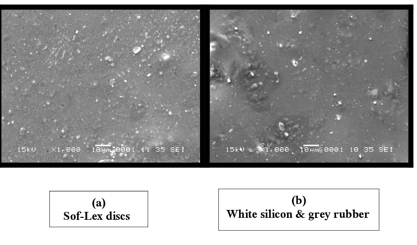
Fig.8a.Sof-Lex discs
Fig.8b.White silicon and grey rubber
Fig.9.Diamond polishing paste
Fig.10.Dental porcelain furnace
Fig.11.Scanning Electron Microscope
Fig.12.Profilometer
Fig.13.Fabrication of the resin patterns
Fig.14.Standardized resin patterns
Fig.15.Casting the resin pattern
Fig.16.Divested casting
Fig.17.Thickness of finished metal substructure
Fig.18.Sandblasted metal substructure
Fig.19.Metal substructure veneered with test porcelain
Fig.21.Schematic representation of thickness of the specimen after veneering the test
porcelain.
Fig.22. Autoglazed samples of Feldspathic and Fluorapatite Leucite
Fig.23.Porcelain samples finished with Sof-Lex discs
Fig.24.Porcelain samples finished with White silicon and grey rubber
Fig.25.Porcelain samples polished with diamond polishing paste
Fig.26.1a.SEM Photomicrographs (l000x magnification) showing surface texture of the autoglazed Feldspathic porcelain specimens.
Fig.27.1b.SEM Photomicrographs (l000x magnification) showing surface texture of the Feldspathic porcelain specimens following abrasion and finishing.
Fig.28.1c.SEM Photomicrographs (l000x magnification) showing surface texture of the Feldspathic porcelain specimens after polishing.
Fig.29.2a.SEM Photomicrographs (1000x magnification) showing surface texture of the autoglazed Fluorapatite leucite porcelain specimens.
Fig.30.2b.SEM Photomicrographs (1000x magnification) showing surface texture of the Fluorapatite leucite specimens following abrasion and finishing.
Fig.31.2c.SEM Photomicrographs (1000x magnification) showing surface texture of the Fluorapatite leucite specimens after polishing.
Fig.32.1d.Profilometer Photomicrographs showing surface texture of autoglazed Feldspathic porcelain specimens.
Fig.33.1e.Profilometer Photomicrographs showing surface texture of Feldspathic porcelain specimens following abrasion and finishing.
Fig.34.1f.Profilometer Photomicrographs showing surface texture of Feldspathic porcelain specimens after polishing.
Fig.35.2d.Profilometer Photomicrograph showing surface texture of autoglazed Fluorapatite leucite porcelain specimens.
Fig.36.2e.Profilometer Photomicrographs showing surface texture of the Fluorapatite leucite specimens following abrasion and finishing.
Fig.37.2f.Profilometer Photomicrograph showing surface texture of the Fluorapatite leucite
INTRODUCTION
Dental ceramics are widely used in dental practice as material of choice for
porcelain-fused-to-metal or all-ceramic restorations in crown and bridge prosthodontics and as laminate
veneers in cosmetic dentistry, because of their natural appearance.31
Porcelain fused to metal restorations account for more than 80% of the restorations
made worldwide. 12 These are popularly used in prosthodontics because of their refractive
nature, hardness, biocompatibility and chemical inertness. These have metal substructures
supporting a ceramic veneer that is mechanically and chemically bonded. Among the various
types of veneering porcelain available for metal ceramic restorations, the traditional
feldspathic porcelain is still widely used despite numerous scientific reports of their harmful
behavior regarding increased wear of the opposing dentition. The fluorapatite leucite
porcelain claims to have smoother surface topography, lower abrasiveness towards the enamel
and improved color which accounts for its increased use in recent times.1, 34 The strong
life-like appearance of the completed metal ceramic restoration results from a surface glaze,
formed on additional firing of the restoration.1, 12, 31, 34
Ideally, ceramic restorations should retain their intact surface glaze. However,
occasions will arise when ceramic restorations require adjustments in circumstances that
preclude reglazing, for example chair side adjustment of ceramic restorations for shape,
contour, occlusion and surface finish.30, 51 In the clinical set up it is not possible to reglaze the
restorations due to practical constraints. In such situations the surfaces tends to become rough.
The Rough ceramic surfaces abrade opposing teeth and/or restorations.14, 15, 32, 53 Rough
porcelain surfaces also significantly reduce the strength of ceramic restorations and make
Roughness of intraoral hard surfaces is a major cause for adhesion and retention of
oral microorganisms.12 This will lead to excessive plaque accumulation, gingival irritation,
increased surface staining, and poor or suboptimal esthetics of the restored teeth and thereby
increasing the risk of dental caries and periodontal disease. In such situations, roughness must
be smoothened to render the surface acceptable to the patient and make it less likely to abrade
opposing tooth structure or restorative materials. The effective finishing and polishing of
dental restorations not only result in optimal aesthetics and longevity of restored teeth, but
also provide for acceptable oral health of soft tissues and marginal integrity of the restorative
interface.25, 26
The adjusting, contouring, and finishing procedures for metal ceramic restorations
play a critical role in achieving both proper function and optimal esthetics. Thus it has
become imperative to consider the various available ceramic finishing systems to recreate the
lost smoothness of the abraded surfaces to obtain optimal biocompatibility. A number of
mechanical polishing techniques are described in the literature and have been compared to the
gold standard given by the original glaze. Some authors initially demonstrated the superior
smoothness of glazed porcelain.11, 41, 42 Others, however, favour mechanical polishing and
concluded that intraoral polishing of porcelain can equal or surpass the smoothness of glazed
porcelain.19, 20, 30, 49, 51 Today, it is recognized that improved esthetic results are obtained by
polishing.3, 9, 22, 39 The ultimate goal of mechanical finishing and polishing is the attainment of
a well polished surface which can substitute for glazed porcelain.
Studies comparing the efficacy of various smoothening and polishing systems for
metal ceramic restorations are carried out either qualitatively or quantitatively. Most studies
have focused on the qualitative analysis of the ceramic surface.2, 11, 44, 45, 47, 51 Fewer studies
quantitative assessment following different finishing procedures.3, 16, 17, 19, 20, 27, 42, 50 The analysis
of the surface both qualitatively and quantitatively can aid in obtaining better inferences.
In light of the above, the present in vitro study was designed to qualitatively and
quantitatively evaluate and compare the effect of two ceramic finishing systems and diamond
polishing paste had on the surface texture of two ceramic materials used for ceramo-metal
restorations.
The objectives of the study included:
1. To qualitatively evaluate and compare the effects of two ceramic finishing systems on the
surface roughness of the test samples of feldspathic porcelain using scanning electron
microscope after Autoglazing, following abrasion and finishing with two test finishing
systems, and finally after polishing.
2. To qualitatively evaluate and compare the effects of two ceramic finishing systems on the
surface roughness of the test samples of fluorapatite leucite porcelain using scanning electron
microscope after Autoglazing, following abrasion and finishing with two test finishing
systems, and finally after polishing.
3. To qualitatively compare the effects of two ceramic finishing systems on the surface
roughness between the test samples of feldspathic and the fluorapatite leucite porcelain using
scanning electron microscope after Autoglazing, following abrasion and finishing with two
test finishing systems, and finally after polishing.
4. To quantitatively evaluate and compare the effects of two ceramic finishing systems on the
surface roughness of the test samples of feldspathic porcelain using profilometer after
Autoglazing, following abrasion and finishing with two test finishing systems, and finally
5. To quantitatively evaluate and compare the effects of two ceramic finishing systems on the
surface roughness of the test samples of fluorapatite leucite porcelain using profilometer after
Autoglazing, following abrasion and finishing with two test finishing systems, and finally
after polishing.
6. To quantitatively compare the effects of two ceramic finishing systems on the surface
roughness between the test samples of feldspathic and the fluorapatite leucite porcelain using
profilometer after Autoglazing, following abrasion and finishing with two test finishing
systems, and finally after polishing.
7. To obtain correlations, if any, of the surface roughness values obtained by quantitative and
qualitative analysis of feldspathic and the fluorapatite leucite porcelain systems
REVIEW OF LITERATURE
Clayton J A et al (1970)13did this study to compare the surface roughness, following
final finishing and polishing, of pontics constructed from cast gold, acrylic resin, and glazed
porcelain. The surface roughness of the sample pontics was measured with a profilometer.
Statistical analysis indicated that the test surfaces of glazed porcelain were significantly
rougher than the polished test surfaces of either acrylic resin or cast gold. There was no
significant difference in surface roughness between the polished acrylic resin and polished
cast gold sample pontic surfaces.
Monasky G E et al (1971)32investigated the wear caused byvarying combinations of
tooth enamel, gold, and porcelain, with particular emphasis on the affect of variations in the
surface finish of the porcelain on the resultant wear. They concluded that: (1) the rougher the
porcelain surface, the more rapid is the rate of tooth wear. Rough ground porcelain surfaces
produced excessive wear rates, and if the glaze is broken on porcelain, it must be repolished.
(2) Porcelain surfaces in contact with tooth structure tend to wear rapidly at first until a
“polish” of the porcelain surface is obtained. This functional polishing of the porcelain
reduced the wear rate on opposing teeth. (3) Gold wears rapidly when in contact with
porcelain. Polishing of porcelain occurs very slowly if at all when in contact with gold. Thus,
if porcelain is to be used in opposition to gold, it is essential that the porcelain be smooth and
well polished.
Morrow R M et al (1973)33 evaluated five commonly used methods for polishing
porcelain denture teeth. Each tooth was rated on a three point scale by comparing its polished
surface to the glazed surface of the control tooth. The statistical analysis found that polishing
additional factors, such as the reduction of tooth thickness, the length of the polishing time,
and the cost, should be considered when selecting the “best” over-all polishing method for a
specific purpose.
Barghi N et al (1975)6 ascertained microscopically, the surface appearance of both
air-fired and fired porcelain following various alterations. They found that
vacuum-fired had a smoother surface than air-vacuum-fired porcelain. The relative absence of bubbles allowed
the vacuum-fired porcelain to be sanded and polished to a significantly superior finish. They
concluded that, regardless of the usual polishing techniques, a final glaze presented the most
acceptable surface.
Barghi N et al (1976)7 conducted this study to determine whether or not the finish
attained prior to glazing affected the final surface texture of glazed porcelain and to examine
for differences between the surface appearance of natural and low-fusing glazes. They
concluded that a smooth surface can be obtained by glazing after grinding and there was no
need for sanding or polishing with a rubber wheel. They also found that a low-fusing glaze
gave a slightly smoother surface than a natural glaze and the low-fusing glaze may be added
at any stage of polishing, as the results will be the same.
Sulik W D et al (1981)51 in their article described a polishing technique for fully
matured porcelain which may be substituted for reglazing and also compared the polished and
glazed surfaces using the Scanning electron microscope. The technique made use of a hard
rubber wheel, fine wet pumice and wet tin oxide. A comparison of the polished and naturally
glazed porcelain surfaces of vacuum-fired porcelain appeared similar both clinically and
under an SEM.
Schlissel E R et al (1980)47 evaluated eleven commonly used methods of adjusting
postadjustment surface roughness, as compared to an unaltered specimen, was made by an
electron micrograph examination of the surface finish of each repolished porcelain denture
tooth. Three methods 7, 10, 11 (7- Abrasive wheel with a slow-speed dental handpiece, hard
rubber wheel with a slow-speed dental handpiece, slurry of medium-grade pumice on a rag
wheel mounted on a dental lathe, and slurry of flour of pumice on a rag wheel mounted on a
dental lathe, 10- Abrasive stone with a slow-speed dental handpiece, hard rubber wheel with a
slow-speed dental handpiece, slurry of medium-grade pumice on a rag wheel mounted on a
dental lathe, and slurry of flour of pumice on a rag wheel mounted on a dental lathe, and 11-
Polishing kit with a sequence of four abrasives in a slow speed dental handpiece) produced
finished surfaces comparable to the unaltered tooth. The best surface finish was produced by
using the proprietary kit in method 11.
Obregon A et al (1981)36 did a study to compare the effects of various opaque and
porcelain surface textures on two different shades of porcelain. A spectrophotometer was used
to measure the color of the porcelain samples when the porcelain and opaque textures were
modified. The results showed that porcelain surface texture, whether rough or smooth, did not
make a difference in Hue. The smooth surface porcelain texture increased the Value of shade
B1 compared to the rough porcelain surface. The interactions that occur between the texture
of porcelain and opaque affecting color are complex phenomena and may be related to the
modification of light by transmission, absorption, refraction, scattering, and reflection.
Smith G A et al (1981)50 did a study to investigate the surface finish which can be
achieved on trimmed porcelain surfaces by the use of a series of discs (Sof-Lex) designed for
finishing composite restorations. The surface finish achieved with the Sof-Lex discs is
compared with that produced by abrasives commonly used for trimming porcelain surfaces.
procedures: (1) Diamond stone, (2) Busch silent wheel, (3) Busch silent wheel followed by
impregnated rubber wheel, (4) Coarse Sof-Lex disc, (5) Busch silent wheel followed by
medium Lex disc, (6) Busch silent wheel followed by medium, fine and superfine
Sof-Lex discs, (7) Diamond stone followed by coarse and medium Sof-Sof-Lex discs, (8) Diamond
stone followed by coarse, medium, fine and superfine Sof-Lex discs. Scanning electron
microscopy and a surfometer have been used to study and compare the appearance and
profiles of the surfaces of glazed specimens of vacuum-fired aluminous porcelain. The results
demonstrate that the surface finish of trimmed porcelain can be improved considerably by the
use of a series of discs designed for finishing composite restorations. The optimum finishing
procedure will do little to improve the strength of trimmed porcelain restorations, but it may
limit surface accumulations of plaque and stain and reduce the friction and abrasion effects on
opposing occlusal surfaces.
Klausner L H et al (1982)30 evaluated four different porcelain polishing sequences,
and the resulting polished surfaces were compared to an unaltered glazed surface. The
sequence included (1) superfine diamond, Dedeco wheels, and levigated alumina; (2) Shofu
porcelain polishing system; (3) superfine diamond, Cratex wheel, Burlew disk, and levigated
alumina; (4) Jelenko porcelain carving and polishing wheels. No significant differences were
found between the final polished surfaces and the initial autoglazed surfaces for any of the
four test sequences. Significant differences were found between comparable abrasives among
the polishing sequences, as well as between steps within a single polishing sequence.
Newitter D A et al (1982)35 compared the effectiveness of commonly available
adjustment (grinding) methods and polishing (finishing) methods used in different
combinations. Six methods for initial reduction of the porcelain on porcelain-bonded-to-metal
Eleven methods for finishing ground porcelain surfaces were evaluated for smoothness of the
surfaces produced. Methods employing finishing wheels followed by pumice or porcelain
polishing paste produced smoother surfaces than other methods. This information can be
helpful in the selection of techniques for initial reduction and finishing of
porcelain-baked-to-metal in the absence of glazing.
Christensen G J (1986)12conducted a survey to determine the attitudes and practices
of dentists regarding porcelain-fused-to-metal restorations. The survey revealed the
following:(1) Porcelain-fused-to-metal crowns are the most commonly used crowns in
dentistry, (2) Cast gold crowns are infrequently placed compared to PFM, (3) Most dentists
consider PFM crowns extremely successful restorations; (4) Although porcelain occlusal
surfaces are considered acceptable by most dentists, dentists prefer metal on occlusal surfaces
for restorations in their own mouths; (5) The anterior ¾ crown is infrequently placed, but the
posterior ¾ crown is commonly used; (6) The Cerestore crown is gaining acceptance; (7)
Porcelain-jacket-crown use is reduced, but still a viable alternative; (8) The most desired
improvement for PFM restorations was less wear on opposing teeth.
Zalkind M et al (1986)55 examined the degrees of roughness of porcelain after
subjecting it to abrasive techniques and natural (self) glazing. In their study they found that
glazing the porcelain surface reduced by an abrasive instrument would not reduce the
resulting roughness. To produce a smooth surface it was needed to sandblast the abraded
surface with aluminum oxide before retiring to produce a natural glaze.
Haywood V B et al (1988)19found that techniques for placement of etched porcelain
laminate veneers require that the glazed porcelain veneer be cemented prior to finishing and
polishing. Using scanning electron microscopy (SEM) and specular reflectance, the surface
placed on those instruments which are suitable for gingival and interproximal finishing.
Finishing with a fine diamond instrument followed only by diamond polishing paste produced
an unacceptable surface. A finish equal or superior in smoothness to glazed porcelain was
achieved through the use of a series of finishing grit diamonds (Micron Finishing System)
followed by a 30-fluted carbide bur and diamond polishing paste. Other finishing
combinations produced surface textures which were not as smooth as glazed porcelain, but
which were better than that attained by the diamond polishing paste alone.
Haywood V B et al (1989)20 evaluated several experimental instruments and materials
to determine if polishing could be done more efficiently. Scanning electron microscopy was
used to evaluate the surface texture produced by different combinations of experimental
instruments applied with high and moderate speed, wet and dry, to porcelain disks. No
sequence matched the polished standard. However, the optimum surface texture was obtained
with diamond instruments (with progressively smaller particle sizes) used at a moderate speed
with water, followed by a 30-fluted carbide bur at high speed and dry, then diamond polishing
paste on a webbed rubber cup. In all polishing sequences tested, the best results were obtained
with each individual instrument when diamond instruments were used at moderate speed wet,
and when carbide instruments were used at high speed dry.
Campbell S D (1989)11 in this study used scanning electron microscopy to evaluate
the effect of polishing procedures on two all-ceramic crown materials (Dicor and
Cerestore).The “as formed,” unpolished specimens of both Dicor and Cerestore materials
presented a rough surface. It was found that any attempt to polish the Cerestore coping
material resulted in an extremely rough surface. Finishing of the Dicor ceramic resulted in a
smoother but pitted surface. Polishing of both ceramic materials resulted in a surface that was
glazed veneer (Cerestore) and shaded porcelain (Dicor) were applied to the all-ceramic
materials.
Brackett S E et al (1989)8 evaluated the flexural strengths of fine porcelains
commonly used in the all-porcelain margin technique and the effect of surface treatment on
the flexural strength. Thirty samples were made by using each of five different porcelain
margin systems. The subgroups received different surface treatments as follows: (1)
autoglaze, (2) overglaze, and (3) autoglaze and polish. A three-point flexural test was used to
test the specimens on a universal testing machine. Crystar shoulder porcelain with distilled
water as the binder was significantly stronger than the other porcelains tested, and porcelain
treated with an overglaze was stronger than porcelain treated with autoglaze or autoglaze and
polish.
Goldstein R E (1989)18 evaluated that regardless of the color, shape or attention to
detail, the qualities and objectives of finishing do require extra time to adequately finish all
restorations. A properly finished restoration should have the following qualities and
objectives. Qualities include: (1) A well finished margin. This implies no overhang, void, or
extension of restorative material that could interfere with tissue health (2) A sufficiently
smooth surface that will not attract bacterial plaque or food stains (3) Suitable surface texture
that blends in or matches adjacent or opposing natural teeth (4) Color matching of the existing
adjacent, opposing, or preselected tooth shade (5) A surface finish devoid of too obvious
contour, finishing bur, or diamond marks. Objectives include: (1) To improve and finalize
restoration margins and contours that will help make the restoration biocompatible with both
tooth and tissue (2) To develop maximum surface luster to enhance esthetics, reduce stain and
Wiley M G (1989)53in this article explored the potentially destructive nature of dental
porcelain placed on the occluding surface of prosthodontic restorations. In depth knowledge
of physical properties of dental porcelains is a necessity. Comprehensive treatment planning
that includes a total evaluation of the patient’s occlusal function and dysfunction is critical.
Finally viable material and treatment options are presented along with methods to help control
the effects of porcelain if its use is mandated. Author suggested that all occluding ceramic
surfaces should be highly polished and glazed after adjustments and before cementation.
Raimondo R L et al (1990)44 compared the finishes on dentalporcelain polished with
four different polishing paste systems with oven reglazing and with a porcelain adjustment kit
without a polishing paste. The polished/reglazed samples were rated according to quality of
finish by independent observers and by scanning electron microscope. On the basis of visual
examination, two of the polishing paste systems tested was found to produce a surface equal
to or better than oven glazing. On the basis of SEM examination, oven glazing was found to
produce a better surface than the other polishing methods. Not all porcelain polishing systems
produce a surface comparable to oven-glazed porcelain, and porcelain polishing systems
should be chosen carefully.
Brewer J D et al (1990)9 in this study determined that whether visual inspection
differences exist between glazed and polished porcelain surfaces. All crowns were initially
autoglazed. For phase 1 observations, six crowns were air abraded and polished and six
retained their glazed surface. For phase 2 observations, the surface treatments were reversed.
Phase 1 polished and glazed crowns had different means for outline form sharpness, porosity,
reflectance, dullness, and general esthetic appearance. Phase 2 crowns were different for
dullness. Polished and glazed crowns alike were duller at phase 1 than at phase 2.Glazed
Significant differences occurred among raters with polished and glazed crowns for several
variables.
Goldstein G R et al (1991)17 According to author, research has indicated that
polishing ground porcelain is essential to control the wear of opposing occlusal surfaces and
reduce the inflammation of contacted soft tissue. Fine popular methods for polishing
porcelain were evaluated by use of a profilometer, SEM, normal vision. Seventy disks, 35
Biobond disks and 35 Ceramco disks were roughened with a green stone and polished with
one of the methods according to the manufacturers’ directions. Brasseler, Dedeco, Dentsply,
and Shofu porcelain polishing systems were suitable for restoring ground porcelain. However,
clinical evaluations correlated to the profilometer and SEM readings revealed that the
Brasseler system was superior for polishing than Ceramco porcelain, whereas the Den-Mat
system was unacceptable.
Patterson C J W et al (1991)41 studied the effect of porcelain refinishing kit on Vita
VMK bonded porcelain qualitatively and quantitatively using scanning electron microscope
and surface profilometer, respectively. The kit proved incapable of restoring a surface glaze to
porcelain adjusted using a fine (red band) diamond bur, but was capable of reducing
significantly the surface roughness (Ra) of adjusted porcelain. The importance of
distinguishing between the integrity of the surface glaze and measurements of surface
roughness was discussed. Confining the application of refinishing procedures to the surface
adjusted is important to avoid unnecessary removal of the original surface glaze.
Palmer D S et al (1991)38 in this study determined the effect of castable ceramic, with
and without shading porcelain applied, on enamel wear. The wear produced by conventional
dental porcelain was used as a control. Enamel wear was calculated from microscopic
found between castable ceramic with and without shading porcelain and between
conventional dental porcelain and castable ceramic with shading. These findings suggest that
castable ceramic with shading porcelain should not be used in regions that will function
against opposing natural teeth.
Jacobi R et al (1991)22 compared a type III gold alloy and six different ceramic
surfaces by securing them in an abrasion machine opposing extracted teeth to determine their
relative abrasiveness and resistance to wear. The rankings of restorative materials from least
abrasive to most abrasive were: gold alloy, polished; cast ceramic, polished; porcelain,
polished; cast ceramic, polished and shaded; porcelain, polished and glazed; cast ceramic,
cerammed skin shaded; and cast ceramic, cerammed skin unshaded. The ranking of materials
from most wear-resistant to least wear-resistant was: gold alloy, cast ceramic cerammed, cast
ceramic cerammed and shaded, porcelain polished, porcelain glazed, cast ceramic polished
and shaded, and cast ceramic polished.
Patterson C J W et al (1992)42 investigates the efficacy of commercial porcelain
refinishing kit, which are claimed to restore the surface finish on porcelain after adjustments
in circumstances that preclude reglazing. In this study, they investigates the efficacy of one
such kit in restoring a Vitadur N porcelain surface finish after grinding with fine (30 µm
grit-red band) and extra-fine (15 µm grit-yellow band) high-speed diamond burs. Randomly
selected examples of surfaces created during refinishing were subjected to scanning electron
microscopy and to surface profilometry tracings. Although refinishing after grinding with a
15 µm grit bur produced surfaces significantly smoother than on specimens previously ground
with the 30 µm grit burs, the surfaces remained significantly rougher than when originally
15 µm grit yellow band types would be appropriate for porcelain adjustments to permit
subsequent refinishing to a surface smoothness comparable to the original glaze.
Scurria M S et al (1994)49 found that conventional and CAD-CAM ceramic
restorations often require adjustments that results in a need to reduce surface roughness.
Surface roughness resulting from fine polishing systems on two ceramics was assessed. Five
profilometer average roughness measurements (Ra) were taken of five replications of each
step in each sequence. Controls were autoglazed. Ceramco II and Dicor MGC ceramic
specimens milled with Cerec diamond wheel. Feldspathic porcelain could be polished
smoother than glazed. Dicor ceramic could be polished smoother than Ceramco II Ceramic.
Finishing diamond points followed by diamond gels produced the smoothest surface. A
30-fluted carbide did not improve smoothness as used. The aluminum oxide point followed by
aluminum oxide pastes was equivalent to finishing diamonds and gels for Dicor ceramic.
Jagger D C et al (1994)23 performed abrasive wear tests on unglazed, glazed, and
polished porcelain stud specimens using human enamel as the opposing plate specimens. The
wear tests were carried out on a wear machine that was specifically designed to simulate the
masticatory cycle. The amount of enamel wear produced by both glazed and unglazed
porcelain was similar; however, that produced by polished porcelain was substantially less.
Investigation of the glazed porcelain surface showed that the glaze was removed in less than 2
hours of wear on the machine.
Ward M T et al (1995)52 found that intraoral porcelain polishing is an important
consideration in many restorative and esthetic procedures. Several porcelain polishing
systems as well as improved ceramics are now commercially available. This study evaluated
the efficacy of eight different intraoral polishing techniques on three opalescent porcelains.
polishing procedures with a profilometer. These results were then compared to self-glazed
and overglazed control groups. Five of the techniques tested produced surfaces smoother than
glazing. The use of a 30-fluted carbide bur before diamond polishing paste produced the
smoothest surfaces.
Fuzzi M et al (1996)16 analyzed the surface roughness of Vita VMK porcelain
following oven glazing and eight grinding/polishing treatments qualitatively using scanning
electron microscope and quantitatively using a profilometer. Scanning electron microscopy
evaluation found oven glazing produced a better surface than other polishing methods. On the
basis of the profilometric examination, the best average roughness value was obtained using
diamond instruments with progressively smaller particle sizes (30, 15, and 8 microns).
Scanning electron microscopy analysis showed that all the treatments left the surfaces
partially porous and cracked; however, the glazed surface yielded the best result. Although no
significant differences were detected for the different treatments, the use of a 30-microns
diamond instrument produced a rougher surface..
Kelly J R et al (1996)29 in this article presents a brief history of dental ceramics and
offers perspectives on recent research aimed at the further development of ceramics for
clinical use, at their evaluation and selection, and very importantly, their clinical performance.
Notable research was highlighted regarding (1) wear of ceramics and opposing enamel, (2)
polishability of porcelains, (3) influence of firing history on the thermal expansion of
porcelains for metal ceramics, (4) machining and CAD/CAM as fabrication methods for
clinical restorations, (5) fit of ceramic restorations, (6) clinical failure mechanisms for
all-ceramic prosthesis, (7) chemical and thermal strengthening of dental all-ceramics, (8) intraoral
strong scientific and collaborative foundations exist for the continued understanding and
improvement of dental ceramic systems.
Jefferies S R (1998)25 presented an overview of basic principles of abrasive science as
they relate to the finishing and polishing of dental restorations. This discussion considers
several commercial products in terms of research into their use and optimal application.
Additional consideration is given to important technique considerations in the application of
various finishing and polishing devices and materials. As proper finishing and polishing of
dental restorations are important aspects of clinical restorative procedures that enhance both
esthetics and longevity of restored teeth. Effective use of rotary cutting burs and bonded,
coated, and loose abrasives can greatly simplify and improve the effectiveness of finishing
and polishing procedures.
Al-Wahadni A M et al (1998)3 presented a review of a number of studies that have
examined the visual and microscopic appearance and roughness of glazed, unglazed and
polished porcelain surfaces using techniques such as, scanning electron microscopy and
surface Profilometry. They agreed that glazed porcelain provides a smooth and dense surface.
Others have shown that polishing can produce an equally smooth surface, which may even be
esthetically better. Still others supported the use of polishing as an alternative to glazing.
However, reports have shown that unglazed porcelain is more abrasive than glazed.
Al-Hiyasat A S et al (1999)1 in this in vitro study, investigated the wear of human
enamel and 3 dental ceramics: a conventional porcelain (Vitadur Alpha), a low-fusing
hydrothermal ceramic (Duceram-LFC), and a machinable ceramic (Vita Mark II) in a 3-body
wear test. The abrasiveness of Alpha porcelain and Duceram-LFC ceramic was similar, yet
both were significantly more abrasive than Vita Mark II ceramic. In addition, Vita Mark II
Al-Wahadni A M et al (1999)4 performed an in vitro investigation into the wear
effects of glazed, unglazed and refinished dental porcelain on an opposing material. The
investigation confirmed that the best finish and least abrasive surface were produced by
glazing of porcelain. The finish produced by intermediate components of the proprietary
finishing kit did not reduce the abrasiveness of the porcelain surface. It was necessary to
complete the polishing sequence with diamond paste to achieve a surface which approached
the wear characteristics of glazed porcelain. The authors recommended that any adjusted
porcelain restoration should be re-glazed or subjected to a finishing sequence which is
followed through to a final stage of polishing with a diamond paste.
Derand P et al (1999)15performed an in vitro investigation to ranka number ofdental
porcelains with respect to their wear-resistance properties. The surface hardness and surface
roughness were also considered. Results obtained showed that the resistance of wear was
lowest for Finesse porcelain, and highest for Creation porcelain. Surface hardness values of
the porcelain were quite similar for all porcelains but could be classified into 3 groups with
Finesses and Vita Alpha porcelains as the softest and Creation porcelain as the hardest
material. The low-fusing porcelain Finesse showed less abrasion resistance in comparison
with Ducera Gold and Ti-Ceram porcelains.
Pascal Magne et al (1999)40 conducted this in vitro study to compare the wear of
enamel against 3 types of ceramics with high esthetic potential. Laboratory finishing
(glazing/polishing) and chairside polishing with a Dialite kit were simulated to compare their
respective effects on wear. Quantitative changes were measured in terms of depth and
volume of wear. Qualitative wear characteristics were assessed by SEM. Duceram-LFC
generated increased volume loss of enamel compared with Creation and Vitadur α. Creation
Duceram-LFC and Vitadur α. The most significant differences among materials were
observed in volume loss, not in depth of wear. For all 3 ceramic systems, qualitative SEM
evaluation revealed an abrasive type of wear. Duceram-LFC was the most abrasive ceramic
for the antagonistic. Creation ceramic was the least abrasive material and most resistant to
wear. Laboratory and chairside finishing procedures generated similar results.
Kawai K et al (2000)28 compared the amount of adhesion of plaque components
(bacterial cells and glucans) on the porcelain disks with various degrees of surface roughness
to assess the effects of surface roughness on the amount of plaque accumulation. The amount
of cells and glucans adhered on porcelain increased with incubation time. The surface
roughness value and the amount of plaque adhesion decreased with the increase in polishing
level. However, the greatest amount of plaque was adhered on glazed surfaces, although their
surfaces were smoother than the surfaces polished with 120-or 600-grit abrasive papers. With
the exception of glazed surfaces, a positive correlation between surface roughness and the
amount of plaque accumulation was observed. Repolishing with a diamond paste would not
induce problems of plaque accumulation, compared with an intact glazed surface.
Jordi Martinez G et al (2003)27 compared the effect of four finishing systems and
diamond paste on ceramic roughness. The initial roughness of all the samples was increased
with a diamond point. The four finishing systems used were white silicon and black rubber,
Shofu kit, diamond burs and Sof-Lex disks. Then all the samples were polished using Yeti
diamond paste for 30 seconds. Study proved that all the four methods reduced the average
roughness of the samples. The most effective system was the Sof-Lex disks. The Yeti
diamond polishing paste reduced the height of the maximum peaks of the surface, but it did
Alkhiary Y M et al (2003)2 evaluated by means of indentation technique the effects
of acid hydrolysis and mechanical polishing on the surface residual stresses of low-fusing
ceramic materials. Scanning electron microscopy (SEM) was used to study surface texture
before and after hydrolysis and polishing. SEM showed obvious surface flaws as a result of
hydrolysis on Duceram-LFC Enamel and Dentin specimens. When comparing polished
groups and non-polished groups the mean crack lengths were significantly shorter for the
polished specimens of Duceram-LFC Enamel, Finesse Enamel, and Finesse Dentin porcelains
compared with there control groups respectively. Hydrolysis did not improve surface residual
stresses of Duceram-LFC and Finesse ceramic materials. Mechanical polishing improved
surface residual stresses of all materials tested, except Duceram-LFC Dentin porcelain.
Clelland N L et al (2003)14 evaluated the wear of human enamel opposing 5
low-fusing dental porcelains and a traditional feldspathic control. The effect of ceramic over firing
on enamel wear was also evaluated. Scanning electron micrographs were made using
representative ceramic samples from each group. The results indicate that none of the
fusing ceramics resulted in significantly less wear than the VMK control. In fact, 3 of the
low-fusing porcelains (OM, RP, LFC) resulted in significantly greater enamel wear than VMK.
There was significantly less enamel wear opposite DS than LFC. Enamel wear was not
significantly affected by the increased firing temperature. This work suggests that variations
in ceramic composition and microstructure may affect the opposing enamel wear, but that
low-fusing temperatures do not necessarily guarantee low enamel wear.
Camacho G B et al (2006)10evaluated the efficiency of different vehicles associated
with diamond pastes indicated for dental ceramic polishing. Surface roughness means (Ra) of
the ceramic specimens were determined with a rugosimeter. It was concluded that: 1)
association with a diamond polishing paste; 2) The use of rubber cup as a vehicle showed
poor efficiency for mechanical polishing of the ceramic surfaces; 3) Both pastes provided
similar and efficient polishing and may be recommended for use with an appropriated vehicle.
Sarac D et al (2006)45 conducted this in vitro study was to compare the effect of
different porcelain polishing methods on the color and surface texture of a feldspathic
ceramic. Quantitatively the surface roughness (Ra) (µm) of the specimens was evaluated
using a profilometer. To evaluate the effects of the polishing systems on the ceramic surfaces
at a microscopic level, specimens were examined under a scanning electron microscope
(SEM). Results showed that the polishing techniques significantly affected the color of the
feldspathic ceramic. All specimens polished with the various techniques showed significantly
different Ra values than the control specimens, except for the groups polished using the
adjustment kit. The highest Ra and DeltaE values were obtained with the use of polishing
paste and polishing stick alone. The SEM observations demonstrated that the polishing
techniques affected the smoothness of the porcelain surface. The authors concluded that the
use of an adjustment kit alone or preceding polishing paste or polishing stick application
created surfaces as smooth as glazed specimens. The use of polishing paste alone did not
improve the smoothness of the porcelain surface.
Jarvis J et al (2006)24 conducted this study to evaluate the alteration in surface
characteristics after orthodontic debonding of two types of porcelain systems commonly used
in prosthetic dentistry. Surface roughness, color, and gloss were evaluated using profilometry,
color shade index, and gloss study. Bonding and debonding increased all roughness
parameters tested; however, no change was revealed between the two polishing protocols.
Similarly, gloss and color index changes were significantly altered after grinding, regardless
with respect to roughness, color index, or gloss. Orthodontic bonding alters the porcelain
surfaces, and postdebond polishing does not restore the surface to the prebond state.
Olivera A B et al (2006)37compared the effect of glazed and polished dental ceramic
on the wear of human enamel. Fine ceramics were tested under standard load after 150,000
and 300,000 simulated chewing cycles. Wear was determined from collected digital data and
analyzed before and after loading. Statistical comparisons were analyzed. Polished ceramics
produced less enamel wear. The amount of enamel wear for opposing IPS Empress Ceramic
was significantly higher (P < .001) than wear provoked by the other ceramics. The enamel
wear rate was higher at the first 150,000 cycles, and polishing increased ceramic roughness,
except for the IPS Empress ceramic. Polishing of dental ceramics at the contact area produces
less antagonistic enamel wear.
Sarac S et al (2007)46 did to compare the surface roughness produced by three
polishing techniques by polishing 2 all-ceramic materials after surface conditioning. Air
particle abrasions (APA) with 25-μm aluminum trioxide, 9.6% hydrofluoric acid (HFA), and
APA + HFA were applied for ceramic surface conditioning. Subsequently, the ceramics were
subjected to 3 polishing techniques: polishing kit, polishing paste, and polishing kit +
polishing paste. Surface roughness (Ra) was evaluated profilometrically. The highest ∆Ra
values were obtained with the polishing kit and polishing kit + paste for the APA + HFA
groups. No significant differences were observed among the polishing paste groups.
Combining a polishing kit and polishing paste produced the smoothest ceramic surfaces.
Jefferies S R (2007)26 in this article provides a clinically useful, outcome-supported
discussion of existing and well-known products, and also provides a glimpse into new and
emerging concepts in optimal surface finishing, polishing, and surface maintenance in
in dental restorative procedures is to create restorations that are aesthetically natural and
harmonize both in function and appearance with the surrounding natural tooth structure.
Highly effective and efficient finishing and polishing procedures achieve this objective by
producing restorations with a surface smoothness and light reflectivity similar to natural tooth
structure. Optimal surface properties and smoothness are also important for maintaining the
tooth-restorative interface-appropriate oral hygiene procedures.
MATERIALS AND METHOD
This study was conducted to investigate in vitro, the effect of two ceramic finishing
systems and diamond polishing paste had on the surface texture of two ceramic materials used
for ceramo-metal restorations:
The following materials were used for the study:
• Metallic mold for obtaining standardized test specimens (Custom – made) (Fig.1,2)
• Pattern resin, Acrylic resin for patterns (GC Corporation, Tokyo, JAPAN) (Fig.3)
• White petroleum jelly (Tejpa & co, INDIA)
• Sprue wax, 2.5mm and 3.5mm diameter (Bego, GERMANY) (Fig.4a)
• Surfactant spray (Silikon & waches entspanner, GERMANY) (Fig.4b)
• Ring liner (Flex Vest liner, Ivoclar Vivadent, GERMANY) (Fig.4c)
• Crucible former (Whip Mix, USA) (Fig.4d)
• Alloy casting rings of 4cm diameter and 5cm length (Whip Mix, USA) (Fig.4e)
• Phosphate bonded Investment (MOLDAVEST exact, Heraeus Kulzer GmbH,
GERMANY) (Fig.4f)
• Investment BS Liquid 1 (Colloidal silica, Heraeus Kulzer GmbH, Germany) (Fig.4g)
• Base metal nickel chromium alloy (HERAENIUM-S, Heraeus Kulzer GmbH,
GERMANY) (Fig.4h)
• Aluminum oxide powder for sandblasting (Delta, INDIA)
• Separating Discs (Dantaurum, New York,USA) (Fig.4i)
a. Feldspathic porcelain (Ivoclar-IPS Classic, Ivoclar Vivadent AG, Liechtenstein,
GERMANY), D3, shade (Fig.5a)
b. Fluorapatite leucite porcelain (Ivoclar-d sign, Ivoclar Vivadent AG, Liechtenstein,
GERMANY), D3, shade (Fig.5b)
• Ceramic Slab (Vita, Bad Sackingen, GERMANY) (Fig.6a)
• Ceramic Holder (Ivoclar Vivadent Ag, Liechtenstein, GERMANY)
(Fig.6b)
• Ceramic Honeycomb tray (vita, Bad Sackingen, GERMANY) (Fig.6c)
• Ceramic Brushes (Ivoclar Vivadent AG, Liechtenstein, GERMANY)
(Fig.6d)
• Tissues (Premier Aryco, INDIA) (Fig.6e)
• Sintered Diamond (Diatech Dental AG, Heerbrugg, Switzerland) (Fig.7)
• Sof-Lex discs: coarse, medium, fine and extrafine (3M ESPE, Dental
Products Division, St.Paul, MN) (Fig.8a)
• White silicon and grey rubber (Dentsply/Caulk, Milford, U.S.A) (Fig.8b)
• Diamond polishing paste (YETI Dental Products, GmbH, GERMANY)
(Fig.9)
• Rubber prophy cup (Webbed Latch; DentAmerica Ind, Bedford Circle, CA, U.S.A)
(Fig.9)
The following equipments were used for the study:
Laboratory equipments:
• Vacuum power mixer (The Continental, Whip Mix, Kentucky, USA)
• Burn-out furnace (Technico, Technico laboratory products PVT, LTD. Chennai,
INDIA.
• Sand blaster (Basic Professional, Renfert GmbH, GERMANY)
• Alloy grinder (Demco, Dental Maintenance Co., INC, California, USA)
• Dental porcelain furnace – Vita Vacument 100 (vita, Bad Sackingen, GERMANY)
(Fig.10)
• Micromotor (Micromotor Strong series Saeshin precision Find. co, KOREA)
• Physical Balance (Mettler Toledo Weighing Machine, Ohio, USA)
Testing equipments:
• Scanning Electron Microscope (JEOL, ASM 6360, JAPAN) (Fig.11)
• Profilometer (Taylor Hobson, Talysurf, UK) (Fig.12)
Scanning Electron Microscopy (SEM) for surface texture analysis:
In the present study the surface texture of the ceramic test specimens was analyzed
qualitatively using the Scanning Electron Microscope (JEOL, ASM 6360, Japan Electronic).
(fig.11)
Electron microscopes use a beam of highly energetic electrons (1keV- 1MeV) to
examine objects on a very fine scale (0.2 nm upwards). They can reveal the fine structure of a
variety of materials. As the name suggests, SEM uses a scanned beam rather than a fixed
beam. It is used primarily for the examination of thick (i.e. electron opaque) samples. The
specimens to be magnified may have some conductivity and may get charged up. So they are
coated with a platinum layer to prevent the charging up and in order to increase the secondary
emissions. Sometimes the specimens may be coated with tungsten with tungsten when a
higher magnification is essential.
The incident electron probe scans the sample surface and the signals produced are
electron which are back scattered from the specimen are collected to provide (i) topographical
information (i.e. detailed shape of the specimen surface) if low energy secondary electrons (<
50 eV) are collected; (ii) atomic number and reorientation information if the higher energy,
back scattered electrons are used, or if the leakage current to the earth is used. The
magnification is given immediately by the ratio of the CRT scan size to the specimen scan
size.
Profilometry for surface texture analysis:
In the present study the surface texture of the ceramic test specimens was analyzed
quantitatively using the Profilometer (Taylor Hobson, Talysurf, UK) (FIG.12).
Profilometer is a contact stylus instrument used to measure surface textures (profiles,
roughness) with a resolution (Z) of 16nm/1mm. A two axis laser interferometric transducer
coupled to a pivoted stylus is used to precisely measure both vertical and horizontal data of a
surface using ultra software.
A diamond stylus (2-μm tip radius) was used under a constant measuring force of
4mN to measure the Ra value. The instrument was calibrated using a standard reference
specimen, then set to travel at a speed of 0.5mm/sec for traveling length of 0.1 to 50 mm
during testing. A mean roughness profile (Ra) was determined of each specimen to describe
the overall roughness of the surface.
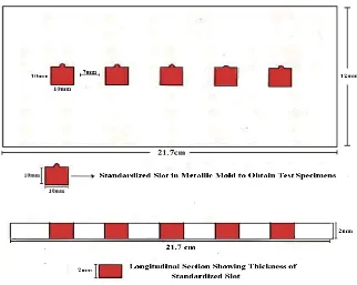
Description of custom – made Metallic mold:
The present study was conducted with test specimens having a metal substructure
overlaid with porcelain. To obtain standardized test specimens, a custom metallic mold (fig.1,
2) was fabricated to the dimensions as required by the testing equipment employed in this
study. The custom metallic mold is a three piece unit consisting of [a] Base (fig.1a) [b]
Middle flat plate (fig.1b) [c] Lid (fig.1c) made up of stainless steel. Four rivets are present at
to aid in seating these two parts over the base precisely. The middle flat plate of the unit has a
thickness of 2 mm (fig.2). Five square slots, each measuring 10x10mm were cut out in the
middle flat plate. When the middle flat plate was seated onto the base, five equal dimension
slots were formed, to obtain resin patterns (fig.14). The five slots each measuring 10mm in
length and 10mm in width and 2mm in thickness were separated by and equal distance of
7mm (fig.2). Patterns of standardized dimensions were prepared using this custom mold.
These were subsequently cast to obtain the metal substructure of all the test specimens for
each test group.
METHODOLOGY
The methodology adopted for this study has been divided into the following stages:
I. Preparation of the porcelain fused to metal test samples:
a. Pattern fabrication
b. Casting and finishing of metal substructure of test specimens
c. Veneering of the metal substructure with the test porcelain systems:
i. Veneering with Feldspathic porcelain
ii. Veneering with Fluorapatite leucite porcelain
II. Surface texture evaluation of the Autoglazed test samples qualitatively and
quantitatively.
III. Surface texture evaluation of the Autoglazed test samples following abrasion and finishing with two different ceramic finishing systems (Sof-Lex discs and White silicon and grey rubber) qualitatively and quantitatively.
GROUP I: Feldspathic porcelain (20 samples)
SUBGROUP-IA, finished with Sof-Lex discs (coarse, medium, fine, extra fine)
SUBGROUP-IB, finished with White silicon & grey rubber.
SUBGROUP-IIA, finished with Sof-Lex discs (coarse, medium, fine, extra fine)
SUBGROUP-IIB, finished with White silicon & grey rubber.
IV. Surface texture evaluation of the finished test samples after polishing with diamond polishing paste qualitatively and quantitatively.
I. Preparation of the porcelain fused to metal test samples: a. Pattern fabrication:
The custom – made metallic mold (Fig.1, 2) as described previously, was used to fabricate
standardized resin patterns. A thin coat of whit petroleum jelly (Tejpa & co, INDIA) was
applied over all the components of the metal mold on all sides. The middle plate was placed
on the base and auto polymerizing pattern resin was mixed and poured into the slots. The
upper lid of the metallic mold was placed into the rivets over the resin and was precisely
closed using a bench press (Fig.13). After the pattern resin set, the upper lid was removed and
thus plastic resin patterns were obtained. The dimensions of the patterns were 10mm width
and 2mm thickness. In this manner a total of forty resin patterns were fabricated to provide
twenty specimens for each test porcelain system employed in the study.
b. Casting and finishing of metal substructure of test specimens:
All the forty patterns were sprued with preformed wax sprue ( Bego, GERMANY) (Fig.4a) of
2.5 cm lengths and 2.5 mm diameter and invested using phosphate bonded investment
( MOLDAVEST exact, Heraeus Kulzer GmbH, GERMANY) (Fig.4f) mechanically mixed
with colloidal silica ( investment BS Liquid 1 – Heraeus Kulzer) (Fig.4g) according to the
manufacture’s instructions under vacuum using vacuum power mixer ( Whip Mix. Inc. Co.
U.S.A.) After a 20 minute bench set time the patterns were subsequently burnt out in a
burnout furnace (Technico, Technico laboratory products pvt. Ltd., Chennai, India.) and cast
machine ( Fornax GEU, Bego, GERMANY) followed by divesting and finishing the casting
to obtain test specimens of uniform dimensions of 10x10x2mm. The dimension of each test
specimen was verified by measuring the length and breadth using a stainless steel ruler and
the thickness with an Iwanson’s gauge. (Essago, GERMANY) (Fig.17). The acceptable
specimens were then air abraded and subsequently steam cleaned to remove surface
impurities. In this manner a total of forty metal substructures of standardized dimensions were
obtained. These were randomly assigned into two main groups (Group I & Group II) with
twenty samples in each test group. Group I & Group II samples were subsequently veneered
with feldspathic porcelain and fluorapatite leucite porcelain systems respectively.
c. Veneering the metal substructure with test porcelain: In this study, Feldspathic porcelain (Ivoclar-IPS Classic, Ivoclar Vivadent AG, Liechtenstein, GERMANY) and
Fluorapatite leucite porcelain (Ivoclar- d sign, Ivoclar Vivadent AG, Liechtenstein,
GERMANY) employed were assigned as group I and II respectively. A common basic D3
shade was selected for both the porcelain systems. All the test specimens were fired in Dental
porcelain furnace – Vita Vacumat 100 (Vita, Bad Sackingen, GERMANY) (Fig.10)
i) Veneering with Feldspathic porcelain:
On the group I comprising of twenty specimens opaque of the chosen D3 shade was painted
onto the prepared metal substructure. The wash opaque was fired in Dental porcelain
furnace-Vita Vacumat 10 (furnace-Vita, Bad Sackingen, GERMANY) (Fig.10) according to manufacturer’s
instructions given in table 1 (Fig.21a) and a second layer of opaque was applied to completely
mask the metal. The second firing was done as per the chart given in table 1. The thickness of
opaque layer was between 0.3 – 0.4mm after two firings (Fig.20a) for all specimens. This was
followed by the application of dentine porcelain of D3 shade on the opaque layer excess
was fired according to manufacturer’s instruction given in table 1. The thicknesses of samples