Copyright © 2000, American Society for Microbiology. All Rights Reserved.
Increased Neutralization Sensitivity and Reduced Replicative Capacity
of Human Immunodeficiency Virus Type 1 after Short-Term
In Vivo or In Vitro Passage through Chimpanzees
TIM BEAUMONT,1,2SILVIA BROERSEN,1,2ADVANNUENEN,1,2HAN G. HUISMAN,2,3 ANA-MARIADERODA HUSMAN,1,2† JONATHAN L. HEENEY,4ANDHANNEKE SCHUITEMAKER1,2*
Department of Clinical Viro-Immunology1and Department of Pathophysiology of Plasma Proteins,3CLB, and
Laboratory for Experimental and Clinical Immunology, University of Amsterdam, Academic Medical Center,2
Amsterdam, and Department of Virology, Biomedical Primate Research Centre, Rijswijk,4The Netherlands
Received 10 November 1999/Accepted 19 May 2000
Development of disease is extremely rare in chimpanzees when inoculated with either T-cell-line-adapted neutralization-sensitive or primary human immunodeficiency virus type 1 (HIV-1), at first excluding a role for HIV-1 neutralization sensitivity in the clinical course of infection. Interestingly, we observed that short-term in vivo and in vitro passage of primary HIV-1 isolates through chimpanzee peripheral blood mononuclear cells (PBMC) resulted in a neutralization-sensitive phenotype. Furthermore, an HIV-1 variant reisolated from a chimpanzee 10 years after experimental infection was still sensitive to neutralization by soluble CD4, the CD4 binding site recognizing antibody IgG1b12 and autologous chimpanzee serum samples, but had become relatively resistant to neutralization by polyclonal human sera and neutralizing monoclonal antibodies. The initial adaptation of HIV-1 to replicate in chimpanzee PBMC seemed to coincide with a selection for viruses with low replicative kinetics. Neither coreceptor usage nor the expression level of CD4, CCR5, or CXCR4 on chimpanzee PBMC compared to human cells could explain the phenotypic changes observed in these chim-panzee-passaged viruses. Our data suggest that the increased neutralization sensitivity of HIV-1 after repli-cation in chimpanzee cells may in part contribute to the long-term asymptomatic HIV-1 infection in experi-mentally infected chimpanzees.
Chimpanzees (Pan troglodytes) are the most commonly used nonhuman primates infected with human immunodeficiency virus type 1 (HIV-1). Upon inoculation of chimpanzees with HIV-1, virus isolation from peripheral blood mononuclear cells (PBMC) was repeatedly successful, and the development of HIV-1-specific antibodies together with an HIV-1-specific cytotoxic-T-lymphocyte response could be demonstrated (1, 2, 4, 17, 44, 45, 52). However, despite persistent HIV replication in vivo, clinical progression to AIDS-defining illnesses was generally not observed (17, 24, 44, 64), which reduced the relevance of this animal model for pathogenesis studies. A number of mechanisms have been postulated to explain the absence of disease progression. These include the inability of chimpanzee CD4⫹cells to support HIV-1-induced syncytium
formation (8), the inability of chimpanzee macrophages to support 1 replication (18, 57, 64), and reduced HIV-induced apoptotic cell death in these animals (20, 23, 57). However, HIV-1 viruses highly adapted to replicate in chim-panzee PBMC through multiple in vivo or repetitious in vitro passages could form syncytia, replicated in macrophages, and induced apoptosis in CD4⫹ T cells (14, 18, 47, 59). Other
proposed mechanisms for long-term survival of chimpanzees are the lack of impaired CD4⫹ T-cell renewal (24), major
histocompatibility complex polymorphisms (2), differences in production of CD8⫹T-cell factor (22), and the role of
infil-tration of CD8⫹ T cells in lymphoid tissue (29), which all
remain to be clarified (25).
During HIV-1 infection in humans, increased replicative capacity of the virus is associated with an increase in viral load and subsequent disease progression. In 50% of the cases, a switch from non-syncytium- to syncytium-inducing variants (from CCR5 to CXCR4 usage) (12, 63) can be observed, which correlates with an accelerated CD4 T-cell decline and a more rapid disease progression (30). Although no correlation be-tween disease progression and serum-neutralizing antibody re-sponses were found (10, 21), a more broadly effective antibody response was found during the course of infection, which was most pronounced in long-term nonprogressors (37, 41, 48, 54, 67). This may indicate that normal regulation of the antibody response in HIV-1 infected individuals is impaired and there-fore development of escape mutants cannot be prevented (5). It has been suggested that in long-term nonprogressors a more vigorous antibody response can develop, possibly because of reduced viral replication (54, 68).
Biological properties of HIV-1 such as replication rate and neutralization sensitivity, both considered relevant for AIDS pathogenesis, have not been extensively studied in chimpan-zees. Here we studied primary HIV-1 variants after short-term in vivo and in vitro passage through chimpanzee cells and compared them with an HIV-LAI variant reisolated 10 years after experimental infection of a chimpanzee.
MATERIALS AND METHODS
Primary cells.PBMC were isolated from buffy coats obtained from healthy blood donors or from heparinized vena punctures of healthy HIV-1-negative chimpanzees by Ficoll-Isopaque density gradient centrifugation. Cells (5⫻106/
ml) were stimulated for 3 days in Iscove modified Dulbecco medium (IMDM) supplemented with 10% fetal calf serum (FCS), penicillin (100 U/ml), strepto-mycin (100g/ml), and phytohemagglutinin (PHA; 5g/ml). Subsequently, cells * Corresponding author. Mailing address: Department of Clinical
Viro-Immunology, CLB, Plesmanlaan 125, 1066 CX Amsterdam, The Netherlands. Phone: 31-20-5123317. Fax: 31-20-5123310. E-mail: J_Schuitemaker@CLB.nl.
† Present address: Department of Microbiological Laboratory for Healthcare, RIVM, Bilthoven, The Netherlands.
7699
on November 9, 2019 by guest
http://jvi.asm.org/
(106/ml) were grown in the absence of PHA, in medium supplemented with
recombinant interleukin-2 (10 U/ml, a kind gift of R. Rombouts, Chiron Benelux). Prior to use of chimpanzee PBMC in 50% tissue culture infective dose (TCID50) or neutralization assays, CD8⫹T cells were depleted using
anti-CD8-coated magnetic beads (Dynal).
Viruses.Virus isolation and virus stock preparation were performed on PHA-stimulated PBMC according to standard procedures (56). The HAN2 isolate was obtained from the European Programme for a Vaccine against AIDS (Pro-gramme EVA, Potters Bar, United Kingdom). Isolation and properties of the virus have previously been described (53). An inoculum of the HIV-LAI strain prepared on the H9 T-cell line was used to experimentally infect a chimpanzee. HIVAms37 was obtained from an AIDS patient visiting the Academic Medical
Center in Amsterdam. The HIV-IIIB stock was also prepared on human PBMC. Each week, virus production in the supernatant was monitored in an in-house p24 antigen capture enzyme-linked immunosorbent assay (ELISA). If sufficient p24 antigen production could be demonstrated, the titer of the virus stock was quantified by determination of the TCID50in PHA-stimulated healthy donor
PBMC.
In vivo chimpanzee passage of a T-cell line-adapted (TCLA) and primary HIV-1 isolate.All chimpanzees studied were housed in the Biomedical Primate Research Center in Rijswijk, The Netherlands. Ten years after the experimental infection with HIV-LAI (infected in 1984), biological virus clones were reisolated from chimpanzee Maya (ch-Ma). Experimental infection with the primary isolate HAN2 was performed as previously described (4). A female chimpanzee was inoculated with 100 TCID50(as determined on chimpanzee PBMC) of a virus
stock prepared on human PBMC. Blood samples were drawn every week and revealed a peak in HIV-1 RNA serum levels at 4.5 weeks postinoculation, and after 6 weeks antibodies could be detected (4). From a PBMC sample collected 4.5 weeks after inoculation, biological HIV-1 clones (HAN2/ch-in vivo) were obtained by cocultivation with human PHA-stimulated PBMC according to stan-dard procedures (56).
Neutralizing agents.Viruses were tested for their relative neutralization sen-sitivity against increasing concentrations of recombinant sCD4, HIV-1 immuno-globulin (HIVIG), pooled human sera (Amshps), and the human monoclonal
antibodies (MAbs) gp13, gp68, IgG1b12, and 1577. HIVIG is a preparation of purified polyclonal immunoglobulin derived from plasma of multiple HIV-in-fected donors who had more than 400 CD4⫹T cells/l of blood (13). Amshpsis
a nonimmunoglobulin purified pooled plasma of 34 seropositive patients from the Amsterdam Cohort whose CD4⫹T-cell counts ranged from 40 to 820 cells/
l. The gp13 and gp68 antibodies recognize epitopes surrounding the CD4bs of gp120 (58), and IgG1b12 recognizes the CD4bs of gp120 (7). MAb 1577 recog-nizes a highly conserved epitope of gp41 (residues 735 to 752) (16). Autologous serum neutralization of LAI/ch-Ma was studied with three serum samples that were obtained in March 1992, June 1994, and March 1996.
Neutralization sensitivity of HIV-1 variants.From each virus isolate, a final inoculum of 10 TCID50in a volume of 100l was incubated for 1 to 2 h at 37°C
with increasing concentrations of the neutralizing agents. Subsequently, the mix-tures of virus with sCD4, sera, or antibodies were added to 1053-day
PHA-stimulated PBMC of either human or chimpanzee origin in 96-well microtiter plates. The following day, plates incubated with HIVIG, Amshps, or chimpanzee
serum were washed extensively. On days 7 and 14, virus production in culture supernatants was analyzed by an in-house p24 antigen capture ELISA. Means of quadruplicate experiments of each agent, tested at least twice, were plotted. The percent neutralization was calculated by determining the reduction in p24 pro-duction in the presence of the agent compared to the cultures with virus only. When possible 50% (IC50) and 90% (IC90) inhibitory concentrations were
de-termined by linear regression. When sensitivity to neutralization was measured on chimpanzee PBMC, p24 production was monitored until day 10.
In vitro characterization of virus replication kinetics.Analysis of replication kinetics was performed as described previously (63). Briefly, PHA-stimulated PBMC (5.0⫻106cells/0.5 ml) were incubated with 102TCID
50(1 ml) in a total
volume of 1.5 ml for 2 h at 37°C. Virus supernatant was removed, and cells were incubated at a concentration of 1.0⫻106cells/ml. Every day, 50l of
superna-tant was collected to measure p24 production. Fresh PHA-stimulated PBMC were added on days 5, 8, and 11.
125I radiolabeling of V3 peptides and HIV envelope protein.In brief, 50-l
solutions of phosphate-buffered saline (PBS) containing either 20g of peptides or 5g of protein was used for labeling with Na-125I using chloramine T for 30 s
(27). The radiolabeled preparations were purified from free iodine by dialysis and subsequently aliquoted in the presence of protease inhibitor phenylmethyl-sulfonyl fluoride and bovine serum albumin (BSA; fraction V; Sigma) in a final concentration of 0.1% and stored until use at⫺20°C. The peptide V3-IIIB (tip loop, SP104) was prepared as described earlier (35), and the circular V3 peptides were purchased from Zeneca (Cambridge Research Biochemicals) and are based on an isolate obtained from patient 168 from the Amsterdam Cohort (ACH.168). The recombinant gp160 from strain IIIB was expressed via a baculovirus expres-sion system in insect cells and was kindly provided by Phage, La Jolla, Calif.
Binding of HIV envelope protein and V3 peptides to immobilized antibody (radioimmunoassay format).The binding studies of antibodies to V3 peptides and gp160 protein were performed as follows: a serial dilution of serum was mixed with an excess of protein A-Sepharose beads suspended in PBS containing 0.1% BSA and 0.05% NP-40. After 2 h of incubation head over tail, the beads
were washed four times with PBS containing 0.01% Tween 20. Antibodies bound to protein A-Sepharose beads were incubated head over tail with radiolabeled gp160 or V3 peptide (ca. 100,000 cpm) in PBS containing 0.1% BSA and 0.05% NP-40 for 16 h at room temperature. After washing the beads with PBS–0.01% Tween 20, radioactivity was measured in a gamma counter.
Determination of coreceptor use by HIV-1.U87 cells stably expressing CD4 alone or in combination with CCR5 and CXCR4 (a kind gift of D. Littman) were seeded at 104cells per well in 96-well plates in IMDM supplemented with 5g
of Polybrene and 1g of puromycin per ml. Occasionally, 200g of G418 per ml was added to select for CD4-expressing cells. The next day, cells were washed in PBS, and 102to 104TCID
50of virus/ml were added in a 100-l final volume.
After 24 h, cells were washed twice with PBS, and 200l of fresh medium was added. Supernatants were harvested on days 7, 14, 21, and 28 and tested for the presence of p24 antigen by ELISA.
Cell surface expression of CD4, CCR5, and CXCR4.Flow cytometry was used to analyze the expression of CD4, CCR5, and CXCR4. Human and chimpanzee PBMC (0.5⫻106) were incubated either with 5g of anti-CCR5 MAb 2D7
(kindly provided by C. Mackay) or with 5g of IgG2a isotype-matched control MAb (CLB, Amsterdam, The Netherlands) per ml. Cells were then washed and resuspended in 50l of fluorescein isothiocyanate-conjugated affinity-purified F(ab⬘)2goat anti-mouse IgG (CLB). Subsequently, cells were incubated first with
normal mouse serum (CLB) to diminish background staining, followed by an incubation with PerCP-labeled anti-CD4 MAbs (Becton Dickinson, San Jose, Calif.) and phycoerythrin-labeled anti-CXCR4 MAbs (Pharmingen). Cells were analyzed on the FACScan to determine the levels of cell surface expression. All incubation steps were performed at 4°C for 20 min. Between two incubation steps, two wash steps were performed with PBS supplemented with 0.5% BSA.
RESULTS
Phenotypic changes in HIV-1 variants induced by in vitro
passage through chimpanzee cells.We first studied the effect
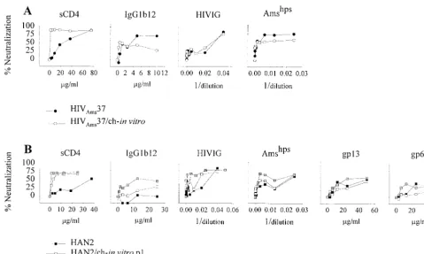
of in vitro passage of primary viruses through chimpanzee PBMC on neutralization sensitivity. PHA-stimulated chimpan-zee PBMC were inoculated with virus stocks prepared on hu-man PBMC of the primary isolates HIVAms37 and HAN2. When p24 production in the HAN2-infected chimpanzee PBMC culture could be demonstrated, supernatant (HAN2/ ch-in vitro-p1) was harvested and in part used for cell-free inoculation of fresh PHA-stimulated chimpanzee PBMC. In this way, p2 and p3 isolates were also obtained. For further studies only the p1 and p3 HAN2 isolates were used. Virus production in HIVAms37-infected chimpanzee PBMC became evident after 3 weeks of culture, and this supernatant was used for further study. In parallel, parental viruses were passaged on human PBMC. Virus stocks from HAN2/ch-in vitro-p1 and -p3, HIVAms37/ch-in vitro, and all the parental viruses were prepared using a mixture of human PBMC derived from dif-ferent donors. On the same cells, a TCID50 assay was per-formed for quantification of the virus titer. Original and pas-saged viruses were then analyzed for their neutralization sensitivity on human PBMC. For comparison the TCLA IIIB virus was included in the experiments.
The sCD4 neutralization resistance of HIVAms37 decreased more than 20-fold from an IC50of 35.0g/ml and an IC90of 67.2g/ml for the primary virus to an sCD4 IC50of 1.3g/ml and an IC90of 3.9g/ml after in vitro passage through chim-panzee cells (Fig. 1A, Table 1). The same was observed for the HAN2 isolate, which originally showed an sCD4 IC50of 51.9 g/ml and an IC90of 98.6g/ml but a 13-fold-decreased IC50 of 3.9g/ml and an IC90of 29.0g/ml after a single passage through chimpanzee cells (Table 1, Fig. 1B).
Differences in neutralization of HAN2 by antibody and pooled sera following one passage through chimpanzee PBMC was not observed. However, the HAN2/ch-in vitro-p3 isolate had become even more sensitive for sCD4 and also an in-creased sensitivity for IgG1b12, Amshps, HIVIG, and gp13 MAb could be measured (Table 1, Fig. 1B).
For HIVAms37/ch-in vitro there was no change in neutral-ization sensitivity for IgG1b12, HIVIG, and Amshps, a finding which may be related to the fact that the primary HIVAms37
7700 BEAUMONT ET AL. J. VIROL.
on November 9, 2019 by guest
http://jvi.asm.org/
isolate was already sensitive for neutralization by IgG1b12 and Amshps(Fig. 1A).
Neutralization sensitivity of a primary HIV-1 variant after
short-term in vivo passage through a chimpanzee. The
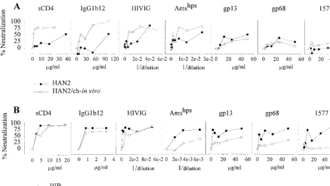
in-creased neutralization-sensitive phenotype of the primary iso-lates after in vitro passage through chimpanzee PBMC suggest that adaptation to replicate in these cells selects for a neutral-ization-sensitive envelope configuration. We next compared the neutralization sensitivity of the primary neutralization-re-sistant HAN2 virus and the HIV-1 biological clone reisolated from PBMC from a chimpanzee that had been experimentally infected with this HAN2 virus for 4.5 weeks (4). The neutral-ization sensitivity of the reisolated HAN2 variant was in-creased 20- and 5-fold, respectively, for sCD4 and IgG1b12 (Fig. 2A). IC50values for HIVIG and Amshpswere decreased respectively by 5- and 7.5-fold (Table 1). There were no dif-ferences for gp13 and gp68 neutralization sensitivity between the primary and in vivo-passaged HAN2 virus.
Neutralization sensitivity of an HIV-1 variant reisolated 10
years after experimental infection of a chimpanzee.The effect
on HIV-1 neutralization sensitivity upon long-term in vivo passage in a chimpanzee was then studied. The LAI variant (LAI/ch-Ma) that was reisolated from chimpanzee Maya 10 years after experimental infection was analyzed for its sCD4 and antibody neutralization sensitivity. Soluble CD4 concen-trations that resulted in a 50% (IC50) or 90% (IC90) reduction in titer of the LAI/ch-Ma isolate were 2.3 and 7.2 g/ml, respectively. This was in the same range as the concentrations required for neutralization of the TCLA IIIB isolate (IC50⫽
3.5g/ml and IC90⫽8.7g/ml). The MAb IgG1b12, known to recognize an epitope within the gp120 CD4 binding domain, could also neutralize the LAI/ch-Ma variant at an IC50 con-centration of 1.4g/ml and an IC90concentration of 3.5g/ml (Fig. 2B, Table 1). The LAI/ch-Ma virus had become relatively resistant for neutralization by HIVIG, Amshps, the anti-gp41 MAb 1577, and the two MAbs gp13 and gp68. Resistance for HIVIG and Amshps increased 5- and 16-fold, respectively, while 15 and 8 times more gp13 and gp68 antibodies, respec-tively, were required for 50% inhibition of the LAI/ch-Ma variant. The LAI/ch-Ma variant was very well neutralized by autologous serum obtained at time points before, at, and after the moment the LAI/ch-Ma variant was isolated (Fig. 3A). The same efficient neutralization was observed for IIIB but not for HAN2 and HAN2/ch-in vivo, indicating the specificity of the humoral immune response.
The specificity was confirmed in a radioimmunoassay with radiolabeled env-derived antigens and the serum of chimpan-zee Maya (Fig. 3B). Serum antibodies, present in a 1991 sam-ple recognized unprocessed gp160, monomeric gp120 (also from MN, data not shown) or the V3 loop of IIIB in a dose-dependent manner. A consensus V3 loop of a non-syncytium-inducing (CCR5 coreceptor-using) virus was not recognized. Furthermore, the linear and folded V3 peptide of a virus iso-late from patient ACH.168, a dual-tropic primary isoiso-late, were also not recognized.
[image:3.612.61.540.70.357.2]The presence of a relatively neutralization-sensitive virus variant 10 years after infection in the presence of specific
FIG. 1. Neutralization sensitivity as determined on human PHA-stimulated PBMC. (A) One hundred TCID50of the primary isolate HIVAms37/ml, before (F) and
after (E) in vitro passage through chimpanzee PBMC (HIVAms37/ch-in vitro). Viruses were incubated with increasing concentrations of sCD4, IgG1b12, gp68, and gp13
or increasing dilutions of HIVIG and Amshpsas indicated. P24 production was measured, and mean OD values were calculated from quadruplicate cultures from at
least duplicate experiments. The percent neutralization was calculated by determining the reduction in supernatant p24 production in the presence of the neutralizing
agents relative to control cultures lacking these agents. (B) Neutralization sensitivity of the primary isolates HAN2 (■) and HAN2 after one (HAN2/ch-in vitro-p1;䊐)
or three (HAN2/ch-in vitro-p3;o) in vitro passages through chimpanzee PBMC.
on November 9, 2019 by guest
http://jvi.asm.org/
antibodies may imply that full escape from humoral immunity is not necessary for HIV persistence in the chimpanzee.
Sensitivity to neutralization as measured on chimpanzee
PBMC.Next, we wanted to exclude that the increased
neutral-ization sensitivity after passage through chimpanzee PBMC in vivo and in vitro is due to the type of target cell used for the neutralization assay. We compared the sensitivities of IIIB, LAI/ch-Ma, HAN2, and HAN2/ch-in vivo to IgG1b12 neutral-ization on chimpanzee PBMC. First, the TCID50was deter-mined on CD8 T-cell-depleted chimpanzee PHA-stimulated PBMC before 100 TCID50was used in the neutralization assay. We found the same neutralization sensitivity irrespective of the use of chimpanzee or human PBMC. Both on chimpanzee PBMC and on human PBMC, HIV-IIIB and LAI/ch-Ma were highly sensitive, and the primary HAN2 isolate was resistant, whereas relative to the parental HAN2, the HAN2/ch-in vivo exhibited an increased sensitivity for neutralization by IgG1b12 (Fig. 4).
Influence of in vivo and in vitro passage of HIV-1 through
chimpanzee PBMC on replication kinetics.It has been
hypoth-esized that viral adaptation to a new environment would select for virus variants who most efficiently enter and duplicate in these new target cells (28, 46, 60). This process could influence replication kinetics, which in this case coincides with a neutral-ization-sensitive HIV-1 envelope glycoprotein. We therefore analyzed how this neutralization-sensitive phenotype associ-ated with the viral replicative capacity.
As demonstrated in Fig. 5, serial passage of the HAN2 isolate through chimpanzee cells reduced its replicative capac-ity on human PBMC. The same was observed for the virus reisolated after 4.5 weeks of in vivo replication in a chimpan-zee. The LAI/ch-Ma isolate, which was still sCD4 and IgG1b12 sensitive but resistant to pooled sera, showed relatively high replication kinetics compared to the IIIB isolate and the HAN2 isolates. The reduced replicative capacity of chimpan-zee-passaged HIV-1 was also evident when chimpanzee PBMC were used as target cells (data not shown).
Coreceptor usage of HIV-1 variants before and after
pas-sage through chimpanzee cells.The increased neutralization
sensitivity of HIV-1 upon passage through chimpanzee PBMC closely resembles the phenotypic changes associated with ad-aptation to grow in T-cell lines. Passage of primary isolates through T-cell lines or cell lines expressing one of the different coreceptors (55) does not normally induce a change in core-ceptor usage during transition from neutralization resistance to neutralization sensitivity (33, 40, 62). To exclude a role for coreceptor use during chimpanzee PBMC passage in vitro and in vivo, we tested whether passage was associated with changes in coreceptor use.
The neutralization-resistant isolates HIVAms37 and HAN2 both used CCR5 and CXCR4. In vitro passage of these isolates through chimpanzee cells did increase the neutralization sen-sitivity and reduced the replicative capacity but did not change the capacity to use both CCR5 and CXCR4 (Table 2). In vivo passage of the HAN2 isolate through chimpanzee cells, which also resulted in a highly neutralization-sensitive progeny virus, also did not change the capacity to use both CCR5 and CXCR4. The LAI/ch-Ma isolate could only use CXCR4 as a coreceptor.
Expression of CD4, CCR5, and CXCR4 on human and
chimpanzee PBMC.It has been suggested that the low
expres-sion of CD4 on cell lines compared to the expresexpres-sion of CD4 on primary T cells would select for viruses with the highest affinity for CD4 (31, 49), thus explaining why TCLA viruses are highly sensitive to sCD4 neutralization. Since passage through chimpanzee cells exerted the same effect as T-cell-line
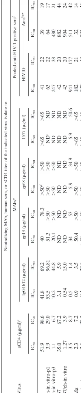
adap-TABLE 1. ICs of HIV-1 isolates passaged through chimpanzee PBMC Virus Neutralizing MAb, human sera, or sCD4 titer of the indicated virus isolate to: sCD4 ( g/ml) a MAbs a Pooled anti-HIV-1-positive sera b IgG1b12 ( g/ml) gp13 ( g/ml) gp68 ( g/ml) 1577 ( g/ml) HIVIG Ams hps IC50 IC90 IC50 IC90 IC50 IC90 IC50 IC90 IC50 IC90 IC50 IC90 IC50 IC90 HAN2 51.9 98.6 41.8 69.2 40 78.3 ⬎ 50 d ⬎ 50 ⬎ 65 ⬎ 65 41 22 39 19 HAN2/ch-in vitro-p1 3.9 29.0 53.5 96.81 51.3 ⬎ 50 ⬎ 50 ⬎ 50 ND c ND 63 22 64 17 HAN2/ch-in vitro-p3 1.1 6.7 10.2 44.8 20.1 ⬎ 50 ⬎ 50 ⬎ 50 ND ND 247 38 480 21 HIV Ams 37 35.0 67.2 3.1 5.9 ND ND ND ND ND ND 42 20 882 44 HIV Ams 37/ch-in vitro 1.27 3.9 0.54 15.0 ND ND ND ND ND ND 43 20 904 24 IIIB 3.5 8.7 0.5 1.4 34 53 5.9 34.9 5.9 50.6 941 177 511 42 LAI/ch-Ma 2.3 7.2 0.9 3.5 50.4 ⬎ 50 ⬎ 50 ⬎ 50 ⬎ 65 ⬎ 65 182 21 32 14 HAN2/ch-in vivo 2.5 23.3 9.4 32.5 ⬎ 50 ⬎ 50 ⬎ 50 ⬎ 50 ⬎ 65 ⬎ 65 187 43 291 45 aThe 50 and 90% neutralization titers were determined by assaying p24 inhibition. bReciprocal 50 and 90% neutralization titers. cND, not done. dInstances in which no neutralization was observed with the highest concentration of MAb used are indicated with a “ ⬎ ” sign.
7702 BEAUMONT ET AL. J. VIROL.
on November 9, 2019 by guest
http://jvi.asm.org/
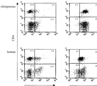
[image:4.612.96.230.79.729.2]tation, we analyzed the expression of CD4 on PBMC of both human and chimpanzee origin. In addition, we examined the expression of the HIV-1 coreceptors CCR5 and CXCR4. Anal-ysis of the membrane expression of the different molecules did not reveal a difference between PBMC originating from either humans or chimpanzees (Fig. 6).
DISCUSSION
We observed here that short-term passage through chimpan-zee cells of primary neutralization-resistant viruses resulted in HIV-1 progeny with increased neutralization sensitivity for soluble CD4 (sCD4), pooled human sera, and the CD4 binding site (CD4bs) recognizing antibody IgG1b12. This neutraliza-tion-sensitive phenotype appeared to be relatively stable since an HIV-1 variant reisolated from a long-term asymptomatic chimpanzee that had been infected with HIV for 10 years was still sensitive to sCD4, IgG1b12, and autologous serum even in a retrospective fashion, suggesting that within a timeframe of 2 years no neutralization-resistant escape variants developed. By using radiolabeled HIV antigens, we showed that this autolo-gous serum had IIIB-envelope-specific reactivity which most likely occurred in the context of the highly antigenic -QR-GPGR motif in the V3 loop of HIV-IIIB (26). This QR motif is absent in the NSI consensus and in the ACH.168 V3 loop. The increased neutralization sensitivity after short-term pas-sage through chimpanzee PBMC coincided with a reduced replicative capacity. The efficient replication of the long term in vivo-passaged isolate may suggest a process of ongoing ad-aptation to growth in chimpanzee PBMC, although neutraliza-tion resistance to CD4bs-directed agents was not regained. The immediate increase in neutralization sensitivity for CD4bs-recognizing agents of primary HIV-1 variants upon passage
through chimpanzee PBMC may indicate that the loss of sCD4 and IgG1b12 resistance was due to selection for or rapid de-velopment of viruses with a gp120-gp41 configuration that could efficiently support entry into chimpanzee CD4⫹T cells.
It can be stated that this phenotypic change both in vivo and in vitro occurred in the absence of neutralizing antibodies since in vivo the first antibodies were detected 6 weeks after infection. It therefore cannot be excluded that in the absence of neutral-izing antibodies viruses are selected that are sensitive for neu-tralization. However, this is different from observations in hu-mans, where the development of neutralization-sensitive viruses during primary infection does not occur (36). The ad-aptation to grow in chimpanzee cells may result in reduced levels of protection against a chimpanzee CD4bs-directed neu-tralization. Long-term passage in a chimpanzee did not fully revert the neutralization sensitivity of LAI/ch-Ma. It thus seems that not merely the presence of HIV-1-neutralizing an-tibodies in vivo (3, 6, 15, 19) but also the sensitivity of circu-lating HIV-1 variants to CD4bs-recognizing antibodies may correlate with the benign clinical course of infection in HIV-1-infected chimpanzees.
[image:5.612.64.541.74.343.2]It could be that the affinity of chimpanzee CD4 for HIV-1 gp120 is different. Although 5-amino-acid differences have been observed between human and chimpanzee CD4, there was no difference in affinity and association rate for TCLA monomeric gp120 between human or chimpanzee CD4 (8). Also, no differences in infection of HIV-IIIB on human or chimpanzee CD4-transfected cells were found. Our observa-tion that neutralizaobserva-tion sensitivity was not dependent on the use of chimpanzee or human PBMC confirmed these results. Furthermore, no differences between human and chimpanzee CD4 glycosylation have been found, and CD4 epitopes recog-nized by a panel of 19 different anti-CD4 MAbs showed that
FIG. 2. Neutralization sensitivity of the HAN2 isolate (■) and HAN2 reisolated 4.5 weeks after experimental infection of a chimpanzee (HAN2/ch-in vivo;䊐) (A)
and the TCLA IIIB virus (F) and the virus reisolated 10 years after experimental infection of a chimpanzee (LAI/ch-Ma;E) (B). The assay was performed as described
in the legend to Fig. 1 except that, along with the other agents, MAb 1577 neutralization sensitivity was also determined.
on November 9, 2019 by guest
http://jvi.asm.org/
epitopes were equally expressed on human and chimpanzee cells (38), which seems to exclude major conformational dif-ferences between human and chimpanzee CD4. Therefore, higher CD4 affinity as a general mechanism for increased neu-tralization sensitivity seems unlikely. Another possibility could be a differential expression or functioning of coreceptors CCR5 and CXCR4 on human and chimpanzee CD4⫹T cells.
We showed here that the expression of coreceptors is compa-rable in cells of human and chimpanzee origin. Moreover, human and chimpanzee CXCR4 show complete sequence ho-mology, while in CCR5 2-amino-acid substitutions have been reported that do not disturb coreceptor functioning (50, 61). Altogether, these results do not suggest that differences in expression or function of coreceptors influence the neutraliza-tion sensitivity of progeny virus.
The phenotypic changes as observed after adaptation of HIV-1 variants to chimpanzee PBMC resemble the alterations observed after T-cell-line passage. The increased neutraliza-tion sensitivity during T-cell-line adaptaneutraliza-tion may be related to
shielding of relevant epitopes on primary viruses (32, 66), re-sulting in a less-than-optimal binding efficiency to CD4 cells but creating an optimal adaptation for replication in the pres-ence of neutralizing antibodies in vivo (43). TCLA HIV-1 isolates would then be optimally adapted to replicate in vitro, by trading of protection from neutralizing antibodies for an envelope configuration that allows more efficient CD4 binding and subsequent cell fusion (31, 42, 62). Since the neutraliza-tion-sensitive phenotype of the LAI/ch-Ma persisted in the presence of high neutralizing antibody titers (1, 19, 45, 46), the phenotypic similarities between HIV-1 adaptation to FIG. 3. Analysis of the neutralizing capacity of diluted autologous
chimpan-zee serum. (A) A standard neutralization assay was performed with serum obtained at different time points (March 1992, June 1994, and March 1996)
of chimpanzee Maya and used to test the viruses HIV-IIIB (F), LAI/ch-Ma
(reisolated from chimpanzee Maya in 1994) (E), HAN2 (Œ), and HAN2/ch-in
vivo (‚). Serum and viruses were washed away 24 h after incubation on human
[image:6.612.53.292.550.667.2]PHA-stimulated PBMC. The percent neutralization compared with that of the control is shown. (B) Serum of chimpanzee Maya obtained in 1991 was tested for binding to different HIV-envelope-related antigens using a radioimmunoassay (see Materials and Methods).
FIG. 4. Sensitivity to neutralization by the IgG1b12 MAb of parental and chimpanzee passaged isolates as measured on CD8 T-cell-depleted
PHA-stim-ulated chimpanzee PBMC. (A) HIV-IIIB (F) and LAI/ch-Ma (E). (B) HAN2
(■) and HAN2/ch-in vivo (䊐). The TCID50of the on human PBMC-grown virus
stocks was first determined on the same CD8 T-cell-depleted chimpanzee PBMC. Depicted in the graph is the percent neutralization compared to that of untreated controls.
FIG. 5. Replication kinetics of progeny viruses before and after passage
through chimpanzee PBMC. (A) HIV-IIIB (F) and LAI/ch-Ma (E). (B) HAN2
(■), HAN2/ch-in vivo (䊐), and HAN2/ch-in vitro-p3 ( ).
7704 BEAUMONT ET AL. J. VIROL.
on November 9, 2019 by guest
http://jvi.asm.org/
[image:6.612.314.551.586.700.2]zee PBMC and T-cell lines may be based on different mecha-nisms; otherwise, the hypothesis needs to be adjusted.
With respect to HIV-1 infection in humans, we reisolated a slowly replicating IIIB variant that was sensitive to neutraliza-tion by CD4bs-recognizing agents, 3 years after accidental in-fection of a laboratory worker with the IIIB virus (65). An isolate obtained 4 years later had become neutralization resis-tant and possessed an increased replicative capacity (unpub-lished data). It cannot be excluded that in chimpanzees, the same evolution of biological properties is ongoing, but with delayed kinetics due to low-level replication and consequently slow accumulation of required mutations (11). In favor of this hypothesis is the observation that serial passage of HIV-1 through chimpanzees, and thus adaptation to this host, indeed seems to result in a virus that is more pathogenic for chimpan-zees (14, 47, 59). In these studies, changes in the neutralization sensitivity of the virus were not considered. However, based on
our observations in the accidentally infected laboratory worker and on recent observations in macaques (9, 28), it is tempting to speculate that HIV-1 variants that cause AIDS in chimpan-zees are neutralization resistant for CD4bs-recognizing agents, with a fully restored replicative capacity (9, 39).
The increased neutralization sensitivity of primary HIV-1 isolates upon passage through chimpanzee cells, as demon-strated here, argues for caution in the interpretation of HIV-1 vaccine studies in this animal. The ability of experimental vac-cines to protect from infection may be overestimated in chim-panzees since at least the HIV-1 isolates tested here, immedi-ately became neutralization sensitive in this species. A vaccine that is considered to induce protective humoral immunity based on experiments in chimpanzees could possibly fail in humans since the level of neutralizing antibodies and the dif-ference in neutralization sensitivity of the viruses that provide protection in chimpanzees might be insufficient against pri-mary neutralization-resistant HIV-1 variants present in hu-mans. Finally, a correlation between neutralization sensitivity and the clinical course of an HIV-1 infection may indeed exist. Elucidation of the molecular basis for neutralization resistance of primary HIV-1 variants may reveal strategies to prevent infection (32, 34, 51, 66).
ACKNOWLEDGMENTS
We thank R. Sweet (SmithKline Beecham) for providing recombi-nant sCD4 and A. Prince for HIVIG. Amshpswas obtained from J.
Goudsmit, gp13 and gp68 were a kind gift of M. Schutten, and the IgG1b12 MAb was kindly provided by P. W. H. I. Parren and D. R. Burton. The 1577 gp41 MAb was obtained through the AIDS Re-search and Reference Reagent Program, NIH, and was contributed by M. Ferguson. We thank Aran Labrijn, Rene van Lier, and Frank Miedema for critically reading the manuscript.
FIG. 6. CCR5 and CXCR4 expression on CD4-positive lymphocyte populations of chimpanzee (top panels) or human (bottom panels) PBMC. A three-color
staining protocol was used to assess CCR5 expression (xaxis of left panels) and CXCR4 expression (xaxis of right panels) on the total number of CD4-positive cells
[image:7.612.151.463.73.349.2](yaxis in all plots).
TABLE 2. Coreceptor usage of primary and chimpanzee-passaged HIV-1 variants
Virus
CD4 expressing U87 cells cotransfected with:
CXCR4 CCR5
IIIB ⫹ ⫺
LAI/ch-MA ⫹ ⫺
HAN2 ⫹ ⫹
HAN2/ch-in vivo ⫹ ⫹
HAN2/ch-in-vitro-p1 ⫹ ⫹
HIVAms37 ⫹ ⫹
HIVAms37/ch-in vitro ⫹ ⫹
on November 9, 2019 by guest
http://jvi.asm.org/
This work was supported by The Dutch AIDS Foundation (grant number 1304).
REFERENCES
1.Alter, H. J., J. W. Eichberg, H. Masur, W. C. Saxinger, R. C. Gallo, A. M. Macher, H. C. Lane, and A. S. Fauci.1984. Transmission of HTLV-III infection from human plasma to chimpanzees: an animal model for AIDS. Science226:549–552.
2.Balla-Jhagjhoorsingh, S. S., G. Koopman, P. Mooij, T. G. M. Haaksma, V. J. P. Teeuwsen, R. E. Bontrop, and J. L. Heeney.1999. Conserved CTL epitopes shared between HIV-infected human long-term survivors and chim-panzees. J. Immunol.162:2308–2314.
3.Berman, P. W., T. J. Gregory, L. Riddle, G. R. Nakamura, M. A. Champe, J. P. Porter, F. M. Wurm, R. D. Hershberg, E. K. Cobb, and J. W. Eichberg. 1990. Protection of chimpanzees from infection by HIV-1 after vaccination with recombinant glycoprotein gp120 but not gp160. Nature345:622–625. 4.Bogers, W. M. J. M., W. Koornstra, R. Dubbes, P. J. F. ten Haaft, B. E.
Verstrepen, S. Jhagjhoorsingh, T. Haaksma, H. Niphuis, J. D. Laman, S. Norley, H. Schuitemaker, J. Goudsmit, G. Hunsmann, J. L. Heeney, and H. Wigzell.1998. Characteristics of primary infection of a European human immunodeficiency virus type 1 clade B isolate in chimpanzee. J. Gen. Virol. 79:2895–2903.
5.Bradney, A. P., S. Scheer, J. M. Crawford, S. P. Buchbinder, and D. C. Montefiori.1999. Neutralization escape in human immunodeficiency virus type 1-infected long-term nonprogressors. J. Infect. Dis.179:1264–1267. 6.Bruck, C., C. Thiriart, L. Fabry, M. Francotte, P. Pala, O. Van Opstal, J.
Culp, M. Rosenberg, M. De Wilde, and P. Heidt.1994. HIV-1 envelope-elicited neutralizing antibody titres correlate with protection and virus load in chimpanzee. Vaccine12:1141–1148.
7.Burton, D. R., J. Pyati, R. Koduri, S. J. Sharp, G. B. Thornton, P. W. H. Parren, L. S. W. Sawyer, R. M. Hendry, N. Dunlop, P. L. Nara, M. Lamac-chia, E. M. Garratty, E. R. Stiehm, Y. J. Bryson, Y. Cao, J. P. Moore, D. D. Ho, and C. F. Barbas III.1994. Efficient neutralization of primary isolates of HIV-1 by a recombinant human monoclonal antibody. Science266:1024– 1027.
8.Camerini, D., and B. Seed.1990. A CD4 domain important for HIV-medi-ated syncytium formation lies outside the virus binding site. Cell60:747–754. 9.Cayabyab, M., G. B. Karlsson, B. A. Etemad-Moghadam, W. Hofmann, T. Steenbeke, M. Halloran, J. W. Fanton, M. K. Axthelm, N. L. Letvin, and J. G. Sodroski.1999. Changes in human immunodeficiency virus type 1 envelope glycoproteins responsible for the pathogenicity of a multiple pas-saged simian-human immunodeficiency virus (SHIV-HXBc2). J. Virol.73: 976–984.
10. Cecilia, D., C. A. Kleeberger, A. Munoz, J. V. Giorgi, and S. Zolla-Pazner. 1999. A longitudinal study of neutralizing antibodies and disease progression in HIV-1-infected subjects. J. Infect. Dis.176:1365–1374.
11. Coffin, J. M.1995. HIV population dynamics in vivo: implications for genetic variation, pathogenesis, and therapy. Science267:483–489.
12. Connor, R. I., and D. D. Ho.1994. Human immunodeficiency virus type 1 variants with increased replicative capacity develop during the asymptomatic stage before disease progression. J. Virol.68:4400–4408.
13. Cummins, L. M., K. J. Weinhold, T. J. Matthews, A. J. Langlois, C. F. Perno, R. M. Condie, and J. P. Allain.1991. Preparation and characterization of an intravenous solution of IgG from human immunodeficiency virus-seroposi-tive donors. Blood77:1111–1117.
14. Davis, I. C., M. Girard, and P. N. Fultz.1998. Loss of CD4⫹T cells in human
immunodeficiency virus type 1-infected chimpanzees is associated with in-creased lymphocyte apoptosis. J. Virol.72:4623–4632.
15. Emini, E. A., W. A. Schleif, J. H. Nunberg, A. J. Conley, Y. Eda, S. Tokiyoshi, S. D. Putney, S. Matsushita, K. E. Cobb, C. M. Jett, J. W. Eichberg, and K. K. Murthy.1992. Prevention of HIV-1 infection in chimpanzees by gp120 V3 domain-specific monoclonal antibody. Nature355:728–730.
16. Evans, D. J., J. McKeating, J. M. Meredith, K. L. Burke, K. Katrak, A. John, M. Ferguson, P. D. Minor, R. A. Weiss, and J. W. Almond. 1989. An engineered poliovirus chimaera elicits broadly reactive HIV-1 neutralizing antibodies. Nature339:385–388.
17. Fultz, P. N., H. M. McClure, B. Swenson, C. R. McGrath, A. Brodie, J. P. Getchell, F. C. Jensen, D. C. Anderson, J. R. Broderson, and D. P. Francis. 1986. Persistent infection of chimpanzees with human T-lymphotropic virus type III/lymphadenopathy associated virus: a potential model for acquired immunodeficiency syndrome. J. Virol.58:116–124.
18. Gendelman, H. E., G. D. Ehrlich, L. M. Baca, S. Conley, J. Ribas, D. C. Kalter, M. S. Meltzer, B. J. Poiesz, and P. Nara.1991. The inability of human immunodeficiency virus to infect chimpanzee monocytes can be overcome by serial viral passage in vivo. J. Virol.65:3853–3863.
19. Girard, M., M.-P. Kieny, A. Pinter, F. Barre-Sinoussi, P. Nara, H. Kolbe, K. Kusumi, A. Chaput, T. Reinhart, E. Muchmore, J. Ronco, M. Kaczorek, E. Gomard, J.-C. Gluckman, and P. N. Fultz.1991. Immunization of chimpan-zees confers protection against challenge with human immunodeficiency virus. Proc. Natl. Acad. Sci. USA88:542–546.
20. Gougeon, M., and L. Montagnier.1993. Apoptosis in AIDS. Science260: 1269–1270.
21. Hay, C. M., D. J. Ruhl, N. O. Basgoz, C. C. Wilson, J. M. Billingsley, M. P. DePasquale, R. T. D’Aquila, S. M. Wolinsky, J. M. Crawford, D. C. Monte-fiori, and B. D. Walker.1999. Lack of viral escape and defective in vivo activation of human immunodeficiency virus type 1-specific cytotoxic T lym-phocytes in rapidly progressive infection. J. Virol.73:5509–5519. 22. Heeney, J., W. Bogers, L. Buijs, R. Dubbes, P. ten Haaft, W. Koornstra, H.
Niphuis, P. Nara, and V. Teeuwsen.1996. Immune strategies utilized by lentivirus infected chimpanzees to resist progression to AIDS. Immunol. Lett.51:45–52.
23. Heeney, J., R. Jonker, W. Koornstra, R. Dubbes, H. Niphuis, A. M. Di Rienzo, M. L. Gougeon, and L. Montagnier.1993. The resistance of HIV-infected chimpanzees to progression to AIDS correlates with absence of HIV-related T-cell dysfunction. J. Med. Primatol.22:194–200.
24. Heeney, J. L.1995. AIDS: a disease of impaired Th-cell renewal? Immunol. Today16:515–519.
25. Heeney, J. L., C. Bruck, J. Goudsmit, L. Montagnier, A. Schultz, D. Tyrrell, and S. Zolla-Pazner.1997. Immune correlates of protection from HIV in-fection and AIDS. Immunol. Today18:4–8.
26. Huisman, J. G., A. Carotenuto, A. F. Labrijn, C. H. M. Papavoine, J. D. Laman, M. M. Schellekens, M. G. H. M. Koppelman, and C. W. Hilbers. Recognition properties of V3-specific antibodies to V3-loop peptides de-rived from HIV-1 gp120 presented in multiple conformations. Biochemistry, in press.
27. Huisman, J. G., I. N. Winkel, P. N. Lelie, M. Tersmette, J. Goudsmit, and F. Miedema.1987. Detection of early anti-p24 HIV responses in EIA- and immunoblot-negative individuals. Implications for confirmatory testing. Vox Sang53:31–36.
28. Kimata, J. T., L. Kuller, D. B. Anderson, P. Dailey, and J. Overbaugh.1999. Emerging cytopathic and antigenic simian immunodeficiency virus variants influence AIDS progression. Nat. Med.5:535–541.
29. Koopman, G., A. G. Haaksma, J. ten Velden, C. E. Hack, and J. L. Heeney. 1999. The relative resistance of HIV type 1-infected chimpanzees to AIDS correlates with maintenance of follicular architecture and the absence of infiltration by CD8⫹cytotoxic lymphocytes. AIDS Res. Hum. Retrovir.15:
365–373.
30. Koot, M., I. P. M. Keet, A. H. V. Vos, R. E. Y. De Goede, M. Th. L. Roos, R. A. Coutinho, F. Miedema, P. Th. A. Schellekens, and M. Tersmette.1993. Prognostic value of human immunodeficiency virus type 1 biological pheno-type for rate of CD4⫹cell depletion and progression to AIDS. Ann. Intern.
Med.118:681–688.
31. Kozak, S. L., E. J. Platt, N. Madani, F. E. Ferro, K. Peden, and D. Kabat. 1997. CD4, CXCR-4, and CCR5 dependencies for infections by primary patient and laboratory-adapted isolates of human immunodeficiency virus type 1. J. Virol.71:873–882.
32. Kwong, P. D., R. Wyatt, J. Robinson, R. W. Sweet, J. Sodroski, and W. A. Hendrickson.1998. Structure of an HIV gp120 envelope glycoprotein in complex with the CD4 receptor and a neutralizing human antibody. Nature 393:648–659.
33. LaCasse, R. A., K. E. Follis, T. Moudgil, M. Trahey, J. M. Binley, V. Planelles, S. Zolla-Pazner, and J. H. Numberg.1998. Coreceptor utilization by human immunodeficiency virus type 1 is not a primary determinant of neutralization sensitivity. J. Virol.72:2491–2495.
34. LaCasse, R. A., K. E. Follis, M. Trahey, J. D. Scarborough, D. R. Littman, and J. H. Nunberg.1999. Fusion-competent vaccines: broad neutralization of primary isolates of HIV. Science283:357–362.
35. Laman, J. D., M. M. Schellekens, Y. H. Abacioglu, G. K. Lewis, M. Ters-mette, R. A. M. Fouchier, J. P. M. Langedijk, E. Claassen, and W. J. A. Boersma.1992. Variant-specific monoclonal and group-specific polyclonal human immunodeficiency virus type 1 neutralizing antibodies raised with synthetic peptides from the gp120 third variable domain. J. Virol.66:1823– 1831.
36. Lewis, J., P. Balfe, C. Arnold, S. Kaye, R. S. Tedder, and J. A. McKeating. 1998. Development of a neutralizing antibody response during acute primary human immunodeficiency virus type 1 infection and the emergence of anti-genic variants. J. Virol.72:8943–8951.
37. Loomis-Price, L. D., J. H. Cox, J. R. Mascola, T. C. VanCott, N. L. Michael, T. R. Fouts, R. R. Redfield, M. L. Robb, B. Wahren, H. W. Sheppard, and D. L. Birx.1998. Correlation between humoral responses to human immu-nodeficiency virus type 1 envelope and disease progression in early-stage infection. J. Infect. Dis.178:1306–1316.
38. McClure, M. O., Q. Sattentau, P. C. L. Beverley, J. P. Hearns, A. K. Fitzger-ald, A. J. Zuckerman, and R. A. Weiss.1987. HIV infection of primate lymphocytes and conservation of the CD4 receptor. Nature330:487–489. 39. Mo, H., L. Stamatatos, J. E. Ip, C. F. Barbas, P. W. H. I. Parren, D. R.
Burton, J. P. Moore, and D. D. Ho.1997. Human immunodeficiency virus type 1 mutants that escape neutralization by human monoclonal antibody IgG1b12. J. Virol.71:6869–6874.
40. Montefiori, D. C., R. G. Collman, T. R. Fouts, J. Y. Zhou, M. Bilska, J. A. Hoxie, J. P. Moore, and D. P. Bolognesi.1998. Evidence that antibody mediated neutralization of human immunodeficiency virus type 1 by sera from infected individuals is independent of coreceptor usage. J. Virol.72: 1886–1893.
7706 BEAUMONT ET AL. J. VIROL.
on November 9, 2019 by guest
http://jvi.asm.org/
41.Moog, C., H. J. A. Fleury, I. Pellegrin, A. Kirn, and A. M. Aubertin.1997. Autologous and heterologous neutralizing antibody responses following ini-tial seroconversion in human immunodeficiency virus type 1-infected indi-viduals. J. Virol.71:3734–3741.
42. Moore, J. P., and D. D. Ho.1995. HIV-1 neutralization: the consequences of viral adaptation to growth on transformed T cells. AIDS9(Suppl. A):S117– S136.
43. Moore, J. P., J. A. McKeating, W. A. Norton, and Q. J. Sattentau.1991. Direct measurement of soluble CD4 binding to human immunodeficiency virus type 1 virions: gp120 dissociation and its implications for virus-cell binding and fusion reactions and their neutralization by soluble CD4. J. Vi-rol.65:1133–1140.
44. Nara, P., W. Hatch, J. Kessler, J. Kelliher, and S. Carter.1989. The biology of human immunodeficiency virus-1 IIIB infection in the chimpanzee: in vivo and in vitro correlations. J. Med. Primatol.18:343–355.
45. Nara, P. L., W. G. Robey, L. O. Arthur, D. M. Asher, A. V. Wolff, C. J. Gibbs, D. C. Gajdusek, and P. J. Fischinger.1987. Persistent infection of chimpan-zees with human immunodeficiency virus: serological responses and proper-ties of reisolated viruses. J. Virol.61:3173–3180.
46. Nara, P. L., L. Smit, N. Dunlop, W. Hatch, M. Merges, D. Waters, J. Kelliher, R. C. Gallo, P. J. Fischinger, and J. Goudsmit.1990. Emergence of virus resistant to neutralization by V3-specific antibodies in experimental human immunodeficiency virus type 1 IIIB infection of chimpanzees. J. Vi-rol.64:3779–3791.
47. Novembre, F. J., M. Saucier, D. C. Anderson, S. A. Klumpp, S. P. O’Neal, C. R. Brown II, C. E. Hart, P. C. Guenthner, R. B. Swenson, and H. M. McClure.1997. Development of AIDS in a chimpanzee infected with human immunodeficiency virus type 1. J. Virol.71:4086–4091.
48. Pilgrim, A. K., G. Pantaleo, O. J. Cohen, L. M. Fink, J. Y. Zhou, J. T. Zhou, D. P. Bolognesi, A. S. Fauci, and D. C. Montefiori.1997. Neutralizing anti-body responses to human immunodeficiency virus type 1 in primary infection and long-term-nonprogressive infection. J. Infect. Dis.176:932.
49. Platt, E. J., N. Madani, S. L. Kozak, and D. Kabat.1997. Infectious prop-erties of human immunodeficiency virus type 1 mutants with distinct affinities for the CD4 receptor. J. Virol.71:883–890.
50. Pretet, J. L., A. C. Zerbib, M. Girard, J. G. Guillet, and C. Butor.1997. Chimpanzee CXCR4 and CCR5 act as coreceptors for HIV type 1. AIDS Res. Hum. Retrovir.13:1583–1587.
51. Rizzuto, C. D., R. Wyatt, N. Herna´ndez-Ramos, Y. Sun, P. D. Kwong, W. A. Hendrickson, and J. Sodroski.1998. A conserved HIV gp120 glycoprotein structure involved in chemokine receptor binding. Science280:1949–1953. 52. Santra, S., P. N. Fultz, and N. L. Letvin.1999. Virus-specific cytotoxic T
lymphocytes in human immunodeficiency virus type 1-infected chimpanzees. J. Virol.73:7065–7069.
53. Sauermann, U., J. Schneider, J. Mous, U. Brunckhorst, I. Schedel, K. D. Jentsch, and G. Hunsmann.1990. Molecular cloning and characterization of a German HIV-1 isolate. AIDS Res. Hum. Retrovir.6:813–823.
54. Scarlatti, G., T. Leitner, V. Hodara, M. Jansson, A. Karlsson, J. Wahlberg, P. Rossi, M. Uhle´n, E. M. Fenyo¨, and J. Albert.1996. Interplay of HIV-1 phenotype and neutralizing antibody response in pathogenesis of AIDS. Immunol. Lett.51:23–28.
55. Scarlatti, G., E. Tresoldi, Å. Bjo¨rndal, R. Fredriksson, C. Colognesi, H. K. Deng, M. S. Malnati, A. Plebani, A. G. Siccardi, D. R. Littman, E. M. Fenyo¨, and P. Lusso.1997. In vivo evolution of HIV-1 co-receptor usage and sensitivity to chemokine mediated suppression. Nat. Med.3:1259–1265. 56. Schuitemaker, H., M. Koot, N. A. Kootstra, M. W. Dercksen, R. E. Y. De
Goede, R. P. Van Steenwijk, J. M. A. Lange, J. K. M. Eeftink Schattenkerk, F. Miedema, and M. Tersmette.1992. Biological phenotype of human im-munodeficiency virus type 1 clones at different stages of infection: progres-sion of disease is associated with a shift from monocytotropic to T-cell-tropic virus populations. J. Virol.66:1354–1360.
57. Schuitemaker, H., L. Meyaard, N. A. Kootstra, S. A. Otto, R. Dubbes, M. Tersmette, J. L. Heeney, and F. Miedema.1993. Lack of T-cell dysfunction and programmed cell death in human immunodeficiency virus type-1 in-fected chimpanzees correlates with absence of monocytotropic variants. J. Infect. Dis.168:1140–1147.
58. Schutten, M., A. McKnight, R. C. Huisman, M. Thali, J. A. McKeating, J. Sodroski, J. Goudsmit, and A. D. Osterhaus.1993. Further characterization of an antigenic site of HIV-1 gp120 recognized by virus neutralizing human monoclonal antibodies. AIDS7:919–923.
59. Shibata, R., M. D. Hoggan, C. Broscius, G. Englund, T. S. Theodore, A. Buckler-White, L. O. Arthur, Z. Israel, A. Schultz, C. H. Lane, and M. A. Martin.1995. Isolation and characterization of a syncytium-inducing, mac-rophage/T-cell line-tropic human immunodeficiency virus type 1 isolate that readily infects chimpanzee cells in vitro and in vivo. J. Virol.69:4453–4462. 60. Sullivan, N., Y. Sun, J. Li, W. Hofman, and J. Sodroski.1995. Replicative function and neutralization sensitivity of envelope glycoproteins from pri-mary and T-cell line-passaged human immunodeficiency virus type 1 isolates. J. Virol.69:4413–4422.
61. ten Haaft, P. J. F., K. K. Murthy, B. E. Verstrepen, J. W. Eichberg, and J. L. Heeney.1997. Intact CCR-5 coreceptors in HIV-1 infected chimpanzees. AIDS11:1291–1293.
62. Trkola, A., T. Ketas, V. N. KewalRamani, F. Endorf, J. M. Binley, H. Katinger, J. Robinson, D. R. Littman, and J. P. Moore.1998. Neutralization sensitivity of human immunodeficiency virus type 1 primary isolates to an-tibodies and CD4-based reagents is independent of coreceptor usage. J. Vi-rol.72:1876–1885.
63. Van’t Wout, A. B., H. Blaak, L. J. Ran, M. Brouwer, C. Kuiken, and H. Schuitemaker.1998. Evolution of syncytium inducing and non-syncytium inducing biological virus clones in relation to replication kinetics during the course of HIV-1 infection. J. Virol.72:5099–5107.
64. Watanabe, M., D. J. Ringler, P. N. Fultz, J. J. MacKey, J. E. Boyson, C. G. Levine, and N. L. Letvin.1991. A chimpanzee-passaged human immunode-ficiency virus isolate is cytopathic for chimpanzee cells but does not induce disease. J. Virol.65:3344–3348.
65. Weiss, S. H., J. J. Goedert, S. Gartner, M. Popovic, D. Waters, P. Markham, F. Di Marzo Veronese, M. H. Gail, W. E. Barkley, J. Gibbons, F. A. Gill, M. Leuther, G. M. Shaw, R. C. Gallo, and W. A. Blattner.1988. Risk of human immunodeficiency virus (HIV-1) infection among laboratory workers. Sci-ence239:68–71.
66. Wyatt, R., P. D. Kwong, E. Desjardins, R. W. Sweet, J. Robinson, W. A. Hendrickson, and J. Sodroski.1998. The antigenic structure of the HIV gp120 envelope protein. Nature393:705–711.
67. Zhang, P. F., X. Chen, D. W. Fu, J. B. Margolick, and G. V. J. Quinnan.1999. Primary virus envelope cross-reactivity of the broadening neutralizing anti-body response during early chronic human immunodeficiency virus type 1 infection. J. Virol.73:5225–5230.
68. Zhang, Y. J., C. Fracasso, J. R. Fiore, A. Bjo¨rndal, G. Angarano, A. Gringeri, and E. M. Fenyo¨.1997. Augmented serum neutralizing activity against pri-mary human immunodeficiency virus type 1 (HIV-1) isolates in two groups of HIV-1-infected long-term nonprogressors. J. Infect. Dis.176:1180–1187.