STUDY ON THE CONVERSION OF LAPAROSCOPIC
CHOLECYSTECTOMY OWING TO PER OPERATIVE
COMPLICATIONS
Submitted in partial fulfillment of
the requirement for the award of the degree of
M.S. BRANCH
–
I
(GENERAL SURGERY)
DEPARTMENT OF GENERAL SURGERY
GOVT. STANLEY MEDICAL COLLEGE & HOSPITALS
THE TAMILNADU Dr. M.G.R. MEDICAL UNIVERSITY,
CHENNAI – TAMILNADU.
S.VISHNUVARTHAN
M.S.GENERAL SURGERY
REG NO 22101072
1
INTRODUCTION
In the modern medical era, laparoscopic approach to surgical
conditions have reached to a height that it is performed even for
malignant conditions. The Minimal access Surgery which has grown up
from minimally invasive surgery has given us the faith that nearly all
surgeries can be done by laparoscopy.
HISTORY OF LAPAROSCOPIC CHOLECYSTECTOMY:
The first laparoscopic cholecystectomy was done by Prof. Dr.
Med Erich Muhe of Germany in the year 1985 . He has been awarded
the German surgical society anniversary award in the year 1992.
The laparoscopic cholecystectomy is performed by two different
techniques,
(A) The DA VINCI system
(B) The BONATI system
About ten lakh cholecystectomies are performed ever year , of
which 96% are done by laparoscopic method . laparascopic
cholecystectomy is the most common laparoscopic procedure that is
2
By number of studies and research works , it is stated that
laparoscopic cholecystectomy is superior to conventional open method
because of its advantages like smaller incisions , early recovery, less
post operative pain and hospital stay, better cosmesis. However
laparoscopic cholecystectomy also has got its own disadvantages and
complications .
In 1992, a NIH consensus conference held in Bethesda approved
laparoscopic cholecystectomy as the treatment of choice for
symptomatic cholelithiasis.
Conversion to open technique is a major morbidity of laparoscopy
as it looses its supremacy over open technique once the conversion takes
place.With growing experience of laparoscopic cholecystectomy and
completion of the learning curve, the indications for laparoscopic
cholecystectomy have been extended approaching that of opaen
3
Complications of laparoscopic cholecystectomy have been
minimized to as low as 2-6%.3 However, a substantial proportion of
patients had to be converted to open operation because of technical
difficulties or intraoperative complication. Conversion rates of 2.6% to
14% had been described in different studies. The factor to be considered
with conversion is that it should never be considered a complication, but
4
AIM OF THE STUDY
The aim of the present study is
1. To study the incidence of conversion of laparoscopic
Cholecystectomy
2. To analyse the reasons for the conversion of laparoscopic
cholecystectomy owing to per operative complications like ,
(A) Arterial injury
(B) Hepato biliary tract injury
(C) Dense adhesions
(D) Aberrant anatomy
5
REVIEW OF LITERATURE
(1) Raad S Al –saafar et al of Al –najaf medical university after
analyzing 300 cases from 2007 to 2009 stated that the conversion rate in
laparoscopic cholecystectomy was 1.66% , of which dense adhesions
being the most common cause for conversion.
(2) Muhammed Shamim et al of Fatima and Baqai hospital after
observing 1238 patients concluded that the conversion rate in
laparoscopic cholecystectomy was 6%(81 patients) of which Calots
triangle
adhesions-44
Arterial injury-11
Wide cystic duct -7
CBD injury-5
Equipment failure-9
6
(3) Changiz Gholipur et al Tabriz medical college , Iran in 2009
concluded that the conversion rate in laparoscopic cholecystectomy
was 9% and it can be predicted pre operatively by ANN(Artificial
Neural Network)
(4) Iftirkhar A. Khan et al after analysis of 44 patients who
underwent laparoscopic cholecystectomy for acute cholecystitis
observed that the conversion rate in laparoscopic cholecystectomy was
23 %.
(5) Ajay Anand et al of India observed 176 patients from 2002 to
2003 and stated that the conversion rate in laparoscopic
cholecystectomy was 11.93%. (21 patients)
Adhesions - 8 (3.54%)
Bleeding - 5 (2.84%)
Unclear anatomy - 3 (1.70%)
CBD injury - 2 (1.14%)
7
(6) Volkan Genc et al of Ankara medical university, Turkey after
analyzing 5164 patients from 1999 to 2010 stated that the conversion
rate in laparoscopic cholecystectomy was 3.16% and most of it was
due adhesions of Calots triangle
(7) Pavlidis et al in 2007 stated after studying 1263 patients that the
conversion rate in laparoscopic cholecystectomy was 6.3%.
(8) S K Biswas et al of Bangladesh after analyzing 760 patients
from 2006 to 2011 observed that the conversion rate in laparoscopic
cholecystectomy was 2.5% ( 19 patients )
Calots Adhesions - 6 (2.5 %)
Bleeding - 2 (0.26 %)
Acute cholecystitis - 5 (0.66 %)
CBD injury - 3(0.40 %)
Equipment failure - 3 (0.40 %)
(9) Jeremy M Lipman M D in surgery vol 142, issue 4, oct 2002 ;
556-565 quoted that the conversion rate in laparoscopic
8
(10) A. Alponet et al ; world journal surgery 1997 ; july-august; 21(6)
; 629 - 633 that the conversion rate in laparoscopic cholecystectomy
was 4%
(11) Nadim Khan et al PGMI, Pakistan after observing 340 patients
from 2006 to 2009 analyzed that the conversion rate in laparoscopic
cholecystectomy was 6.4 % (22 patients)
Calots Adhesions – 8 (2.35 %)
Bleeding - 6 (1.59 %)
Bowel injury - 1 (0.29 %)
Hepato-biliary injury - 4 (1.16 %)
Equipment failure - 3 (0.87 %)
(12) Michael Rosen M P et al ; American journal of surgerysep 2002
; issue 3; vol 184 ; 254-258 quoted that the conversion rate in
laparoscopic cholecystectomy was 14%
(13) Singh Kuldhip et al ; Indian journal of surgery 2006; 68 ; 205 –
208 stated quoted that the conversion rate in laparoscopic
9
(14) Malik Arshad et al in journal of minimal access surgery 2007;
vol 3 ; issue 2 ; 52- 56 quoted quoted that the conversion rate in
laparoscopic cholecystectomy was 19.4 %
(15) Rooh-al-Muqim et al at Khyber hospital observed 351 patients
from 2005 – 2007 and concluded that the conversion rate in
laparoscopic cholecystectomy was 3.13 % (11 patients)
Adhesions – 3 (0.85%)
Altered anatomy – 3 ( 0.85 % )
Bleeding – 5 ( 1. 43 % )
(16) H.J.J Van der steeg et al , Netherland analysed 972 cases from
2000-2006 concluded that the conversion rate in laparoscopic
cholecystectomy was 12% (121 patients)
(17) Saeed Hadi Al –Bahlooli of Yemen observed 650 patients and
stated that the conversion rate in laparoscopic cholecystectomy was 8.3
% ( 59 patients), adhesions being the most common cause
(18) In NEJM 1991;324;1074-1078 , 20 surgical groups analysed
1518 laparoscopic cholecystectomies and concluded that the conversion
10
Adhesions – 36 ( 2.3 % )
Aberrant anatomy – 6 ( 0.4% )
Bleeding – (5 ( 0.3 % )
CBD injury – 4( 0.3 % )
Bowel injury – 4 ( 0.3 % )
(19) Gabriel R Kumar of India in 2005 stated that the conversion
rate in laparoscopic cholecystectomy was 26 %
(20) Chi – Leung Liu et al in 1996 after observing 500 patients
concluded that the conversion rate in laparoscopic cholecystectomy
was 9 %.
(21) Rosen et al in their study of 1347 patients in 2002 stated that the
conversion rate in laparoscopic cholecystectomy was 5.3 %
(22) Mossad Morshad et al 1999 of Mansoura faculty of medicine
observed that the conversion rate in laparoscopic cholecystectomy
was 6 %
(23 ) Muhammed Rafique Memon of Pakistan, in 2010 stated that
the conversion rate in laparoscopic cholecystectomy was 0.65 % of
11
(24) Malla BR , Katmandu university in 2009 stated that the
conversion rate in laparoscopic cholecystectomy was 3.92%
(25) Vikas Gupta et al SKIMS-India stated that the conversion rate
in laparoscopic cholecystectomy was 7.5 %
(26) Waseem et al in 2008 observed that the conversion rate in
laparoscopic cholecystectomy was 4% after study of 216 patients .
(27) Ishizaki et al of Japan 2006 stated that the conversion rate in
12
ANATOMY PERTAINING TO CHOLECYSTECTOMY
Gallbladder is a pear shaped organ of size 7 to 10 cm , lies in the
gallbladder fossa of visceral surface of right lobe of liver and covered
by peritoneum. Its capacity is about 50 ml . it is attached to the liver
by means of a fibrous capsule of the liver It has three parts ,
Fundus- the wide blunt end that projects from inferior surface of
the liver at the tip of 9th rib in the mid clavicular line.
Body- main portion that contacts the liver , transverse colon,and
the duodenum
Neck- narrow , tapering end and is directed towards the porta
hepatis through which bile drains into cystic duct which is 3 to 4
cm long which in turn join with common hepatic duct to form
the common bile duct.
The mucosa of the neck has the spiral valve which keeps the
cystic duct open so that the bile is converted to the gall bladder when the
13
The blood supply of the gall bladder and the cystic duct is from
the cystic artery which arises from the right hepatic artery in the
triangular area between common hepatic duct , inferior surface of the
liver and the cystic duct known as CYSTOHEPATIC TRIANGLE OF
CALOT’S .
The cystic vein drains the neck of the gall bladder and the cystic
duct which in turn joins the liver directly or through tne portal vein .the
venous drainage from the fundus and the body drains directly to the
hepatic sinusoids .
Lymphatic drainage from the gall bladder is to the cystic lymph
node and to the hepatic nodes . nerve supply to the gall bladder is from
the celiac plexus , the vagius , the right phrenic nerve.
Parasympathetic stimulation causes the gall bladder to
contractand relaxes the sphincter at the ampulla of Vater.
Hartmann’s pouch is an abnormal sacculation at the neck of the
gall bladder and the cystic duct which appears in the diseased state and
14
ANATOMICAL ABERRATIONS :
Only 30 % of the individual show classical anatomy. There are
various anomalies of GB, cystic duct and cystic artery which should be
kept in mind to avoid injury to those structures.. Aberrant anatomy is a
well-recognized risk factor for injury to those vital structures
Anomalies can be in GallBladder, Cystic Duct, Hepatic Ducts,
Blood Vessels
Cystic artery, a branch of Right hepatic artery is usually given off
behind the common hepatic duct supplies the gall bladder.
Anatomy of calots triangle is very important during
Laparoscopic cholecystectomy. It is important to clearly identify
the structures forming the sides of Calot's triangle. The
boundaries of Calot's triangle include the cystic duct, cystic
artery, and the common hepatic duct, which is different from
hepatocystic triangle proper, which is the anterior aspect of the
area bounded by the gallbladder wall and cystic duct, the liver
edge, and the common hepatic duct; the cystic artery lies within
15 The cystic duct may join the common hepatic duct at an acute
angle, travel parallel to the common duct for several centimeters
prior to insertion or insert into the right hepatic duct, or
sometimes be congenitally absent. Sometimes cystic duct might
join the common duct very distally (low union) and in some cases
more proximally ( high union)
Hepatic ducts may be aberrant and assume dangerous positions
like an accessory duct joining thecommon duct outside the liver.
The cystic artery usually arises from the right hepatic artery .
The right hepatic artery may occasionally will loop up onto the
surface of the gallbladder, and a very short cystic artery will arise
and it may arise from another source other than hepatic artery
proper, most commonly from the Superior Mesenteric Artery
The left hepatic artery may aberrantly arise from the left gastric
artery.
In most individuals the right hepatic artery crosses runs posterior
16
cross in front of the common hepatic duct and in some it may not
cross at all if it is arising from the superior mesenteric artery.
There can often be a posterior cystic artery, which can easily be
injured if not recognized .
The aberrant right hepatic duct anomaly is the most common
problem. The most dangerous variant is when the cystic duct joins
a low-lying aberrant right sectional duct
17
LAPAROSCOPIC ANATOMY
The advent of laparoscopic cholecystectomy has opened a new
way in the biliary anatomy particularly in the area of calot’s
triangle.laparoscopic anatomy is different from the anatomy texts we
read .the method of retraction that is done in the laparoscopic
cholecystectomy tend sto distort the anatomy of calot’s triangle.and the
dissection of posterior aspect of the calot’s which is not done in the open
method usually, as the gall bladder is not flipped over in the open
method.
The Rouviere’s sulcus, is a fissure between the caudate process
and the right lobe of liver and is clearly seen only in the laparoscopic
cholecystectomy while performing the posterior dissection of calot’s
triangle.
The Rouviere's sulcus’s is an area which corresponds to the porta
hepatis and marks the site where the right duct enters the liver and thus
it is advised that all the dissections should be done anterior to this
18
The calot’s anatomy is not always exactly the same as text books
in the patients, as there will be adhesions / fibrosis due to inflammatory
conditions.and most of the times the boundaries are not seen clearly
because of this reason.
In order to prevent injury to the vital structures , clear definition
of anatomy per operatively and pre operative imaging studies may help
19
CHOLELITHIASIS [ GALL STONE DISEASE ]
Symptomatic cholelithiasis is the most common indication for
cholecystectomy . about 86% of patients are asymptomatic who needs
conservative line of management.
Gall stones are classified into
o Cholesterol stones
o Pigment stones which may be
Brown pigment stones or Black pigment stones
PATHOPHYSIOLOGY OF CHOLELITHIASIS
A. Most important is the supersaturated bile
B. Gall bladder malfunction
C. Cholesterol nucleating factors
20
Bile facilitates absorption of lipids and fat soluble Vitamins from
the intestines and is the main route of excretion of bilirubin and
cholesterol. Bile salt solubilise lipids and promotes its absorption.
Phospholipids are synthesized in the liver in conjugation with bile salt.
Cholesterol is non-polar, insoluble in water and in bile.
Gall stone represents a malfunction of the gall bladder to
maintain certain biliary solutes, cholesterol and calcium salts in a
ionised state. One of the most important biliary precipitate in gallstone
disorder is “bile sludge”, which is a combination of cholesterol
crystals, calcium bilirubinate granules and mucin gel.. Biliary sludge is
also known to occur in patients who are in prolonged fasting states or
21
CHOLESTEROL GALLSTONES :
The formation of cholesterol gallstones is due to multiple factors
which progress through three stages.
Cholesterol supersaturation
Stone growth
Crystal nucleation
The motor function of the gall bladder and its mucosa plays key
role in gallstone formation. Formation of both micelles, a bile salt –
phospholipid – cholesterol complex and cholesterol – phospholipid
vesicles is the key factor for maintaining cholesterol in solution . The
solubility cholesterol depends on the relative concentrations of
cholesterol, bile salts and phospholipid.
Cholesterol supersaturation can occur even in normal persons
Without formation of gallstones. Cholesterol supersaturation results in
metastable state in which cholesterol precipitation may take place and
additional factors in bile must be present to enhance or inhibit
22
Nucleation is the process in which cholesterol monohydrate
crystals form and conglomerate. As bile gets concentrated in the gall
bladder, a gross transfer of phospholipids and chlolesterol from vesicle
to micelles occurs. The phospholipids are transferred more effectively
than the cholesterol, leading to cholesterol enrichment of the left out
vesicles which aggregate to form multi lamellar liquid vesicles which
then precipitates cholesterol monohydrate crystals. The nucleating
factors like mucin ,glycoproteins , transferring and , immunoglobulins
accelerate precipitation of cholesterols in the bile.
For gallstones to cause symptoms, they must be large enough to
produce mechanical injury to gall bladder or obstruction of the hepato
biliary tree.
Stones may enlarge progressively in two ways.
By deposition of insoluble precipitate at the bile – stone interface
Fusion of individual crystals or stones to form a larger
23
In addition to above said mechanisms , defects in gall bladder
motor function will increase the storage time of bile in the gall bladder ,
thereby facilitating stone formation. This is the reason why gallstones
forms in clinical states with gallbladder stasis, as with prolonged
fasting, long term parenteral nutrition, after vagotomy and in patients
with somatostatin – producing tumors or in patients receiving
somatostatin therapy.
PIGMENT GALLSTONES
The precipitation of calcium salts with anions, bilirubin,
carbonate, phosphate or palmitate forms an insoluble calcium salts and it
serves as a nidus in which pigment stone forms. Furthermore, calcium
bilirubinate, and calcium palmitate also forms major components of
pigment gallstones.
Pigment stones may be brown or black. Black pigment stones are
tarry and are associated with hemolysis where bilirubin load and
concentration of unconjucated bilirubin increases and also in cirrhosis
24
Brown stones are earthy in consistency and found usually in the
bile ducts.. These stones contain more cholesterol and calcium
palmitate and occurs in patients with disorders of gall bladder motility
25
COMPLICATIONS OF GALLSTONES
a) In the Gall Bladder
1. Acute Cholecystitis
2 . Chronic Cholecystitis
3. Gangrene of the gall bladder
4. Perforation / fistula
5. Empyema gall bladder
6. Mucocele, especially in diabetic patients
7. Carcinoma,rare, but should be considered in mind
b) In the Bile Duct
1. Obstructive Jaundice which may be progressive
2. Cholangitis
3. Acute Pancreatitis
c) In the bowel
1. Acute intestinal Obstruction , condition known as gall stone
ileus, mostly in the region of ileo-caecal valve.
26
Silent stones are those which do not produce symptoms, that are
found incidentally during examination for other pathology
PROPLYLACTIC CHOLECYSTECTOMY is the surgery done in
asymptomatic patients and is indicated in the following high risk
groups.
Diabetic patients, for fear of mucocele
Immunosuppressed patients
Renal transplant candidates
Stones > 2 cm
Multiple small stones
Increased risk of gall bladder carcinoma.
Porcelain gall bladder
Cholesterosis gall bladder.
INCIDENTAL CHOLECYSTECTOMY is done in patients
undergoing abdominal surgery for some other pathology with indicental
finding of gall stones, only if the general condition of the patient
27
INDICATIONS FOR LAPAROSCOPIC CHOLECYSTECTOMY:
Symptomatic cholelithiasis
Incapacitating Biliary colic
Acute cholecystitis
Recurrent pancreatitis due to gallstone
Sickle cell disease
Patients on long term Total parenteral nutrition
Chronic immunosuppression
Incidental cholecystectomy in patients undergoing surgery
for other indications
Acalculous cholecystitis (biliary dyskinesia)
Gallbladder polyps >1 cm in diameter
28
ABSOLUTE CONTRA INDICATIONS FOR LAPAROSCOPIC
CHOLECYSTECTOMY
Unable to tolerate general anesthesia
Bleeding diathesis
Gallbladder carcinoma
RELATIVE CONTRA INDICATIONS FOR
LAPAROSCOPIC CHOLECYSTECTOMY
Cholangitis
Peritonitis
Cirrhosis
Chronic obstructive pulmonary disease
Cholecystoenteric fistula
Morbid obesity
29
LAPAROSCOPIC INSTRUMENTS
OPERATING ROOM SETUP
The operating room setup includes equipment which properly
positions the patient. Operative laparoscopic and video equipment and
well coordinated assistant, Anasthesiologist, and nursing team are all
required.
There are two techniques of performing laparoscopic
cholecystectomy
In the "American" technique, the surgeon stands to the left of the
patient, the first assistant stands to the patient's right and camera
operator stands to the left of the surgeon.
In the "French" technique, the patient's legs are abducted and the
surgeon stands between the legs.
The camera operator must always maintain the proper orientation
of the camera and keep the operating instruments in the center of the
30
OPTICAL INSTRUMENTS
Laparoscope 5mm, 10mm – 0 and 30 degree
Computed chip video camera
Light source
Video monitor
ABDOMEN ACCESS EQUIPMENTS
Veress needle or Hasson cannula
Gas cylinder (C02) with Insufflators
Trocar and cannulas
LAPAROSCOPIC INSTRUMENTS
Atraumatic grasping forceps
Bipolar coagulation forceps
Maryland Dissecting forceps
Scissors
Clip applicators
Endo pouches (or) Sacs
Sutures and needles
Needle holder
31
VIDEO CAMERA
The video camera is attached directly to the eye piece of the
laparoscope and contains both manual focus mechanism and zoom
capability.
LAPAROSCOPES
Commonly used laparoscopes are rigid instruments that employ
the Hopkins rod lens system of optics. It comes in sizes ranging 26
between 3mm to 10mm in diameter and variety of viewing angles. The 0
degree or end/ forward viewing is easy to use and results in least amount
of image distortion. Recently, flexible scopes have been developed.
INSUFFALATORS
Insuffalators used to create working space within the abdominal
cavity by delivering C02 via an automatic high flow pressure –
regulatorsystem. C02 is currently the agent of choice because of low
toxicity, low risk of gas embolism, rapid reabsorption, low cost and ease
of use. Ideal insuffalator should be able to deliver 8 to 10L/min with a
minimum acceptable flow rate of 6L/min. It regulates flow rate,
monitors intra abdominal pressure and stops delivering C02 whenever
32
PUNCTURE INSTRUMENTS
Pneumo peritoneum can be created by
1. Veress needle achieve pneumoperitoneum in a “Closed” fashion.
It has outer sharp cutting needle and inner blunt spring loaded obturator.
Once cutting needle enter peritoneal cavity blunt stylet springs forward
thereby reducing injury.
2. Hasson cannula is used to create pneumoperitoneum in a
“opened” fashion. Its use avoids inadverdent injury to the bowel and
vessels which may occur occasionally.
The laparoscopic port consists of an outer hollow sheath that has
a valve which prevents gas escape, port for insuffalation and a port for
instrument access. The commonly used trocars are 5 mm and 10 mm in
diameter.
ENDO SUTURING
They are inserted via a hollow reducing sleeve. The suture then
33
THERMAL INSTRUMENTS
The modality commonly used for coagulation and the
hemostasis is electrocautery – monopolar or bipolar. The entire tip of the
instrument must be well visualized before cauterizing to avoid contact
34
LAPAROSCOPIC CHOLECYSTECTOMY
INSTRUMENTS REQUIRED
10 mm direct laparoscope
Two 5 mm and two 10 mm trocars
Two 5 mm Maryland dissectors
Two 5mm babcocks and graspers
One 10 mm grasping ‘Crocodile’ forceps
One 10 mm curved dissector
One straight cutting scissor
One vessel sealer with foot pedal
One 5 mm irrigation – suction cannula
Dissecting hook with monopolar or bipolar cautery
35
POSITIONING
The patient is firmly strapped to the table so as to permit
adjustment of the table with reverse trendelenburg position and table is
tilted towards the surgeon who stands on the left side . The first assistant
stands on the right side of the patient. Person handling the camera
stands caudal to the surgeon.
PORTS
Umbilical 10mm - Camera port
Epigastric 10mm - Working port
Right subcostal 5 mm - Infundibulam grasper
Right ant. axillary 5 mm – for cephalad traction of gall bladder
OPERATIVE TECHNIQUE - American approach
There are 2 approaches French and American approach.
American approach is detailed here. After creating
pneumoperitoneum by veress needle, first umbilical trocar introduced
36
The following steps are done,
Exposure of porta hepatis
Adhesion release
Decompression
Dissection of calot’s triangle
Cystic pedicle skeletonisation
Clipping and division of cystic pedicle
GB dissection from its bed
Hemostasis and drain placement
Extraction of GB.
Peritoneal lavage.
37
EXPOSURE OF THE PORTA HEPATIS
Exposure of the porta hepatis requires maximal elevation of the
gallbladder fundus and liver edge. This elevation is usually achieved by
the placement of a ratcheted, aggressive clamp on the fundus of the
gallbladder from the most lateral trocar, and cephalad displacement is
initiated until the infundibulum of the gallbladder, the duodenum, and
the porta hepatis are well exposed . If exposure of the porta hepatis is
inadequate, the patient can be placed in a more reverse Trendelenburg
position, the fundic grasper can be moved farther down the gallbladder
to better elevate the gallbladder, or a fifth trocar can be introduced from
the patient's left side to push down on the duodenum. This last technique
38
STRIPPING THE PERITONEUM
Using a two-handed technique, the surgeon grasps the gallbladder
infundibulum with an instrument in his or her left hand and retracts it
laterally. With a fine dissector, the peritoneum is torn at the interface
between the gallbladder and periportal fat. The peritoneum is teased
toward the common duct until the cystic duct, cystic artery, or lymph
node of Calot is identifiable. Complete stripping of the posterior cystic
duct is facilitated if the surgeon pushes the infundibulum medially to
strip the peritoneum off the posterior aspect of the gallbladder and cystic
duct.This to-and-fro retraction of the infundibulum ensures
circumferential visualization and dissection of the gallbladder
39
RELEASING GALL BLADDER ADHESIONS
Releasing the adhesions around the calot's triangle is the foremost and
40
PEDUNCULATION OF THE GALLBLADDER
Stripping of the peritoneum over the gallbladder will reveal the
insertion of the cystic duct into the gallbladder. Continued dissection at
this interface, first with a fine dissector and then with L-hook between
the cystic artery and cystic duct, provides the anatomic definition of
important cystic duct anatomy. The cystic artery and lymphatics that
cross the Calot triangle may be divided near the gallbladder . It is
unnecessary to continue the dissection any farther down the cystic duct
than is needed to place two clips on the structure. The CBD is usually
seen with the angled scope, and it is almost never necessary to dissect
41
CONTROL OF THE CYSTIC DUCT AND CYSTIC ARTERY
Generally, the cystic duct is narrow enough that an 300 or 400
Hemoclip can be passed around it and slid up to the infundibulum of the
gallbladder, where it is closed . Two clips are placed on the cystic duct
immediately below its junction with the gallbladder, and the cystic duct
is divided. A long cystic duct remnant is not a concern as long as no
stones are retained in this remnant. Two Hemoclips are placed on the
cystic artery as it crosses onto the gallbladder, and the cystic artery is
42
CYSTIC DUCT DISSECTION
Cystic duct is dissected either by blunt or sharp dissection. Care
43
CLIPS APPLIED TO THE CYSTIC DUCT
After confirming that it is cystic duct 300 (or) 400 clips are
44
CYSTIC ARTERY IDENTIFICATION
Next step is identification of cystic artery by blunt or sharp
45
CYSTIC ARTERY DISSECTION
After dissection 300 or 400 clips are applied to cystic artery and
46
CLIPPING THE CYSTIC ARTERY PRIOR TO DIVISION
After clipping the cystic artery the stump should be visualized for
47
RESECTION OF THE GALLBLADDER
If adequate pedunculation has been performed before cystic duct
division, the gallbladder should already be dissected off the liver a
quarter of the way to the fundus. Gallbladder resection is facilitated by
strong use of the retracting (left) hand to pull the gallbladder away from
the liver. As the gallbladder is pulled away from the liver, the
monopolar electrode is used to coagulate the small bridging veins and
areolar tissue connecting the gallbladder to the liver . If hemorrhage
occurs during this dissection, it usually means that the surgeon is not in
the right tissue plane, most frequently in the hepatic parenchyma. When
the fundus of the gallbladder is reached, the majority of the gallbladder
is flipped over onto the anterior surface of the liver, and hemostasis of
the liver bed is checked. The remaining peritoneum connecting the
gallbladder and liver is then divided with electrosurgery to disconnect
48
REMOVAL OF THE GALLBLADDER
At this point, the telescope is moved to the epigastric trocar, the
gallbladder is grasped with a 10-mm grasper introduced through the
umbilicus, and the gallbladder and trocar are removed. If the gallbladder
does not come out easily, the bile is generally removed from the
gallbladder with a small suction device passed into the gallbladder
below the level of the fascia.The umbilical fascia is then closed with an
interrupted or figure-eight suture and the abdomen is reinflated. With
the telescope through the epigastric trocar, the right upper quadrant is
thoroughly irrigated, and all fluid is removed from the subphrenic space
and a sub hepatic drain tube may be kept. Then the remaining trocars are
all removed under direct vision, the skin is closed.
49
OPEN CHOLECYSTECTOMY
The location of the gallbladder on the postero-inferior surface of
the liver makes exposure a key aspect in the easy performance of a
cholecystectomy. The right subcostal incision provides direct access to
the liver, gallbladder and also the biliary tree. Exposure to lower
abdominal organs is limited in this incision . whenever access to the
whole of the abdominal cavity is needed , a midline incision is a better
option as it can be easily extended superiorly or inferiorly.
Retraction of the right costal margin isdone with the help of self
retaining retractor. The patient is positioned in a reverse Trendelenburg
position which brings the liver down from the costal margin or moist
gauze packs may be placed behind the right hepatic lobe in order to
bring the liver forward. A retractor is used to lift the inferior aspect of
the liver up may be used taking care not to injure the liver capsule.
Moist packs are used to pack away adjacent structures .A nasogastric
tube is used for decompressing the stomach which enhances the
exposure. Dense adhesions to the colon or duodenum must be dissected..
Dissection in all situations should be performed close to the gallbladder.
The fundus of the gall bladder is grasped with a clamp. A
50
manipulate . During acute inflammation, there will be net secretion
into the gallbladder with no excretion. This produces hydrops of the
gallbladder.
There are two methods available to remove the gallbladder.
1. NECK TOWARD THE FUNDUS APPROACH
It is used for clear cut cases in which there is minimal or no
inflammation and adhesions, and structures of the Calot's triangle are
easily identifiable. In laparoscopic cholecystectomy this method is
adopted usually . When there is intense inflammation and dense
adhesions in which there is no clear visualization of the triangle
51
Incising the peritoneum overlying the hepatoduodenal ligament
52
The surgery begins with incising the undersurface of the
gallbladder and extending to the anterior aspect of the hepatoduodenal
ligament . A grasper may be placed on the infundibulum of the
gallbladder to give traction laterally and anteriorly so as to delineate
the cystic duct away from the common bile duct. Blunt dissection of the
triangle is performed to identify the cystic duct and its junction with the
common bile duct. The surgeon has to palpate the duct and if any stones
53
Cholecystectomy commences with adequate exposure of the
54
At this point an intraoperative cholangiogram is performed if available
when there is a suspicion for a common bile duct stone . The common
bile duct should be opened, if a stone is palpable within it or detected on
55
The cystic duct is sharply divided between clamps as close as
possible to the gallbladder to prevent injury to the common bile duct.
The cystic artery lies superior to the cystic duct and it is dissected back
to the gallbladder for confirmation. Once the cystic artery has been
dissected and distinguished from a right hepatic artery, it is divided
56
Intraoperative cholangiogram can be performed to identify
57
After the cystic artery and cystic duct division , the neck of the
gallbladder is now free and dissection of the gallbladder from its gall
bladder fossa can be started. Cephalad traction on the neck of the
gallbladder exposes the investing peritoneum around the gallbladder and
the liver. The gallbladder is dissected from its fossa by sharp or blunt
or using electrocautery. This continues in all margins until the
gallbladder is free . Sometimes there may be aberrant bile duct
branches(ducts of Luschka ) from the right hepatic or common hepatic
ducts communicating directly with the cystic fossa. These when present
it should be recognized , clipped and divided. In cases of postoperative
bile leak, these ducts often cease draining spontaneously. The liver bed
58
FUNDUS DOWN TECHNIQUE:
The fundus down method is especially useful in the cases of
acute cholecystitis where there is dense adhesions and the neck of the
gallbladder, cystic duct, cystic artery, and the hepatoduodenal ligament
are obscured . In this technique the gallbladder is released from the
liver first then the identification of ductal and vascular structures is
done subsequently which reduces the rate of inadvertent injury to those
structures.
An incision is made in the gallbladder serosa at the tip of the
fundusand a subserosal plane is created between the gallbladder and
the liver on both sides .The fundus is grasped with a clamp to give a
caudal traction and the gallbladder is taken out of the fossa by sharp or
59
When the neck of the gall bladder is reached, the cystic artery
will be seen entering the gallbladder wall. The cystic artery is then
divided between clamps and ligated. The cystic duct, common bile duct,
and common hepatic duct should be identified subsequently .The cystic
60
The gallbladder fossa is inspected for hemostasis. The use of a
closed suction drain is only indicated if the surgeon is suspicious about
bile leak. The drain is placed in the gallbladder fossa and brought out
through a separate lateral stab incision. The abdominal incision is closed
61
MATERIALS AND METHODS
The study was conducted in the patients who underwent
laparoscopic cholecystectomy from November 2011 to November 2012
in the department of general surgery, Government Stanley Hospital .
There were totally 100 patients of which are males and are
females. The study has been done after the patients informed consent .
All these patients were evaluated in a proper manner as given in the
proforma and have been assessed pre operatively and operated under
perfect anaesthetic fitness.
This study mainly focusses on the patients who have been
converted to open method . They were analysed further regarding the
reasons for conversion to open method due to per-operative
complications.
1. INCLUSION CRITERIA:
i. All patients with syptomatic gallstone disease
ii. Asymptomatic gallstone disease in patients with type 2 DM
62
2. EXCLUSION CRITERIA:
i. Patients who had undergone previous upper GI surgeries
ii. Patients with known liver diseases
3. METHODOLOGY :
All patients admitted in SMC-GS ward with a diagnosis of
cholelithiasis in the time period of November 2011 – November
2012 are included in this study. Thorough history and clinical
examination was done.
Admission baseline blood investigations was done. Liver
function test was done in all patients.
As per the standard protocol all patients were treated with medical
and surgical care as available in institution.All patients were
subjected to ultrasonogram abdomen and upper GI endoscopy .
CECT abdomen was done in patients with suspected
63 MRCP was done in patients with elevated alkaline phosphatase
and dilated CBD/associated CBD pathology.
Consent regarding conversion if necessary was also obtained in
64
OBSERVATION AND RESULTS
From November 2011 – November 2012 , a total of 98 patients
had undergone laparoscopic cholecystectomy in the Department Of
General Surgery ,Government Stanley Hospital
Out of 98 patients who underwent laparoscopic cholecystectomy
13 patients ( 13.26 % ) were converted to open cholecystectomy owing
to per-operative complications .The reasons for conversions are listed in
the ( Table 1and Chart 1).
Of these 98 patients 36 were males and 62 were females, out of
which 6 males and 7 females were converted into open cholecystectomy
i.e.,6.12% males and 7.14% females of total cases .when taken in terms
of number of males and females who got converted male and female
percentage were 16.66% and 11.29% ( Table 2 and Chart 2 )
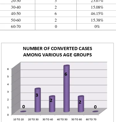
respectively. The least age patient in our study is 16 and the highest age
patient is 70. The conversion was higher in 40-50 age group (46.15% of
total conversion) and was low in 30-40 age group (15.38% of total
conversion) patients. Conversion was not done in 10-20 age group and
65
laparoscopic cholecystectomy were in 30-40 age group. ( Table 3 and
Chart 3 )
All patients underwent cholecystectomy as an elective case .
Though some morbidity was there in terms of post operative
66
TABLE 1
REASONS FOR CONVERSION OF LAPAROSCOPIC
CHOLECYSTECTOMY
REASONS NO OF
CASES
% OF TOTAL (98)
CASES
% OF
CONVERTED(13) CASES
DENSE
ADHESIONS 5 5.10% 38.46%
DIFFICULT ANATOMY AROUND
CALOT’S 3 3.06% 23.07%
ARTERIAL
INJURY 3 3.06% 23.07%
HEPATO BILIARY
INJURY 1 1.02% 7.69%
TECHNICAL
PROBLEMS 1 1.02% 7.69%
67
CHART
–
1
This is the chart showing the reasons for conversion of
laparoscopic cholecystectomy due to conditions arising
68
.
TABLE
–
2
INCIDENCE OF CONVERSION AMONG MALES AND
FEMALES
CONVERTED CASES
% OF TOTAL CASES
MALES 6 6.12%
FEMALES 7 7.14%
6.12%
7.14%
SEX DISTRIBUTION
MALES
FEMALES
69
TABLE
–
3
CONVERSION OF LAPAROSCOPIC CHOLEYSTECTOMY
AMONG VARIOUS AGE GROUPS
AGEGROUP
NUMBER OF CASES CONVERTED
% OUT OF CONVERTED
CASES
10-20 0 0%
20-30 3 23.07%
30-40 2 15.08%
40-50 6 46.15%
50-60 2 15.38%
60-70 0 0%
0 1 2 3 4 5 6
10 TO 20 20 TO 30 30 TO 40 40 TO 50 50 TO 60 60 TO 70
0
3
2
6
2
0
NUMBER OF CONVERTED CASES
AMONG VARIOUS AGE GROUPS
[image:71.596.107.508.280.704.2]
70
DISCUSSION
Laparoscopic cholecystectomy is the gold standard treatment for
cholelithiasis.the superiority of laparoscopic cholecystectomy over open
method ha sbeen analysed and results were drawn in favour of
laparoscopic cholecystectomy, even in developing countries like ours
nowadays cholelithiasis is treated by laparoscopic method.
Studies from various countries reported a conversion rate of 5 to
14 %. In our study 98 patients were operated over a period of 1 year
from november 2011- november 2012 and the conversion rate was
13.26%.several prospective studies done by Michael Rosen et al, Singh
Kuldip et al , H.J.J Van Der Steeg et al , Ajay Anand et al have
drawn results similar to our study.
Because of several and repeated analysis and research in this
particular aspect of laparoscopic cholecystectomy , it should not be
considered neither as a failure nor as an inexperience of the surgeon. It
is for the safety and goodness of the patient conversions are being made
and it is the only reason almost always.hence it should be emphasized
that the conversion of laparoscopic cholecystectomy indicates accuracy
71
By analyzing the data from our study the following events
occuring during laparoscopic cholecystectomy may need conversion to
open method. Some of the complications may be repaired
laparoscopically by experienced hand and some of them definitely
needs conversion.anyway . By all means it is the decision of the surgeon
to decide conversion .
DENSE ADHESIONS :
The conversion of laparoscopic cholecystectomy due to dense
adhesions in our study is 5.1% of total cases that were operated and is
38.46% of cases that got converted .even though the patients in our
study have not undergone any previous upper abdomen surgeries,
adhesions remained the most common cause for conversion. In all
patients who got converted due to adhesions , laparoscopic adhesiolysis
was tried initially and cases which failed to achieve adhesiolysis got
converted.studies by Nadim Khan et al, NEJM Surgical Study Group
,Saeed Hadi Al-Bahlooli quoted that adhesions are the most common
cause for conversion of laparoscopic cholecystectomy .in our study
group , adhesions were found in 5 cases, of which adhesions between
72
and duodenum in 1 case, greater omentum and anterior abdominal wall
in 2 cases,adhesions around porta hepatis in one case.
DIFFICULT
ANATOMY
AROUND
CALOT’S
TRIANGLE:
The conversion of laparoscopic cholecystectomy due to difficult
anatomy around calot’s triangle in our study is 3 case (3.06% ). Of
total cases that were operated and is 23.07% of cases that got converted
. The conversion of laparoscopic cholecystectomy is extremely
important when there is difficult anatomy around calot’s triangle as
there is more chance of bile duct and cystic artery injury in this
situation. In studies by Malla B R , Muhammed Shamim et al , S K
Biswas Et Al , Vikas Gupta et al , it was concluded that the conversion
of laparoscopic cholecystectomy due to difficult anatomy around calot’s
triangle is the most common cause for conversion of laparoscopic
cholecystectomy . Anatomy can get altered due to any cause like acute
cholecystitis, aberrations in cystic artery , right hepatic artery, common
hepatic duct and common bile duct.in our case series there was
excessive fat near the calot’s triangle in two cases , cystic artery
identification was difficult in one case due to adhesions around calot’s
73
ARTERIAL INJURY:
The conversion of laparoscopic cholecystectomy due to arterial
injury in our study is 3 case (3.06% ) of total cases that were operated
and is 23.07% of cases that got converted . It is one of the most
important cause for conversion as it produce immediate hypotension and
even death if immediate intervention was not done .usually laparoscopic
repair of the bleeding site is not done because the field will become
messed up with blood and most of the times it is impossible to identify
the bleeding vessel in such a situation. Therefore only option that saves
the patient in such condition is conversion .blind application of clips or
cauterization in unclear area is absolutely contraindicated, because of
the potential danger of misplacing the clips or cautery to vital structures
the results of which may go hazardous to the patients life.in our study
cystic artery was injured in two cases because of difficult dissection
near calots triangle and 1 case is due to aberrant origin of cystic artery
from common hepatic artery. Another case is converted due to torrential
74
HEPATO-BILIARY INJURY:
The conversion of laparoscopic cholecystectomy due to
hepato-biliary injury in our study is 1 case (1.02% ) of total cases that were
operated and is 7.69% of cases that got converted .nadim et al and
studies by surgical study group nejm concluded that hepato biliary
injury also plays an important role in conversion of laparoscopic
cholecystectomy.it occurs mostly due to blind dissection near calots
when anatomy is unclear .it requires high level of expertise and
experience to identify hepato biliary injury as most of the times it is not
identified per operatively and diagnosed only in the post operative
period . But when there is suspicion intra operatively , cholangiogram
is to be done whenever there is a possibility and repair to be done in the
most appropriate way.at any cost bile leak should be prevented as it
75
TECHNICAL ISSUES:
The conversion of laparoscopic cholecystectomy due to technical
issues in our study is 1 case (1.02% ) of total cases that were operated
and is 7.69% of cases that got converted.very few of the conversions
are due to technical issues like poor lighting, insuffalator defects,
unclear monitor/ cameras, defective dissectors / graspers,diathermy
handles. Conversion due to technical issues are going down even in the
developing countries like ours . It assumes importance because it is one
of the easily correctable causes to avoid a conversion . Studies from the
eastern part of the world showed technical issues as a reason for
conversion in some of the cases. In our study one case got converted to
open technique because of the problem with insuffalator and non
replacement can be done at that time and hebce proceeded to open
method even before the dissection of gall bladder and cystic duct,
artery.it is entirely hazardous to operate with defective instruments and
conversion is the better option in terms of patient safety.
Though many studies have drawn results citing inferior vena cava
injury, portal vein injury,hepatic artery injury, bowel injury , incidental
intra operative diagnosis of gall bladder and cholangiocarcinoma,
76
any those situation and thus not included or explained in this study.but
these conditions are also extremely important and conversion of
laparoscopic cholectstectomy is needed in almost all cases of above
said conditions.
Again it is highlighted that the conversion of laparoscopic
cholecystectomy should be viewed as a good cause in terms of patient
77
CONCLUSION
After analyzing the results of our study,
We conclude that the incidence of conversion of laparoscopic
cholecystectomy is 13.26 %.
The reasons for conversion in descending order of frequency are
1. Dense adhesions ( 5.1% )
2. Difficult anatomy around calot’s triangle ( 3.06 % )
3. Arterial injury ( 3.06 % )
4. Hepato-biliary injury ( 1.02 % )
PRIYA 25 F 4127 YES
VALLIAMMAL 52 F 42388
KALPANA 27 F 44405
THANSIVA 48 M 42684
SULOCHANA 57 F 44850
LAKSHMI 16 F 45286
KANDASAMY 59 M 43835
GOWRI 50 F 4387 YES
AMEENABEE 56 F 44183
VEMBU 48 M 45321
DURGADEVI 26 F 1935
KAVINISHA 48 F 2095
MUSTAQ 47 M 2080 YES
VASANTHA 44 F 2232
BALAMANI 64 M 2237
ASHIQ 43 M 4180
GEETHA 35 F 10092
GAJALAKSHMI 39 F 3949
GOWRI 50 F 4387
JASMINE 45 F 5140
SHANMUGAM 60 M 11211 YES
RASIGABEGUM 55 F 39760
MAHALAKSMI 25 F 37231
SAGAYAM 28 F 15873
VALLIAMMAL 55 F 36702
VENKAT 68 M 36236
RANI 50 F 35349
SOUNDARYA 34 F 17957 YES
VASANTHA 59 F 30842
SHALINI 35 F 29639
KANNAN 32 M 26665
BUVANESHWARI 28 F 40439 YES
RAMAN 25 M 27218
MERCY 25 F 26865
SAMABEGAM 37 F 25974
PORKODI 51 F 27026
LOGU 37 M 41287 YES YES
SELVAM 37 M 26306
DEVAPILLAI 60 F 25963
ANITHA 30 F 25964
FATIMA 25 F 24760
RENUKA 34 F 20390
ANJALAI 32 F 20450
NAVEEN 30 M 19282
JAYAPRIYA 36 F 19182
SUBBU 26 F 18221
ELUMALAI 64 M 17167
SHANMUGAM 64 M 17162
ARUMUGAM 47 M 40815 YES
REVATHY 26 F 16039
SALIMA 69 M 10321
HABUB 60 F 10480
GEETHA 35 F 10092
VEMANNA 56 M 8369
PARTHASARATHY 56 M 9046
PUSHPA 45 F 8839
RAGAVAN 44 M 4388 YES YES
POPPY 41 F 8817
AMUDHA 30 F 8142
PUSHPA 42 F 8182
PONNUSAMY 55 M 7510
SIVAGAMY 63 F 6348
DEEPA 27 F 5349
KARPAGAM 26 F 5356
SAMEEER 50 M 4089
JANAKI 70 F 2415
KALYANI 45 F 4440 YES
SANTHIYA 35 F 43022
THAMARAI 48 F 41491
SKRAJA 44 M 43594
KAGARNISHA 38 F 42493
PADMA 37 F 43473
PRABAKAR 30 M 42603
RAMALINGAM 58 M 10190 YES YES
ANURADHA 32 F 42151
CHINNAPA 38 M 41746
MUNIYAMMA 55 F 40908
MADHAN 35 M 3921
GOVINDAMMAL 50 F 41772
SAROJA 60 F 38884
KATEEJA 38 F 39493
GULSARBEE 50 F 38552
DURGA 26 F 1935 YES
ABDUL 38 M 51465
SENGUTTUVAN 53 M 49997
SELVI 38 F 52742
BANU 59 F 51402
DASS 62 M 51694
KALAIVANI 17 F 51005
SUMATHY 38 F 49175
MANIKUMAR 50 M 49046
YES
YES
YES
YES
YES
YES