Copyright01993, American Society forMicrobiology
Sindbis
Virus Membrane Fusion Is Mediated
by
Reduction
of
Glycoprotein
Disulfide
Bridges
at
the
Cell Surface
BARBARAA. ABELLANDDENNIST. BROWN*
The CellResearch Institute and Department of
Microbiology,
TheUniversity
of TexasatAustin, Austin, Texas 78713-7640Received 21 April 1993/Accepted 7 June 1993
Wehaveexamined the role of thiol-disulfideexchangereactions duringthepenetrationof cellsbySindbis
virus.Theprotein-protein associations that form the rigid icosahedral latticeofthe Sindbisvirusenvelope have
been shown tobe stabilized by disulfidebridges, and reduction of these critical disulfide bridges duringcell
penetrationmaybethemechanism by which the rigid protein latticeisdisrupted priortofusion (R.Anthony and D. T. Brown, J. Virol. 65:1187-1194, 1991; R. Anthony, A. Paredes, and D. T. Brown, Virology 190:330-336, 1992). Reduction of disulfide bridges occurs at near neutral pHs viathiol-disulfide exchange reactions, and these reactionscanbe blocked bycovalentmodificationof the thiolinvolved.Inthisstudy,the effects of thereducingagent2-mercaptoethanolonSindbisvirus-mediatedcell-cellfusion from without and the effects of the membrane-impermeable thiol-alkylating reagent5,5'-dithiobis(2-nitrobenzoic acid) on Sindbis virus penetrationweredetermined.Thepresenceofexogenousreducingagentwasfoundtoinduce fusion from
without under conditions unfavorable to both typical Sindbis virus-mediated fusion from without and
cysteine-mediated thiol-disulfide exchange reactions. In addition, the thiol-alkylating reagent wasfound to inhibit Sindbisvirusentrywhenpresentduringinfection. Theseresultsareconsistentwithamodel for Sindbis
virusentryinwhichreductionofcriticaldisulfide bridgesatthe cell surfacedisrupts the rigid protein-protein associationsof theenvelope,allowing membrane fusionandrelease of the viralgenomeinto the cell.
Sindbis virus is a structurally well defined alphavirus composed of 240 copies each of the three virally encoded
proteinsEl,E2, and C. The positive-strandRNAgenomeis protectedbya T=4icosahedral nucleocapsid composed of theviral protein C (4, 5, 19). This nucleocapsid is in turn surroundedbya host-derived lipidenvelope containing the viral glycoproteins El and E2. Sindbis virus is a unique enveloped virus in that its envelope proteins are rigidly organizedas aT=4icosahedrallattice(1, 14).Incontrast to other viral envelopes, which are regarded as membranes
with associated protein, the Sindbis virusenvelope canbe consideredarigid proteinlattice with associatedlipid.El-E2
heterodimers trimerizetoform theenvelopecapsomeresvia El-El associations which are stabilized by intramolecular disulfide bridges (1). Additional El-El and E2-E2 interac-tions organize these capsomeres into a T=4 icosahedral lattice (see Fig. 7 in reference 1), which appears to be
maintained primarily by disulfide bridge-stabilized El-El associations(2).Theunique rigid organizationof theSindbis virus envelope proteinsmakes itimperative that these
pro-tein-protein interactions bedisrupted duringentry.
Thedisulfidebridgeswhich maintain the structural integ-rity of the envelope protein lattice are protected from
exogenous reducing agents by the conformation of the
capsomere (2). Conformational changes induced by
ex-tendedtreatmentwith dithiothreitol (DTT)orbyverybrief
treatment atlowpHexposethese critical disulfide bridgesto reductionby DTT. Thisreduction results in disassembly of
the envelope and release ofthe nucleocapsid (2). In vitro disassembly ofSindbis virus following low-pH (5.3)
treat-mentrequires a return toneutral pH asreduction byDTT
occursviaathiol-disulfideexchange reaction which ishighly
pH dependent (26). In this two-step process, the in vitro
* Correspondingauthor.
disassemblyof theSindbis virusenvelope byDTTparallels
the pH requirements demonstrated forSindbis
virus-medi-ated fusionfrom without(FFWO);fusion inducedbythe HR strain ofSindbis virusrequiresaninitialexposuretopH5.3 followedbyareturn toneutralpH (9).
The observation thatsomeenvelopedvirusesdemonstrate
low-pH-induced FFWO is believedtosupportthe
hypothe-sis that these viruses enter cells via an acidic, endosomal
route (16). For Sindbis virus, such coupling of
low-pH-inducedFFWO and entry fromanacidic endosome has been putinquestion byseveral observations.Agentswhichblock
endocytosis do not block penetration of cells by Sindbis virus (3, 6). In addition, a temperature-sensitive mutant CHO cell line defective in the acidification of endosomes
undergoesFFWOatthe normal acidic thresholdpHand yet is efficiently infected with Sindbis virus at temperatures whichpreventendosome acidificationto thispH (10).
Fur-thermore, Sindbis virus-mediated FFWOdoesnotoccurat the low-pHthreshold requiredtoinduce fusion butinstead
requiresareturn to aneutral-pH environment (9). Finally,
different cell lines show different pH optima for fusion, includingone atpH 4.6,well belowthe acidification
capa-bilityof thecells; these cellsarealsoefficientlyinfectedby
Sindbis virus (8). The above observations suggest that the
low-pH threshold required during FEWO is not required during natural entry, and thus penetration of cells is not restrictedto the acidicenvironment ofthe endosome.
The observation thatDTTcaninduceSindbis virus enve-lope disassembly in vitro suggests that cleavage or
rear-rangementofthe intramolecular disulfidebridges stabilizing
thecritical El-El associationsmayberequiredtodisruptthe
envelope protein lattice during entry (2). Thiol-disulfide
exchange reactions arebiologically important for reducing and/or rearrangingdisulfidebridgesinproteins. Theyrequire neutral pH conditions and can be blocked by covalent
modification ofthe thiol involved (26). An envelope
disas-5496
on November 9, 2019 by guest
http://jvi.asm.org/
sembly mechanism including such a reaction could explain the two-step nature of the FFWO phenomenon, as the low-pH step would induce conformational changes in the virions, allowing critical reductive events to occur upon a return to neutral pH. During the natural route of cell penetration, conformational changes in the envelope pro-teins induced by binding to the receptor at the plasma membrane (13) could trigger the thiol-disulfide exchange reactions, bypassing the need for low-pH-induced conforma-tional changes. Such a reductive event could provide a mechanism for thedisruption of the protein-protein associ-ations of theenvelope and allowdirectpenetration of cells at the plasma membrane. In an attempt to investigate this
possibility, the virus-mediated phenomenon of FFWO has been reexamined and theeffect of the
membrane-imperme-able thiol-alkylating reagent 5,5'-dithiobis(2-nitrobenzoic acid) (DTNB)onviruspenetration has beenstudied.
MATERIALSANDMETHODS
Cells, virus, and media. BHK-21 cells were cultured at
37°C in Eagle's minimal essential medium (MEM) supple-mented with 10% fetal calf serum (GIBCO), 5% tryptose phosphate broth, and 2 mM L-glutamine. Heat-resistant Sindbis virus(SVHR),originally providedby E. Pfefferkorn (DartmouthMedicalCollege),waspassagedatlow
multiplic-ity and titrated on BHK-21 cells as described previously
(21).
Sindbisvirus-mediatedFFWO.Variations ofSindbis virus-mediated FFWO described previously were performed (9, 18). Briefly, FFWO at low pH was induced by adsorbing
1,000 PFUof SVHR per cell to BHKmonolayersat4°C for 60 min; the viral inoculum with then replaced with 37°C
fusion medium, consisting ofEagle's MEMwithout bicar-bonate, 10 mMMES [2(N-morpholino)ethanesulfonic acid],
and 10 mM HEPES
(N-2-hydroxyethylpiperazine-N'-2-ethanesulfonic acid) adjusted to pH 5.3 with and without various concentrations of2-mercaptoethanol (2-ME).Fusion
was assayed with a phase-contrastmicroscope after 5 h of incubationat37°C and scoredasdescribed previously(-,no
fusion; +, less than 25% of cells fused; ++, 25 to 50%of cells fused; +++,50 to 95% of cells fused; ++++, more
than95%of cellsfused);thedegreeof fusionwascalculated
as1 - (numberofcells/numberofnuclei) (18). Two-pH-step
FFWOwasinducedasdescribed above except that various
multiplicities of SVHRwere used, the fusion medium
con-tained 0.05 nM 2-ME, and the virus-cell complexes were
returned to growth medium after 5 min of incubation in
low-pHfusion medium. Themonolayers were photographed underphase-contrast illumination after 1 h of incubation at
37°C andscoredasdescribed above (18).
Drugs. DTNB (Pierce) was solubilized in serum-free MEM. DTNBconcentrationsashighas5 mM didnotaffect cell viability, as assayed by incorporation of
[3Hjuridine
(ICN Biochemicals)intotrichloroacetic acid
(TCA)-precipi-table cellular RNA, trypan blue exclusion, or neutral red
uptake (datanotshown).Direct effects of DTNBonSindbis
virusinfectivitywere determinedby incubating the reagent with the virus for various periods of time, quenching the incubation by serial dilution intophosphate-buffered saline
containing 3% fetal calf serum
(GIBCO),
and titrating onBHK cells in a standard plaque assay as described previ-ously(21).
Penetration assays. Viral RNAsynthesiswas assayed by determining incorporation of
[3H]uridine
(ICNBiochemi-cals) into TCA-precipitable material in BHK cells treated
UH'
Concentrationof 2-ME(Mol)
FIG. 1. One-step FWO inBHKcells at pH 5.3 in the presence of 2-ME. Virus-cell complexes were exposed to low-pH fusion medium including various concentrations of the reducing agent 2-ME as described in the text. Fusion was scored as described previously (18)andin thetext.
with 4p,gofdactinomycin per ml for 90 min prior to infection and maintained indactinomycin throughouttheexperiment. Monolayers of equal numbers of BHK cells were treated
with 1 mM DTNB for 30 min at 37°C at threedifferent times with respect to a 1-hadsorption of 5 PFU of SVHR per cell
at4°C.Afteradsorption, allofthemonolayerswerewashed
with serum-free MEM and incubated at 37°C to allow
penetration of the adsorbedvirus. The30-minDTNB
treat-ments were done either immediately prior to adsorption, during the periodofpenetrationat37°C, orimmediately after
this penetration period. After the DTNB treatments, the monolayers were washed.[3H]uridine (10
Ci/fml)
wasadded 90 min after the period of penetration, and at 5 h, the monolayers were lysed with sodium dodecyl sulfate andprecipitatedwith 10% ice-coldTCA, and radioactivitywas
counted. Penetrationwas alsoassayed by plaqueformation
onBHKmonolayersthat had beenexposedto1 mMDTNB for60 min under the treatmentconditions describedabove. Plaque formation was determined as described previously (21).
RESULTS
FEWO
at low pH. The rate of Sindbis virus-mediated FFWO is dependent upon the pH to which the cells arereturnedafter exposuretolowpH.Fusionproceedsfasterat
higherpHs and doesnot occur atthelow-pHthreshold of 5.3
requiredtoestablish conditions for fusion upon a return to
neutrality (9). Thiol-disulfide exchange reactions are
simi-larlypH dependent in that the reaction rates are directly
affected by pH, occurring efficiently only in neutral to
alkaline environments. If reduction of critical viraldisulfide bridges is necessary to disrupt the rigid envelope protein
latticepriortomembranefusion,it would be blockedatlow pH.
To examine a role for sucha reductive event in Sindbis virus-mediated FFWO ofBHKcells,fusionwas attempted
at low pH in the presence of the
reducing
agent 2-ME as described in Materials and Methods(Fig. 1).
Unlike DTTandcysteine,2-MEis aneffective
reducing
agent atpH
5.0 and above (26). Treatment with a wide range of 2-ME concentrations was found to induce FFWO atpH
5.3,
although this range ofconcentrations varied between
on November 9, 2019 by guest
http://jvi.asm.org/
[image:2.612.355.523.71.225.2]100
la
uz
80 60 40 20
0 200 400 600 800 1000
MOI (pfu/cell)
FIG. 2. Multiplicity (MOI) dependence of two-step FFWO in BHKcells exposedto2-ME.Virus-cell complexeswereexposedto
low-pH fusion medium with 0.05 nM 2-ME (-)orwithout2-ME (A)
for 5min, returnedtoneutral pH, and scored for fusionasdescribed
in thetext.
vidual experiments.2-ME-induced fusionatlow pH is highly
sensitive to a number of factors, including the passage number of the cells and the degree of confluency of the monolayers, whicharenotascriticaltotheprocessof fusion
afterareturn toneutral pH. Treatmentofmonolayerswith 2-ME in the absence ofvirus and incubation ofvirus-cell complexes atpH 5.3in the absence of 2-ME donotinduce fusion.Theextremelylowconcentrationsofreducingagent
necessarytoinduceFFWOsuggestthat thecriticaldisulfide
bridges being reduced are highly strained. High concentra-tions of2-ME donotpromote fusion, possibly because the viral envelope is destroyed before the fusion event can
occur,ina manneranalogoustothecompletedestructionof
the viralenvelope duringinvitroDTTreduction of
low-pH-treated virus (2). The observation thatSindbis virus-medi-ated FFWO occurs at low pH in the presence of low concentrations of 2-ME is consistent with the hypothesis
that reduction of critical viral disulfide bridges may be
important forvirus-cell fusion. In addition, itmay explain theFFWO requirementforareturn toaneutral-pH
environ-ment.
Effect of 2-MEonthemultiplicityrequirementforFFWO.
Theobservation that2-ME inducesfusionatlowpH implies
that the efficiency of Sindbis virus-induced FFWO may dependupon areductiveevent.Figure2shows the effect of 2-ME on the multiplicity requirement for typical Sindbis virus-mediated FFWO. This reducing agent, when present duringthe briefexposureof thevirus-cellcomplexes tolow pHduringFFWO, increases theefficiency of fusion after a
return to neutrality. Optimal fusion in the 2-ME-treated monolayersis obtainedatmultiplicitiesaslow as100PFU/ cell, whereas multiplicities of 500 to 1,000 PFU/cell are typically requiredinthe absenceof2-MEtreatment(9, 18).
The high-multiplicity requirement for FFWO must in part reflect the frequency with which attaching virions form multiplecellcontacts,astericrequirement for the
cell-virus-cell fusion event. If this fusion phenomenon is dependent upon athiol-disulfide exchange reaction, the high-multiplic-ity requirement may also reflect the frequency with which
thecritical viraldisulfidebridges associate with appropriate thiol donors.An increase in theavailability of thiols due to
theexogenousreducingagent 2-MEwould reduce the
num-ber ofvirionsrequired toproducemaximal cell-cell fusion.
Such a model is supported by the decrease in multiplicity required formaximal fusion after briefexposure to 2-ME.
._
a
V._
100'
80-
60-40 20
0 15 30 45
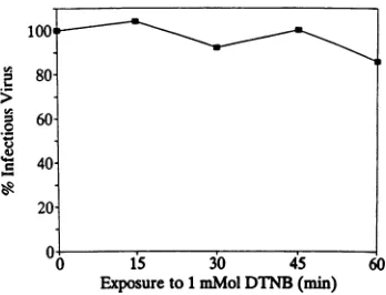
[image:3.612.345.519.71.204.2]Exposureto1mMolDTNB(min) 60
FIG. 3. Directeffect ofDTNBonSindbisvirusinfectivity.Virus
was exposed to 1 mM DTNB for various periods of time, and
remaining infectious virus was titrated on BHK monolayers as
describedin thetext.Infectiousvirus isexpressedas apercentage of
thatinthe untreated control.
Role ofthiol-disulfide exchange reactions in Sindbis virus
penetrationof BHK cells. The datapresentedabove suggest that the reduction of disulfidebridgesin the rigid envelope protein lattice of Sindbisvirus may be required for
mem-branefusion duringentry.It has beendemonstratedthat cell surfaces have a reductive capability mediated by thiol-disulfide exchange reactions (11). Such plasma membrane-associated activity has been implicated in the reduction of the disulfide bridge connecting the A and B chains of
diphtheriatoxin(31).Thishypothesishas received dramatic supportfromexperimentsinwhich it has been demonstrated that chemical reagents such as DTNB which block thiol
groups on the cell surface prevent the cleavage of the
diphtheria toxin disulfide bridge and render sensitive cells resistant tothe toxin (23). DTNB is a
membrane-imperme-able sulfhydryl-blocking reagent that covalently modifies
sulfhydrylsviaathiol-disulfideexchangereaction. As it does notdirectlyaffect Sindbis virusinfectivity (Fig. 3)or BHK
cellviability (asdescribed inMaterials andMethods),it isan
excellent reagent fortesting thehypothesis that such reac-tions occur at the plasma membrane during Sindbis virus
penetration.
The effect of this reagent on viral penetration at low
multiplicitiesof infectionwasassayed by [3H]uridine
incor-porationinto viral RNA under threetreatmentconditionsas
described in Materials and Methods (Table 1). In the
pres-ence of dactinomycin, BHK cellswere treated for 30min
with 1 mM DTNB eitherpriorto adsorption of thevirusat 4°C, duringpenetrationof the virusat37°C, orimmediately
after the penetration period. In each case, viral RNAwas assayedat5hafter thepenetration period by measuringthe
incorporationof[3H]uridineintoTCA-precipitablematerial. DTNB treatment of cells prior to infection has a slight inhibitory effect on viral RNA synthesis, while treatment duringviruspenetrationinhibits RNAproductionto
approx-imately40% of control levels. The decrease in RNA
synthe-sis seenwithtreatment ofthe cellspriorto infectionlikely
results from a failure to remove all of the drug prior to infection. Treatment with DTNB after an initial period of
penetration has a limited inhibitory effect; however, the
depressioninRNAsynthesisafter thistreatmentmaybe due to the fact thatSindbis virus infection increases the
perme-ability ofcells, allowing some membrane-impermeable
re-agentsdirect accesstothecytoplasm, wheretheycanhave
secondaryeffects (10, 12).
A~1
on November 9, 2019 by guest
http://jvi.asm.org/
[image:3.612.84.272.75.211.2]TABLE 1. Effect of DTNBonpenetration ofcells by Sindbis virusa
Determination Treatment time % of control
ViralRNAsynthesisb Before penetration 82± 11 During penetration 40± 10 After penetration 81 ± 13 Plaque formationc Before penetration 80± 11 During penetration 46± 10 Afterpenetration 110 ± 10
aVirus was adsorbedtoequal numbers ofBHKcells for1h at4°C, replaced
in warm MEM,and incubated at 37°C toallowpenetration. Cells were treated
for 30 min(A) or 60 min(B) with 1 mM DTNBattheindicated times. The
monolayers were then washed to remove the drug prior to further incubation
at37CinDTNB-free medium. Results are the averages of fiveindependent
exferiments
±standard deviation.Total viral RNA level was determined5 h after the cells were warmed to
37°C to initiate penetration.
c Monolayers were overlaid with agarose and stained with neutral red 48 h
afterinfection.
The effect ofDTNB onSindbis viruspenetrationwasalso determined byassaying plaque formation (Table 1). Pretreat-mentof the cells withDTNBresults inaslightinhibition of plaque formation, while treatment during the period of infection inhibitsplaque formation toapproximately 46%of
untreated-control levels. Again, the decrease in plaque for-mation seen when monolayers were treated with DTNB
prior to infection probably results from an inability to removeallof the reagentpriortoinfection. Treatment with
DTNIB
postinfection has no inhibitory effect on plaqueformation, which is consistent with the above conclusion
that the depression in RNA levels with DTNB treatment
after infection isnotdueto adirecteffectonpenetration of
the virus. Although paradoxical,the slightly higher number ofplaques found in cell cultures treated after infection is highly reproducible, and at this time no explanation is
available.
Theinteresting observation that Sindbisviruspenetration
is most effectively inhibitedby DTNB when the reagent is
presentduring theperiod of infectionsuggests thattargeted thiol groups areunmaskedduring entry through a
coopera-tive interaction between the virus and its receptor. The cooperative and rapid nature of this interaction may create
circumstances in which DTNB cannot efficiently compete with a viral disulfide bridge (Fig. 4) undergoing a thiol-disulfide exchange reaction. Such reactions have rapid
reac-tionrates, about10-6 s atpH8.0(7), andaninefficiencyin
alkylating the critical thiols would result in an inability to
completely block Sindbis viruspenetration.
DISCUSSION
Fusion of enveloped viruses with cellular membranes during penetration is protein mediated, as are intracellular
andintercellular fusionevents(30). Theexposureofafusion domain in theenvelopeprotein El has been implicated in the fusion of alphaviruses with cellular membranes (17). In
addition, the observations presented above, together with
thosepublishedpreviously byourlaboratory(1, 2), suggest
that the disulfidebridge-stabilized El-El associations of the envelope mustbe disrupted formembranefusion to occur.
Themost likely mechanism for disassembly of the Sindbis
virus envelope protein-protein interactions during
penetra-tion isthe reduction ofthesestabilizing disulfide bridgesvia
thiol-disulfide exchange reactions.
Athiol-disulfide exchange reaction occursby the nucleo-philicattack of anionizedthiol on a disulfide bridgeand is
highly dependentonenvironmentalpH, the
pK8
ofthethiol,and steric hindrance. Cysteine-mediated thiol-disulfide
ex-change reactions thus require neutral to alkaline pHs
(11,
26). FFWO mediated by the HR strain of Sindbis virus requiresexposure topH 5.3 with asubsequentreturntopHsgreater than 6 to induce fusion (9). The rate of fusion increases as the pHtowhich thevirus-cell complexes are
returnedincreases, and fusion doesnot occur atthelow-pH thresholdrequired for fusion. Likewise, rapid disassembly of the viralenvelope byDTT alsorequiresbriefexposure to
pH 5.3 followed by a return to neutral pH (2). These observations implicate a definite role for a neutral pH
E1
El
El
El
[image:4.612.62.300.95.181.2] [image:4.612.72.556.505.665.2]Functional
Domoins|
Structurol
X
X
Membrone _____ 2
A
B
C
FIG. 4. Modeldepicting conformational changesinEl duringSindbis virus membranepenetrationandlow-pH-inducedmembranefusion. (A)TheElglycoproteininthematurevirion has both functionaland structuraldomains,whichareinvolvedinmembrane fusion andenvelope
integrity, respectively. (B) Conformational changes induced by the receptor-virusinteraction orbyexposure to lowpH unmask critical disulfidebridges,favoringasubsequentreshuffling of disulfide bridges. (C) Reduction of critical disulfide bridgesresponsibleformaintaining
theprotein-protein associations of the envelope disrupts the rigid protein icosahedral lattice,allowing subsequent fusion withacellular membrane. Solidboxes,El-Elassociations; hatchedboxes, fusion peptide.
on November 9, 2019 by guest
http://jvi.asm.org/
environment and in this way reflect the requirements fora
thiol-disulfide exchange reaction described above. This is
consistent withamodel for virus-cellfusion in which
thiol-disulfide
exchange
reactions are critical to the fusionpro-cess.
The process of
low-pH-mediated
FFWO differsfromtheprocess ofinfection inthat itrequires
high
multiplicities ofvirusand isproteinreceptor
independent (15, 18);
however,both processesmustsupplyamechanism forthe disruption
of the
envelope protein
latticeprior
to fusion. Our data suggest that both events aresimilarly dependent
upon athiol-disulfide
exchange
reaction. Addition of very lowcon-centrationsof the
reducing
agent2-ME(which,
unlike DTT andbiological thiols,
reducesefficiently
atpH5.3)
promotes cell fusion in an acidicenvironment,
suggesting
that thechemical reduction ofcritical disulfide
bridges
can induce fusion in an otherwise unfavorable reductive environment.The
extremely
lowconcentrations of2-MEneeded to pro-motethisfusionimply
thatthe critical disulfidebridges
being
reduced are in very strained conformations. Low-pH-in-duced conformationalchanges
in theenvelope proteins
could beresponsible
forexposing
these strained criticaldisulfide
bridges
and mayexplain
the veryrapid
natureof in vitro DTT-mediated virusdisassembly following low-pH
exposure(2).
The process of Sindbis virus
penetration
ofhost cells is receptordependent,
and several possible receptors have beenidentified(24, 27, 29).
The interaction of thevirus withacell surface receptoratneutralpHinducesconformational
changes
in the viralglycoproteins
whichprecede
penetration
(13).
Thestudiespresented
here also suggest that acooper-ativeinteractionbetweenthereceptorandthe virusinduces conformational
changes
whichallow thereductionofcriticaldisulfide
bridges
within theenvelope
proteins by
thiol-disulfide
exchange
reactions. Theprogression
fromthere-ceptor-induced
conformationalchanges
to thedisruption
of theenvelope protein
lattice and fusionlikely proceeds
veryrapidly.
The fact that thestabilizing
disulfidebridges
andcriticalthiolsareinaccessibletomolecules suchasDTTand DTNB while in the native state, combined with the
ex-tremely rapid
rate atwhich the reductiveeventoccursafter thereceptor-induced
conformationalchanges,
mayexplain
theinefficiency
of DTNB inblocking
this process. Thiolsmediating
the reductive events may reside either in the receptorprotein
orwithin the virus itself. In the latter case, areshuffling
ofdisulfidebridges following
attachment would result in therequired disruption
of theenvelope protein
lattice. In such amodel(Fig.
4),
the interaction of the virus withanappropriate
receptoralters the conformation ofEl,
favoring
areshuffling
ofdisulfidebridges
via thiol-disulfideexchange
reactions. Thisreshuffling
of disulfidebridges
leadstodisruption
of therigid
protein-protein
associations in theenvelope,
allowing subsequent
fusion of the viralenve-lope
with theplasma
membrane.Sturman et al.
(25)
havesuggested
thatreshuffling
of disulfidebridges
is involved in the conformationalchanges
occurring
incoronavirusproteinsduringcellpenetration. In collaboration withShinji
Makino(University
of Texas atAustin),
we have obtainedpreliminaryevidence thatmousehepatitis
virus is also inhibitedbyDTNBwhen the reagent is presentduring
infection(unpublished
observation). It ispossible
that suchrearrangements
of disulfidebridges
arerequired
forpenetration by
avariety
ofenveloped
viruses. A critical reductive event in Sindbis viruspenetration
could occur in an unidentifiedneutral-pH
compartmentwithin the
cell,
as isthought
to be thecase for ricin toxin(28). Like diphtheria toxin, ricin is a heterodimeric toxin with a disulfidebridge connecting the A and B chains that
must be reduced for penetration into the cell cytoplasm.
However, unlike that of diphtheria toxin, the ricin disulfide bridge is reducednot atthe cell surface butinsomeinternal compartment at neutral pH, possibly the trans-Golgi
net-work(28). DTNB has no effect on the cytotoxicity of ricin while inhibiting the cytotoxicity of diphtheria toxin (23).
Thus, if a critical reductive event occurs afterinternalization of Sindbis virus by endocytosis, DTNB would not be
ex-pectedtoinhibitentry.
Our data are in agreement with the studies of Flynn et al. (13), whoidentified the exposure oftransitional epitopes in Sindbis virus glycoproteins during attachment and penetra-tion.Antibodies to these epitopes were able to prevent virus infection when present during the process of infection.Flynn
et al. (13) found that these antibodies were capable of blocking penetration of cells by 30% of the added virus, leading them to conclude, as we have concluded, that the rearrangements leading to infection occur very rapidly. Such rapid protein rearrangements would dramatically limit the ability of antibodies or DTNB to arrest the virus particles during entry.
The identification of conformational changes induced in viral structural proteins by interaction with receptors has resulted in a reevaluation of the mechanism of cell penetra-tion by polioviruses (see reviews in references 20 and 22).
An emerging view is that the conformational changes
re-quired for viruspenetration, whichwerepreviously thought to be induced by exposure to acidic endosomal pH, may occur at the cell surface asvirus-receptor interactions take place.These conformational changesmaylead to the direct penetration of polioviruses at the cell surface, as we also propose here for thealphaviruses.
ACKNOWLEDGMENTS
This research was supported by Public Health Service grants AI14710 andAI19545 from the National Institutes of Health and through funds appropriated by the State of Texas to the Cell ResearchInstitute. Barbara Abellwassupportedbyapredoctoral fellowship throughNIHtraininggrant5-T32-GM-08368-03.
REFERENCES
1. Anthony, R. P., and D. T. Brown.1991. Protein-protein inter-actionsinanalphavirusmembrane.J. Virol.65:1187-1194. 2. Anthony, R. P., A. M.Paredes,andD. T. Brown.1992.Disulfide
bonds areessentialfor thestability of the Sindbis virus enve-lope. Virology190:330-336.
3. Cassell, S., J. Edwards, and D. T. Brown. 1984. Effects of lysosomotropic weak bases on infection ofBHK-21 cells by Sindbis virus.J. Virol. 52:857-864.
4. Choi, H., L. Tong, W. Minor, P. Dumas, U. Boege, M. G. Rossmann, and G. Wengler. 1991. Structure of Sindbis virus coreproteinreveals achymotrypsin-like serine proteinase and theorganisation of the virion.Nature(London)354:37-43. 5. Coombs,K., and D. T. Brown. 1987.Organizationof theSindbis
virus nucleocapsid is revealed by bifunctional cross-linking agents. J.Mol.Biol.195:359-371.
6. Coombs, K., E. Mann, J. Edwards, and D. T. Brown. 1981. Effectsofchloroquine andcytochalasin B on the infection of cells bySindbis virus andvesicularstomatitis virus. J.Virol. 37:1060-1065.
7. Creighton, T. E. 1984. Disulfide bond formation in proteins. MethodsEnzymol. 107:305-329.
8. Edwards, J., and D. T. Brown. 1984. Sindbis virus induced fusion of tissue cultured Aedes albopictus (mosquito) cells. Virus Res. 1:705-711.
on November 9, 2019 by guest
http://jvi.asm.org/
9. Edwards, J.,and D. T.Brown.1986. Sindbis virus-mediated cell fusion from without isatwo-step event.J. Gen. Virol. 67:377-380.
10. Edwards, J., and D. T. Brown. 1991. Sindbis virus infection of a Chinese hamster ovary cellmutantdefective in the acidification of endosomes. Virology 182:28-33.
11. Feener, E. P.,W.Shen, and H. J.-P. Ryser. 1990. Cleavage of disulfide bonds in endocytosed macromolecules: a processing not associated with lysosomes orendosomes. J. Biol. Chem. 265:18780-18785.
12. Fernandez-Puentes, C., andL. Carrasco. 1980. Viral infection permeabilizes mammalian cells to protein toxins. Cell 20:769-775.
13. Flynn, D. C., W. J. Meyer, J. M. Mackenzie, Jr., and R. E. Johnston. 1990. Aconformational change in Sindbis virus gly-coproteins El and E2 is detected attheplasma membraneas a consequence ofearly virus-cell interaction. J.Virol. 64:3643-3653.
14. Fuller, S. D. 1987. The T=4 envelope of Sindbis virus is organized by interactions with acomplementary T=3 capsid. Cell 48:923-934.
15. Kielian, M., and A. Helenius. 1984. Role of cholesterol in fusion ofSemliki Forest virus with membranes. J. Virol. 52:281-283. 16. Kielian, M., andA. Helenius. 1986. Entry of alphaviruses, p.
91-119. In S. Schlesinger and M. J. Schlesinger (ed.), The Togaviridae and Flaviviridae. Plenum Press,NewYork. 17. Levy-Mintz, P., and M. Kielian. 1991. Mutagenesis of the
putative fusion domain of theSemliki Forest virus spike protein. J. Virol. 65:4292-4300.
18. Mann, E., J. Edwards, and D. T. Brown. 1983. Polycaryocyte formation mediated by Sindbis virus glycoproteins. J. Virol. 45:1083-1089.
19. Paredes, A.,A. M.Simon,and D. T. Brown.1992.The mass of theSindbis virusnucleocapsid suggestsithasT=4icosahedral symmetry.Virology 187:329-332.
20. Racaniello, V. R. 1992. Interaction of poliovirus with its cell receptor.Semin. Virol. 3:473-481.
21. Renz, D., and D. T. Brown. 1976. Characteristics of Sindbis virus temperature-sensitive mutants in cultured BHK-21 and Aedesalbopictus(mosquito)cells. J. Virol. 19:775-781. 22. Rotbart,H.A.,and K. Kirkegaard. 1992. Picornavirus
patho-genesis: viral access, attatchment and entry into susceptible cells. Semin. Virol. 3:483-499.
23. Ryser, H.J.-P., R. Mandel, and F. Ghani. 1991. Cell surface sulfhydryls are required forthecytotoxicityofdiphtheria toxin butnotof ricin inChinese hamster ovary cells.J. Biol. Chem. 266:18439-18442.
24. Smith,A.L., and G.H.Tignor. 1980. Host cell receptors for twostrainsof Sindbisvirus. Arch.Virol. 66:11-26.
25. Sturman, Lt S., C. S.Ricard,and K. V.Holmes.1990. Confor-mational change of the coronavirus peplomer glycoprotein at pH 8.0and37'C correlates with virus aggregation and virus-induced cell fusion. J. Virol.64:3042-3050.
26. Torchinskii, Y. M. 1974. Sulfhydryl and disulfide groups of proteins. PlenumPublishing, New York.
27. Ubol, S., and D. E. Griffin. 1991. Identification ofa putative alphavirus receptoron mouse neural cells. J. Virol. 65:6913-6921.
28. van Deurs, B.,K.Sandvig, 0.W.Petersen,S.Olsnes,K.Simons, andG.Griffiths.1988. Estimation ofthe amountofinternalized ricin that reaches thetrans-Golgi network.J.Cell Biol. 106:253-267.
29. Wang, K.-S., R. J. Kuhn, E. G. Strauss, S. Ou, and J. H. Strauss. 1992. High-affinity laminin receptor is a receptor for Sindbis virusinmammalian cells. J. Virol. 66:4992-5001. 30. White, J. 1992. Membrane fusion. Science 258:917-924. 31. Wright, H. T., A. W. Marston, and D.J. Goldstein. 1984. A
functional role for cysteine disulfides in the transmembrane transport ofdiphtheria toxin.J.Biol. Chem. 259:1649-1654.