COMPARISON OF SHEAR BOND STRENGTH AFTER
THERMOCYCLING, OF BRACKETS BONDED USING
TWO DIFFERENT MOISTURE INSENSITIVE
PRIMERS – AN IN VITRO STUDY
Dissertation submitted to
THE TAMILNADU DR. M.G.R.MEDICAL UNIVERSITY
In partial fulfillment for the degree of
MASTER OF DENTAL SURGERY
BRANCH V
ACKNOWLEDGEMENTS
Words seem less to express my deep sense of gratitude to my postgraduate teacher and guide Dr. N.R. Krishnaswamy, M.D.S., M.Ortho RCS. (Edin), Diplomat of Indian board of Orthodontics, Professor and Head, Department of Orthodontics and Dentofacial Orthopedics, Ragas Dental College and Hospital, Chennai, for his valuable guidance and suggestions, tireless pursuit for perfection, immense and constant support, encouragement and keen surveillance for the minute details throughout this dissertation. I thank him for all the help that have been conferred upon me without which this dissertation would not have come true.
I gladly utilize this opportunity to express my deep sense of gratitude and indebtedness to my respected ProfessorDr. S. Venkateswaran, M.D.S, Diplomat of Indian board of Orthodontics, for his everlasting inspiration, incessant
encouragement, constructive criticism and valuable suggestions conferred upon me without which this dissertation would not have come true.
I am privileged to express my extreme gratefulness to my respected Professor Dr. Ashwin George, M.D.S, Diplomat of Indian board of Orthodontics,
for being a constant source of support and supervision, who stimulated, enthused and encouraged me in successful completion of this study and throughout my post graduate course.
I am exceptionally gratified and sincerely express the sincere thanks to
ending willingness to render generous help to me in carrying out this work from its inception to its consummation.
My sincere thanks to Professor Prof. Kanakaraj Chairman &
Dr. Ramachandran, Principal, Ragas Dental College for providing me with an opportunity to utilize the facilities available in this institution in successful completion of this study
I am greatly beholden to Dr. Shahul (Associate Professor), Dr. Jayakumar (Reader), Dr.Shakeel Ahmed (Reader), and senior lecturers, Dr. Rajan, Dr. Rekha, Dr. Shobana, Dr Biju and Dr. Prabhu for their support, enthusiasm & professional assistancethroughout my post graduate course.
I also express my sincere and profound thanks to Dr. V. Kalliyana Krishnan, Senior scientist G, Biomedical Technology Wing, Sree Chitra Tirunal Institute for Medical Sciences & Technology, Trivandrum, for permitting me to use the research facilities in his department. I would be failing in my duty if I don’t thank the immense support rendered byMr.RejinandMs. Soumya, Lab technicians in the same department, for rendering the lab procedures for this study.
My heartfelt thanks to my wonderful batch mates, Dr.Subu Thomas, Dr. Amey Rathi , Dr.Fayyaz Ahmed, Dr. Shailendra Vashi , Dr.Ritika Kailey, Dr.
Goutham Kalladka and Dr Thiunavukkarasu Geetha who were cheerfully
available at all times to help me. I wish them a successful career ahead.
Dr Manikandan, Dr Shakthi, Dr Sivasubramanian, Dr Vijay, Dr Aarthi, Dr
Ashwin, Dr Ravanth, Dr Deepakfor all their support.
My thanks to Mr. Ashok, and Mr. Rajendran for helping me with the photographs for the study.
I would like to thank Mrs. Marina , Sister Lakshmi , Sister Rathi, Sister Kanaka, Haseena,Mr. Mani, Mr. Bhaskar, Ms. Divya, Ms Shalini for their co-operation and help during my post-graduate course.
I also take this opportunity to pay my heartfelt gratitude to my family for being a pillar of support during my postgraduate life. I am forever indebted for their sacrifice and prayers.
Special thanks to my children for their affection and understanding.
Last but not the least let me pay my prayerful thanks to the almighty for his boundless blessings.
CONTENTS
Title
Page Number
1.
Introduction
1
2.
Review of Literature
6
3.
Materials and Methods
33
4.
Results
42
5.
Discussion
45
6.
Summary and Conclusion
66
INTRODUCTION
Direct bonding of orthodontic brackets has been advocated since 1960s117. With the introduction of newer adhesive systems as well as photosensitive
(light-cured) restorative materials in dentistry, additional methods have been suggested to
enhance the polymerization of the materials used, including layering and more
powerful light-curing devices. Adhesives have evolved from acrylics to several
other materials like epoxies, epoxy-acrylates25,109,138glass ionomer fluoride releasing cements,158,171to the present resin modified glass ionomer cements . In addition, other factors147 can potentially contribute to the strength of the bond between the enamel and the orthodontic bracket, including
a. type of enamel conditioner,
b. acid concentration,
c. length of etching time,
d. composition of the adhesive,
e. bracket base design,
f. bracket material,
g. oral environment ( presence of moisture) and
h. skill of the clinician.
Of all these moisture contamination after etching is the principle cause of
Over the years a great deal of attention has been paid to improve the
acid-etching technique, primers and adhesives. Nonetheless, adhesive failures still exist
because of contamination during bonding. Traditional bond materials must be
applied in completely dry and isolated fields to achieve adequate bond strength.
Hydrophilic bond systems have been an important development in orthodontic
practice because many routine clinical procedures are not carried out under ideal
conditions. In particular, it can be difficult to bond attachments to hard-to-reach
places such as the gingival area, second molars, and partially erupted or impacted
teeth56,67,77,126. In these cases, bonding failure is common, and rebonding, which consumes chair time and is a burden for both orthodontist and patient, becomes
necessary126. In unsuccessful bonding procedures, most of the porosities produced by the enamel acid-etch procedure become plugged with moisture. As a result, resin
penetration is impaired, and when insufficient numbers and lengths of resin tags
form, bond strength is compromised96,128,135. Contamination of the enamel surface therefore necessitates the etching procedure to be repeated to ensure the adequate
bonding of composites96. For this reason, several studies have evaluated the effects of contaminants like water, saliva, and blood on SBS32, 67,126,169.
Earlier resins were manufactured of hydrophobic monomers, which
performed well only in dry environments. Nowadays hydrophilic monomer(water
soluble), such as 2-hydroxyethyl methacrylate (HEMA), 2-hydroxypropyl
methacrylate (HPMA), 4 -hydroxybutyl methacrylate (HBMA) which are well
known in restorative dentistry for dentine bonding, are used as adhesives for enamel
bracket bonding. These substances allow for greater shear bond strength (SBS) on
formed by moisture on the enamel’s surface. Out of these HEMA is used commonly
in moisture-insensitive primers (MIP) which perform adequately even in the
presence of moisture. However, there are controversial reports on whether MIPs
fulfil the requirements for bond strength in a dry environment39,67.There are some studies which report that moisture contamination did not decrease the shear bond
strength of composite resins with MIP when bonded to etched enamel149. Study by
Hara et al71 reported clinically acceptable bond strength under dry conditions for hydrophilic adhesive systems. A few studies showed that MIP with their respective
adhesives is effective in both wet and dry fields67,149.
Recent studies critically noted the inherent disadvantages of HEMA in
adhesives which include:
1. HEMA retains water within the adhesive layer and adversely affects
the mechanical strength42;
2. 70% of HEMA can be hydrolyzed in acidic solutions within a week
at 37°C122;
3. HEMA provides low photopolymerization reactivity. Moreover,
HEMA causes sensitized delayed allergic reaction89.
Therefore, it could be anticipated that HEMA containing adhesive layers
with high water absorption behaviour will possess low physical properties and result
in poor bonding durability to the tooth substrates.
Glass ionomers were introduced to the profession 38 years ago and have
cements, are materials made of calcium, strontium aluminosilicate glass powder
(base) combined with a water-soluble polymer (acid)114. These cements possess certain unique properties that make them useful as adhesive materials,including.
1. Adhesion to tooth structure and base metals78,
2. Anticariogenic properties due to release of fluoride75, 3. Thermal compatibility with tooth enamel, and
4. Less enamel loss41from debonding compared to composites.
In recent years there have been considerable changes in the formulations,
properties and handling properties of the glass ionomer cements for different clinical
applications. Glass ionomer cements (GICs) were initially introduced as orthodontic
bonding adhesives to take advantage of the fluoride-releasing capabilities of the
material72. But the use of GICs in orthodontics was limited because of their lower bond strengths19,38. It is certain that no material is perfect, but with the current level of intensive research on glass ionomers, the deficiencies that exist seem to be
eliminated or at least reduced, resulting in an ever – improving range of materials of
this type.
Opal seal is ethanol based filled adhesive, comprised of 38% filled with
substantial glass ionomer and nano-fillers and contains hydroxypropyl methacrylate
(HPMA) which is a hydrophilic monomer. Opalseal has been claimed to be effective
in wet environment. There are no studies comparing the shear bond strength of
Transbond MIP (contains HEMA) and Opal seal along with conventional Transbond
However, most in vitro studies on bond strength after saliva contamination
did not use an artificial ageing procedure before testing, despite the fact that
thermocycling of the specimens has been recommended to consider the durability of
the bond. Thermal cycling is the in vitro process through which the adhesive resin
and the tooth are subjected to temperature extremes compatible with the oral
cavity76.Gale and Darvell64 pointed to the lack of consistency between the conclusions of different studies on the SBS of various adhesive systems following
thermocycling34,68. They attributed this to the lack of standardization between the various thermocycling studies they reviewed. Such large variation between the
thermocycling protocols led the International Organization for Standardization to
provide specific criteria for conducting such tests to enable investigators and
industry to interpret and compare results.
Therefore, the purpose of this study was to investigate the effectiveness of
two moisture insensitive primers mainly Transbond MIP and Opal seal (used along
with Transbond XT adhesive) with respect to conventional hydrophobic primer
(Transbond XT) by comparing their shear-peel bond strengths after thermocycling
REVIEW OF LITERATURE
Literature has been reviewed under the following headings
Studies on Bonding
Studies on Glass Ionomer Cement
Studies on Hydrophilic Primer
STUDIES ON BONDING
Buonocore M.G.et al27 (1955), described a method of bonding acrylic resin filling material to enamel surface by etching the enamel with 85% phosphoric
acid and phosphomolybdate oxalic acid treatment to alter enamel surface
chemically, originating the ideas from industrial examples in which phosphoric acid
was used to obtain better adhesion of paint and resin coatings to treated metal
surfaces. He suggested that the increased adhesion could be due to an increase in the
surface area amenable to mechanical adhesion brought about by acid etching.
Bowen et al23 (1962) explored the possibility of using epoxy resins (diglycidyl ether of bisphenol A) mixed with silica particles. The in vitro results
were promising, but the presence of moisture inhibited the polymerization process of
epoxy resin. To overcome this problem, Bowen attached methylmethacrylate groups
to the groups of the epoxy resin, thereby converting the epoxy resin to a
dimethacrylate. The experiment outcome was successful and resulted in a new resin
called Bisphenol. A glycedyl methacrylate or BISGMA or Bowen’ resin.
Buonocore et al (1968)29 studied the penetration of dental materials in to enamel surfaces with reference to bonding. They showed enhanced bonding to acid
conditioned surfaces due to presence of prism-like tags. Poor bonding was
George V. Newman et al65(1973), commented on the bonding of attachments. He mentioned the various systems that have been used. After various
studies he concluded that the ideal adhesive system should readily wet the tooth
surface, polymerize in situ forming a firm bond with tooth structure on one side and
the attachment on the other, especially in shear and tensile strength and be able to be
removed by the orthodontist without destroying the integrity of enamel surface. The
adhesive and attachment together should be aesthetic, adhere to enamel surface for
an adequate treatment period, withstand the forces of mastication, arch wire stress,
resist changes in ph from foodstuff and resist temperature changes and minimal
leakage.
Silverstone et al154 (1974) showed that etching the enamel surface with phosphoric acid resulted in a superficial etched zone and underlying porous zones.
Depth of etched zone depended on the acid concentration, duration of etching,
chemical composition of surface enamel etc.
Retief137 (1975) commented that the depth of etch produced by 50% orthophosphoric acid was more than that obtained with orthophosphoric acid
solution at any other concentration and produced comparable bond strength. The
surface roughness of enamel etched with 50%orthophosphoric acid was greater than
that obtained with more dilute acid solutions.
Reynolds et al138 (1975) reported that clinically, the bonded brackets should be able to withstand forces generated by treatment mechanics and occlusion,
yet allow easy debonding without damage to enamel. He has reported that maximum
but added that in vitro tensile strength levels of 4.9 Mpa have proved clinically
acceptable.
Thompson et al 140(1981) conducted an in vitro study to determine the amount of enamel lost during prophylaxis and during multiple bonding/debonding
procedures. Both bristle brush and rubber cup were used with four different
prophylactic pastes. The enamel loss with the bristle brush (14.38µ) was
significantly greater than the loss with rubber cup (6.9 µ). No significant differences
were associated with use of different pastes. Multiple bonding/debonding procedures
were conducted with filled and unfilled resin adhesive. Total enamel loss was 71.5µ
in the group bonded with a filled resin with prophylaxis and acid etching between
bonds. This was significantly greater than the amount lost (22.3 µ) by the group
bonded with the same resin without prophylaxis and acid etching between bonds.
Total enamel loss was 45.4 µ in the group bonded with an unfilled resin with
prophylaxis and acid etching between bonds. This was significantly greater than the
amount lost (17.8 µ) by the group bonded with the same resin without prophylaxis
and acid etching between bonds.
R. L. Bowen23 (1962) stated that when equilibrated together, water and certain methacrylate monomer formulations form two phases, with the activity of
water being equal with that of normal physiological saline solution. The proportions
of hydrophilic and hydrophobic monomers were adjusted empirically to achieve this
by measuring the solute concentrations in the aqueous phase by osmometric
chemically different monomers. Formulations of this type should be evaluated as
vehicles for adhesion promoting coupling agents.
Artun and Bergland et al9(1984) in a comparative study of conventional acid etching with 37%phosphoric acid and crystal growth conditioning with
polyacrylic acid which contain residual sulphate ion, found that the latter has less
bond strength and debonding was easier since the bond failure occurred at tooth
adhesive interface. He introduced more complex means of defining the site of bond
failure by adhesive remnant index (ARI). The score ranged between 0-3 and criteria
for scoring is as follows:
Score 0-no adhesive left on the tooth
Score1-less than half of the adhesive left on the tooth
Score2-more than half of the adhesive left on the tooth
Score3-all the adhesive left on the tooth with distinct impression of
the bracket mesh.
Fox et al58 (1994) suggested a possible standard technique for bond strength testing. He suggested the following protocol for bond strength testing in
orthodontics.
a. Surface of premolar enamel should be used on teeth extracted from
adolescent patients for orthodontic reasons.
c. After bonding, the specimens should be immersed in water for 24 hrs
at 37°
d. Debonding should take place on an Instron or equivalent machine.
e. The point of application and direction of force should be same for all
specimens.
f. At least 20 specimens should be used per test.
g. Site of failure should be reported.
h. Statistical analysis should include survival analysis to give a
prediction of performance of the material. Bond strength should be
STUDIES ON GLASS IONOMER CEMENT
A. D. Wilson and B. E. Kent (1971)171 introduced a new translucent cement, the product of the reaction between ion-leachable glass and an aqueous
solution of polyacrylic acid is described. Its properties, with particular reference to
dental applications, are reported and a proposed setting mechanism is advanced.
Chadwick SM, Gordon PH (1995)35 Compared the fluoride release from a variety of orthodontic bonding agents. Fluoride release into de-ionized water was
measured over a 20-week period. Material based on the fluoride exchange resin was
also tested in a saline solution. Glass ionomer-based materials showed substantially
greater fluoride release when compared with resin-based materials. The presence of
anions (Cl-) did not improve the release from fluoride exchange resin. The results
proved that Glass ionomer/resin hybrid material (Vitrabond) released the greatest
amount of fluoride.
Millett et al108 (1996) stated in his literature review that orthodontic bonding with glass ionomer cement is comparatively new. The purpose of this
article is to review the current literature covering both in vitroandin vivostudies of various glass ionomer cements that have been used for orthodontic bonding. The
review indicates that there is little support in the literature to suggest that the
currently available conventional glass ionomer cements are suitable for routine
clinical use in orthodontics. Dual- or tri-cured hybrid materials, however,
comprising both glass ionomer and resin components, appear to have greater
A Marcusson, L-I Norevall and M Persson100(1997)studied the benefit from using glass ionomer cement (GIC) instead of a conventional diacrylate in
bracket bonding for the prevention of white spot formation. No additional fluoride
treatment other than fluoride toothpaste was prescribed. It is concluded that the use
of GIC for orthodontic bonding will result in a significant reduction in the number of
white spot surfaces at debonding compared with the use of conventional diacrylate
Vittori Cacciafesta et al165 (1998), evaluated the shear bond strength of Fuji ortho LC, a light cured resin modified glass ionomer, used for direct bonding of
stainless steel and ceramic brackets under 4 different enamel conditions: their results
indicated that shear bond strength of Fuji ortho LC is significantly enhanced by
contaminating the enamel surface with either saliva or water after conditioning,
depending on bracket type used. Even water contamination of non conditioned
enamel surfaces offer clinically acceptable bond strengths for both stainless steel
and ceramic brackets, allowing a safe debonding without enamel damage.
T. J. Gillgrass, P.C.M. Benington, D.T. Millett24 (2001) Compared the time to first failure, the position of band failure at deband, and the change in enamel
white spot lesions of teeth bonded with a modified composite or a conventional
glass ionomer( Band-Lok,Ketac-Cem.) in a randomized half mouth trial over the full
course of orthodontic treatment.The results revealed that overall band failure rates of
5% and 2.8% were recorded for the modified composite and the conventional glass
ionomer, respectively, with no significant difference found between their times to
first band failure. At the end- of-treatment deband, the position of band failure was
predominantly at the enamel-cement interface for the modified composite and at the
of changes in mean enamel white spot lesion scores during treatment did not reveal
significant differences between the cements.
Akira Komori,Iori Kojima2(2003) Studied the fluoride release and uptake characteristics of Fuji Ortho Band Paste Pak (GC Corporation, Tokyo,
Japan), with that of 3 others commonly used to cement orthodontic bands: a
conventional resin-reinforced glass ionomer cement, a polyacid-modified composite
resin, and a conventional glass ionomer cement. Results proved that the fluoride
uptake and release values of Fuji Ortho Band Paste Pak the new 2-paste
resin-reinforced glass ionomer cement were statistically significantly higher than those of
the conventional resin-reinforced glass ionomer cement or the conventional glass
ionomer cement. The new 2-paste resin-reinforced glass ionomer cement might be a
good alternative to conventional products for cementation of orthodontic bands.
Roberto Justus et al142 (2010) stated that by eliminating the organic substances from the enamel surface before etching (deproteinization), orthodontic
bond strength can theoretically be increased because the resulting etch-pattern is
predominantly type 1 and 2, instead of type 3. The objective of this study was to
determine whether deproteinization of human dental enamel surfaces, with 5.25%
sodium hypochlorite (NaOCl) before etching, increases orthodontic bracket shear
bond strength (SBS) of 2 adhesive systems: a composite resin and a RMGI. It was
concluded from this in vitro study that with NaOCl use, bracket bond strength with
Fuji Ortho LC is similar to Transbond XT, so that fluoride-releasing RMGIs may
STUDIES ON HYDROPHILIC PRIMERS
Miura et al111 (1971) described an acrylic resin (orthomite) with linear polymerization using a modified tri-alkyl borane catalyst that proved to be
particularly successful for bonding plastic brackets and for enhanced adhesion in
presence of moisture.
Robert J. Feigal, et al141 (1992)investigated bond strength in vitro, using a bonding agent beneath sealants under varied conditions of contamination. Five
hundred bovine incisor crowns were separated randomly into eight groups and
etched for 60 sec with a 37% phosphoric acid gel under conditions of humidity of
intact saliva, sealant alone showed significant reduction in bond strength. Bonding
agent under sealant on wet contamination yielded bond strengths equivalent to the
bond strength obtained when sealant was bonded directly to clean, etched enamel.
They concluded that bonding agent used without contamination yielded bond
strengths significantly greater than the bond strength obtained when using sealant
alone with contamination. When the saliva was air dried onto the surface, there was
no significant difference in bond strengths whether or not a bonding agent was used
under the sealant.
J. Xie et all174 (1993) studied the in vitro bond strength of human enamel and dentin treated with contaminants like air, water , saliva , plasma and hand piece
lubricant. Two commercial bonding agents, a lower viscosity solvent containing
type - All bond and a higher viscosity hydrophilic monomer type Scotchbond were
evaluated. They concluded that bond strengths to tooth structure with the bonding
assumed. Re- etching without additional mechanical preparation is sufficient to
provide the expected bond strength.
Vargas et al162 (1994) investigated the use of dentin bonding agents containing hydrophilic primers on enamel. Using HEMA (hydroxyl-ethyl
methacrylate) base “scotchbond multipurpose”, equivalent bond strengths to enamel
were achieved in dry or moist conditions.
Andrew L. Sonis7(1994), did an in vitro test to compare the shear bond strength of “scotchbond multipurpose” primer in saliva contaminated enamel surface with conventional primer in uncontaminated enamel and found that the bond
strength were similar.
Wakefield C. W.166 (1996) determined the effect of oral ambient air on shear strength to etched enamel and dentin using OptiBond FL and Prodigy resin
composite. Enamel and dentin specimens of extracted human teeth were treated both
in a dry environment and after exposure to oral humidity. Differences in shear
strengths between the wet and dry enamel or the wet and dry dentin were not
significant. Fracture modes for all specimens were examined under a
stereomicroscope. Within the parameters of this in vitro study, OptiBond FL was not
affected by oral humidity.
El- Kalla51 (1997) compared the shear bond strengths of 4 single- bottle adhesives to enamel and dentin contaminated with human saliva. Materials used
were Prime and Bond 2.1, One Step, Tenure Quick and Syntac single component
not affect the shear bond strength of the adhesives tested except for Syntac Sc with
contaminated dried enamel. Prime and Bond 2.1 showed significantly higher bond
strengths to enamel than the other adhesives, but there was no significant difference
for dentin bond strength under the contaminated condition.
Fritz U. B., J Finger W. J., Stean. 63 (1998) determine the effect of salivary contamination of enamel and dentin on bonding efficacy of an experimental
one- bottle resin adhesive. The control group used was a light- curing urethane
dimethacrylate adhesive. The results showed that there was no negative effect in
Group 3 and 4, compared with control. Air drying after salivary contamination in
Group 2 resulted in lower shear bond strength and wide marginal gaps.
Contamination of the cured adhesive layer group 5 and 6 had no adverse effect on
enamel shear bond strengths, they conclude that the one- bottle adhesive system is
relatively insensitive to salivary contamination, provide that the contamination
occurs prior to the light curing of the adhesive and is carefully rinsed and blot dried.
Salivary contact after adhesive curing must be avoided.
Iwami . Y84 (1998) evaluated the effect of the wetness of human enamel and dentin surfaces on the shear bond strength of composites. Seven dentin bonding
system were used. Shear bond strength was measured with a universal testing
machine. Among the dentin specimens which were tested by two systems with water
based primers, the shear bond strength values of the dry technique group were
higher. Among the acetone based primers the bond strength values for the wet or
semi dry technique groups were higher than those of the dry technique group. It was
obtain high bond strength on the dentin surfaces, but the drying methods did not
affect bond strength to enamel surfaces either before priming or after conditioning.
Miller106 (1998) discussed wet field bonding in the 21st century. He compared the shear bond strength of Transbond XT primer and Transbond MIP on
wet and dry etched enamel. In the dry state, both primers showed comparable bond
strengths; however, under wet conditions, Transbond MIP was vastly superior.
Samir E. Bishara146(1998),did a study on a RMGIC (Fuji Ortho LC). He studied the bond strength on various enamel conditions viz: etched and unetched
surfaces both moistened with saliva and water. He concluded that etching is a
critical variable affecting both the bond strength and the failure location and it
improved the bond strength. With unetched enamel, the shear bond strength of
RMGIC is significantly reduced by a third to a half to that of etched enamel.
Benderli Y15, (1999)examined the shear bond strength of bonding agents to normal or fluoridated enamel following use of weak or strong acids to prepared
enamel surfaces and contaminated with a measured amount of saliva at various
stages of the bonding procedure . When normal enamel surfaces were rinsed and
dried immediately after contamination, there was no significant reduction of shear
bond strength of adhesive to enamel. In the fluoridated specimens, there was no
statistically significant difference between any of the contamination groups and the
control group when maleic acid was used. Saliva contamination may not be a risk
factor for successful bonding between bonding agent and dental tissues for normal
or fluoridated enamel surfaces if they are rinsed and dried immediately after
presence of contamination may provide higher shear bond strength than etching with
maleic acid.
Swift. J. Jr.157 (1999) evaluated bond strengths of six one-bottle bonding agents and a control primer, plus unfilled resin to moist enamel. One-hundred and
five bovine teeth were randomly assigned to seven groups of 15. Enamel was etched
for seconds with 35% phosphoric acid, rinsed, and excess water was blotted with
tissue paper. Following application of the adhesive, composite resin was bonded
using a gelatin capsule technique. Shear bond strengths to enamel were determined
using a universal testing machine. The results of this study suggested that all of the
one-bottle systems tested provided clinically acceptable bond strengths to moist
enamel.
W. Pitakononda173 (1999) evaluated the shear bond strength of three orthodontic bonding agents namely, Concise, Chemical Cure, Rely-a-Bond and
Transbond XT. Ortho-sealant in conjunction with bonding agents resulted in
increased bond strength after moisture exposure for a prescribed period It was
concluded that the time differences of moisture exposure did not have a significant
effect on the shear bond strength of the three commercial brands of bonding agents,
and both Concise and Transbond XT had shear bond strengths statistically
significantly greater than Rely-a-Bond, and the use of Ortho-sealant purported to
prevent moisture interference during polymerization and when it was applied around
the border of bracket after bonding procedure, did not have a significant effect.
etched (10% polyacrylic acid) and unetched human premolar enamel . Two
products: one commercially available product (Fuji Ortho LC) and one experimental
(EX) light-cured glass ionomer. Bond strength was measured after aging for
5 minutes, 15 minutes, and 24 hours. For Fuji Ortho LC, the bond strength of
brackets bonded to, etched and without contamination, was statistically higher than
that of brackets bonded to unetched enamel for all aging times. Contamination by
saliva did not reduced bond strength to unetched enamel. For both etched and
unetched enamel, there was no significant difference between Fuji Ortho LG and EX
after 24 hours for all contamination conditions.
Hara.AT71 (1999) compared the enamel shear bond strength of four hydrophilic adhesive systems: one multiple-bottle (Scotchbond Multi-Purpose Plus),
two one-bottle (Stae, Single Bond) and one self-etching (Etch & Prime) after storing
in humid environment for 1 week. The mean shear bond strength values were:
Single Bond: 24.28 +/- 5.27 (a); Scotchbond Multi-Purpose Plus: 21.18 +/- 4.35
(ab); Stae: 19.56 +/- 4.71 (b); Etch & Prime 3.0: 15.13 +/- 4.92 (c). It could be
concluded that the self-etching adhesive system did not provide as good a bond to
enamel surface, as did the one- and multiple-bottle systems.
Cacciafesta V, Bosch C, Melsen B31. (1999), compared the clinical performance of resin-modified self-cured glass ionomer cement to a standard
composite resin in the direct bonding of orthodontic brackets when bonded onto dry
and saliva contaminated enamel. After bond strength testing, they concluded that,
GC Fuji Ortho shows clinically acceptable bond strength when bonded onto moist
teeth, but not when used on dry enamel. Both bonding agents failed mostly at the
Littlewood et al96 (2000), carried out a study to investigate the bond strength of brackets bonded using a new hydrophilic primer designed to be
insensitive to moisture and compares it with a conventional primer. Although
designed to be moisture insensitive, the directions for use stipulate drying teeth
before bonding. Therefore, for the purpose of comparison with a conventional
primer, the experiment was conducted under dry conditions. The results showed that
the bond strengths obtained with a hydrophilic primer were significantly lower than
the conventional primer.
I.Kirovshi and S. Madzarova79(2000),investigated the effects of etching and 3 different environmental conditions (Distill water, saliva and plasma) on the
bond strength of a light-cured RMGIC (Fuji Ortho LC). The results showed that
etching of enamel produced higher bond strengths when compared to non-etched
surfaces and also, the contamination with saliva or plasma increased the bond
strength. The material provided adequate bond strengths for direct bonding.
Hobson et al77 (2001), did an in-vitro study to evaluate the bond strength of Transbond MIP under dry, moist and blood contaminated conditions. The results
showed higher bond strength in dry followed by moist and blood contaminated
surfaces respectively. It was concluded that bonding with Transbond MIP even
under the difficult conditions of blood contamination and poor moisture control
provides more than adequate bond strength.
Mark J. Webster, Nanda101 (2001)did an in-vitro study to compare the shear bond strengths of two light-cured hydrophilic bonding systems, namely
hydrophobic bonding system Transbond XT with XT primer.Comparison tests were
conducted under four enamel surface conditions: Etched and dry, Etched and
moisture with artificial saliva, Etched, primer and moistened with artificial saliva
and Etched, primed, moistened with artificial saliva and reprimed.
After doing the bond strength tests, they concluded that the enamel etched
and dried and enamel etched, primed, moistened with saliva and reprimed (i.e. a and
d) showed highest mean bond strength.
Littlewood et al96 (2000) did a clinical study to compare the clinical failure rates of brackets bonded using a prototype hydrophilic primer designed to be
insensitive to moisture with brackets bonded with a conventional primer. He
concluded using survival analysis that there was an increased risk of bracket failure
when bonded with the hydrophilic primer compared with the conventional primer
therefore this hydrophilic primer cannot be recommended for routine clinical use.
Douglas Rix et al45 (2001),compared 3 orthodontic adhesives in the areas of shear-peal bond strength, location of adhesive failure, and extent of enamel
cracking before bonding after debonding of orthodontic brackets. The adhesives
included were composite resin control, Fuji Ortho LC and a polyacid modified
composite resin (Assure) under dry and saliva contaminated conditions. They
concluded that, Transbond XT had higher bond strength than Fuji Ortho LC and
Assure (polyacid composite), although the bond strength for all 3 adhesives were
clinically acceptable. The bond strength for the Assure adhesive was not
Grandhi et al67 (2001)did an in-vitro study on bovine enamel to evaluate the shear bond strength of stainless steel brackets bonded in dry and wet field with
the use of MIP and to evaluate the effectiveness of MIP with chemically activated
(concise) and light activated (Transbond XT) composite resin.The results of this
study suggested that MIP should be used with only (Transbond XT) light-cured
resins as this combination produced higher bond strength and suggested the use of
MIP in situations where moisture control is difficult.
Eliades et al50 (2002), did a study to investigate the reactivity with water of Transbond MIP in conjunction with a no-mix adhesive (Unite), and a moisture
insensitive adhesive (Smartbond), and to assess their bond strength to wet and
saliva contaminated enamel relative to the conventional application of the no-mix
adhesive.After the bond strength testing, they concluded that only Smartbond set in
the presence of water. Transbond MIP did not improve bond strength values when
combined with the no-mix adhesive. Most adhesive-enamel condition combinations
showed a trend to present lower bond strength in the presence of saliva; however,
this was not confirmed statistically.
Moll K., Gartner, Haller B.113 (2002), evaluated the effect of moist bonding on strength of resin based composite to enamel using different adhesive
systems: They concluded that moist bonding did not significantly affect shear bond
strength to enamel of the adhesives tested expect for solid bond without primer
application. Primer contamination of the etched enamel did not significantly
influence bond strength, neither in the dry nor in the moist bonding group. Of all 6
adhesives tested in the both group, the highest mean bond strength was observed
Shane Schaneveldt and Foley149 (2002), did an in vitro bonding study to evaluate the effectiveness of 2 moisture insensitive primers, Assure and MIP
compared with a control hydrophobic primer, Transbond XT.
After bond strength testing, they concluded
that:-1. Both bonding systems provide adequate bond strengths whether
saliva contamination occurs before or after the application of the
hydrophilic primers.
2. Comparing saliva contamination after application of primer, both
MIP and Assure had significantly greater shear-peel bond strengths
than when contamination occurred before the application of each
primer.
3. Contamination between 2 layers of primer showed significantly
greater shear-peel bond strengths compared with the other groups.
4. The group with saliva contamination before application of the primer
showed more frequent failure at the enamel-adhesive interface,
suggesting that complete penetration of primer was prevented.
Whereas the groups with saliva contamination after the first
application of primer showed more frequent failures at the
adhesive-bracket interface.
Cacciafesta et al32 (2003), did a study to assess the effect of water and saliva contamination on the shear bond strength and bond failure site of 3 different
Etching Primer) used with a light-cured composite resin (Transbond
XT).Comparison test were conducted under 7 different enamel surface .After bond
strength testing, they concluded that non-contaminated enamel surfaces had the
highest bond strengths for conventional, hydrophilic and Self etching Primers, which
produced the same strength values. In most contaminated conditions, the Self
Etching Primer had higher strength values than either the hydrophilic or
conventional primers. The Self Etching Primer was the least influenced by water and
saliva contamination, except when moistening occurred after the recommended
3-second air burst.
Kula KS., Nash TD., Purk JH93.(2003), determined whether a hydrophilic primer (Transbond MIP) produced a significant difference in shear/peel
bond strength compared with a traditional hydrophobic primer (Transbond XT) in
wet and dry condition and difference in site of bond failure.
They concluded that orthodontists who suspect moisture contamination
should use a hydrophilic primer during bonding procedures to maintain shear/peel
bond strength.
Ashraf E. Nasr11 (2003) evaluated the effectiveness of moisture-insensitive primers and hydrophilic adhesive under dry, moisture contamination and
saliva contamination. One commercially available micro- hybrid composite resin
and compomer dental restorative materials both are light cured and with their
corresponding etching primer and adhesives. The results indicated that moisture
contamination either with saliva or distilled water decrease the shear bond strength
composites, compomers are less sensitive to moisture contamination than resin
composites. This decreased sensitivity mechanism was unclear, while hygroscopic
expansion is suggested to play role, This hygroscopic expansion is high in Dyract
Ap due to the predominance of (poly hydroxyethyl methacrylate) that act as water
binding hydrogel.
Vittoria cacciafesta164 (2004), studied regarding effect of blood contamination on the shear bond strength of conventional and hydrophilic primer.
1. Non contaminated enamel surface had the highest bond strength for
both conventional and hydrophilic primer.
2. Under blood contamination, both primers produces significantly
lower bond strengths. The hydrophilic primer had significantly
higher bond strength values than the conventional primer, but they
both had bond strength values that might not be clinically adequate.
3. Both shows significant debond failure site, depending on enamel
surface conditions.
Rangaswamy Rajagopal et al135 (2004) Shear bond strength was compared among three materials: conventional Transbond XT primer (3M Unitek),
moisture-insensitive primer (MIP, 3M Unitek), and self-etch primer (Transbond
plus, 3M Unitek). Both MIP and self-etch primer showed adequate bond strength
superior to that of conventional primer in case of moisture contamination. All
bracket-adhesive interface or within the bracket-adhesive itself, with the exception of the
conventional primer used with moisture -contaminated enamel.
Ciola36 (2006) investigated, in vivo, the bond strength of two adhesive materials: a moisture insensitive primer (MIP) and a one step self etching primer (SEP), both
used with Transbond XT on dry and wet enamel and an adhesion time of 10-15
minutes.Moisture insensitive primer tested on wet enamel showed the highest mean
bond strength outcomes (8.98 MPa) compared to one step etching primer (5.81
MPa). Since the mean bond strength of SEP proved sufficient for clinical purposes
and its behavior tended to be more homogeneous, it was considered the best choice.
Mehmet Og˘ uz Öztoprak105
(2007) assessed the effect of blood and saliva contamination on the shear bond strength of 4 orthodontic adhesives
(Transbond XT primer ,Transbond Plus self-etch primer , Assure hydrophilic primer
and Smartbond cyanoacrylate )tested under different enamel conditions: dry, and
blood and saliva contamination after priming. Result showed that the shear bond
strength of the Smartbond cyanoacrylate adhesive group was significantly lower
than all other groups; however, it was the only adhesive that was not affected by
contamination. Saliva and blood contamination resulted in significant drops in shear
bond strengths of the Transbond XT and Assure groups. Transbond Plus self-etch
primer was also negatively affected by blood contamination, although it was suitable
for bonding with saliva contamination.
Endo T52(2008) evaluated the effects of different degrees of water contamination on the shear bond strength of orthodontic brackets bonded to dental
the modes of bracket/adhesive failure. They concluded that to achieve clinically
sufficient bond strengths with the hydrophilic MIP adhesive system, excess water
should be blotted from the water-contaminated enamel surface.
Luciana Borges Retamoso et al97(2009)evaluated the influence of saliva contamination on shear bond strength and the bond failure pattern of 3 adhesive
systems (Transbond XT, AdheSE and Xeno III) on orthodontic metallic brackets
bonded to human enamel after storing in distilled water at 37ºC for 24 hour. Each
system was tested under 2 different enamel conditions: no contamination (T, A and
X groups) and contaminated with saliva (TS, AS and XS groups). They concluded
that the control and contaminated groups showed no significant difference in shear
bond strength for the same adhesive system. However, shear bond strength of T
group was significantly higher than that of AS and XS groups.
Santos et al147 ( 2010) compared the shear bond strength (SBS) of two bond systems: Transbond XT/XT primer (TXT/XT) and Transbond Plus Color
Change/Transbond Self Etching Primer (TPCC/TSEP)after thermocycling. Each
system was examined under four enamel surface conditions (dry, water, saliva, and
blood), and 160 bovine teeth were divided into eight groups of 20 according to
enamel surface condition. Group 1 used TPCC/TSEP and Group 2 used TXT/XT
under dry conditions; Group 3 used TPCC/TSEP and Group 4 used TXT/XT with
water; Group 5 used TPCC/TSEP and Group 6 used TXT/XT under saliva; and
Group 7 used TPCC/TSEP and Group 8 used TXT/XT with blood. They concluded
that the presence of blood resulted in the lowest SBS from both bond systems, but
especially from TXT/XT. TPCC/TSEP resulted in a higher SBS than TXT/XT under
STUDIES ON THERMOCYCLING
Bishara et al19 ( 1975) Plastic brackets were bonded to 560 extracted human teeth with use of two orthodontic adhesive systems: (1) methyl methacrylate
resin bonded to a sealant that was polymerized using ultraviolet light, and (2)
self-polymerizing methyl methacrylate resin bonded directly to the etched enamel.
Tensile and shear like tests were performed. Prolonged exposure to heat, moisture,
and severe temperature changes decreased the shear like strength of both adhesives.
Both systems were adequately strong to withstand routine orthodontic and estimated
masticatory forces.
Schaneveldt et al144 (2002) evaluated the effectiveness of 2 moisture-insensitive primers, Assure and MIP compared with a control hydrophobic primer,
Transbond XT . In vitro shear-peel bond strengths were acceptable for all groups,
and the bond strengths for Assure and MIP were not significantly affected by saliva
contamination. Bond failure analysis (adhesive remnant index) mainly showed
adhesive bond failures. An increased frequency of enamel fractures at debond was
noted, with the control group (1) and the MIP groups (3 and 5) having 22.5%,
12.5%, and 15%, respectively. The Assure groups had no enamel fractures.
Bishara et al21( 2003)evaluated the effects of thermocycling on the shear bond strength of a cyanoacrylate adhesive system, specifically 24 hours after
bonding when the adhesive has achieved most of its bond strength and after
thermocycling. The findings indicated that the cyanoacrylate adhesive tested has
about 80% of its strength after thermocycling. The clinician should consider all
properties of the adhesive, including no need for a curing light, working time of 5
seconds before the adhesive starts to set, and the significant decrease in bond
strength after thermocycling.
De Munck et al42(2005)in their review of literature, concluded that after about 3 months, all classes of adhesives exhibited mechanical and morphological
evidence of degradation that resembles in vivo aging effects. A comparison of contemporary adhesives revealed that the three-step etch-and-rinse adhesives remain
the 'gold standard' in terms of durability. Any kind of simplification in the clinical
application procedure results in loss of bonding effectiveness. Only the two-step
self-etch adhesives approach the gold standard and do have some additional clinical
benefits.
J. De Muncket al86 (2005)reviewed the fundamental processes that cause the adhesion of biomaterials to enamel and dentin to degrade with time. Several
laboratory protocols were developed to predict bond durability. A correlation of in
vitro and in vivo data revealed that, currently, the most validated method to assess
adhesion durability involves aging of microspecimens of biomaterials bonded to
either enamel or dentin. Various methods by which aging achieved are,
1. Aging by storage
2. Aging by thermo-cycling
3. Aging by occlusal loading
After about 3 months, all classes of adhesives exhibited mechanical and
morphological evidence of degradation that resembles in vivo aging effects. A
comparison of contemporary adhesives revealed that the three-step etch-and-rinse
adhesives remain the 'gold standard' in terms of durability. Any kind of
simplification in the clinical application procedure results in loss of bonding
effectiveness. Only the two-step self-etch adhesives approach the gold standard and
do have some additional clinical benefits.
Andreas Faltermeier et al6 (2007) evaluated the shear bond strength of a conventional primer (Transbond XT) and a moisture-insensitive primer (Transbond
MIP) under dry conditions and after contamination with saliva, blood and etching
gel remnants after thermocycling(6000× 5°C/55°C) in a mastication device to
simulate temperature changes and the moisture of saliva in the oral cavity. Under
dry conditions Transbond XT and Transbond MIP showed no significant difference
in SBS. However, clinically unacceptable bond strength was observed using
Transbond XT after saliva and blood contamination. In wet conditions only
Transbond MIP showed sufficient bond strength. Blood contamination seems to be a
more serious problem for bond strength than saliva or etching gel contamination.
Oesterle LJ, Shellhart WC (2008) did evaluate and isolate the effect of composite aging, one of many factors that can contribute to decreased bond strength
during a normal 24-month orthodontic treatment period. Two orthodontic bonding
adhesives--Transbond APC II and Quick Cure adhesive were used to bond brackets
to 280 bovine incisors that were stored in distilled water at 37 degrees C for 30
minutes, 24 hours, and 1, 6, 12, 18, and 24 months before shear-peel testing. They
minutes to 24 hours and then tends to decrease over the next 24 months. The
decrease in bond strength due to the effects of composite aging in water appears to
be a major factor in the decreased bond strength seen clinically.
Chatzistavrou E et al35 (2009)assessed the effect of intra-oral aging on the shear bond strength of a composite resin orthodontic adhesive (Transbond XT,
3M Unitek, Monrovia, CA, USA) after 6 months in the oral cavity and to compare it
with control specimens not subjected to intra-oral aging. No statistically significant
differences were found in the shear bond strength between the two groups of
composite resin adhesive systems (with and without clinical aging) or in the
adhesive remnant coverage following debonding. No significant correlation was
detected between the shear bond strength and the adhesive remnant coverage in the
test specimens with and without aging. Laboratory studies of shear bond strength
appear to be clinically relevant.
Toshihiro Yuasa156( 2010)evaluated the effects of 2 years of storage and 6000 thermocycles on the shear bond strength (SBS) of two self-etching adhesive
systems were studied. Two self-etching primer (SEP) systems (Transbond Plus and
Beauty Ortho Bond) and one etch and rinse system (Transbond XT) were used .
There was no significant difference in the mean SBS for the bonding materials
among the three conditions. ARI scores showed that Transbond XT and Beauty
Ortho Bond had less adhesive remaining on the teeth after ageing compared with
storage for 24 hours. Specimens bonded with Beauty Ortho Bond showed leakage
between the resin adhesive and enamel after ageing. Both SEP systems produced
appropriate technique for determining the durability of orthodontic bracket bonding
materials.
Oral Sökücü et al127( 2010)compared the effect of thermocycling on the shear bond strength of brackets bonded using different primers. In all three,
Transbond XT was the bonding adhesive of choice. In the control group (I), the
standard Transbond primer was used, in group II a fluoride-releasing primer
(Reliance fluoride containing light cure bond), and in group III an antimicrobial
fluoride-releasing self-etching primer (Clearfil Protect Bond). The shear bond
strength was determined after thermocycling .They concluded that The group with
the antimicrobial fluoride-releasing self-etching primer had a significantly lower
shear bond strength than all other groups (P < .05). Thermocycling reduced the shear
MATERIALS AND METHODS
The various material, apparatus and the methodology used in the study are
enumerated and briefly described below.
MATERIALS
1. TEETH USED FOR THIS STUDY :
a. 90 sound human adolescent premolars extracted for orthodontic purpose were collected immediately after extraction.
b. The teeth were cleaned of soft tissue debris and blood and washed
with distilled water thoroughly and stored in 10% formalin.
Inclusion Criteria
Intact buccal enamel
No evidence of caries
No development defects
No cracks due to the pressure of the extraction forceps.
Teeth without any restorations
Absence of pre-treatment with chemical agents (such as hydrogen
2. BRACKETS
a. Stainless steel pre-adjusted edgewise, ROTH 0.022 slot premolar
brackets with metallic foil-mesh backing (diamond, Mini 2000
series, Ormco) were used.
b. The average bracket base surface area was determined to be 9.63
mm² (given by manufacturers and also tested).
2. ETCHANT
37% orthophosphoric acid
3. PRIMERS
a. Opal seal – primer containing fluoride releasing and recharging with
drying agent (Opal Ultradent ,Texas)
b. Transbond moisture insensitive primer. (Transbond MIP, 3M Unitek,
Monrovia, California).
c. Transbond XT primer(3M Unitek, Monrovia, California)
4. ADHESIVES
Transbond XT (3M Unitek, Monrovia, Calif).
3M Unitek halogen curing light, visible light range - 400 to 500 nm,
Light power - 450mw/cm².
6. DPI RR COLD CURE ACRYLIC, DPI Wallace Street, Bombay.
7. OTHER MATERIALS AND ARMAMENTARIUM INCLUDED: Moulds for making acrylic blocks into which teeth were fixed.
Distilled water, polishing cup and slurry of pumice.
Bracket positioner, Tweezer & Explorer.
Plastic instrument and spatulas.
Compressed air / water facility with a 3-way syringe.
Dental micromotor unit with hand piece.
Mixing well.
Saliva: artificial saliva-made with international standards that
contains both organic and inorganic components
8. THERMOCYLER
Willitech thermocyler with cooling system, Haake EK 30, Thermoelectron
Corporation, Germany
9. UNIVERSAL TESTING MACHINE
Universal testing machine (INSTRON 3365, UK) was used for measuring
METHODOLOGY
• 90 human adolescent premolars extracted for orthodontic purpose
were collected over a 3-month period.
• The teeth were debrided and examined for caries, pre-existing
fractures, and restorations.
• The criteria included intact buccal and lingual enamel surface with no
cracks caused by pressure of the extraction forceps.
• The teeth were stored in 10% formalin initially and cleaned and
stored in distilled water 48hrs prior to the experiment.
SAMPLE PREPARATION
• 90 premolar teeth were randomly assigned to 3 groups with 30 teeth
per group.
• Each group was given different colour coding
1. Green – for Opal seal ( Group A )
2. Yellow- for Transbond MIP (Group B)
3. Orange- for Transbond XT (Group C )
• Each tooth from the in vitro sample was mounted on acrylic stubs
with roots embedded in a fast set self cure polymethylmethacrylate
resin.
• The teeth were oriented such as to ensure that the height of contour of
the buccal surfaces were perpendicular to the adhesive and bracket
BONDING PROCEDURE
Brackets were bonded to the teeth according to the instructions given by
the manufacturer. All the bonding procedures were carried out by the same operator.
GROUP A(Green- Opal seal)
1. Teeth were pumiced with non-fluoridated pumice, rinsed thoroughly
with water spray and dried.
2. Enamel surface of the teeth were etched with 37% phosphoric acid
for 15 seconds, and then rinsed with water spray for 10 seconds and
gently air dried.
3. The buccal surfaces were coated with thin layer of artificial saliva.
4. Opal seal- one liberal coat was applied on the entire buccal etched
surface avoiding the gingival area. Then it was air bursted gently for
3 seconds perpendicular to the buccal surface.
5. The brackets (Roth 0.22 slot premolar, diamond, miniseries 2000
Ormco) were coated with Transbond XT resin, and placed on the
buccal surface of the tooth and pressed firmly into place to expel the
excessive adhesive.
6. The excess adhesive was removed with a scaler. The resin was cured
for a period of 40 seconds, 10 seconds proximally, 10 seconds
GROUP B(Yellow- Transbond MIP)
1. Teeth were pumiced with non-fluoridated pumice, rinsed thoroughly
with water spray and dried.
2. Enamel surface of the teeth were etched with 37% phosphoric acid
for 15 seconds, and then rinsed with water spray for 10 seconds and
gently air dried.
3. The buccal surfaces were coated with thin layer of artificial saliva.
4. Transbond MIP - one liberal coat was applied on the entire buccal
etched surface avoiding the gingival area. Then it was air bursted
gently for 3 seconds perpendicular to the buccal surface.
5. The brackets (ROTH 0.22 slot premolar, diamond, miniseries 2000
Ormco) were coated with Transbond XT resin, and placed on the
buccal surface of the tooth and pressed firmly into place to expel the
excessive adhesive.
6. The excess adhesive was removed with a scaler. The resin was cured
for a period of 40 seconds, 10 seconds proximally, 10 seconds
GROUP C (Orange - Transbond XT)
1. Teeth were pumiced with non-fluoridated pumice, rinsed thoroughly
with water spray and dried.
2. Enamel surface of the teeth were etched with 37% phosphoric acid
for 15 seconds and then rinsed with water spray for 10 seconds and
gently air dried.
3. A thin layer of Transbond XT primer was applied on the buccal
surface of teeth.
4. The brackets (ROTH 0.22 slot premolar, diamond, miniseries 2000
Ormco) were coated with Transbond XT resin, and placed on the
buccal surface of the tooth and pressed firmly into place to expel the
excessive adhesive.
5. The excess adhesive was removed with a scaler. The resin was cured
for a period of 40 seconds, 10 seconds proximally, 10 seconds
occlusally and 10 seconds gingivally.
After bonding all the samples were stored in distilled water in sealed
container at room temperature for 48 hrs. The specimens were thermocycled (500 x)
between 5ºC and 55ºC, (Haake EK 30, Thermoelectron corporation, Germany) with
a dwell time in each bath of 30 seconds and a transfer time between baths of 15
seconds. Twenty four hours after thermocycling, they were subjected to a shear load
The specimens were placed in a mounting jig in the INSTRON universal
machine (INSTRON 3365, UK) in such a way that the bracket base was parallel to
the shear-peel load. To ensure that all the brackets were mounted in the same
orientation relative to the acrylic cylinder, the teeth were suspended from a stainless
steel wire (0.019×0.025 inch). This mounting procedure ensured consistency for the
point of force application and direction of the debonding force.
A shear debonding force was applied to the bracket base in an
occluso-gingival direction at a crosshead speed of 1mm/ min. The maximum force necessary
to debond or initiate bracket fracture was recorded in Newtons and then converted
into Megapascals (MPa) as a ratio of Newtons to bracket base surface area.
Debonded specimens were randomly examined at 16x magnification to evaluate the
site of bond failure.
TEST FOR DEBONDING CHARACTERISTICS
The surface of debonded specimens (both the tooth surface and the bracket
base surface) were studied under 16 X magnification and the mode of bonding
failure was determined using the Adhesive remnant index (ARI) which describes
the amount of adhesive remaining on the tooth and bracket pad and was expressed
as a percentage of the total bonded area.
Adhesive remnant index developed by Artun and Berglandwere used in
The score ranges for ARI are:
Score 0 - no adhesive left on the tooth in the bonded area
Score 1 - less than half of the adhesive left on the tooth
Score 2 - more than half of the adhesive left on the tooth
Score 3 - the entire adhesive left on the tooth with distinct impression
of the bracket base.
STATISTICAL ANALYSIS
Statistical analysis was performed with SPSS 15.0 software (SPSS Inc,
Chicago). Descriptive statistics of SBS (mean, standard deviation and significance)
were calculated for all groups. One-way analysis of variance (ANOVA) and
Chisqare test were carried out for SBS and ARI, respectively, to determine
significant differences among the groups. The intergroup comparison of SBS was
done with Independent T- test.The statistical significance level was established at P
ARMAMENTARIUM USED IN THE STUDY
3M UNITEK LIGHT CURING UNIT
WILLITECH THERMOCYLER
INSTRON UNIVERSAL TESTING MACHINE
ASSESSMENT OF ARI ON TOOTH SURFACE OPAL SEAL - GROUP A
TRANSBOND XT- GROUP
RESULT
This study was performed on 90 premolars extracted for orthodontic
purpose and free of caries, without enamel damage. They were divided into 3 equal
group and colour coded.
Group A = Green
Artificial saliva +opal Seal primer + Transbond XT adhesive
Group B = Yellow
Artificial saliva + Tran bond MIP primer + Tran bond XT adhesive
Group C = Orange
Transbond XT primer + Transbond XT adhesive
After bonding all the samples were stored in distilled water in sealed
container at room temperature for 48 hrs and then thermocycled (500 x) between
5ºC and 55ºC, with a dwell time in each bath of 30 seconds and a transfer time
between baths of 15 seconds in Willitech thermocyler with cooling system, Haake
EK 30, Thermoelectron corporation, Germany.
INSTRON universal machine (INSTRON 3365, UK) set at a speed of
1mm/minute was used to evaluate shear bond strength and the readings were
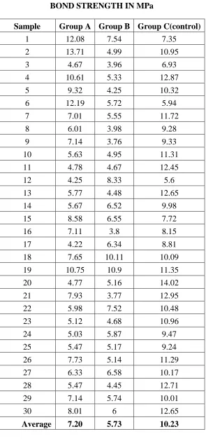
expressed in Megapascals ( Mpa) as shown in Table 1 and Graph 1 depicts the
results in the form of a Bar graph. The bond strength was calculated in megapascals
Force in Newtons Bond strength MPa =
Surface area of brackets in mm2
The surface area of the bracket was 9.63 mm2, as given by the
manufacturer. The results obtained have been shown in the table 1. Descriptive
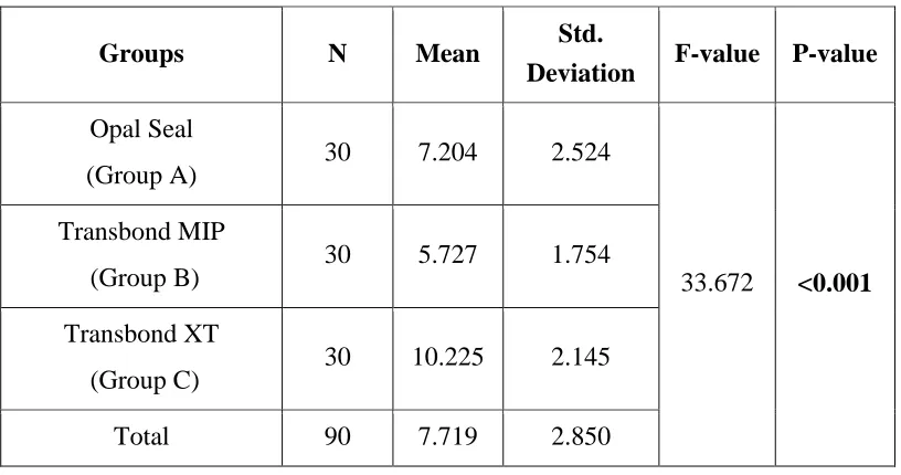
statistics means, and standard deviations were calculated for the shear-peel band
strength data for each adhesive group (Table 3). A one-way analysis of variance
(ANOVA) was performed to test for statistical significance (Table 3, Graph1).
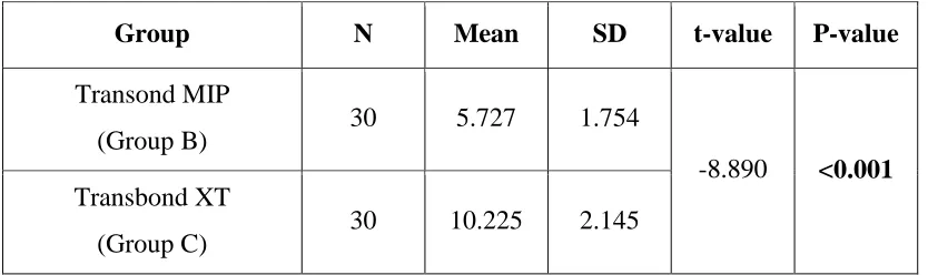
Independent samplet-test was utilized to determine whether there was a significant difference in SBS between Group A and Group B, Group B and group C, and Group
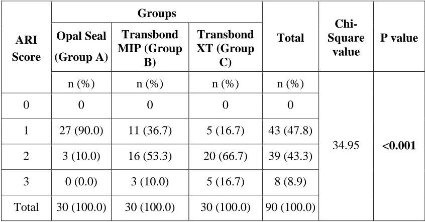
A and group C(Table 4). The chi-square test was used to compare the bond failure
mode (ARI scores) between the groups (Table 5, Graph 2). Significance for all
statistical tests was predetermined atP< 0.05.
STATISTICAL INFERENCE Shear Bond Strength
The mean SBS for the brackets bonded using the Opal seal was 7.204 ±
2.524 MPa; for Transbond MIP 5.727± 1.754 MPa; and for Transbond Primer
10.225± 2.145 MPa. The results of the one-way analysis of variance (F = 33.672) indicated statistically significant differences (P < 0.001) between the groups (Table 3,Graph 1).
Independent sample t-test revealed that there was statistically significant difference in SBS between Opal Seal and Transbond MIP with t- value and p- value
and Transbond MIP (T= -8.890, P < 0.001) showed highly significant statistical difference in SBS when compared with the control group (Transbond XT).
Adhesive Remnant Index
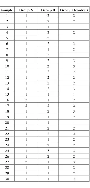
The failure modes of the three groups are presented in Table 2. The
chi-square comparisons (Table 5, Graph 2) of the ARI scores between the three
groups (χ² = 34.95) indicated that bracket failure modes were significantly different
(P < 0.001). In Transbond MIP ( Group B) and control (Group C) groups, most of the bond failure was within the adhesive with more adhesive on enamel (score 2),
while in the Opal Seal group(Group A) the bond failure was similar with less
adhesive on enamel (score 1). The comparisons of the ARI scores between the three
groups (χ2
= 34.95) indicated that bracket failure mode was significantly different
(P < 0.001) with more adhesive remaining on the teeth bonded with Transbond XT (Table 5).
A closer look at the data indicated that there was a greater incidence of
bracket failure towards enamel-adhesive interface with the new Opal Seal Primer,