JOURNAL OFVIROLOGY,
0022-538X/97/$04.0010 June 1997, p. 4544–4554 Vol. 71, No. 6
Copyright © 1997, American Society for Microbiology
Efficient Encapsidation of Human Immunodeficiency Virus Type 1
Vectors and Further Characterization of cis Elements
Required for Encapsidation
M. SCOTT MCBRIDE,† MICHAEL D. SCHWARTZ,ANDANTONITO T. PANGANIBAN*
McArdle Laboratory for Cancer Research, University of Wisconsin Medical School, Madison, Wisconsin 53706
Received 11 November 1996/Accepted 13 March 1997
To determine whether there is a cis-acting effect of translational expression of gag on RNA encapsidation, we compared the encapsidation of wild-type RNA with that of a mutant in which the translation of gag was ablated. This comparison indicated that there is not such a cis effect. To determine what is necessary and sufficient for encapsidation, we measured the relative encapsidation efficiencies of human immunodeficiency virus type 1 vector RNAs containing mutations in domains proximal to the canonical encapsidation signal or containing large deletions in the remainder of the genome. These data indicate that TAR and two additional regions are required for encapsidation and that the 5*end of the genome is sufficient for encapsidation. The Rev-responsive element is required mainly for efficient RNA transport from the nucleus to the cytoplasm. A foreign sequence was found to have a negative effect on encapsidation upon placement within the parental vector. Interestingly, this negative effect was compounded by multiple copies of the sequence.
The sequence within the 59 untranslated leader region of retroviral RNA contains important cis elements for many steps in viral replication. For human immunodeficiency virus type 1 (HIV-1), the terminal repeat (r), the unique 59sequence (u5), and the primer binding site (pbs) have been shown to be important in the initiation and translocation steps of reverse transcription (7, 26, 31, 63–65). Following reverse transcrip-tion, sequences originally derived from u5 are important for integration of the viral DNA (25, 58, 62). After transcription, the major splice donor (59ss), which is used to generate the subgenomic mRNAs, is required for efficient expression of most of the internal genes of HIV-1. The TAR stem-loop within r is important for maximal transcription from the viral promoter (17, 21, 27, 32, 47, 51), and sequences within r and u5 are important for the polyadenylation of transcripts (12). Se-quences throughout the 59untranslated region have been im-plicated in the process of dimerization (4, 18, 57, 60). In par-ticular, a stem-loop called SL1, located between the pbs and the 59ss, has been shown to be important for initiation of the dimerization process in vitro (16, 36, 44, 48, 52, 59).
Encapsidation is the process whereby genomic RNA is in-corporated into the assembling virus particle. Subgenomic RNAs and nonviral RNAs are largely excluded from encapsi-dation (40, 45, 46). The process of encapsiencapsi-dation involves the recognition of cis elements within genomic RNA (E orC) by the product of the gag gene, the trans factor shown to be required for particle formation and encapsidation (33). The most well characterized of the E elements are those that are present downstream of the pbs and that extend into the 59end of the gag gene. The secondary structure of this region consists of four hairpins, SL1, SL2, SL3, and SL4 (5, 15, 28). Three of
these hairpins, SL1, SL3, and SL4, function in encapsidation (45, 46), but they are not sufficient for encapsidation in vivo.
Within the 59 untranslated leader region, sequences up-stream of SL1 are also required for encapsidation (35, 45). Since these sequences are upstream of the 59ss, they are present in both genomic and subgenomic RNAs. In addition, sequences within gag and downstream of SL4 augment encap-sidation (40, 53). Since these sequences are present only in genomic RNAs, they are likely to contribute to the selective encapsidation of genomic RNAs. The exact nature of all of the
cis elements upstream and downstream of SL1 through SL4
and the functions that they serve in the process of encapsida-tion remain to be fully elucidated. Finally, some data have indicated that sequences within env are required for encapsi-dation (34, 55). Some of these sequences within env encompass the Rev-responsive element (rre). However, the rre is required for efficient transport of genomic RNA from the nucleus to the cytoplasm, and therefore it is unclear whether these sequences function directly or indirectly in encapsidation.
Retroviral vectors contain cis elements sufficient for viral replication but usually lack essential trans factors. Retroviral vectors can be used to transduce foreign genes into cells pro-vided that essential trans factors can be supplied by helper cells, helper viruses, or helper plasmids. Encapsidation of HIV-1 vectors by helper plasmids and helper cell lines can be attained, but the encapsidation efficiency of the vectors is low (10, 40, 53). This relatively low encapsidation efficiency may be due to the absence of a full complement of the cis elements required for encapsidation or to the inadvertent disruption of encapsidation elements by mutations designed to ablate trans-lation of the gag gene. Alternatively, E may not have been able to function within the context in which it was expressed be-cause the presence of a foreign sequence may have impinged upon the capability of E to function.
To gain a better understanding of the requirements for ef-ficient HIV-1 vector RNA encapsidation and to identify re-gions of the viral genome outside the canonical E region (SL1 to SL4) that contribute to encapsidation, we examined the encapsidation efficiencies of mutants that lack discrete seg-* Corresponding author. Mailing address: McArdle Laboratory for
Cancer Research, University of Wisconsin Medical School, 1400 Uni-versity Ave., Madison, WI 53706. Phone: (608) 263-7820. Fax: (608) 262-2824. E-mail: [email protected].
† Present address: Department of Molecular Microbiology, Wash-ington University School of Medicine, St. Louis, MO 63130.
4544
on November 9, 2019 by guest
http://jvi.asm.org/
ments of the viral genome. Our results indicate that ablation of any of three regions upstream of E results in substantial re-duction in encapsidation. These three regions are TAR, a contiguous region that likely contains a hairpin structure termed the r-u5 stem-loop, and a third region of undefined secondary structure that surrounds the pbs. In contrast, the rre and the adjacent region do not contribute substantially to en-capsidation in a direct way. Finally, introduction of a foreign gene reduced encapsidation efficiency marginally. However, the introduction of multiple copies of the gene had a synergis-tic negative effect on encapsidation. These data help to define what is sufficient in cis for encapsidation and have implications for successful HIV-1 vector design.
MATERIALS AND METHODS
Construction of plasmid DNAs.Standard techniques were used for molecular cloning (43). The creation of pMSMBA has been described previously (45). Unless otherwise noted, nucleotide designations refer to the DNA sequence of pNL4-3 (1). RNA nucleotide (nt) 1 corresponds to DNA nt 455. pCT is derived from pMSMBA and contains a stop codon within the capsid domain of the gag gene. pCT was created in several steps. First, two fragments obtained by PCR amplification of pMSMBA with two sets of primers, the sense mismatch primer complementary to the sequence of nt 687 to 715 (sense mismatch primer 687-715 EcoRI) (45) with the antisense mismatch primer complementary to the sequence of nt 1483 to 1460 (antisense mismatch primer 1483-1460 XbaI) (59-TTGGTTC TCTCTAGAGGCCTGGTG) and the sense mismatch primer 1460-1483 XbaI (59-CACCAGGCCTCTAGAGAGAACCAA) with the antisense mismatch primer 1999-1976 XbaI (59-TTTTGGCTATCTAGACTTCTTTGC), were di-gested with XbaI and ligated. Next, the ligation mixture was PCR amplified with the sense primer 687-715 EcoRI and the antisense primer 1535-1511 (45). The resulting 848-bp fragment was digested with BssHII and SpeI and ligated into pMSMBA digested with the same two enzymes.
pdTAR was created by a similar protocol. Two fragments obtained by PCR amplification of pCT with two sets of primers, the sense primer225-28 (59-A AGCTTATGCATGCGGCC) with the antisense mismatch primer 464-443 XbaI (59-CCTCTAGACCCAGTACAGGCAA) and the sense mismatch primer 504-528 XbaI (59-GGTCTAGACACTGCTTAAGCCTCAA) with the antisense mis-match primer 774-743 AgeI (45), were digested with XbaI and ligated. This ligation mixture was PCR amplified with the sense primer225-28 and the antisense primer 774-743 AgeI. The resulting 799-bp fragment was digested with NotI and BssHII and ligated into pCT that had been digested with the same two enzymes. A similar protocol was used to create pdR/U5, pR/U5, pR/U5*, pdR/L, and pdPBS with an appropriate set of internal primers, the sense mismatch primer 551-575 XbaI (59-GGTCTAGATCTGCCCGTCTGTTGTG) with the antisense mismatch primer 519-495 XbaI (59-CCTCTAGACCTTCCCTAGTTA GCCA), the sense mismatch primer 510-533 XbaI (59-CCTCTAGATTAAGCC TCAATAAAG) with the antisense mismatch primer 519-495 XbaI (see sequence above), the sense mismatch primer 544-567 (59-GTGCTCAAATTTGGGTGT GCCCGT) with the antisense mismatch primer 567-544 (59-ACGGGCACACC CAAATTTGAGCAC), the sense mismatch primer 675-698 XbaI (59-GAGGA GATCTCTAGACGCAGGACT) with the antisense mismatch primer 519-495, and the sense mismatch primer 624-635/654-665 MfeI (59-AATCTCTAGCAAT TGAAAGCGAAA) with the antisense mismatch primer 665-654/635-624 MfeI (59-TTTCGCTTTCAATTGCTAGAGAT), respectively.
pdU5/L was created by PCR amplification of pCT by using the sense primer
225-28 with the antisense primer 720-697/564-556 (59-CCGTGCGCGCTTCA GCAAGCCGAGGGCACACAC). The resulting 616-bp fragment was digested with NotI and BssHII and ligated into pCT digested with the same two enzymes. pdRRE was created in steps. First, two fragments obtained by PCR amplifi-cation of pMSMBA with two sets of primers, the sense primer 6321-6343 (59-T GTGGGTCACAGTCTATTATGGGG) with the antisense mismatch primer 7773-7750 MluI (59-AGGAACAAAACGCGTATTCCCACT) and the sense mismatch primer 7991-8014 MluI (59-CTGGGGATTACGCGTTGCTCTGGA) with the antisense primer 8927-8903 (59-ATTGCTACTTGTGATTGCTCCAT G), were digested with MluI and ligated. Next, the ligation mixture was PCR amplified with the sense primer 6321-6343 and the antisense primer 8927-8903. The resulting 1,477-bp fragment was digested with NheI and XhoI and ligated into pMSMBA digested with the same two enzymes.
pd(X-N) was created by digesting pCT with XbaI and NheI followed by ligating the larger fragment to generate a plasmid containing a deletion between these two sites. pd(N-H) was created by digesting pCT with NheI and HpaI, treating the DNA with the Klenow fragment of DNA polymerase, and ligating the larger fragment. pB(SVpA) was created by digesting pMSM50 (45) with BamHI and NaeI and ligating a 264-bp fragment containing the simian virus 40 (SV40) poly(A) site into pCT that had been digested with BamHI and SmaI. pd(X-N)B(SVpA) was created by digesting pB(SVpA) with XbaI and NheI and recir-cularizing the larger fragment through ligation. pd(X-X) was created by digesting pCT with XbaI and XhoI, treating the DNA with the Klenow fragment of DNA
polymerase, and ligating. pBSSK(2)CTE was created by digesting pKB504CTE, containing the Mason-Pfizer monkey virus (MPMV) sequence from nt 8039 to 8184 (kindly provided by Kathy Boris-Lawrie), with BamHI and PstI and ligating a fragment into pBSSK(2) (Stratagene) digested with the same two enzymes. pd(X-X)CTE was created by digesting pBSSK(2)CTE with XbaI and XhoI and ligating a fragment containing the MPMV constitutive transport element (cte) into pCT that had been digested with XbaI and XhoI.
pH-1 was created by digesting GB108 (14) with XbaI and NheI and ligating a fragment containing the hygromycin B resistance gene into pCT that had been digested with the same two enzymes. pH-2 was created by digesting pH-1 with XbaI and NheI and ligating the fragment containing the hygromycin B resistance gene into pH-1 digested with NheI. pH-4 was created by digesting pH-2 with XbaI and NheI and ligating the fragment containing two copies of the hygromycin B resistance gene into pH-2 digested with NheI.
pGEM(1247-1523) and pGEM(1247-1523TAG) were created by PCR ampli-fication of pMSMBA and pCT, respectively, with the sense primer 687-715 EcoRI (45) and the antisense mismatch primer 1535-1512 BamHI (59-CATCC TATTGGATCCTGAAGGGTA), respectively. The resulting 848-bp fragments were digested with BamHI and NsiI and ligated into pGEM11zf(2) (Promega) digested with the same two enzymes.
Transfections.Twenty-four hours before transfection, 293 cells were seeded at a density of 1.03107cells per 150-mm-diameter plate in 30 ml of Dulbecco modified Eagle medium supplemented with 10% fetal bovine serum and were incubated at 37°C in 5% CO2. Transfections were carried out with 15mg of each of the two plasmid DNAs per 150-mm-diameter plate for a total of 30mg of DNA. The calcium phosphate precipitation method was used as described pre-viously (3).
RNA isolation. After 48 to 72 h, the media and cytoplasmic RNA were concurrently collected. The cytoplasmic RNA, nuclear RNA, and total cell RNA were harvested as previously described (43) and the concentrations were deter-mined by spectrophotometric absorption at a wavelength of 260 nm. Virus was harvested from the media by first removing cellular debris by centrifugation at 4,000 rpm in a Beckman AccuspinFR tabletop centrifuge. The virus was then pelleted from the clarified media by centrifugation through a 20% sucrose cush-ion for 2.5 h at 25,000 rpm in an SW28 rotor. The viral pellet was then resus-pended in 500ml of TNE buffer (10 mM Tris-HCl [pH 7.4], 100 mM NaCl, 1 mM EDTA). The physical virus titer was determined by using an antigen-capture assay to quantitate p24 (Coulter Cytometry). To isolate RNA, virions were disrupted by the addition of sodium dodecyl sulfate to 1% and by incubation in a boiling water bath for 5 min. The sample was then treated with proteinase K (500mg/ml) at 37°C for 30 min followed by phenol (pH 4.5)-chloroform extrac-tion, chloroform extracextrac-tion, and ethanol precipitation in the presence of 20mg of poly(C) RNA. The nucleic acids isolated both from the cytoplasm of the trans-fected cells and from the virus were treated with DNase I by resuspending the isolated nucleic acids in 50ml of a buffer containing 10 mM Tris-HCl (pH 7.4), 10 mM MgCl2, 40 U of RNasin (Promega), 1 mM dithiothreitol, and 10 U of RNase-free DNase I (Boehringer-Mannheim). The nucleic acids were incubated at 37°C for 30 min. To terminate the reaction, the samples were again treated with proteinase K, phenol-chloroform extracted, chloroform extracted, and pre-cipitated with ethanol.
RNase protection.Antisense probe (;1.03108cpm/mg) was synthesized by linearizing pGEM(1247-1523) or pGEM(1247-1523TAG) with HindIII. T7 RNA polymerase (Promega) was then used to transcribe the template in the presence of [a-32P]CTP (43). Either 25mg of cytoplasmic RNA or one-fifth of the virion RNA preparation supplemented with 25mg of poly(C) RNA was mixed with 106 Cerenkov counts of32P-labeled antisense RNA (;200 fmol) and was precipi-tated with ethanol. Samples were washed with 70% ethanol and resuspended in 20ml of hybridization buffer (100 mM sodium citrate [pH 6.4], 300 mM sodium acetate [pH 6.4], 1 mM EDTA in 80% formamide), heated at 95°C for 3 min, and hybridized at 42°C for 16 h. Then, 200ml of RNase digestion mixture (10 mM Tris-HCl [pH 7.4], 300 mM NaCl, 5 mM EDTA, 2mg of RNase T1per ml, 5mg of RNase A per ml), was added for a 30-min incubation at 37°C. Sodium dodecyl sulfate was added to 1% and proteinase K was added to 0.5 mg/ml. Samples were incubated for 30 min at 37°C, phenol-chloroform extracted, chloroform ex-tracted, and precipitated with ethanol in the presence of 25mg of poly(C) RNA carrier. Pellets were dissolved in 10ml of formamide-loading buffer (2 mM EDTA [pH 8.0] in 80% formamide), heated at 95°C for 3 min, and subjected to polyacrylamide gel electrophoresis (6% acrylamide, ratio of bisacrylamide to acrylamide, 19:1; 8 M urea). To create size markers on denaturing polyacryl-amide gels, we labeled pGEM11zf(2) (Promega) digested with HpaII by treat-ment with the Klenow fragtreat-ment in the presence of [a-32P]dCTP (43). The quantitation of the various protected RNA species was determined by Phosphor-Imager analysis (Molecular Dynamics).
RESULTS
Translation of gag does not increase RNA encapsidation in
cis.Retroviral vectors contain essential cis elements for viral replication but lack essential trans factors. However, at least some HIV-1 vectors exhibit relatively low encapsidation
on November 9, 2019 by guest
http://jvi.asm.org/
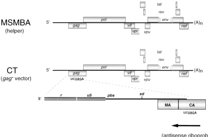
ciencies (40), presumably because the mutations introduced into these vectors, which had been created to disrupt transla-tion of Gag mRNA, had the inadvertent effect of disrupting cis elements essential for encapsidation. To systematically exam-ine the criteria required for efficient vector encapsidation, we first created an HIV-1 vector by disrupting translation of Gag mRNA at a location distal to cis elements known to be essen-tial for encapsidation (53). pCT is a derivative of the proviral clone pMSMBA carrying the mutant env gene (45) and con-tains an XbaI site at nt 1469 and a stop codon at nt 1472 within the capsid domain (CA or p24) of the gag gene (Fig. 1). pCT efficiently expresses genomic RNA at a level similar to that of pMSMBA (Fig. 2). However, the truncated Gag derivative would not be expected to be proficient for particle assembly. As expected, the truncated version of Pr55 could not be de-tected in the media of pCT-transfected cells as evidenced by Western blot analysis (data not shown).
To determine the relative encapsidation efficiency of CT RNA, we cotransfected 293 cells with pCT and pMSMBA and performed an RNase protection assay on RNA isolated from the cytoplasm of the transfected cells and on RNA isolated from virions (Fig. 2). In this experiment, pMSMBA served as the source of Gag protein for encapsidation of both MSMBA RNA and CT RNA. The probe used in the RNase protection analysis was capable of detecting and distinguishing CT RNA and MSMBA RNA. Thus, it was possible to determine the relative encapsidation efficiencies of these two RNAs by de-termining the ratio of CT to MSMBA RNA within the virion and dividing by the ratio of these two RNAs within the cyto-plasm. The CT genomic RNA was encapsidated at an efficiency of 0.93 relative to the MSMBA RNA. As expected, we did not detect genomic RNA within the media of cells transfected with pCT alone (Fig. 2). Since CT RNA was efficiently encapsi-dated, the stop codon that was created within pCT does not disrupt any cis elements necessary for encapsidation. More-over, since CT RNA is encapsidated nearly as well as MSMBA
RNA, the trans factors necessary for particle formation and encapsidation do not exhibit a cis effect; there is not preferen-tial encapsidation of the mRNA that functions as mRNA for the translation of gag.
Requirement of sequences at the 5*end of HIV-1 RNA for encapsidation.We previously showed that two stem-loops, SL1 and SL3, and potentially a third, SL4, are important in encap-sidation (45). However, our results and the results of others suggest that sequences upstream of SL1, SL3, and SL4, are also required for encapsidation (35, 45). Using a model of the 59 end of HIV-1 RNA (56) (Fig. 3A), we made derivatives of pCT containing precise deletions of regions containing known or suspected secondary structures at the 59end of the RNA and determined the effect of these mutations on encapsidation. The 59end of the HIV-1 genome is composed of TAR and two regions that are likely to contain ordered structure. TAR is the primary cis-acting element that is recognized by the viral Tat protein. Based on a progressive comparison of probable RNA secondary structure among diverse replication-competent HIV-1 strains (Mfold-Phylo program; University of Wisconsin, Genetics Computer Group), it is likely that the nucleotides immediately distal to TAR form a structure termed the r-u5 stem-loop. The disposition of the nucleotides distal to the r-u5 stem-loop is unclear. However, based on a comparison of the corresponding region in type C retroviruses, Aiyar et al. (2) have argued that there is likely to be conservation of a struc-ture that helps to define a region called the u5-leader (u5-1) stem-loop.
[image:3.612.126.479.69.302.2]pdTAR, pdR/U5, and pdU5/L contain deletions of the TAR stem-loop, the putative r-u5 stem-loop, and the putative u5-l stem-loop, respectively (Fig. 3B to D). pdR/L contains an exact deletion of both the putative r-u5 stem-loop and the u5-1 stem-loop (Fig. 3E). Deletion of TAR resulted in an approx-imately fourfold decrease in the steady-state level of cytoplas-mic pdTAR RNA (Fig. 2). All other mutants expressed cyto-plasmic RNA at a level similar to that of pCT. The relative FIG. 1. Schematic representation of MSMBA RNA (helper) and CT RNA (vector). pMSMBA contains a stop codon at DNA nt 6359 within env (RNA nt 5905) and a deletion from DNA nt 6365 to 7252 (RNA nt 5911 to 6798) (40). pCT is a derivative of pMSMBA that contains a stop codon at DNA nt 1472 (RNA nt 1018) within the capsid domain of gag. The antisense riboprobe used in the experiments described in the text hybridizes to a portion of the gag gene in pMSMBA or pCT as noted. sd, splice donor, 59ss.
4546 MCBRIDE ET AL. J. VIROL.
on November 9, 2019 by guest
http://jvi.asm.org/
encapsidation efficiency of each of these mutants was deter-mined as described above and was found to be reduced (Table 1). We think it unlikely that the fourfold reduction in cytoplas-mic pdTAR RNA accounts for the decrease in RNA encapsi-dation. An examination of other HIV-1 vectors that are re-duced in cytoplasmic accumulation to levels similar to that of pdTAR but that retain an intact E region indicates that these RNAs are still encapsidated efficiently (data not shown). Thus, reduction of intracellular viral RNA does not by itself diminish the encapsidation efficiency of that RNA.
The pbs is the sequence present on genomic RNA to which the tRNA primer anneals and is the initiation site of reverse transcription. Upon the annealing of the tRNA primer to genomic RNA, the secondary structure of genomic RNA changes in vitro (31). Since the deletion within encapsidation-defective mutant pdU5/L would remove the pbs, we decided to see whether the pbs was required for encapsidation. We cre-ated a mutant, pdPBS, in which the pbs was deleted and an
MfeI site was created (Fig. 3F). The relative encapsidation
efficiency of this mutant was substantially higher than that of pdU5/L (Table 1). Thus, it is unlikely that the dramatic de-crease in the encapsidation of pdU5/L is due to the absence of the pbs. Moreover, it is unlikely that the stable association of the tRNA primer with the viral RNA is required in cis for encapsidation.
To determine whether disruption of the r-u5 stem-loop af-fects encapsidation, we created a mutant (pR/U5) which con-tains a set of base substitutions designed to disrupt base pairing within the putative r-u5 stem-loop, and a mutant (pR/U5*) which contains these same mutations in conjunction with sec-ond site mutations designed to restore base pairing (Fig. 4). We measured the relative encapsidation efficiencies of these two mutants and found that both were similarly low (Table 1). Since the second site mutations within pR/U5* do not function
as compensatory mutations, it is unclear whether the r-u5 stem-loop forms and whether this is an encapsidation element. However, these data show that the sequence within the puta-tive r-u5 stem-loop is critical for encapsidation.
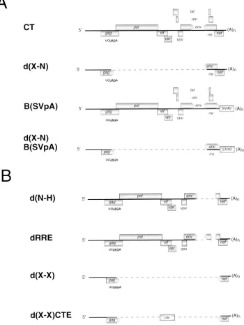
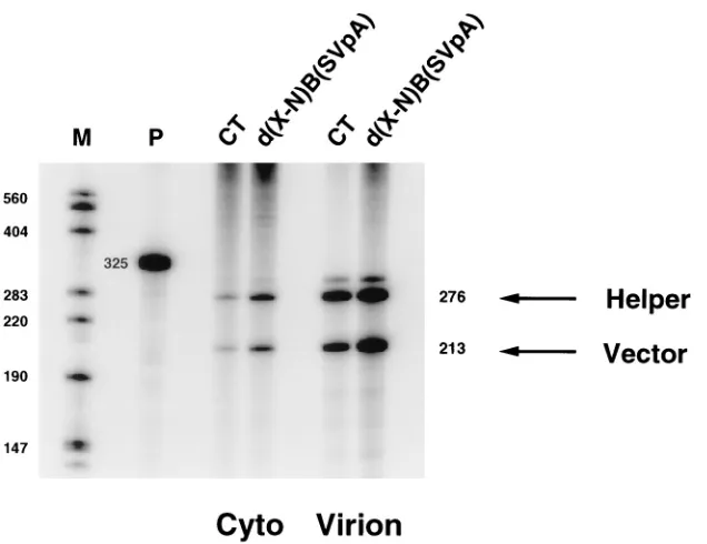
The untranslated leader region and the 5* end of gag are sufficient for encapsidation.Some data indicate that the 59end of HIV-1 RNA may not be sufficient for encapsidation (10) and that sequences within env may be necessary for encapsi-dation (34, 55). We attempted to assess whether regions out-side the 59 end of the RNA contribute to encapsidation by determining whether deletions and substitutions throughout the genome affect encapsidation. Several of these mutations are shown in Fig. 5A and B. pd(X-N) contains a deletion between the XbaI site of pCT at nt 1469 in gag and an NheI site at nt 7250 in env. pB(SVpA) contains a replacement of se-quences downstream of the BamHI site at nt 8465 in env with sequences containing the SV40 polyadenylation signal. pd(X-N)B(SVpA) has both the deletion present in pd(X-N) and the replacement present in pB(SVpA). pd(N-H) contains a dele-tion spanning the rre between the NheI site and the HpaI site at nt 8648 in env. The relative encapsidation efficiencies of each of these mutants were determined by an RNase protection assay. We found that all HIV-1 RNA sequences downstream of the XbaI site were dispensable for encapsidation with the ex-ception of the sequence between nt 7254 and 8651 within the
env gene. pd(X-N)B(SVpA) RNA was encapsidated efficiently,
while pd(N-H) RNA was not encapsidated efficiently (Fig. 6, Table 2).
[image:4.612.142.476.70.311.2]The rre is required in most cell types for efficient transport of genomic viral RNA from the nucleus to the cytoplasm (20, 42). Since, the deletion in pd(N-H) removes the rre, we at-tempted to determine whether the encapsidation defect of pd(N-H) RNA could be attributed solely to the absence of the rre. We created rre-negative mutant pdRRE (Fig. 5B), which FIG. 2. The 59end of HIV-1 RNA is required for encapsidation. pCT or derivatives containing mutations throughout the 59end of the viral RNA were cotransfected into 293 cells along with pMSMBA. The cytoplasmic RNA from the transfected cells and the virion RNA were subjected to RNase protection analysis. The marker (M) was generated as described in Materials and Methods. The probe (P) used was generated from pGEM(1247-1523TAG). This probe is complementary to a portion of the gag gene from pCT and is capable of distinguishing vector and helper RNAs. The amount of probe loaded on the gel is 1/50 of the amount of probe used in the experiment. The diagnostic bands for vector RNA and helper RNA are 276 and 213 nt, respectively. pCT was also transfected alone to verify the dependence of pCT on pMSMBA for particle production.
on November 9, 2019 by guest
http://jvi.asm.org/
contains a deletion of nt 7762 to 8002 spanning the rre in env. In addition, we generated an rre-negative mutant which con-tains a deletion between the XbaI site in gag and the XhoI site at nt 8887 in nef designated X) and a derivative of
[image:5.612.59.556.69.603.2]pd(X-X), which contains the cis-transport element (CTE) from MPMV located between the same XbaI and XhoI sites. The CTE can function in place of Rev and the rre to facilitate RNA transport from the nucleus to the cytoplasm (13), but the RNA
FIG. 3. Organization of the 59end of HIV-1 RNA. (A) MSMBA (wild type). The secondary structure shown was generated by using the Mfold-Phylo program available from the University of Wisconsin Genetics Computer Group. Since the pbs ultimately is annealed with the 39end of tRNA3Lys, we specified that the pbs remain
not base paired in generating the possible secondary structure. The TAR stem-loop, putative r-u5 stem-loop, and putative u5-1 stem-loop are shown. In addition, the pbs, stem-loop 1 (SL1), and the polyadenylation signal (pA) in the 59r sequence are shown. While the polyadenylation signal is present in both the 59and 39r sequences, only the 39polyadenylation signal is efficiently utilized (12). (B) dTAR contains an XbaI sequence in place of the TAR stem-loop from DNA nt 456 to 511 (RNA nt 2 to 57). (C) dR/U5 contains an XbaI sequence in place of the putative r-u5 stem-loop from DNA nt 512 to 558 (RNA nt 58 to 104). (D) The putative u5-1 stem-loop is deleted in dU5/L from DNA nt 565 to 696 (RNA nt 111 to 242). (E) dR/L contains an XbaI sequence in place of the putative r-u5 and u5-l stem-loops from DNA nt 512 to 689 (RNA nt 58 to 235). (F) dPBS contains an MfeI sequence in place of the pbs from DNA nt 633 to 656 (RNA nt 179 to 202).
4548 MCBRIDE ET AL. J. VIROL.
on November 9, 2019 by guest
http://jvi.asm.org/
secondary structures of the rre and the CTE are not similar (30, 42, 61). The relative encapsidation efficiencies of pdRRE and pd(X-X) were low, while the CTE significantly restored encapsidation (Fig. 7, Table 2). The fact that the CTE restored encapsidation in the absence of the rre indicates that the en-capsidation defect of RNA lacking the rre is mainly due to inefficient or aberrant transport of this RNA from the nucleus to the cytoplasm. Moreover, these data indicate that the 59 segment of the viral genome is sufficient for encapsidation provided that the RNA is transported efficiently and properly to the cytoplasm.
Effect of a foreign sequence on HIV-1 RNA encapsidation. Retroviral vectors are used as tools to transduce foreign se-quences into cells. We attempted to determine how the pres-ence of a foreign sequpres-ence, the hygromycin B resistance gene, would affect encapsidation of HIV-1 RNA. Starting with a vector, pd(X-N), that lacks most of the viral genome but is encapsidated efficiently (Table 2), we created derivatives of pd(X-N) containing one, two, or four inserts of the hygromycin B resistance gene (pH-1, pH-2, and pH-4, respectively) (Fig. 8). Each of these vectors was cotransfected along with the helper plasmid pMSMBA, and the relative encapsidation effi-ciency of each of these vectors was determined by RNase protection analysis of cytoplasmic and virion-associated RNA. The presence of one copy of the hygromycin B resistance gene (pH-1) reduced relative encapsidation efficiency moderately (Table 3). However, the presence of two (pH-2) or four (pH-4) copies of the hygromycin B resistance gene reduced relative encapsidation efficiency more profoundly. Since all of these vectors express RNAs that are shorter than genomic RNA, simple exclusion of these RNAs for encapsidation is unlikely.
DISCUSSION
Our data indicate that the 59end of HIV-1 RNA extending from the transcription start site to position 1017 within the genomic RNA is sufficient for encapsidation provided that the rre is present to effect efficient nuclear transport. In addition to the region generally viewed as E, the upstream region contain-ing TAR, the r-u5 stem-loop, and the u5-1 stem-loop function in encapsidation. Moreover, the magnitude of the effect of mutations in this upstream region is similar to that of muta-tions in the SL1 to SL4 region. Thus, for HIV-1, it is probably appropriate to consider the entire 59 untranslated leader re-gion and the 59end of gag as E. The exact function of TAR and the two putative stem-loops, r-u5 and u5-1, in encapsidation remains to be determined. Some have shown that this region of
HIV-1 RNA is preferentially bound by Gag in vitro (24), but others have shown that Gag preferentially binds a region con-taining SL1, SL2, SL3, SL4, and sequences within gag (8, 9, 15, 39, 57). The TAR stem-loop appears to be a multifunctional element. In addition to its roles in transcription and encapsi-dation, TAR has recently been shown to be required for re-verse transcription (26). To see whether the role of TAR in encapsidation was Tat dependent, we introduced a nonsense mutation at the ninth codon of the tat gene. This tat mutation was then introduced in parallel into MSMBA and dTAR, each of which also contained a human cytomegalovirus promoter-enhancer in place of the U3 portion of the HIV long terminal repeat. Measurement of the relative encapsidation efficiencies of the RNAs expressed from these constructs indicated that TAR plays a role in encapsidation that is independent of Tat. The exact portion of gag that is required for encapsidation and the nature of the signals within gag have not been deter-mined. Luban and Goff found that sequences from nt 335 to 503 within genomic RNA were required for encapsidation (40). Our laboratory found that sequences from nt 389 to 1053 within the genomic RNA enhanced the ability of vectors to be propagated (14). Parolin et al. observed no enhancement in the ability of vectors to be propagated when sequences past RNA nt 990 were included (53). In fact, they found that the ability of these vectors to be propagated was reduced upon the addition of gag sequence downstream of RNA nt 990. From their work, it appears that the 39end of the HIV-1 encapsidation signal lies between RNA nt 797 and 990 within gag.
We observed efficient encapsidation of an HIV-1 vector when the proteins essential for particle formation were pro-vided in trans. Other HIV-1 vectors have exhibited inefficient encapsidation. For instance, Luban and Goff created an HIV-1 vector by mutating the Kozak consensus sequence upstream of the gag start codon such that translation was inefficient (40). When transfection was performed with a helper plasmid, the
[image:6.612.365.504.444.656.2]FIG. 4. Base substitutions within the putative r-u5 stem. pR/U5 contains a group of five mutations, three of which disrupt the base pairing within the stem at RNA nt 59, 61, and 63. These mutations correspond to DNA nt 513, 515, and 517, respectively. Base pairing would still occur in the presence of the mutations at RNA nt 58 and 60. pR/U5* contains the same mutations present in pR/U5 in conjunction with a group of three mutations designed to restore base pairing within the stem at RNA nt 99, 101, and 103. These latter mutations correspond to DNA nt 553, 555, and 557, respectively. pA, polyadenylation signal.
TABLE 1. Relative encapsidation efficiencies of vectors containing mutations within the 59end of HIV-1 RNA
Vectora Rel. Enc. Eff.b
pCT ... 0.9360.16 pdTAR... 0.2660.05 pdR/U5 ... 0.1160.01 pdU5/L... 0.0460.02 pdR/L... 0.0260.01 pdPBS ... 0.5360.06 pR/U5 ... 0.1060.01 pR/U5* ... 0.1160.02
aAll vectors are derivatives of the parental vector, pCT.
bThe relative encapsidation efficiency (Rel. Enc. Eff.) of each vector was
calculated by dividing the ratio of vector RNA to helper RNA in the virion by the ratio of vector RNA to helper RNA in the cytoplasm as determined by RNase protection analysis (Fig. 2). The results represent at least three independent experiments.
on November 9, 2019 by guest
http://jvi.asm.org/
relative encapsidation efficiency of this HIV-1 vector was found to be low, presumably because the mutations created to ablate translation inadvertently affected E. Berkowitz et al. (10) created an HIV-1 vector that expresses most of the se-quence that we have defined here as being sufficient for en-capsidation. However, that vector lacks 19 nt at the 59end of the RNA and is not encapsidated efficiently when expressed in a helper cell line. Our results indicate that the inability of that vector to be encapsidated can probably be attributed to at least
two defects. First, since the 19 nt at the 59end of the RNA are not present, the vector does not contain the entire TAR stem-loop. We observed that the relative encapsidation efficiency of a vector lacking TAR is approximately 0.26. Second, that vec-tor expresses a foreign gene. We observed that the presence of the hygromycin B resistance gene reduced the relative encap-sidation efficiencies of pH-1, pH-2, and pH-4 (Table 3), and it is possible that a similar reduction in the relative encapsidation efficiency may result from the presence of other foreign se-FIG. 5. Mutants containing deletions that eliminate substantial segments of the HIV-1 genome. (A) pd(X-N) has a deletion from the XbaI site at DNA nt 1469 (RNA nt 1015) in gag to the NheI site at DNA nt 7250 (RNA nt 6796) in env. pB(SVpA) has a replacement of sequences downstream of the BamHI site at DNA nt 8465 (RNA nt 8011) in env with sequences spanning the SV40 polyadenylation signal. pd(X-N)B(SVpA) has both the deletion from the XbaI site to the NheI site and the replacement sequences downstream of the BamHI site. (B) Derivatives of pCT lacking the rre. pd(N-H) has a deletion from the NheI site in env to the HpaI site at DNA nt 8648 (RNA nt 8194) in env. pdRRE has a deletion from DNA nt 7762 to 8002 (RNA nt 7308 to 7548) spanning the rre. pd(X-X) has a deletion from the
XbaI site in gag to the XhoI site at DNA nt 8887 (RNA nt 8433) in nef. pd(X-X)CTE contains the MPMV CTE cloned between the XbaI site in gag and the XhoI site
in nef.
4550 MCBRIDE ET AL. J. VIROL.
on November 9, 2019 by guest
http://jvi.asm.org/
[image:7.612.134.484.78.543.2]quences. Interestingly, the helper cell line used by Berkowitz et al. (10) expresses RNA that is not encapsidated, so competi-tion for encapsidacompeti-tion between vector RNA and helper RNA does not occur. The fact that they did not observe encapsida-tion of their vector implies that cis elements, TAR in particu-lar, are required for encapsidation even in the absence of competition by encapsidation-competent RNA.
Although we were unable to genetically demonstrate that the secondary structure of the r-u5 stem-loop is required for encapsidation, its conservation among the primate lentiviruses coupled with the low level of sequence identity throughout this region suggests that the secondary structure is real (6). Among the primate lentiviruses, the only widely conserved sequences throughout the r-u5 stem-loop are the polyadenylation signal and a GCUU tetranucleotide sequence located from RNA nt 62 to 65 within the stem (6). Our pR/U5 and pR/U5* mutants, which are substantially reduced in encapsidation efficiency, contain groups of base substitutions that change the GCUU
tetranucleotide sequence. Interestingly, the r-u5 hairpin loop contains a palindromic sequence reminiscent of SL1. A palin-dromic sequence is also present in the loops of the putative r-u5 stem-loops of strain cpz of simian immunodeficiency virus (SIVcpz) and SIVsyk (6). Recent electron microscopic data and computer modeling indicate that the r-u5 stem-loop may have an important role in RNA-RNA association within the dimer (29).
[image:8.612.151.469.66.311.2]In contrast to the situation for the r-u5 stem-loop, there is FIG. 6. Sequences within env in conjunction with sequences at the 59end of HIV-1 RNA are sufficient for encapsidation. 293 cells were cotransfected with pMSMBA and pCT or pd(X-N)B(SVpA). RNA isolated from the cytoplasm (Cyto) of the transfected cells and from virions (Virion) was subjected to RNase protection analysis as described in the legend for Fig. 2 except that the probe used here was generated from pGEM(1247-1523), which contains a portion of gag from pMSMBA. The diagnostic bands for helper RNA (276-nt band) and for vector RNA (213-nt band) are reversed from those of Fig. 2. The amount of probe loaded on the gel is 1/50 of the amount of probe used in the experiment.
FIG. 7. The MPMV CTE restores encapsidation in the absence of the rre. 293 cells were cotransfected with pMSMBA and pCT, pd(X-X) or pd(X-X)CTE. Cytoplasmic RNA (Cyto) and virion RNA (Virion) were subjected to RNase protection analysis as described in the legend for Fig. 6.
TABLE 2. Relative encapsidation efficiencies of vectors lacking substantial segments of the HIV-1 genome
Vectora Rel. Enc. Eff.b
pCT ... 0.9360.16 pd(X-N) ... 1.1560.15 pB(SVpA) ... 0.9060.08 pd(X-N)B(SVpA)... 1.5260.08 pd(N-H)... 0.0360.01 pdRRE... 0.0460.02 pd(X-X) ... 0.0360.02 pd(X-X)CTE... 0.5160.11
aAll vectors are derivatives of the parental vector, pCT.
bThe relative encapsidation efficiency (Rel. Enc. Eff.) of each vector was
calculated as described for Table 1. The results represent at least three inde-pendent experiments.
on November 9, 2019 by guest
http://jvi.asm.org/
[image:8.612.319.557.493.691.2]not wide agreement on the secondary structure surrounding the pbs that we have presented here as the u5-1 stem-loop (5, 56). However, at the top of the u5-1 stem-loop there is a subregion called the u5-IR stem-loop, and the secondary struc-ture of this stem-loop appears to be generally conserved among different retroviruses (2). Similarly, sequences within u5 are important for encapsidation of murine leukemia and spleen necrosis virus RNAs (19, 49).
In addition to the pbs, the u5-1 stem-loop contains other cis elements that are important for the annealing of the tRNA3Lys
and the initiation of reverse transcription (63, 65). Although the secondary structure of genomic RNA changes upon the annealing of the tRNA3Lysto genomic RNA in vitro (31), this
change in secondary structure may not be relevant to encapsi-dation. First, the processes of tRNA incorporation into the assembling virus particle and genomic RNA encapsidation may be completely separate, since genomic RNA encapsidation is not required for selective incorporation of tRNAs into the assembling virus particle in murine leukemia or mouse mam-mary tumor viruses (37, 54). Additionally, the reverse tran-scriptase domain of Gag-Pol is required for selective incorpo-ration of tRNA3Lys into the virus particle and annealing (38,
41), but reverse transcriptase is not required for genomic RNA encapsidation (33). Accordingly, we find that relative encapsi-dation efficiency is only modestly reduced in the absence of the pbs in agreement with a previous report (50). However, it will
be interesting to determine if the sequences flanking the pbs that are important in annealing and initiation of reverse tran-scription are important for genomic RNA encapsidation.
Others have suggested that sequences within env and span-ning the rre may be directly required for encapsidation (34, 55). Our results indicate that the role of these sequences in encapsidation is mainly indirect; they function to enhance the transport of genomic RNA from the nucleus to the cytoplasm where encapsidation occurs. Our rre-negative, CTE-containing RNA could be encapsidated at an efficiency approximately half that of the parental vector RNA, so the rre has a direct effect on encapsidation of twofold at most. It is possible that genomic RNA may have to be transported from the nucleus to the cytoplasm by a specific pathway for optimal encapsidation. Interestingly, genomic and subgenomic RNAs are localized to different subregions within the nucleus (11), and the Rev/rre transport pathway is likely to be different from the mRNA transport pathway (22, 23).
We observed that the presence of a foreign sequence, the hygromycin B resistance gene, has a negative effect on the ability of HIV-1 RNA to be encapsidated. The negative effect of the hygromycin B resistance gene on encapsidation may not be specific but rather may reflect a general reduction in en-capsidation of HIV-1 RNA in the presence of any foreign sequence. It will be interesting to determine whether other heterologous sequences have a similar negative effect on en-capsidation and whether placing direct repeats within a vector decreases encapsidation efficiency. Since the 59end of HIV-1 RNA is sufficient for encapsidation, it is interesting that re-placing a nonessential viral sequence with a foreign sequence can have a negative effect on encapsidation. Obviously, the foreign sequence does not behave in a neutral way; it may be that the foreign sequence influences the secondary structure of the encapsidation signal at the 59end of the RNA or impinges on other viral processes that occur prior to encapsidation.
ACKNOWLEDGMENTS
We thank Katrin Talbot, Diccon Fiore, and Kate Gerten for tech-nical assistance and Dan Loeb for helpful discussion.
[image:9.612.137.478.67.294.2]M.S.M. was supported in part by a National Science Foundation FIG. 8. Derivatives of pd(X-N) containing one, two, or four copies of the hygromycin B resistance gene.
TABLE 3. Relative encapsidation efficiencies of vectors containing the hygromycin B resistance gene
Vectora Rel. Enc. Eff.b
pd(X-N) ... 1.1560.15 pH-1 ... 0.5360.15 pH-2 ... 0.2460.06 pH-4 ... 0.0660.02
aAll vectors are derivatives of pd(X-N).
bThe relative encapsidation efficiency (Rel. Enc. Eff.) of each vector was
calculated as described for Table 1. The results represent at least three inde-pendent experiments.
4552 MCBRIDE ET AL. J. VIROL.
on November 9, 2019 by guest
http://jvi.asm.org/
(NSF) predoctoral fellowship and a Wisconsin Alumni Research Foun-dation (WARF) predoctoral fellowship. M.S.M. and M.D.S. are Cre-mer Scholars. This work was supported by NIH grant R01 AI34733.
REFERENCES
1. Adachi, A., H. E. Gendelman, S. Koenig, T. Folks, R. Wiley, A. Rabson, and
M. A. Martin.1986. Production of acquired immunodeficiency syndrome-associated retrovirus in human and nonhuman cells transfected with an infectious molecular clone. J. Virol. 59:284–291.
2. Aiyar, A., D. Cobrinik, Z. Ge, H.-J. Kung, and J. Leis. 1992. Interaction between retroviral U5 RNA and the TCC loop of the tRNATrpprimer is required for efficient initiation of reverse transcription. J. Virol. 66:2464– 2472.
3. Aldovini, A., and B. D. Walker. 1990. Techniques in HIV research. Stockton Press, New York, N.Y.
4. Awang, G., and D. Sen. 1993. Mode of dimerization of HIV-1 genomic RNA. Biochemistry 32:11453–11457.
5. Baudin, F., R. Marquet, C. Isel, J. L. Darlix, B. Ehresmann, and C.
Ehres-mann.1993. Functional sites in the 59region of human immunodeficiency virus type 1 RNA form defined structural domains. J. Mol. Biol. 229:382– 397.
6. Berkhout, B., B. Klaver, and A. T. Das. 1995. A conserved hairpin structure predicted for the poly(A) signal of human and simian immunodeficiency viruses. Virology 207:276–281.
7. Berkhout, B., J. van Wamel, and B. Klaver. 1995. Requirements for DNA strand transfer during reverse transcription in mutant HIV-1 virions. J. Mol. Biol. 252:59–69.
8. Berkowitz, R. D., and S. P. Goff. 1994. Analysis of binding elements in the human immunodeficiency virus type 1 genomic RNA and nucleocapsid pro-tein. Virology 202:233–246.
9. Berkowitz, R. D., J. Luban, and S. P. Goff. 1993. Specific binding of human immunodeficiency virus type 1 gag polyprotein and nucleocapsid protein to viral RNAs detected by RNA mobility shift assays. J. Virol. 67:7190–7200. 10. Berkowitz, R. D., M. L. Hammarskjold, C. Helga-Maria, D. Rekosh, and
S. P. Goff.1995. 59regions of HIV-1 RNAs are not sufficient for encapsida-tion: implications for the HIV-1 packaging signal. Virology 212:718–723. 11. Berthold, E., and F. Maldarelli. 1996. cis-acting elements in human
immu-nodeficiency virus type 1 RNAs direct viral transcripts to distinct intranu-clear locations. J. Virol. 70:4667–4682.
12. Bohnlein, S., J. Hauber, and B. R. Cullen. 1989. Identification of a U5-specific sequence required for efficient polyadenylation within the human immunodeficiency virus long terminal repeat. J. Virol. 63:421–424. 13. Bray, M., S. E. Prasad, E. Dubay, E. Hunter, K. T. Jeang, D. Rekosh, and
M. L. Hammarskjold.1994. A small element from the Mason-Pfizer monkey virus genome makes human immunodeficiency virus type 1 expression and replication Rev-independent. Proc. Natl. Acad. Sci. USA 91:1256–1260. 14. Buchschacher, G. L., Jr., and A. T. Panganiban. 1992. Human
immunode-ficiency virus vectors for inducible expression of foreign genes. J. Virol. 66: 2731–2739.
15. Clever, J., C. Sassetti, and T. G. Parslow. 1995. RNA secondary structure and binding sites for gag gene products in the 59packaging signal of human immunodeficiency virus type 1. J. Virol. 69:2101–2109.
16. Clever, J. L., M. L. Wong, and T. G. Parslow. 1996. Requirements for kissing-loop-mediated dimerization of human immunodeficiency virus RNA. J. Virol. 70:5902–5908.
17. Cullen, B. R. 1986. Trans-activation of human immunodeficiency virus occurs via a bimodal mechanism. Cell 46:973–982.
18. Darlix, J.-L., C. Gabus, M. T. Nugeyre, F. Clavel, and F. Barre-Sinoussi. 1990. cis-Elements and trans-acting factors involved in the RNA dimeriza-tion of the human immunodeficiency virus HIV-1. J. Mol. Biol. 216:689–699. 19. Embretson, J. E., and H. M. Temin. 1987. Lack of competition results in efficient packaging of heterologous murine retroviral RNAs and reticuloen-dotheliosis virus encapsidation-minus RNAs by the reticuloenreticuloen-dotheliosis virus helper cell line. J. Virol. 61:2675–2683.
20. Felber, B. K., M. Hadzopoulou-Cladaras, C. Cladaras, T. Copeland, and
G. N. Pavlakis.1989. Rev protein of human immunodeficiency virus type 1 affects the stability and transport of the viral mRNA. Proc. Natl. Acad. Sci. USA 86:1495–1499.
21. Feng, S., and E. C. Holland. 1988. HIV-1 tat trans-activation requires the loop sequence with tar. Nature 334:165–167.
22. Fischer, U., J. Huber, W. C. Boelens, I. W. Mattaj, and R. Luhrmann. 1995. The HIV-1 Rev activation domain is a nuclear export signal that accesses an export pathway used by specific cellular RNAs. Cell 82:475–483. 23. Fridell, R. A., U. Fischer, R. Luhrmann, B. E. Meyer, J. L. Meinkoth, M. H.
Malim, and B. R. Cullen.1996. Amphibian transcription factor IIIA proteins contain a sequence element functionally equivalent to the nuclear export signal of human immunodeficiency virus type 1 Rev. Proc. Natl. Acad. Sci. USA 93:2936–2940.
24. Geigenmuller, U., and M. L. Linial. 1996. Specific binding of human immu-nodeficiency virus type 1 (HIV-1) Gag-derived proteins to a 59 HIV-1 genomic RNA sequence. J. Virol. 70:667–671.
25. Goodarzi, G., G. J. Im, K. Brackmann, and D. Grandgenett. 1995. Concerted
integration of retrovirus-like DNA by human immunodeficiency virus type 1 integrase. J. Virol. 69:6090–6097.
26. Harrich, D., C. Ulich, and R. B. Gaynor. 1996. A critical role for the TAR element in promoting efficient human immunodeficiency virus type 1 reverse transcription. J. Virol. 70:4017–4027.
27. Harrich, D., G. Mavankal, A. Mette-Snider, and R. B. Gaynor. 1995. Human immunodeficiency virus type 1 TAR element revertant viruses define RNA structures required for efficient viral gene expression and replication. J. Vi-rol. 69:4906–4913.
28. Harrison, G. P., and A. M. L. Lever. 1992. The human immunodeficiency virus type 1 packaging signal and major splice donor region have a conserved stable secondary structure. J. Virol. 66:4144–4153.
29. Hoglund, S., J. Gonclaves, A. Ohagen, J.-Y. Sgro, A. T. Panganiban, and D.
Gabuzda.1996. Unpublished data.
30. Holland, S. M., N. Ahmad, R. K. Maitra, P. Wingfield, and S. Venkatesan. 1990. Human immunodeficiency virus Rev protein recognizes a target se-quence in Rev-responsive element RNA within the context of RNA second-ary structure. J. Virol. 64:5966–5975.
31. Isel, C., C. Ehresmann, G. Keith, B. Ehresmann, and R. Marquet. 1995. Initiation of reverse transcription of HIV-1: secondary structure of the HIV-1 RNA/tRNA(Lys,3) (template/primer). J. Mol. Biol. 247:236–250. 32. Kao, S. Y., A. Calman, P. A. Luciw, and M. B. Peterline. 1987.
Anti-termi-nation of transcription within the long terminal repeat of HIV by tat gene product. Nature 330:489–493.
33. Kaye, J. F., and A. M. L. Lever. 1996. trans-Acting proteins involved in RNA encapsidation and viral assembly in human immunodeficiency virus type 1. J. Virol. 70:880–886.
34. Kaye, J. F., J. H. Richardson, and A. M. L. Lever. 1995. cis-Acting sequences involved in human immunodeficiency virus type 1 RNA packaging. J. Virol.
69:6588–6592.
35. Kim, H.-J., K. Lee, and J. J. O’Rear. 1994. A short sequence upstream of the 59major splice site is important for encapsidation of HIV-1 genomic RNA. Virology 198:336–340.
36. Laughrea, M., and L. Jette. 1994. A 19-nucleotide sequence upstream of the 59major splice donor is part of the dimerization domain of human immu-nodeficiency virus 1 genomic RNA. Biochemistry 33:13464–13474. 37. Levin, J. G., and J. G. Seidman. 1979. Selective packaging of tRNAs does not
require genomic RNA. J. Virol. 29:328–335.
38. Litvak, S., L. Sarih-Cottin, M. Fournier, M. Andreola, and L.
Tarrago-Litvak.1994. Priming of HIV replication by tRNA(Lys3): role of reverse transcriptase. Trends Biochem. Sci. 19:114–118.
39. Luban, J., and S. P. Goff. 1991. Binding of human immunodeficiency virus type 1 (HIV-1) RNA to recombinant HIV-1 gag polyprotein. J. Virol. 65: 3203–3212.
40. Luban, J., and S. P. Goff. 1994. Mutational analysis of cis-acting packaging signals in human immunodeficiency virus type 1 RNA. J. Virol. 68:3784– 3793.
41. Mak, J., M. Jiang, M. A. Wainberg, M.-L. Hammarskjo¨ld, D. Rekosh, and L.
Kleiman.1994. Role of Pr160gag-polin mediating the selective incorporation
of tRNALysinto human immunodeficiency virus type 1 particles. J. Virol. 68: 2065–2072.
42. Malim, M. H., J. Hauber, S. Y. Le, J. V. Maizel, and B. R. Cullen. 1989. The HIV-1 Rev trans-activator acts through a structured target sequence to activate nuclear export of unspliced viral mRNA. Nature 338:254–257. 43. Maniatis, T., E. F. Fritsch, and J. Sambrook. 1982. Molecular cloning: a
laboratory manual. Cold Spring Harbor Laboratory, Cold Spring Harbor, N.Y.
44. Marquet, R., J. C. Paillart, E. Skripkin, C. Ehresmann, and B. Ehresmann. 1994. Dimerization of human immunodeficiency virus type 1 RNA involves sequences located upstream of the splice donor site. Nucleic Acids Res. 22: 145–151.
45. McBride, M. S., and A. T. Panganiban. 1996. The human immunodeficiency virus type 1 encapsidation site is a multipartite RNA element composed of functional hairpin structures. J. Virol. 70:2963–2973.
46. McBride, M. S., and A. T. Panganiban. 1997. Position dependence of func-tional hairpins important for human immunodeficiency virus type 1 RNA encapsidation in vivo. J. Virol. 71:2050–2058.
47. Muesing, M. A., D. H. Smith, and D. J. Capon. 1987. Regulation of mRNA accumulation by a human immunodeficiency virus trans-activator protein. Cell 48:691–701.
48. Muriaux, D., P. M. Girard, B. Bonnet-Mathoniere, and J. Paoletti. 1995. Dimerization of HIV-Lai RNA at low ionic strength. An autocomplementary sequence in the 59leader region is evidenced by an antisense oligonucleo-tide. J. Biol. Chem. 270:8209–8216.
49. Murphy, J. E., and S. P. Goff. 1989. Construction and analysis of deletion mutations in the U5 region of Moloney murine leukemia virus: effects on RNA packaging and reverse transcription. J. Virol. 63:319–327.
50. Nagashunmugam, T., A. Velpandi, C. S. Goldsmith, S. R. Zaki, A.
Kaly-anaraman, and A. Srinivasan.1992. Mutation in the primer binding site of the type 1 human immunodeficiency virus genome affects virus production and infectivity. Proc. Natl. Acad. Sci. USA 89:4114–4118.
51. Okamoto, T., and F. Wong-Staal. 1986. Demonstration of virus-specific
on November 9, 2019 by guest
http://jvi.asm.org/
scriptional activator(s) in cells infected with HTLV-III by an in vitro cell-free system. Cell 47:29–35.
52. Paillart, J. C., R. Marquet, E. Skripkin, B. Ehresmann, and C. Ehresmann. 1994. Mutational analysis of the bipartite dimer linkage structure of human immunodeficiency virus type 1 genomic RNA. J. Biol. Chem. 269:27486– 27493.
53. Parolin, C., T. Dorfman, G. Palu, H. Gottlinger, and J. Sodroski. 1994. Analysis in human immunodeficiency virus type 1 vectors of cis-acting se-quences that affect gene transfer into human lymphocytes. J. Virol. 68:3888– 3895.
54. Peters, G. G., and J. Hu. 1980. Reverse transcriptase as the major determi-nant for selective packaging of tRNAs into avian sarcoma virus particles. J. Virol. 36:692–700.
55. Richardson, J. H., L. A. Child, and A. M. L. Lever. 1993. Packaging of human immunodeficiency virus type 1 RNA requires cis-acting sequences outside the 59leader region. J. Virol. 67:3997–4005.
56. Rizvi, T. A., and A. T. Panganiban. 1993. Simian immunodeficiency virus RNA is efficiently encapsidated by human immunodeficiency virus type 1 particles. J. Virol. 67:2681–2688.
57. Sakaguchi, K., N. Zambrano, E. T. Baldwin, B. A. Shapiro, J. W. Erickson,
J. G. Omichinski, G. M. Clore, A. M. Gronenborn, and E. Appella.1993. Identification of a binding site for the human immunodeficiency virus type 1 nucleocapsid protein. Proc. Natl. Acad. Sci. USA 90:5219–5223.
58. Sherman, P. A., M. L. Dickson, and J. A. Fyfe. 1992. Human immunodefi-ciency virus type 1 integration protein: DNA sequence requirements for
cleaving and joining reactions. J. Virol. 66:3593–3601.
59. Skripkin, E., J. C. Paillart, R. Marquet, B. Ehresmann, and C. Ehresmann. 1994. Identification of the primary site of the human immunodeficiency virus type 1 RNA dimerization in vitro. Proc. Natl. Acad. Sci. USA 91:4945–4949. 60. Sundquist, W. I., and S. Heaphy. 1993. Evidence for interstrand quadruplex formation in the dimerization of human immunodeficiency virus 1 genomic RNA. Proc. Natl. Acad. Sci. USA 90:3393–3397.
61. Tabernero, C., A. S. Zolotukhin, A. Valentin, G. N. Pavlakis, and B. K.
Felber.1996. The posttranscriptional control element of the simian retrovi-rus type 1 forms an extensive RNA secondary structure necessary for its function. J. Virol. 70:5998–6011.
62. Vicenzi, E., D. S. Dimitrov, A. Engelman, T. S. Migone, D. F. Purcell, J.
Leonard, G. Englund, and M. A. Martin.1994. An integration-defective U5 deletion mutant of human immunodeficiency virus type 1 reverts by elimi-nating additional long terminal repeat sequences. J. Virol. 68:7879–7890. 63. Wakefield, J. K., A. G. Wolf, and C. D. Morrow. 1995. Human
immunode-ficiency virus type 1 can use different tRNAs as primers for reverse tran-scription but selectively maintains a primer binding site complementary to tRNA3Lys. J. Virol. 69:6021–6029.
64. Wakefield, J. K., and C. D. Morrow. 1996. Mutations within the primer binding site of the human immunodeficiency virus type 1 define sequence requirements essential for reverse transcription. Virology 220:290–298. 65. Wakefield, J. K., S. M. Kang, and C. D. Morrow. 1996. Construction of a type
1 human immunodeficiency virus that maintains a primer binding site com-plementary to tRNAHis. J. Virol. 70:966–975.
4554 MCBRIDE ET AL. J. VIROL.