Copyright © 1998, American Society for Microbiology. All Rights Reserved.
The Human Homolog of HAVcr-1 Codes for a Hepatitis A
Virus Cellular Receptor
DINO FEIGELSTOCK, PETER THOMPSON, PRAVINA MATTOO, YUAN ZHANG,
ANDGERARDO G. KAPLAN*
Laboratory of Hepatitis Viruses, Division of Viral Products, Center for Biologics Evaluation
and Research, Food and Drug Administration, Bethesda, Maryland 20892
Received 12 March 1998/Accepted 13 May 1998
The hepatitis A virus cellular receptor 1 (HAVcr-1) cDNA was isolated from a cDNA expression library of
African green monkey kidney (AGMK) cells by using protective monoclonal antibody (MAb) 190/4, which
blocks the binding of hepatitis A virus (HAV) to AGMK cells. The HAVcr-1 cDNA codes for havcr-1, a
451-amino-acid class I integral-membrane mucin-like glycoprotein of unknown natural function. To determine
the existence of a human homolog(s) of HAVcr-1 (huHAVcr-1), we used HAVcr-1-specific primers to amplify
cDNAs from human liver and kidney mRNA by reverse transcription-PCR. Nucleotide sequence analysis
revealed that the amplified liver and kidney huHAVcr-1 cDNAs were identical and that they coded for a
359-amino-acid glycoprotein, termed huhavcr-1, which was approximately 79% identical to havcr-1. The six Cys
residues of the extracellular domain of havcr-1 and its first N-glycosylation site were conserved in huhavcr-1.
However, the number of hexameric repeats of the mucin-like region was reduced from 27 in havcr-1 to 13 in
huhavcr-1. In addition, 12 C-terminal amino acids in the cytoplasmic domain of huhavcr-1 were deleted.
Northern blot analysis of poly(A) RNA showed that huhavcr-1 is expressed in every organ analyzed, including
the liver, small intestine, colon, and spleen, and that it is expressed at higher levels in the kidney and testis.
Although dog cells transfected with the huHAVcr-1 cDNA did not express the protective 190/4 epitope, they
bound hepatitis A virus (HAV) and gained limited susceptibility to HAV infection. Treatment with MAb 190/4
did not protect AGMK cell transfectants expressing huhavcr-1 against HAV, suggesting that HAV infected
these cells via the huhavcr-1 receptor and not the endogenously expressed havcr-1, which was blocked by MAb
190/4. Our data demonstrate that huhavcr-1 is a binding receptor for HAV and suggest that it is also a
functional receptor for HAV.
Hepatitis A virus (HAV), a hepatotropic picornavirus,
causes a medically important acute hepatitis. The first step
in the life cycle of HAV is binding to a cell surface receptor
that in African green monkey kidney (AGMK) cells is coded
by HAVcr-1 (7). Monoclonal antibody (MAb) 190/4, which
was used as a probe to molecularly clone the HAVcr-1 cDNA
from an expression cDNA library of AGMK cells, blocks the
binding of HAV and protects AGMK cells against HAV
in-fection (7). Nucleotide sequence analysis showed that
HAVcr-1 cDNA codes for a class I integral membrane
glyco-protein, termed havcr-1, of unknown natural function. The
extracellular domain of havcr-1 consists of an N-terminal
cys-teine-rich (Cys-rich) region followed by a threonine-, serine-,
and proline-rich (TSP-rich) region. The havcr-1 Cys-rich
re-gion displays homology to members of the immunoglobulin
(Ig) superfamily (7), and the TSP-rich region has the
charac-teristics of mucin-like glycoproteins (15). The putative
“lolli-pop-on-a-stick” structure (6) of havcr-1 suggested that the
extended O-glycosylated TSP-rich region presents the Cys-rich
globular domain above the cell surface and makes it accessible
for interactions with extracellular molecules. We have recently
shown that the Cys-rich region of havcr-1 and its first
N-gly-cosylation site are required for the binding of HAV and
pro-tective MAb 190/4 (19), and we are currently analyzing
whether the Cys-rich region of havcr-1 is sufficient for HAV
receptor function.
Receptor-negative cell lines that are otherwise fully
sus-ceptible to HAV replication have not yet been identified.
Although we devoted considerable effort to isolating one, we
have not been able to do so (4). We have previously shown that
mouse and dog cells, which contain internal blocks to HAV
replication, gain limited susceptibility to HAV infection upon
transfection with the HAVcr-1 cDNA (7, 19). Infection of
these mouse and dog cell transfectants with HAV resulted in
low levels of the characteristic granular cytoplasmic
fluores-cence of HAV-infected cells, which lasted for several days but
became undetectable after 1 month postinfection. This limited
level of susceptibility of the mouse and dog cell transfectants to
HAV infection resulted in only a
,
10-fold increase in HAV
titers and a
,
2-fold increase in HAV-specific RNA (7, 19);
therefore, its is likely that the input virus internalized through
havcr-1 contributed to the HAV-specific fluorescence observed
in the mouse and dog cell transfectants. Although further
anal-ysis of the HAV receptor function of havcr-1 awaits the
isola-tion of nonsusceptible cells that could fully support HAV
rep-lication upon transfection of the HAVcr-1 cDNA, the mouse
and dog cell transfectants allowed us to characterize the
havcr-1-mediated binding of HAV, its internalization, and the
lim-ited level of susceptibility to HAV infection (5, 7, 19).
Initial studies revealed that protective MAb 190/4 reacted
with the cell surfaces of clone GL37 AGMK cells but not with
the cell surfaces of HeLa cells (7). Therefore, it was of great
interest to ascertain the existence of the human homolog of
HAVcr-1 and determine its function as an HAV receptor and
its role in the pathogenesis of HAV in humans. In this report,
we describe the molecular cloning of the cDNA coding for the
human homolog of HAVcr-1 (huHAVcr-1). Nucleotide
se-* Corresponding author. Mailing address: Division of Viral
Prod-ucts, CBER-FDA, 8800 Rockville Pike, Bldg. 29A-NIH, rm. no. 1D10,
HFM-448, Bethesda, MD 20892. Phone: (301) 827-1870. Fax: (301)
480-5326. E-mail: gk@helix.nih.gov.
6621
on November 9, 2019 by guest
http://jvi.asm.org/
quence analysis revealed that the huHAVcr-1 cDNA codes
for a glycoprotein, termed huhavcr-1, that is 79% identical
to havcr-1. Northern blot analysis showed that huHAVcr-1 is
expressed in every human organ analyzed, including the liver,
small intestine, colon, and spleen, and that it is expressed at
higher levels in the kidney and testis. Dog cells transfected with
the huHAVcr-1 cDNA gained limited susceptibility to HAV
infection, whereas dog cells transfected with vector alone or
HAVcr-1 cDNA with the Cys-rich region deleted were
resis-tant to HAV infection. These results suggest that huhavcr-1 is
a receptor for HAV which may play a role in the pathogenesis
of HAV in humans.
MATERIALS AND METHODS
Cells and viruses.Continuous clone GL37 AGMK cells, termed GL37 cells, were selected for supporting optimal growth of HAV (20). Canine osteogenic sarcoma D-17 cells, obtained from the American Type Culture Collection, were cotransfected with pCMVEBNA and pSV2neo (Clontech Laboratories). A G418-resistant cell clone, termed Perro6D, that had 10- to 100-times-higher transfection efficiency with pDR2 (14, 18) than parental D-17 cells was isolated (19) and used for transfection with HAVcr-1 cDNA constructs cloned into the pDR2 vector. Cell lines were grown in Eagle’s minimal essential medium (EMEM) containing 10% fetal bovine serum (FBS) at 37°C in a CO2incubator.
Human tissue culture-adapted HAV strain HM175 of genotype 1B was de-rived from infectious cDNA (3) and grown in GL37 cells.
Antisera.Anti-havcr-1 antiserum was obtained from rabbits immunized with recombinant protein GST-2 consisting of the mucin-like region of havcr-1 fused to the C terminus of glutathione S-transferase expressed in Escherichia coli (19). Murine IgG1 subtype MAbs P1B5 raised against the humana3 integrin (Gibco BRL, Inc.), 190/4 directed against havcr-1 (7), and M2 directed against the FLAG peptide DTKDDDDK (IBI, Inc.) were purified through protein A-agarose columns. Unlabeled and125I-labeled human HAV polyclonal
anti-sera were obtained from the HAVAB kit (Abbott Laboratories). Fluorescein isothiocyanate (FITC)-labeled goat anti-human IgG and IgM Abs (Accurate Inc.) were used to detect HAV by indirect immunofluorescence (IF). Alkaline phosphatase-labeled goat rabbit Abs and peroxidase-labeled goat anti-mouse Abs were used as suggested by the manufacturer (Kirkegaard and Perry Laboratories, Inc.).
Indirect IF analysis.Growth of HAV was assessed by indirect IF analysis. Monolayers of GL37 cells and dog cell transfectants grown in eight-well Per-manox culture slides (Nunc, Inc.) were fixed with cold acetone for 20 min, treated with a 1:1,000 dilution of human anti-HAV Ab for 1 h at room temperature, and stained with a 1:400 dilution of FITC-labeled goat anti-human IgG and IgM. IF micrographs were taken with a Zeiss Axioscope microscope at31,000 with an oil immersion objective.
Southern blot analysis.Genomic DNA extracted from cell lines and human leukocytes was digested with restriction enzyme PstI, fractionated in a 1% TAE-agarose gel, transferred to a nylon membrane (Zeta-Probe; BioRad, Inc.), dried, and irradiated with 120 mJ of UV light (254 nm) in an auto-cross-linker (Strat-agene, Inc.). Blots were hybridized in 50% formamide–53SSC (13SSC is 0.15 M NaCl plus 0.015 M sodium citrate) at 45°C with a full-length HAVcr-1 cDNA probe which had been cut from pDR2GL37/5 (7) with restriction endonucleases
BamHI and XbaI, purified by 1% TAE-agarose gel electrophoresis, and labeled
with32P by random priming with the Prime-a-Gene kit as recommended by the
manufacturer (Promega Corp.). After 20 h of hybridization, all blots were washed with 23SSC–0.1% SDS at 65°C and exposed for 48 h to X-ray film by using intensifying screens.
Northern blot analysis.Human multiple-tissue Northern blots (MTN blots; Clontech Laboratories), which contain poly(A) RNA (2mg per well) purified from different normal human tissues and blotted onto a nylon membrane, were hybridized in 50% formamide–53SSC at 42°C with the above-mentioned32
P-labeled full-length huHAVcr-1 cDNA probe. MTN blots were washed with 23
SSC–0.1% sodium dodecyl sulfate (SDS) at 65°C and autoradiographed for 1 day or 3 weeks in the presence of intensifying screens. MTN blots were stripped and rehybridized under the same conditions to ab-actin probe (Clontech Laborato-ries) labeled with32P as recommended by the manufacturer.
Slot blot analysis.Confluent monolayers of GL37 cells in six-well plates were treated with 0.5 ml (10mg/ml) of MAb 190/4 or MAb P1B5 in EMEM–5% FBS for 1 h at 37°C and inoculated at a multiplicity of infection (MOI) of 0.1 50% tissue culture infectious doses (TCID50) of HAV per cell for 1 h at room
temperature. It should be pointed out that both MAbs react against the cell surfaces of GL37 cells. After being washed three times, monolayers were placed in a 35°C incubator under 5% CO2, and cytoplasmic extracts were prepared at
72 h postinfection. Total cell RNA was extracted with phenol–chloroform–1% SDS, applied to nitrocellulose with a slot blotter (Schleicher & Shuell, Inc.), baked at 80°C for 2 h, and hybridized with a32P-labeled HAV cDNA probe (4).
To control for loading, the same blots were stripped and rehybridized under the same conditions with the32P-labeledb-actin probe described above.
PCR.The huHAVcr-1 cDNA was amplified from 1 ng of human kidney or liver cDNA (Quick-Clone; Clontech, Inc.) by using 1mg of synthetic oligonucle-otides cr196-218(1) and cr1548-1525(2) (Table 1) and a mixture of Taq and Pwo DNA polymerases in 30 cycles as recommended by the manufacturer (Expand High Fidelity PCR System; Boehringer Mannheim). PCR was initiated by the hot-start technique in a 50-ml reaction mixture without MgCl2but containing wax
beads which, upon melting, provided a final concentration of 1.5 mM MgCl2
(HotWax Mg1beads; Invitrogen). A 1.1-kb cDNA fragment was amplified from both kidney and liver cDNA and purified by TAE–1% low-melting-point agarose gel electrophoresis. To determine the sequence of the 59untranslated region (59UTR), huHAVcr-1 cDNA was amplified with synthetic oligonucleotides 136(1) and cr425-403(2) (Table 1) under the above-mentioned PCR conditions. Similarly, the huhavcr-1 mRNA 39UTR was amplified with synthetic oligo-nucleotides cr487-509(1) and 1794(2) (Table 1) under the above-mentioned PCR conditions. As expected, we did not detect a poly(A) tract in any of the huHAVcr-1 PCR fragments, which confirmed that the very 39 end of the huHAVcr-1 mRNA was not amplified.
Cycle sequencing analysis.The nucleotide sequences of the PCR fragments, cloned cDNAs, and plasmid constructs were obtained by automatic sequencing using an ABI Prism model 377 automatic sequencer and the ABI PRISM Dye terminator cycle sequencing ready reaction kit (Perkin-Elmer Cetus, Inc.). Both strands of the PCR products were sequenced by using positive- and negative-sense synthetic oligonucleotides spaced 300 to 400 bases apart.
Plasmid constructs.Recombinant DNA manipulations were done by standard methods (17). Constructions were verified by automatic nucleotide sequencing. All plasmids were grown in E. coli DH5aand purified by chromatography with plasmid preparation kits as recommended by the manufacturer (Qiagen, Inc.). The nucleotide positions of the HAVcr-1 cDNA and the corresponding amino acid positions are in accordance with the previously published sequence (7), and those of the huHAVcr-1 cDNA and corresponding amino acids are in accor-dance with the sequence obtained from the PCR products (Fig. 2).
(i) pDR2huHAVcr.The 1.1-kb PCR cDNA fragment amplified from human kidney cDNA with synthetic oligonucleotides cr196-218(1) and cr1548-1525(2) was purified by TAE–1% low-melting-point agarose gel electrophoresis, phos-phorylated with ATP and T4 polynucleotide kinase (NEB, Inc.), and cloned into the SmaI site of pUC19. It should be pointed out that due to the use of synthetic oligonucleotide cr196-218(1) in the PCR, the amplified huHAVcr-1 cDNA codes for a P-to-L change at amino acid position 3 of the signal sequence which corresponds to the sequence found in havcr-1. The nucleotide sequences of three independent clones were determined, and one of the clones, whose correspond-ing amino acid sequence was identical to that of the uncloned huhavcr-1 PCR cDNA product except for the P-to-L change at amino acid residue 3, was named pUChuHAVcr. This plasmid was linearized with EcoRI, filled in with DNA polymerase I Klenow fragment (Klenow enzyme), and cut with SalI. The result-ing 1.1-kb huHAVcr-1 cDNA fragment was subcloned into the XbaI and filled-in
BamHI sites of pDR2. This construct, termed pDR2huHAVcr, contains the
full-length huHAVcr-1 cDNA under the control of a Rous Sarcoma Virus pro-moter.
(ii) pDR2huHAVcrFlagBstBI.To introduce a tag epitope into the extracellular domain of huhavcr-1, synthetic oligonucleotides FLAG4(1) and FLAG(42) (Table 1) were treated with polynucleotide kinase and ATP, annealed, and inserted into the unique BstBI site of pUChuHAVcr at nucleotide 482 of the huHAVcr-1 cDNA. The nucleotide sequences of several clones were analyzed, and a clone containing the inserted oligonucleotides in the correct orientation was selected and termed pUChHAVcrFlagBstBI. This plasmid was cut with
PvuII, and the resulting 943-bp fragment was subcloned into the PvuII sites of
pDR2huHAVcr. The resulting construct, pDR2huHAVcrFlagBstBI, codes for a FLAG-tagged receptor termed huflagBst under the control of a Rous sarcoma virus promoter.
(iii) pHook3hHAVcrFlagBstBI.The 1.1-kb KpnI-XbaI fragment of pDR2hu HAVcrFlagBstBI coding for huflagBst was subcloned into KpnI-XbaI-cut pHook3 (Invitrogen, Inc.). The resulting plasmid, pHook3hHAVcrFlagBstBI, codes for huflagBst under the control of a cytomegalovirus promoter.
[image:2.612.312.549.78.168.2]Transfections and selection of antibiotic-resistant cells. Cell monolayers grown in 25-cm2 flasks were transfected with 1mg of plasmid and 10ml of
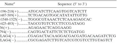
TABLE 1. Oligonucleotides used in this study
Namea Sequence (59to 39)
cr196-218(1) ...ATGCATCTTCAAGTGGTCATCTT cr487-509(1) ...TCTGACAGTGGCATATATTGTTG cr1548-1525(2) ...TGGGCGTAAACTCTCAAAGAGCAC cr425-403(2) ...TAGCGTGTCTCCTTCCGATAGG 136(1)...AGAGAACTCAGGAAGG 1794(2)...CAGTGACTATTCTCTCTGATGG
FLAG4(1) ...CGAGACTACAAGGACGACGATGACAAGATCTCG
FLAG4(2) ...CGCGAGATCTTGTCATCGTCGTCCTTGTAGTCT
aThe1or2in the name indicates the polarity.
on November 9, 2019 by guest
http://jvi.asm.org/
DOSPER as recommended by the manufacturer (Boehringer Mannheim, Inc.) in a final volume of 3 ml of OptiMEM (Gibco BRL, Inc.). After overnight incubation at 37°C under 5% CO2, 3 ml of EMEM–10% FBS was added to each
25-cm2flask.
Perro6D cells were transfected with constructs based in pDR2, a shuttle vector which contains the Epstein-Barr virus P1 origin of replication that allows the episomal maintenance of these plasmids in dog cells and that codes for a hygro-mycin resistance selectable marker. At 48 h posttransfection, the medium was changed to EMEM–10% FBS containing 250mg of hygromycin (Boehringer Manheim, Inc.) per ml. After 7 days of treatment with hygromycin, approxi-mately 20 to 30% of the transfected cells survived, whereas none of the mock-transfected cells resisted the antibiotic selection. Hygromycin-resistant Perro6D cells transfected with pDR2HAVcrFlag, which codes for a FLAG-tagged havcr-1, and pDR2HAVcrD12, which codes for a FLAG-tagged havcr-1 with the Cys-rich region deleted, were described previously (19) and were named flag and d12
cells, respectively. Hygromycin-resistant Perro6D cells transfected with vector pDR2 alone were named DR2 cells. Hygromycin-resistant Perro6D cells trans-fected with pDR2huHAVcr and pDR2huHAVcrFlagBstBI, constructs which are described in this paper, were named huhavcr-1 and huflagBst cells, respectively. The expression of huhavcr-1 and huflagBst in the dog cell transfectants grown in EMEM–10%FBS–250mg of hygromycin per ml was stable for 2 weeks but decreased thereafter to undetectable levels as judged by Western blot analysis using an anti-GST2 Ab.
GL37 cells were transfected with constructs based in pHook3, a shuttle vector which contains the simian virus 40 origin of replication and which codes for a zeomycin resistance selectable marker. At 48 h posttransfection, the transfection medium was changed to EMEM–10% FBS containing 500mg of zeomycin (In-vitrogen, Inc.) per ml. Two zeomycin-resistant cell clones, GL37huflagBst 2 and GL37huflagBst 3, which expressed the FLAG epitope as determined by a cell surface enzyme-linked immunosorbent assay (ELISA) and Western blot analysis using MAb M2 (data not shown), were isolated.
Cell surface ELISA.Expression of HAVcr-1 in GL37 cells and dog cell trans-fectants was analyzed by a cell surface ELISA as described previously (7). Briefly, duplicate wells of unfixed cells grown in 96-well plates were treated with twofold dilutions of 190/4 or M2 MAbs for 1 h at room temperature, washed extensively, and treated with a 1:1,000 dilution of affinity-purified peroxidase-labeled anti-mouse Ab for 1 h at room temperature. After the cells were washed extensively, 100ml of the one-component tetramethyl-benzidine (TMB) substrate (Kirke-gaard and Perry Laboratories, Inc.) was added per well. The reaction was stopped with 1% H2SO4, and absorbance was read at 450 nm. The difference in
the optical densities at 450 nm (OD450) of the duplicate wells were within the
experimental error of 5 to 10%, the average values were highly reproducible, and backgrounds were below 0.1 OD450units. The mean OD450of duplicate wells
was plotted versus the log10of the antibody dilution.
Western blot analysis.Confluent monolayers of dog cell transfectants grown in 25-cm2flasks were scraped into 1 ml of phosphate-buffered saline (PBS),
pel-leted, resuspended in 0.2 ml of reticulocyte standard buffer (RSB; 10 mM NaCl, 10 mM Tris-HCl [pH 7.2]) containing 1% Nonidet P-40 (NP-40), and incubated for 2 min at room temperature. After the nuclei were removed by centrifugation at 12,0003g, the total amount of protein in the supernatant was determined by
the Bradford method using the Bio-Rad protein assay kit. The cytoplasmic extracts were used immediately or stored at270°C. Approximately one-third of the cytoplasmic extract obtained from a 25-cm2cell monolayer (20 to 25mg of
total protein) was loaded per well and fractionated in SDS–10% polyacrylamide gels. Proteins were transferred to polyvinylidene difluoride membranes (Immo-bilon-P; Millipore, Inc.), probed with a 1:1,000 dilution of rabbit anti-GST2 Ab, and stained with a 1:5,000 dilution of alkaline phosphatase-labeled goat anti-rabbit Ab. The substrate 5-bromo-4-chloro-3-indolylphosphate–nitroblue tetra-zolium (BCIP-NBT substrate) was used as recommended by the manufacturer (Kirkegaard & Perry Laboratories).
HAV binding assay.Binding of HAV to the dog cell transfectants was quan-titated by radioimmunoassay as reported previously (7) but with minor modifi-cations. HAV was purified from 20 15-cm2dishes containing confluent
mono-layers of AGMK cells infected at a MOI of 1 TCID50of HAV per cell for 1 week
at 35°C in a CO2incubator. Cytoplasmic extracts were prepared as described
above in 5 ml of RSB–1% NP-40 and treated with 1% SDS and 1% Sarkosyl overnight at room temperature. HAV present in the cytoplasmic extracts was pelleted through a 4-ml-thick cushion of 40% sucrose–NTE (150 mM NaCl, 10 mM Tris-HCl [pH 7.4], 1 mM EDTA [pH 8.0]) by centrifugation at 40,000 rpm for 4 h at 16°C in a Beckman SW40 rotor. The pelleted virus was resuspended in 2 ml of NTE, aliquoted, and stored at270°C. Purified HAV had a titer of approximately 1010TCID
50/ml, as assessed in 96-well plates containing confluent
monolayers of FRhK-4 cells (4). The purified HAV was bound to 80%-confluent monolayers of dog cell transfectants grown in 96-well plates. Duplicate wells were treated with 50ml of 1:10, 1:20, and 1:40 dilutions of purified HAV in EMEM–10% FBS for 1 h at 35°C in a CO2incubator. Monolayers were washed
four times with EMEM–10% FBS, fixed with 80% methanol, blocked with 5% bovine serum albumin in PBS, incubated with 50ml of 125I-labeled human
anti-HAV Ab, washed four times with PBS, and exposed to X-ray film (XAR-2; Kodak) with an intensifying screen for 24 to 96 h. After exposure, the 96-well plates were stained with 1% crystal violet and absorbance at 595 nm was deter-mined with an ELISA plate reader (Bio-Rad Laboratories); this assay indicated
that similar numbers of cells, within a 5 to 10% range, were present in each well. Densitometric analysis of the autoradiography was performed on a Macintosh Quadra950 computer by using the public-domain NIH Image program (written by Wayne Rasband at the National Institutes of Health).
HAV infectivity assay.Dog cell transfectants and GL37 cells grown in eight-well Permanox culture slides (Nunc, Inc.) were infected with 107to 108TCID
50
of HAV purified as described above or were mock infected for 6 h at 35°C under 5% CO2. After being washed three times with EMEM–10% FBS, cells were
incubated for 3 days at 35°C under 5% CO2. Cell monolayers were fixed with cold
acetone and analyzed by indirect IF as described above.
Nucleotide sequence accession number.GenBank accession no. AF043724 was obtained for the huHAVcr-1 cDNA.
RESULTS
Molecular cloning of the human homolog of HAVcr-1.
Pre-liminary results indicated that the 190/4 epitope was not
ex-pressed in HeLa cells (7); therefore, it was of interest to
deter-mine whether human cells coded for an HAVcr-1 homolog(s).
To do so, genomic DNA was extracted from clone GL37
AGMK cells, from Ltk
2cells transfected with HAVcr-1 cDNA
(Lcr5 cells) or pDR2 vector (LDR2 cells), from human male
and female peripheral blood leukocytes, and from mouse cells.
Southern blot analysis of PstI-digested genomic DNA
hybrid-ized with a
32P-labeled full-length HAVcr-1 cDNA probe (Fig.
1) revealed the presence of several HAVcr-1-specific bands in
AGMK (lane 1), Lcr5 (lane 2), and human (lanes 4 and 5) cells
but not in vector-transfected LDR2 cells (lane 3) and
untrans-fected mouse cells (lane 6). To assess whether these HAVcr-1
homolog sequences were expressed in human cells, we
per-formed an reverse transcription-PCR analysis of mRNA
ex-tracted from human kidney and liver. Using synthetic
oligonu-cleotides cr196-218(
1
) and cr1548-1525(
2
) corresponding to
the N- and C-terminal sequences of havcr-1, respectively, we
amplified cDNA fragments of 1.1 kb from human kidney and
liver cDNA. Nucleotide sequence analysis revealed that the
1.1-kb PCR cDNA fragments amplified from human kidney
and liver cDNA were identical and contained the complete
coding sequence of a human homolog of HAVcr-1 that we
FIG. 1. Southern blot analysis of the huhavcr-1 gene. Genomic DNA extracted from simian AGMK cells, mouse Ltk2cells transfected with pDR2GL37/5 (Lcr5
cells) or pDR2 (LDR2 cells), human male (M) and female (F) peripheral blood leukocytes, and mouse cells was digested with PstI and examined by Southern blot analysis using a full-length32P-labeled HAVcr-1 cDNA probe. The positions
of size markers in kilobase pairs (kb) are illustrated.
on November 9, 2019 by guest
http://jvi.asm.org/
termed huHAVcr-1 (Fig. 2). To verify that the initiation and
termination codons of the amplified 1.1-kb huHAVcr-1 cDNA
were not artificially introduced by the synthetic
oligonucleo-tides used in the PCR, we amplified cDNA fragments
corre-sponding to the 5
9
and 3
9
ends of the huHAVcr-1 mRNA with
synthetic oligonucleotides 136(
1
) and cr425-403(
2
) or
cr487-509(
1
) and 1794(
2
), respectively. Nucleotide sequence
anal-ysis of the 5
9
-end PCR product showed that the ATG initiation
codon was present in the huHAVcr-1 mRNA but that synthetic
oligonucleotide cr196-218(
1
) introduced a C-to-T change in
the huHAVcr-1 cDNA which resulted in an P-to-L amino acid
change at position 3 in the signal sequence. Nucleotide
se-quence analysis of the 3
9
-end PCR product showed that PCR
primer cr1548-1525(
2
) did not introduce any change into the
huHAVcr-1 cDNA. Nucleotide sequence analysis (Fig. 2)
re-vealed that the huHAVcr-1 cDNA encodes a polypeptide of
359 amino acids, termed huhavcr-1, which is 79.11% identical
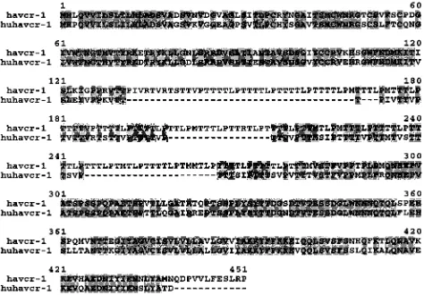
to havcr-1 (Fig. 3). Like havcr-1, huhavcr-1 has the typical
features of a type I integral-membrane glycoprotein, with two
distinctive hydrophobic regions: a putative 17-amino-acid
sig-nal sequence with a hydrophobic core following the initiating
methionine and a putative transmembrane domain of 22
resi-dues between amino acids 290 and 311, which is the major
hydrophobic region of the protein. A conserved cysteine
resi-due (Cys
296) found within the transmembrane domain of
hu-havcr-1 is possibly used for the addition of fatty acids that may
stabilize the receptor attachment to the membrane (10).
Be-tween the signal sequence and the transmembrane domain,
there is a predicted extracellular domain of 272 residues
com-prising two distinctive regions: a Cys-rich N-terminal region of
109 residues and a C-terminal segment of 163 residues
con-taining a TSP-rich region. The six Cys residues and the length
of the Cys-rich region of havcr-1 are conserved in huhavcr-1.
The first but not the second N-glycosylation site of the
[image:4.612.148.458.77.327.2]havcr-1 Cys-rich region is conserved in huhavcr-havcr-1. Only havcr-13 of the 27
consecutive repeats of the consensus sequence PTTTTL of the
TSP-rich region of havcr-1 remained in huhavcr-1 (Fig. 3). The
two N-glycosylation sites of the TSP-rich region of havcr-1 are
conserved in huhavcr-1, which also contains a third putative
N-glycosylation site at amino acid residues 258 to 260. The
huhavcr-1 intracellular domain contains 48 amino acids and is
12 residues shorter than the intracellular domain of havcr-1.
Consequently, the havcr-1 and huhavcr-1 putative structures
FIG. 2. cDNA sequence and predicted amino acid composition of huhavcr-1. The nucleotide sequence of the huHAVcr-1 cDNA was determined on both strands by automatic sequencing using specific oligonucleotides. The white box indicates the putative signal sequence. The six cysteines of the Cys-rich region are marked with solid lines. Potential N-linked glycosylation sites are marked with dashed lines. The arrowhead indicates the putative beginning of the TSP-rich region. The putative transmembrane domain is boxed in grey. The termination codon is indicated by asterisks.
FIG. 3. Alignment of havcr-1 and huhavcr-1. Alignment of amino acid se-quences predicted from the HAVcr-1 and huHAVcr-1 cDNAs was done with the Clustal W program. Gaps introduced in the sequences for the alignment are indicated by dashes; numbers of residues starting with the respective initiating methionine codons are indicated. Amino acids identical to those of the havcr-1 sequence are in grey background.
on November 9, 2019 by guest
http://jvi.asm.org/
[image:4.612.310.547.509.674.2]are very similar, resembling a lollipop-on-a-stick model (6) in
which the Cys-rich region of havcr-1 will most likely be
ex-tended further above the cell surface than that of huhavcr-1.
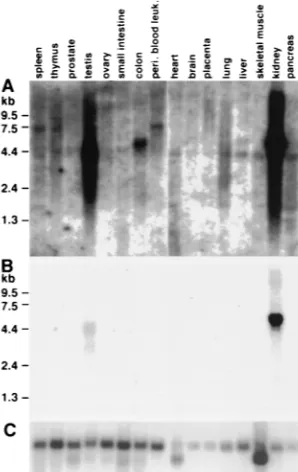
Expression of huhavcr-1 in different human tissues.
To study
the pattern of expression of huHAVcr-1 in different human
tissues, MTN blots were hybridized to a
32P-labeled full-length
huHAVcr-1 cDNA probe (Fig. 4). After 3 weeks of exposure,
the autoradiogram of the MTN blots (Fig. 4A) showed that
huHAVcr-1 was expressed in every organ analyzed and
ex-pressed at higher levels in the kidney and testis than in other
organs. An huHAVcr-1-specific 4.4-kb band was present for
almost every organ, a 5.5-kb band was observed for the colon
and liver, and a 7.5-kb band was observed for the spleen and
thymus and for peripheral blood leukocytes. Several bands of
lower intensity and smaller than 4.4 kb were also observed for
the spleen, lung, skeletal muscle, and pancreas. A shorter
ex-posure of the MTN blots for 24 h (Fig. 4B) revealed that the
kidney expressed 5.5-kb huHAVcr-1 mRNA at higher levels
than those of the 3- and 4.4-kb huHAVcr-1-specific mRNAs
expressed in the testis. Since the huHAVcr-1 1.1-kb cDNA
fragments amplified from the kidney and liver contain the
whole coding sequence of huhavcr-1, it is possible that the
huHAVcr-1 mRNA species detected in the MTN blots code
for long 3
9
and/or 5
9
nontranslated regions. However, we
can-not rule out the possibility that additional coding sequences
are contained in these huHAVcr-1 mRNAs. To control for
loading and integrity of the mRNA, MTN blots were
rehybrid-ized to a
32P-labeled
b
-actin probe (Fig. 4C), which showed
that similar levels of undegraded mRNA were loaded into each
lane except for lanes for the pancreas, placenta, and brain,
which contained three- to fourfold less mRNA.
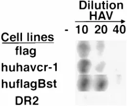
Characterization of the receptor encoded by the huHAVcr-1
cDNA.
To characterize huhavcr-1, we transfected Perro6D cells
with pDR2huHAVcr and selected hygromycin-resistant cells
that expressed huhavcr-1. Because there are no available Abs
capable of detecting the expression of huhavcr-1 at the cell
surface, we introduced a tag epitope into the mucin-like region
of huhavcr-1 to monitor its expression at the cell surfaces of the
dog cell transfectants. The resulting construct, termed huflagBst,
contains the FLAG octapeptide DTKDDDDK inserted
be-tween amino acid residues 145 and 146 of the TSP-rich region
of huhavcr-1. Dog cells transfected with pDR2HAVcrFlag
(flag cells), which express a FLAG-tagged havcr-1, or vector
pDR2 (DR2 cells) were previously characterized (19) and were
used as controls for our studies.
Western blot analysis showed that the anti-GST2 Ab (Fig. 5)
reacted with a 64-kDa protein in flag cells (lane 1),
correspond-ing to the major form of the FLAG-tagged havcr-1 (19), and
cross-reacted with a 54-kDa protein in huflagBst cells (lane 2)
and a 53-kDa protein in huhavcr-1 cells (lane 3). The
anti-GST2 Ab did not recognize havcr-1-specific bands in DR2 cells
(lane 4), which indicated that the 64-, 54-, and 53-kDa proteins
corresponded to HAV cellular receptors. It should be pointed
out that the 54-kDa huflagBst band migrated with the expected
molecular weight of a FLAG-tagged huhavcr-1.
To determine whether huhavcr-1 was presented on the cell
surfaces of the dog cell transfectants, we performed a cell
surface ELISA of huhavcr-1, huflagBst, flag, and DR2 cells
with the anti-FLAG M2 MAb and anti-havcr-1 190/4 MAb
(Fig. 6). As expected, flag cells reacted with both MAbs and
DR2 cells reacted with neither MAb, which confirmed that the
cell surface ELISA specifically recognized the M2 and 190/4
epitopes. MAb M2 did not react with huhavcr-1 cells but
re-acted with huflagBst cells (Fig. 6A), which indicated that the
FLAG-tagged huhavcr-1 was expressed at the cell surfaces of
the dog cell transfectants and suggested the same for untagged
huhavcr-1. Unfortunately, we could not confirm this with the
anti-GST2 Ab because this Ab recognized neither havcr-1 nor
FIG. 4. Expression of huhavcr-1 in human tissues. MTN blots containing poly(A) RNA from different human tissues were probed with32P-labeled
full-length huHAVcr-1 cDNA at high stringency and autoradiographed for 3 weeks (A) or 1 day (B). (C) The same blots were stripped and rehybridized with a
32P-labeledb-actin probe. The positions of size markers in kilobases are
[image:5.612.91.240.70.306.2]illus-trated. peri. blood leuk., peripheral blood leukocytes.
FIG. 5. Western blot analysis of the expression of huhavcr-1 in dog cell transfectants. Cytoplasmic extracts of dog cell transfectants expressing FLAG-tagged havcr-1 (flag), FLAG-FLAG-tagged huhavcr-1 (huflagBst), and unFLAG-tagged huhavcr-1 (huhavcr-1) and dog cells transfected with vector pDR2 alone (DR2) were prepared in RSB–1% NP-40, resolved by SDS–10% polyacrylamide gel electrophoresis, transferred to polyvinylidene difluoride membranes, and probed with rabbit anti-GST2 Ab directed against the TSP-rich region of huhavcr-1. The positions of prestained molecular weight markers and their sizes in kilodaltons are shown on the left.
on November 9, 2019 by guest
http://jvi.asm.org/
huhavcr-1 when they were presented at the cell surface. MAb
190/4 did not react with the huhavcr-1 and huflagBst cells (Fig.
6B), which confirmed that huhavcr-1 does not code for the
190/4 epitope.
Although similar levels of flag and huflagBst were detected
by Western blot analysis of the dog cell transfectants (Fig. 5,
compare lanes 1 and 2), the cell surface ELISA showed that
huflagBst cells expressed higher levels of the M2 epitope than
flag cells (Fig. 6A). Therefore, it is possible that the human
homolog of havcr-1 is expressed at a higher level at the cell
surface than the simian havcr-1 receptor. However, since the
FLAG tag was inserted at different positions in the flag and
huflagBst receptors, we cannot rule out the possibility that the
difference in the level of reaction of the dog cell transfectants
with MAb M2 was due to steric hindrance.
Binding of HAV to dog cell transfectants.
To determine
wheth-er HAV binds specifically to huhavcr-1, 96-well plates
contain-ing subconfluent monolayers of flag, huhavcr-1, huflagBst, and
DR2 cells were infected with 1:10, 1:20, and 1:40 dilutions of
purified HAV or were mock infected for 1 h at 35°C. After
being washed extensively, monolayers were fixed, and bound
HAV was detected with
125I-labeled human HAV
anti-body. This assay showed a concentration-dependent binding of
HAV to flag, huhavcr-1, and huflagBst cells and a low
back-ground level of binding of HAV to DR2 cells (Fig. 7). No
signal was observed for mock-infected cells, which indicated
that the
125I-labeled Ab reacted specifically against HAV
bound to the cells. These results indicated that huhavcr-1 is a
binding receptor for HAV and that the insertion of the FLAG
tag into its TSP-rich region did not affect the binding of HAV.
Susceptibility of dog cells expressing huHAVcr-1 to HAV
infection.
Receptor-negative but otherwise fully
HAV-suscep-tible cell lines have not been identified yet. We previously
showed that dog cells do not support HAV growth upon
inoc-ulation or transfection of infectious HAV RNA, which
indi-cated that dog cells contain an internal block(s) to HAV
rep-lication (4). However, we have recently shown that HAV
inoculation of dog cell transfectants expressing havcr-1
re-sulted in the characteristic granular cytoplasmic fluorescence
of HAV-infected cells, which lasted approximately 1 week (19),
indicating that these dog cell transfectants gained a limited
level of susceptibility to HAV infection. Therefore, we used
this dog cell system to analyze the HAV receptor function of
huhavcr-1. To do so, we infected monolayers of dog cell
trans-fectants with a MOI of 100 to 1,000 TCID50
of HAV per cell
and analyzed the presence of HAV antigen by indirect IF
staining with human anti-HAV Ab (Fig. 8). At 3 days
postin-fection, HAV-specific IF was detected in control GL37 (Fig.
8A) and flag (Fig. 8C) cells but not in DR2 cells (Fig. 8B) and
d1
2
cells (which contain FLAG-tagged havcr-1 constructs with
the Cys-rich regions deleted; Fig. 8D) (19). HAV-specific IF
was also detected in huhavcr-1 and huflagBst cells (Fig. 8E and
F), which indicated that huhavcr-1 functions as an HAV
re-ceptor that mediates a limited level of susceptibility to HAV
infection, similar to what was previously reported for
havcr-1 (7, havcr-19). It should be pointed out that in huhavcr-havcr-1 and
huflagBst cells we detected a low level of HAV-specific IF at 0
days postinfection; this level increased approximately twofold
at 3 days postinfection, as judged by microscopic examination
(data not shown). Mock-infected GL37 cells and dog cell
trans-fectants did not fluoresce (data not shown), which showed that
the human HAV Ab specifically detected the HAV
anti-gen present in the infected cells.
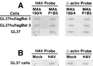
MAb 190/4 protects GL37 cells but not GL37 transfectants
expressing huhavcr-1 against HAV infection.
To further
ana-lyze the function of huhavcr-1 as a receptor for HAV, we used
protective MAb 190/4 to block the endogenous havcr-1
ex-FIG. 7. Binding of HAV to dog cells transfected with huHAVcr-1 cDNA. Dog cell transfectants expressing FLAG-tagged huhavcr-1 (flag), FLAG-tagged huhavcr-1 (huflagBst), and untagged huhavcr-1 (huhavcr-1) and dog cells trans-fected with vector pDR2 (DR2) were grown in 96-well plates and intrans-fected with 1:10, 1:20, and 1:40 dilutions of purified HAV or were mock infected (2) for 1 h at 35°C. After being extensive washed, monolayers were fixed, and HAV bound to the cells was detected with125I-labeled human anti-HAV Ab.
[image:6.612.135.462.69.222.2]Autoradiogra-phy of the 96-well plate showing a single well for each treatment is presented. FIG. 6. Expression of huhavcr-1 at the cell surfaces of dog cell transfectants. Expression of the M2 (A) and 190/4 (B) epitopes at the cell surfaces of dog cell transfectants expressing FLAG-tagged huhavcr-1 (flag cells; solid circles), FLAG-tagged huhavcr-1 (huflagBst cells; solid squares); and untagged huhavcr-1 (huhavcr-1 cells; open squares) and dog cells transfected with vector pDR2 (DR2 cells; solid triangles) was determined by ELISA using twofold dilutions of the MAbs. Absorbance at 450 nm was plotted versus the log10of the MAb dilution. Data are means of duplicate wells; duplicate values varied by less than 10%. The results correspond to one experiment which was repeated two times with approximately 5 to 10% experimental error.
on November 9, 2019 by guest
http://jvi.asm.org/
[image:6.612.361.491.549.658.2]pressed at the cell surfaces of GL37 cell transfectants. Since
huhavcr-1 does not contain the 190/4 epitope (Fig. 6), we
reasoned that MAb 190/4 will not protect the GL37 cell
trans-fectants expressing huhavcr-1 against HAV infection. To test
this hypothesis, we treated GL37hflagBst 2 and GL37huflagBst
3 cells, two zeomycin-resistant GL37 cell clones that expressed
a FLAG-tagged huhavcr-1 construct at the cell surface, and
GL37 cells with control MAb P1B5 or MAb 190/4 for 1 h and
then inoculated these cells at a MOI of 0.1 TCID50
of HAV
per cell. Three days after infection, the protective effect of the
MAb treatment was determined by slot blot analysis of total
cellular RNA probed with
32P-labeled HAV cDNA (Fig. 9A).
Treatment of GL37 cells with MAb 190/4 resulted in a lower
level of HAV-specific signal than treatment with MAb P1B5,
which indicated that MAb 190/4 protected GL37 cells against
HAV infection (7). Similar levels of HAV-specific signal were
observed in GL37huflagBst 2 and GL37huflagBst 3 cells
treated with MAbs 190/4 or P1B5, which indicated that MAb
190/4 did not protect these cell lines against HAV infection.
Untreated GL37 cells were infected with HAV or were mock
infected in parallel under the same conditions mentioned
above. Samples of total cellular RNA extracted from these
control cells were included in the slot blot analysis (Fig. 9B),
which showed that only HAV-inoculated cells reacted with the
HAV probe. To control for RNA loading, the same blot was
stripped and rehybridized with a
32P-labeled
b
-actin cDNA
FIG. 8. Detection of HAV antigen in the cytoplasm of dog cell transfectants by indirect IF analysis. Monolayers of GL37 (A), DR2 (B), flag (C), d12(D), huhavcr-1 (E), and huflagBst (F) cells grown in eight-well slides were infected with a MOI of huhavcr-100 to huhavcr-1,000 TCID50of purified HAV per cell for 6 h, washed extensively, and
incubated at 35°C for 3 days. Cells were fixed with cold acetone and stained with human anti-HAV Ab and FITC-labeled goat anti-human IgG and IgM Abs. Mock-infected cells did not immunofluoresce (data not shown).
FIG. 9. Slot blot analysis of MAb 190/4-mediated protection of GL37 cell transfectants against HAV infection. (A) Slot blot analysis of protection of GL37 cells and GL37 cell transfectants expressing FLAG-tagged huhavcr-1 (GL37hu flagBst 2 and GL37hflaBst 3 cells) against HAV infection. Confluent monolayers of GL37 cells in six-well plates were treated with MAb 190/4 or anti-humana3 integrin MAb P1B5 for 1 h at 35°C, washed, and inoculated at a MOI of 0.1 TCID50of HAV per cell for 1 h at 35°C. Monolayers were washed and incubated
at 35°C, cytoplasmic extracts were prepared at 3 days postinfection, and total RNA was extracted. HAV-specific RNA was detected with a32P-labeled HAV
cDNA probe. To control for RNA loading, the slot blot was stripped and re-hybridized with a32P-labeledb-actin cDNA probe. (B) Control GL37 cells were
infected with HAV or were mock infected (Mock) without prior MAb treatment. RNA samples were prepared in parallel with the samples shown in panel A and hybridized under the same conditions. The autoradiogram was exposed for 2.5 h with intensifying screens.
on November 9, 2019 by guest
http://jvi.asm.org/
[image:7.612.353.504.492.600.2]probe, which showed that similar levels of total cellular RNA
were loaded in each slot (Fig. 9A and B). Considered together,
these results further suggested that huhavcr-1 is a functional
receptor for HAV.
DISCUSSION
Our initial studies on the identification of cellular receptors
for HAV showed that the protective epitope 190/4 was not
expressed in HeLa cells (7) and raised the possibility that the
HAVcr-1 gene itself was not conserved in humans. In this work,
we identified a human homolog of havcr-1 that has approximately
79% homology with its simian counterpart and showed that it did
not express the 190/4 epitope. This antigenic variability among
primates of protective epitope 190/4 contrasts to the high degree
of conservation of protective epitope D171 of the poliovirus
re-ceptor, which has been found in all the cell lines of primate
origin tested (13, 16). Moreover, we recently showed that
an-tigenic variants of havcr-1 expressed in BS-C-1 and CV-1 cells,
two widely used AGMK cell lines, contain a K108Q change in
the Cys-rich region that is responsible for their lack of reaction
with MAb 190/4 (5). Taken all together, these data suggested
that the HAV–havcr-1 interaction tolerates some degree of
variability in protective epitope 190/4. Mutagenesis of havcr-1
and further mapping of the 190/4 epitope will allow us to
understand how HAV interacts with its cellular receptor.
The receptor function of huhavcr-1 was analyzed by a
bind-ing assay which showed that dog cell transfectants expressbind-ing
huhavcr-1 bound HAV (Fig. 7). Further analysis of the
tor function of huhavcr-1 is complicated by the lack of
recep-tor-negative fully HAV-susceptible cell lines (4, 7, 19). To
overcome this limitation, we performed two kinds of
experi-ments, which provided further evidence that huhavcr-1 is a
functional receptor for HAV. First, we showed that dog cell
transfectants expressing huhavcr-1 developed the
characteris-tic cytoplasmic granular fluorescence of HAV-infected cells,
whereas dog cell transfectants expressing an havcr-1 construct
with the Cys-rich region deleted and vector-transfected dog
cells did not fluoresce. This experiment suggested that dog cell
transfectants expressing huhavcr-1 gained limited susceptibility
to HAV infection (Fig. 8). Second, MAb 190/4 did not protect
GL37 cell transfectants expressing huhavcr-1 against HAV
in-fection (Fig. 9), which suggested that HAV entered the cells
via huhavcr-1 and not the endogenous havcr-1 blocked by MAb
190/4. Although these two lines of evidence strongly suggested
that huhavcr-1 is a functional receptor for HAV, further
con-firmation of its functionality will have to wait for the isolation
of a receptor-negative fully HAV-susceptible cell lines.
The pathogenesis of HAV is poorly understood, and
extra-hepatic sites of HAV replication are not well defined. HAV is
transmitted through the fecal/oral route and, after ingestion, it
probably travels from the gut to the liver where it replicates
before being excreted with bile to the intestine. Detection of
HAV in saliva and in the throat of an experimentally infected
chimpanzee suggested that the initial viral replication might
occur in the oropharynx and salivary glands (2). Replication of
HAV in the gastrointestinal tract has been difficult to
deter-mine (9, 11, 12), but HAV antigen was found in the intestinal
mucosa of two marmosets inoculated intravenously with HAV
(8). HAV antigen was also detected in the kidneys, spleens,
and lymph nodes of experimentally infected primates (2, 8, 11).
Recently, HAV has been detected by IF analysis in the
epithe-lial cells of the intestinal crypts and in cells of the lamina
propria of the small intestines of orally inoculated owl
mon-keys (1). The same study showed that HAV antigen, besides
being detected in the liver, was also detected in the kidneys and
spleen but not in pharyngeal tissues of the owl monkey. These
data on the detection of HAV antigen in different organs of
nonhuman primates correlated well with our data on the
ubiq-uitous expression of huHAVcr-1-specific messages in humans.
ACKNOWLEDGMENTS
We thank Stephen Feinstone for encouragement and helpful advice
and Barry Falgout and Robin Levis for comments on the manuscript.
We also thank Michael Klutch for automatic sequencing of DNA
samples.
This research was supported in part by the appointment of D.F. to the
Postgraduate Research Participation Program at the Center for Biologics
Evaluation and Research administered by the Oak Ridge Institute for
Science and Education through an interagency agreement between the
U.S. Department of Energy and the U.S. Food and Drug Administration.
REFERENCES
1. Asher, L. V., L. N. Binn, T. L. Mensing, R. H. Marchwicki, R. A. Vassell, and
G. D. Young. 1995. Pathogenesis of hepatitis A in orally inoculated owl monkeys (Aotus trivirgatus). J. Med. Virol. 47:260–268.
2. Cohen, J. I., S. Feinstone, and R. H. Purcell. 1989. Hepatitis A virus infection in a chimpanzee: duration of viremia and detection of virus in saliva and throat swabs. J. Infect. Dis. 160:887–890.
3. Cohen, J. I., J. R. Ticehurst, S. M. Feinstone, B. Rosenblum, and R. H.
Purcell.1987. Hepatitis A virus cDNA and its RNA transcripts are infectious in cell culture. J. Virol. 61:3035–3039.
4. Dotzauer, A., S. M. Feinstone, and G. Kaplan. 1994. Susceptibility of non-primate cell lines to hepatitis A virus infection. J. Virol. 68:6064–6068. 5. Feigelstock, D., P. Thompson, P. Mattoo, and G. G. Kaplan. 1998.
Polymor-phisms of the hepatitis A virus cellular receptor 1 in African green monkey kidney cells result in antigenic variants that do not react with protective monoclonal antibody 190/4. J. Virol. 72:6218–6222.
6. Jentoft, N. 1990. Why are proteins O-glycosylated? Trends Biochem. Sci. 15: 291–294.
7. Kaplan, G., A. Totsuka, P. Thompson, T. Akatsuka, Y. Moritsugu, and S. M.
Feinstone.1996. Identification of a surface glycoprotein on African green mon-key kidney cells as a receptor for hepatitis A virus. EMBO J. 15:4282–4296. 8. Karayiannis, P., T. Jowett, M. Enticott, D. Moore, M. Pignatelli, F. Brenes,
P. J. Scheuer, and H. C. Thomas.1986. Hepatitis A virus replication in tamarins and host immune response in relation to pathogenesis of liver cell damage. J. Med. Virol. 18:261–276.
9. Krawczynski, K. K., D. W. Bradley, B. L. Murphy, J. W. Ebert, T. E.
Anderson, I. L. Doto, A. Nowoslawski, W. Duermeyer, and J. E. Maynard.
1981. Pathogenetic aspects of hepatitis A virus infection in enterally inocu-lated marmosets. Am. J. Clin. Pathol. 76:698–706.
10. Magee, A. I., L. Gutierrez, C. J. Marshall, and J. F. Hancock. 1989. Targeting of oncoproteins to membranes by fatty acylation. J. Cell Sci. Suppl. 11:149–160. 11. Mathiesen, L. R., J. Drucker, D. Lorenz, J. A. Wagner, R. J. Gerety, and
R. H. Purcell.1978. Localization of hepatitis A antigen in marmoset organs during acute infection with hepatitis A virus. J. Infect. Dis. 138:369–377. 12. Mathiesen, L. R., S. M. Feinstone, R. H. Purcell, and J. A. Wagner. 1977.
Detection of hepatitis A antigen by immunofluorescence. Infect. Immun. 18: 524–530.
13. Minor, P. D., P. A. Pipkin, D. Hockley, G. C. Schild, and J. W. Almond. 1984. Monoclonal antibodies which block cellular receptors of poliovirus. Virus Res. 1:203–212.
14. Murphy, A. J. M., A. L. Kung, R. A. Swirski, and R. T. Schimke. 1992. cDNA expression cloning in human cells using the plDR2 episomal vector system. Methods (Orlando) 4:111–131.
15. Neutra, M. R., and J. F. Forstner. 1987. Gastrointestinal mucus: synthesis, secretion, and function, p. 975–1009. In L. R. Johnson (ed.), Physiology of the gastrointestinal tract. Raven Press, New York, N.Y.
16. Nobis, P., R. Zibirre, G. Meyer, J. Ku¨hne, G. Warnecke, and G. Koch. 1985. Production of a monoclonal antibody against an epitope of HeLa cells that is the functional poliovirus biding site. J. Gen. Virol. 66:2563–2569. 17. Sambrook, J., E. F. Fritsch, and T. Maniatis. 1989. Molecular cloning: a
laboratory manual, 2nd ed. Cold Spring Harbor Laboratory, Cold Spring Harbor, N.Y.
18. Swirski, R. A., D. Van Den Berg, A. J. M. Murphy, C. M. Lambert, E. C.
Fried-berg, and R. T. Schimke.1992. Improvements in the Epstein-Barr-based shuttle vector system for direct cloning in human tissue culture cells. Methods (Orlan-do) 4:133–142.
19. Thompson, P., J. Lu, and G. G. Kaplan. 1998. The Cys-rich region of the hepatitis A virus cellular receptor 1 (HAVcr-1) is required for binding of hep-atitis A virus and protective monoclonal antibody 190/4. J. Virol. 72:3751–3761. 20. Totsuka, A., and Y. Moritsugu. 1994. Hepatitis A vaccine development in Japan, p. 509–513. In K. Nishioka, H. Suzuki, S. Mishiro, and T. Oda (ed.), Viral hepatitis and liver disease. Springer-Verlag, Tokyo, Japan.
on November 9, 2019 by guest
http://jvi.asm.org/