White Rose Research Online URL for this paper:
http://eprints.whiterose.ac.uk/1483/
Article:
Ejbjerg, B., McQueen, F., Lassere, M. et al. (10 more authors) (2005) The
EULAR-OMERACT rheumatoid arthritis MRI reference image atlas: the wrist joint. Annals
of the Rheumatic Diseases, 64 (Suppl ). i23-i47. ISSN 0003-4967
https://doi.org/10.1136/ard.2004.031823
Reuse
See Attached
Takedown
If you consider content in White Rose Research Online to be in breach of UK law, please notify us by
The EULAR–OMERACT rheumatoid arthritis MRI reference
image atlas: the wrist joint
B Ejbjerg, F McQueen, M Lassere, E Haavardsholm, P Conaghan, P O’Connor, P Bird, C Peterfy,
J Edmonds, M Szkudlarek, H Genant, P Emery, M Østergaard
. . . .
Ann Rheum Dis2005;64(Suppl I):i23–i47. doi: 10.1136/ard.2004.031823
This paper presents the wrist joint MR images of the
EULAR–OMERACT rheumatoid arthritis MRI reference
image atlas. Reference images for scoring synovitis, bone
oedema, and bone erosions according to the OMERACT
RA MRI scoring (RAMRIS) system are provided. All grades
(0–3) of synovitis are illustrated in each of the three wrist
joint areas defined in the scoring system—that is, the distal
radioulnar joint, the radiocarpal joint, and the
intercarpal-carpometacarpal joints. For reasons of feasibility,
examples of bone abnormalities are limited to five selected
bones: the radius, scaphoid, lunate, capitate, and a
metacarpal base. In these bones, grades 0–3 of bone
oedema are illustrated, and for bone erosion, grades 0–3
and examples of higher grades are presented. The
presented reference images can be used to guide scoring
of wrist joints according to the OMERACT RA MRI scoring
system.
. . . .
T
he wrist joints are very frequently involved in rheumatoid arthritis (RA), including early RA, and assessment of wrist joints is included in conventional radiological and clinical scoring systems.1–4Numerous studies using magnetic resonance imaging (MRI) in RA have examined the wrist joint, either alone or in combination with the metacarpophalangeal (MCP) joints. A predictive value of magnetic resonance imaging (MRI) findings (synovitis, bone oedema, and MRI bone erosions) in the wrist joint with respect to short term (one
The OMERACT 2002 RA MRI scoring system includes assessment of wrist joints.16
The aim of this section of the EULAR–OMERACT RA MRI reference image atlas is to provide wrist joint reference images for scoring according to the OMERACT RA MRI scoring (RAMRIS) system, described in more detail by Østergaard et al in this supplement.17
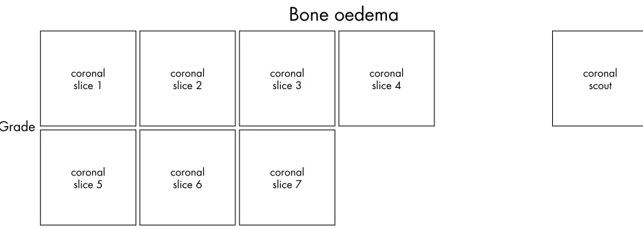
THE WRIST JOINT REFERENCE IMAGES
This atlas illustrates synovitis in the three regions of the wrist that are recommended for assess-ment when using the OMERACT scoring method—that is, the distal radioulnar joint, the radiocarpal joint and the intercarpal-carpometa-carpophalangeal joints. Furthermore, example images are provided for semiquantitative scor-ing of bone erosions and bone oedema in five selected bones of the wrist: the radius, scaphoid, lunate, capitate and a metacarpal base. Representative examples of each grade of synovitis and a selection of grades for bone abnormalities are presented. For reasons regard-ing feasibility not all bones and grades are included.
The examples for this atlas were selected by consensus in the OMERACT MRI in RA group. Details of the selection process and applied MRI sequences can be found in the paper by Birdet al in this supplement.18
A description of the reference image sheets presented on the following pages, and how to use them, is provided in figs 1–3 (see page 46–47).
We hope the presented reference images will be useful to guide scoring of wrist joints according to the OMERACT RA MRI scoring system.
ACKNOWLEDGEMENTS
Grade
0-low
–Gd
+Gd
0-high
–Gd
+Gd
1-low
–Gd
+Gd
1-high
–Gd
Grade
2-low
–Gd
+Gd
2-high
–Gd
+Gd
3-low
–Gd
+Gd
3-high
–Gd
Grade
0-low
–Gd
+Gd
0-high
–Gd
+Gd
1-low
–Gd
+Gd
1-high
–Gd
Grade
2-low
–Gd
+Gd
2-high
–Gd
+Gd
3-low
–Gd
+Gd
3-high
–Gd
Grade
0-low
–Gd
+Gd
0-high
–Gd
+Gd
1-low
–Gd
+Gd
1-high
–Gd
Grade
2-low
–Gd
+Gd
2-high
–Gd
+Gd
3-low
–Gd
+Gd
3-high
–Gd
Grade
0
1
2
Grade
1
0
2
3
Grade
1
0
2
Grade
1
0
2
3
Grade
1
0
2
3
Grade
1
0
2
3
Grade
5
7
9
Grade
1
0
2
3
Grade
5
9
Grade
1
0
2
3
Grade
4
6
Grade
1
0
2
3
Grade
5
8
Grade
1
0
2
3
Grade
9
5
Grade
no Gd axial slice 1 with Gd axial slice 1 –Gd +Gd no Gd axial slice 2 with Gd axial slice 2 no Gd axial slice 3Synovitis
with Gd axial slice 3 no Gd coronal with Gd coronalFigure 1 Synovitis reference image sheets (pages i24–i29, total 6). Reference image sheets for synovitis in the distal radioulnar joint, the radiocarpal joint, and the intercarpal-carpometacarpophalangeal joints are illustrated on two single-page sheets each. Examples are provided from the low end and high end of each grade (0–3). Synovitis is graded 0–3 (normal, mild, moderate, severe) as estimated by thirds of the presumed maximum volume of enhancing tissue as described in the OMERACT RAMRIS (see table 1, reference 11). The MRI set to be assessed should be compared with the axial precontrast and postcontrast T1 weighted reference images and the joint assigned the score of the best possible match. The first carpometacarpal joint should not be scored. All axial slices covering the joint should be taken into account. A total score (range 0–9) can be calculated. The diagram above describes the positions and types of images included.
[image:25.612.70.537.485.652.2]Authors’ affiliations
. . . .
B Ejbjerg,Departments of Rheumatology, Radiology and MRI, Copenhagen University Hospital at Hvidovre, Copenhagen, Denmark F McQueen,Department of Molecular Medicine and Pathology, Faculty of Medicine and Health Sciences, University of Auckland, Auckland, New Zealand
M Lassere,Department of Rheumatology, St George Hospital, University of NSW, Sydney, Australia
E A Haavardsholm,Department of Rheumatology, Diakonhjemmet Hospital, University of Oslo, Oslo, Norway
P Conaghan,Academic Unit of Musculoskeletal Disease, University of Leeds, Leeds, UK
P O’Connor,Department of Radiology, Leeds General Infirmary, Leeds, UK
P Bird,Department of Rheumatology, St George Hospital, University of NSW, Sydney, Australia
C Peterfy,Synarc Inc, San Francisco, CA, USA
J Edmonds,Department of Rheumatology, St George Hospital, University of NSW, Sydney, Australia
M Szkudlarek,Department of Rheumatology, Copenhagen University Hospital at Hvidovre, Copenhagen, Denmark
H Genant,Department of Radiology, University of California at San Francisco, San Francisco, CA, USA
P Emery,Academic Unit of Musculoskeletal Disease, University of Leeds, Leeds, UK
M Østergaard,Departments of Rheumatology, Copenhagen University Hospitals at Herlev and Hvidovre, Copenhagen, Denmark
REFERENCES
1 Fleming A, Benn RT, Corbett M, Wood PH. Early rheumatoid disease. II. Patterns of joint involvement.Ann Rheum Dis1976;35:361–4. 2 van der Heijde DMFM. Plain X-rays in rheumatoid arthritis: overview of
scoring methods, their reliability and applicability.Baillieres Clin Rheumatol
6 Østergaard M, Hansen M, Stoltenberg M, Gideon P, Klarlund M, Jensen KE,
et al.Magnetic resonance imaging-determined synovial membrane volume as a marker of disease activity and a predictor of progressive joint destruction in the wrists of patients with rheumatoid arthritis.Arthritis Rheum
1999;42:918–29.
7 Lindegaard H, Hørslev-Petersen K, Vallø J, Junker P, Østergaard M. Baseline MRI erosions in early rheumatoid arthritis MCP and wrist joint bones markedly increase the risk of radiographic erosions at 1 year follow-up.Arthritis Rheum
2002;46:S521.
8 Østergaard M, Hansen M, Stoltenberg M, Jensen KE, Szkudlarek M, Pedersen-Zbinden B,et al.New radiographic bone erosions in the wrists of patients with rheumatoid arthritis are detectable with magnetic resonance imaging a median of two years earlier.Arthritis Rheum2003;48:2128–31. 9 McQueen FM, Benton N, Perry D, Crabbe J, Robinson E, Yeoman S,et al.
Bone edema scored on magnetic resonance imaging scans of the dominant carpus at presentation predicts radiographic joint damage of the hands and feet six years later in patients with rheumatoid arthritis.Arthritis Rheum
2003;48:1814–27.
10 Ejbjerg B. Magnetic resonance imaging in rheumatoid arthritis. A study of aspects of joint selection, contrast agent use and type of MRI unit [PhD dissertation]. Copenhagen, University of Copenhagen, 2005 (in press). 11 Østergaard M, Klarlund M, Lassere M, Conaghan P, Peterfy C, McQueen F,
et al.Interreader agreement in the assessment of magnetic resonance images of rheumatoid arthritis wrist and finger joints—an international multicenter study.J Rheumatol2001;28:1143–50.
12 Conaghan P, Lassere M, Østergaard M, Peterfy C, McQueen F, O’Connor P,
et al.OMERACT rheumatoid arthritis magnetic resonance imaging studies. Exercise 4: an international multicenter longitudinal study using the RA-MRI Score.J Rheumatol2003;30:1376–9.
13 Lassere M, McQueen F, Østergaard M, Conaghan P, Shnier R, Peterfy C,
et al.OMERACT rheumatoid arthritis magnetic resonance imaging studies. Exercise 3: an international multicenter reliability study using the RA-MRI Score.J Rheumatol2003;30:1366–75.
14 Bird P, Lassere M, Shnier R, Edmonds J. Computerized measurement of magnetic resonance imaging erosion volumes in patients with rheumatoid arthritis: a comparison with existing magnetic resonance imaging scoring systems and standard clinical outcome measures.Arthritis Rheum
2003;48:614–24.
15 Haavardsholm EA, Kvan NP, Østergaard M, Ejbjerg B, Lillea˚s FG, Kvien TK. Reliability of the OMERACT Rheumatoid Arthritis MRI Score (RAMRIS) in a
[image:26.612.71.547.53.216.2]