lmmunoglobulln Signal Transduction Guides the
Specificity of B CelI-T Cell Interactions and
Is Blocked in Tolerant Self-reactive B Cells
By Michael P. Cooke,* Andrew W. Heath,~ Kevan M. Shokat,*
Yongjun Zeng,* Fred D. Finkelman,$ Peter S. Linsley, II
Maureen Howard,$ and Christopher C. Goodnow*
From the *Howard Hughes Medical Institute, and the Department of Microbiology and Immunology, Stanford University, Stanford, California 94305; tDNAX Research Institute, Pala Alto, California 94306; the SDepartment of Medicinc; Uniformed Services University of the Health Sciences, Bethesda, Maryland 20814; and IlBristol-Meyers Squib~ Pharmaceutical Research Institute, Seattle,
Washington 98121
S u m m a r y
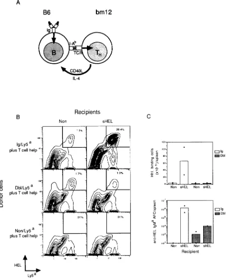
The specifidty of antibody (Ab) responses depends on focusing helper T (Th) lymphocyte signals to suitable B lymphocytes capable of binding foreign antigens (Ags), and away from nonspecific or self-reactive B cells. To investigate the molecular mechanisms that prevent the activation of self-reactive B lymphocytes, the activation requirements of B cells specific for the Ag hen egg lysozyme (HEL) obtained from immunoglobulin (Ig)-transgenic mice were compared with those of functionally tolerant B cells isolated from Ig-transgenic mice which also express soluble HEL. To eliminate the need for surface (s)Ig-mediated Ag uptake and presentation and allow the effects of slg signaling to be studied in isolation, we assessed the ability of allogeneic T cells from bm12 strain mice to provide in vivo help to C57BL/6 strain-transgenic B cells. Interestingly, non- tolerant Ig-transgenic B cells required both allogeneic Th cells and binding of soluble HEL for efl~dent activation and Ab production. By contrast, tolerant self-reactive B cells from Ig/HEL double transgenic mice responded poorly to the same combination of allogeneic T cells and soluble HEL. The tolerant B cells were nevertheless normally responsive to stimulation with interleukin 4 and anti-CD40 Abs in vitro, suggesting that they retained the capacity to respond to mediators of T cell help. However, the tolerant B cells exhibited a proximal block in the slg signaling pathway which prevented activation of receptor-assodated tyrosine kinases in response to the binding of soluble HEL. The functional significance of this slg signaling defect was confirmed by using a more potent membrane-bound form of HEL capable of triggering slg signaling in tolerant B cells, which markedly restored their ability to collaborate with allogeneic Th cells and produce Ab. These findings indicate that Ag-specific B cells require two signals for mounting a T cell-dependent Ab response and identify regulation of slg signaling as a mechanism for con- trolling self-reactive B cells.
T
he ability to distinguish between self and foreign Ags is a central feature of immune recognition, allowing im- munity to be acquired against foreign organisms while avoiding destructive autoimmunity. Studies of self-nonself discrimination in recent years (for reviews see references 1-3) have confirmed the clonal selection theory proposed by Bumet (4), which hypothesized that foreign Ags provoke immunity by triggering clonal expansion and differentiation of Ag- binding B and T lymphocytes, whereas self-Ags induce toler- ance by triggering elimination or inactivation of self-reactive cells. Although these advances provide a cellular framework for understanding self-nonselfdiscrimination, the molecular mechanisms that dictate the choice between lymphocyte ac-tivation or tolerance neverthdess remain to be defined. Un- derstanding the molecular basis for these cellular decisions will be important for controlling the immunogenicity or tolerogenicity of vaccines, tumors, and tissue transplants, and for understanding the breakdown of stir-tolerance in auto- immune diseases.
The decision between lymphocyte activation and tolerance seems particularly approachable in B lymphocytes, since much is already known about the cellular and molecular events causing B cells to proliferate and differentiate into Ab-secreting plasma cells in response to foreign Ags. For many foreign Ags, Th cells play a key role in promoting B cell prolifera- tion and Ab production, by adhering to suitable B cells and
transmitting a set of B cell growth and differentiation signals. One of the most important signals is an integral membrane protein, CD40-1igand (CD40L) 1, that is transiently dis- played on the surface of activated Th cells and triggers B cell proliferation through a receptor, CD40, constitutively ex- pressed on B cells (5, 6). Other molecules secreted by acti- vated Th cells, such as IL-4 and -10, synergize with CD40L in promoting B cell proliferation and in triggering isotype switching and differentiation into plasma cells (for a recent review see reference 7).
Since the receptors for CD40L, IL-4, and IL-IO are con- stitutively expressed on all B cells, development of an effec- tive Ab response depends on preferential delivery of these helper signals to useful B cells whose surface Ig (sIg) bind foreign Ag, and not to useless B calls that do not bind Ag or to potentially harmful B cells that bind sdf-Ags. The specificity of T cell help is directed, at least in part, by the requirement that Th cells recognize a complex composed of foreign Ag peptide fragments and class II MHC molecules, that is dis- played on the surface of potential B cell targets (8-12). Rec- ognition of peptide-MHC complexes by the TCR strengthens the interaction between T and B cells (13), triggers CD40L expression (6, 14), and polarizes IL secretion to the point of synapsis between T and B cells (15, 16). Since B cells that bind foreign Ags are more efficient at internalizi,ng these Ags than other nonbinding B cells, Ag-specific B cells are most likely to present foreign peptide fragments and trigger delivery of T cell help (for a review see reference 12). Other receptor/ ligand pairs also contribute to the synapsis of T and B cells, such as the binding of LFA-1 on both T and B cell to inter- cellular adhesion molecule 1 (ICAM-1) on the opposing cell (13, 17, 18) or the binding of CD28 on the T cell to B7 on the B cell (19), but their role in guiding the specificity of T-B cell interaction is not known.
The molecular mechanisms responsible for B cell tolerance are not well understood, especially in situations where self- reactive B cells become functionally inactivated (anergic) but are not physically eliminated. In this paper, we have explored the basis for tolerance in anergic self-reactive B cells by using a model developed previously (20) in which Ig gene trans- genic mice, whose B lymphocytes express surface IgM and IgD specific for the Ag hen-egg lysozyme (HEL), were mated with transgenic mice expressing the soluble form of HEL. In the resulting soluble lysozyme/anti-lysozyme double (Dbl)- transgenic mice, self-reactive anti-lysozyme B cells developed normally and populated the peripheral lymphoid organs but were functionally tolerant to lysozyme. Since the tolerant B cells were present in normal numbers and expressed all of the developmentally regulated cell surface molecules thought to contribute to B cell activation, the explanation for toler- ance was obscure. Here, we describe that tolerant B cells from these mice remain responsive to T cell-derived signals, but
1Abbr~tions used in this pat~': CD4OL, CD40-ligand; Dbl, double; HEL. hen-egg lysozyme; ICAM-I, intercellular adhesion molecule 1; slg, sur- face Ig; SA-PE, streptavidin-PE.
exhibit a proximal block in B cell slg signaling after Ag binding that precludes effective collaboration with T cells. Moreover, the sIg signaling block can be partially overcome by very ex- tensive receptor cross-linking, and this markedly restored col- laboration with Th cells. These findings demonstrate a piv- otal role for sIg signaling in guiding T cell-dependent Ab responses and that regulation of sIg signaling provides a mech- anism for controlling self-reactive B cells.
Materials and Methods
Mice. Anti-HEL/sHEL Dbl-transgenic animals were obtained by mating transgenic animals from the MD4 anti-HEL IgM + IgD line and the ML5 soluble HEL line, which were produced and maintained on a C57BL/6J (B6) background as described (20). To mark the Ig- or Dbl-transgenic cells with Ly5 ~, B6 MD4 x ML5 Dbl-transgenic mice were mated with C57BL/6-Ly5" con- genic mice (the generous gift of Dr. I. Weissman, Stanford Univer- sity, Stanford, CA), and the Ft-hybrid offspring used as spleen cell donors. Membrane (m)HEL transgenic mice were from the line KLK4 expressing lysozyme fused to class I transmembrane and cy- toplasmic regions (21). B6 and bin12 mice were obtained from The Jackson Laboratory (Bar Harbor, ME). All mice used were between
8 and 16 wk of age.
Adoptive Transfers. Recipient mice were age matched or litter- mate C57BL/6J nontransgenic, sHEL transgenic (ML5 line), or mHEL transgenic (KLK4 line) mice, sublethally irradiated with 750 tad x-irradiation and injected intravenously with 107 trans- genic splenocytes and 5 x 10⁷ splenocytes from B6 or bm12 mice. Cotransfer experiments (e.g., see Fig. 2, A-C) used the same pro- tocol except that 107 B6-Dbl-transgenic splenocytes were injected along with 107 Ig/Ly5' splenocytes and 5 x 10⁷ splenocytes from B6 or bin12 mice. 5 d after transfer, spleen cells from the recipients were harvested, counted by hemocytometer, stained for FACS| - ),sis as described below, and the number of anti-HEL IgM' Ab- secreting cells enumerated by spot ELISA (22).
In Vitro Cultures. For MLCs (see Fig. 2 D), 10 s Ly5~-bearing splenocytes from either lg-, Dbl-, or non-transgenic mice were cul- tured with 5 x 10 s splenocytes from B6 mice and 5 x 100 spleno- cytes from bm12 mice. Cells were cultured in round-bottomed 96- well plates (Falcon) at 37"C, in 5% CO2 in 0.2 ml of RPMI media supplemented with 10% FCS, 2 mM glutamine, 50/zM 2- MEK, with or without 100 ng/ml HEL (Sigma Chemical Co., St. Louis, MO). After 4 d of culture, cells were washed free of residual HEL and the number of anti-HEL IgM ~ secreting cells determined by spot ELISA. For LPS stimulation, 20/zg/ml LPS
(Escherichia coli 0111:B4; Difco Laboratories, Detroit, MI) was added to parallel cultures containing 10 s Ig- or Dbl-transgenic spleen cells. Proliferation assays used 10 s splenocytes cultured in flat- bottomed 96-well plates (Linbro) in a final volume of 0.1 ml RPMI media (see Fig. 3) or 0.2 ml (see Figs. 4 and 6) supplemented with the indicated concentrations of rat antisera to recombinant extracel- luhr domain of mouse CD40 (Heath, A., W. Wu, and M. Howard, manuscript submitted for publication), HEL, LPS, recombinant mouse I1.-4 (DNAX), polydonal goat anti-mouse IgD Ab (23), or ionomycin (Calbiochem-Novabiochem Corp., LaJolla, CA) and PMA (Sigma Chemical Co.). To assess proliferative responses to ionomycin and PMA, splenocytes were depleted of T cells using sheep anti-FITC coated magnetic bea__ds (Advanced Magnetics, Cam- bridge, MA) after staining with FITC-conjugated Abs to CD4 (clone GK1.5, Becton Dickinson & Co., Mountain View, CA) and CD8 (clone TYS169.4, Becton Dickinson & Co.). T cell-depleted
splenocytes contained (1% CD4- or CD8-positive cells. Prolifer- ating cells were detected by a 12-16 h pulse with [3H]thymidine 48 h after stimulation as described (24). [3H]uridine incorporation was determined by pulsing cells from 0-24 h of culture with 1 /zCi/well [3H]uridine (Amersham Corp., Arlington Heights, IL) and harvesting the cells for scintillation counting. Values represent the means of triplicate determinations. For FACS | analysis (Becton Dickinson & Co.) of stimulated cells, splenocytes from Ig or Dbl- transgenic mice were cultured at 106/ml in 1 ml for 12 or 24 h at 37~ in medium alone or with rlL-4 (100 U/ml), anti-CD40 (final dilution 1:104), lysozyme (100 ng/ml), or goat anti-IgD (10 /zg/ml).
Flow Cytoraetry Staining and Analysis. Cell staining and three- color FACS | analysis were conducted as described (25). Ly5 ~ was detected using FITC-conjugated AS20 (25) and HEL-binding cells were revealed by incubating with 100 ng/ml HEL followed by HyHEL5-biotin and streptavidin-Tricolor (Caltag Laboratories, San Francisco, CA) as described (25). Syndecan was detected with mAb 284.1 (26) followed by F(ab)2 anti-rat IgG conjugated to PE (Caltag Laboratories). Class II was detected using mAb 7-16.17 conjugated to biotin (PharMingen, San Diego, CA) followed by streptavidin-PE (SA-PE; Cahag Laboratories). CTLA4-Ig ligand was detected using 10 ~g/ml CTLA4-Ig fusion protein (27) fol- lowed by anti-human IgG-FITC (Tago Inc., Burllngame, CA). ICAM-1 was detected using mAb 3E2 conjugated to biotin (Phar- Mingen) followed by SA-PE. Staining for B220 used Ab RA3-6B2 conjugated to either FITC or PE (Caltag Laboratories).
Calcium Analysis. Calcium analysis was conducted essentially as described (24). Briefly, splenocytes were isolated, washed, and resuspended at 107 cells/ml in 10% FCS/RPMI and loaded with the calcium indicator Indo-lAM (1 #M, final concentration; Mo- lecular Probes, Inc., Eugene, OR) for 30 win at 37~ Cells were subsequently washed and stained with FITC-conjugated Abs to CD4 and CD8. Indo-1 loaded cells were suspended at 5 x 10⁶/rrd and prewarmed to 37~ immediately before analysis. Analysis was conducted at 37~ unstimulated ceils were collected for 45 s, Ag added at I min, and data collection continued for a total of 7 rain, with a flow rate of 200-400 cells/s. For mHEL stimulation (see Fig. 7 A) cells were collected for 45 s to establish basal calcium levels, cens removed, and mHEL-bearing or control thymocytes were added to a concentration of 10Vml. The mixture was pelleted for 5 s using a microfuge, resuspended using a micropipette, and data collection resumed. Thymocytes were excluded from analysis by electronically gating on Indo-1 loaded cells. CD4- or CDg-positive cells were excluded from analysis by electronic gating on FITC- negative cells. Detection of Indo-1 and FITC fluorescence used a dual hser FACStar Plus | flow cytometer (Becton Dickinson & Co.). Conversion of Indo-1 violet-blue fluorescence ratios to calcium levels was determined as described (28, 29). Transfer of Ig or Dbl- transgenic B cells into nontransgenic recipients was perforraed by intravenous injection of 107 spleen ceLls into 750 tad x-irradiated recipients. "Parked" cells were recovered from the spleen 36 h after transfer, loaded with Indo-1, and analyzed as described above.
Pkospkotyrosine lmmunoblotting. For analysis of phosphotyrosine levels, freshly isolated sphnocytes were washed, resuspended at 107/ml in serum-free RPMI, and stimulated with the indicated re- agents for 5 rain at 37~ Cells were then pelleted, lysed, and ly- sates from 10 ~ cell equivalents were resolved on an 11% SDS- PAGE gel and immunoblotted with antiphosphotyrosine mAb 4(310 (Upstate Biotechnology Inc., Lake Placid, NY) (final concentra- tion 1:2,000), using enhanced chemiluminescent detection as de- scribed previously (24).
Results
Tolerant B Cells Fail to Respond to T Cells Plus Ag. In previous experiments, self-reactive B cells from the lyso- zyme/anti-lysozyme Dbl-transgenic mice produced 10-50- fold less Ab than nontolerant B cells from Ig-transgenic mice lacking lysozyme, when assayed in cell transfer experiments after stimulation with Th cells primed to SRBC Ags and an antigenic conjugate of lysozyme and SRBC (20, 21, 30). The failure of self-reactive B cells from Dbl-transgenic mice to collaborate with SRBC-specific T cells could have reflected either a defect in their ability to take up and present lyso- zyme-SRBC Ag conjugates to Th cells, or a defect in the subsequent ability to interact with and receive growth and differentiation factors from T cells. To explore which of these steps was altered, the requirement for Ag binding and pro- cessing into MHC-associated peptides was bypassed by providing a constitutively expressed TCR-ligand on the B cells, using unique determinants on class II M H C molecules themsdves. B cells in C57BL/6 (]36) strain mice display I-A b M H C molecules that differ at three residues from I-A mole- cules in the coisogenic strain, bm12 (31). Because of this al- ldic difference, a proportion of Th calls in bin12 mice carry Ag receptors that are triggered by I-A b (Fig. 1 A; 31). Since I-A b molecules are displayed at normal levels on tolerant and nontolerant B ceils from B6 strain-transgenic mice (see Fig. 2 A), successful collaboration between tolerant B6 B cells and bin12 Th cells would be expected if the B cell defect lay in the ability to take up and present Ags, but not if the defect lay dsewhere.
Following the approach diagrammed in Fig. 1 A, spleen cells from B6 strain Dbl- and Ig-transgenic mice were marked with an alldic Ly5' cell surface marker and an IgM' Ig allo- type marker to allow unequivocal identification, and were transferred to recipient animals of the B6 strain. Spleen cells from bin12 mice, including anti I-A b Th cells, were cotrans- ferred in numbers determined to cause optimal B cell prolifer- ation and Ab production. In the absence of any cotransferred B cells, activation of the bm12-derived T cells occurred in response to I-A b radioresistant calls in the recipients, pro- ducing marked splenomegaly and other symptoms of GVHD within 5-7 d (data not shown). When bin12 T ceils and non- tolerant Ig-transgenic B cells were cotransferred into mice expressing lysozyme, the lysozyme-binding B cells were trans- formed into large blast cells (data not shown) and increased their numbers 10-50-fold (Fig. 1, B top right, and C top). The apparent proliferation of lysozyme-binding B cells was accompanied by differentiation into Ab-secreting phsma ceUs, detected by ELISA-spot assay of spleen calls (Fig. 1 C, bottom). The presence of both lysozyme and I-AS-reactive T cells was required for proliferation and differentiation of the nontolerant lysozyme-binding B cells, since neither occurred when (a) the same cell mixtures were transferred into nontransgenic recipients lacking lysozyme (Fig. 1, B, top left, and C); or (b) Ig-transgenic cells were transferred to lysozyme-expressing mice without bm12 T cells (Fig. 2, A-C, top, and additional data not shown). By contrast with the response of nontolerant
A
B6
bm12
IL-4
B
Recipients
N o n s H E L
C
(1) o
c- o
E3
Ig/Ly5 a plus T cell hell:
D b l / L y 5 a plus T cell hell:
N o n / L y 5 a plus T cell hel~
Ly5 a
~o 100
- w IZ2] Ig
8o ~ Dbl
o ~ _
c u~
L6 o 40
-r- 211
, g. , . . . . . - . L . ~. .
Non sHEL Non sHEL
~L r 10 ~ ~U- tO 5
%
._~ 104 103 10 2
Non sHEL Non sHEL
Recipient
IZ] Ig
B Dbl
Figure 1. In vivo collaboration between nontolerant or tolerant lysozyme-binding B cells expressing I-A b and l-Ab-specific helper T cells (TH) from bin12 strain mice. (.4) Experimental design to dissociate B cell Ag binding from T cell stimulation, using constitutively expressed I-Ab molecules to engage the TCK on bm12 Th cells. (B and C) LyS~-marked splenocytes from B6-LyS* Ig-transgenic (nontolerant; Ig/Ly5"), double-transgenic (tolerant; Dbl/Ly5"), or nontransgenic (Non/L?5=) mice were transferred with splenocytes from bin12 mice (plus T cell help) into irradiated B6-strain nontrans- genic mice (Non) or transgenic mice expressing soluble HEL (sHEL). 5 d after transfer, spleen cells were stained for HEL binding and LyS* expression and the frequency of positively stained cells measured by FACS ~ (B). The number of HEbbinding cells (C, top) and anti-HEL IgM* Ab-secreting cells (C, bottom) in the spleen of each recipient was determined. (Dots) The number of cells in individual recipients; (bars) arithmetic means. The data shown are from a single experiment and are representative of three independent experiments involving a total of 10 separate Ig- and 10 separate Dbl- transgenic donors.
[image:4.612.63.522.57.618.2]o
0
t- O
A
Ig/Ly5a+ Dbl no T ceil help
~on
1 , 5 %
0 . 9 % 0 . 8 %
Recipients
1.
9
Ig/Ly5a+ Dbl %
plus T cell help / ~ - - - - . ~ ' ~
~,--~ ~ ....
HEL ~ ,0 ,.o
Ly5 a
C
no T cell help
sHEL
1 . 5 % 0 . 5 %
... " 5'.o:,o
1 10 ~ao
plus T cell help
nu mbe r ~lk of ceils L ~
Syndecan
Non sHEL
0 . 4 %
lg-Tg
D b I - T g . . .
Ig-Tg
i
Ig-Tg I i Ig-Tg
[I Dbl-Tg ... -~ Dbl-Tg ...
1 to loft 1 10 1o0
B
<D
C
. _ u~
~ b
"1-
- 1 -
D
,o ~ ,
~ ,o 2
101 12,
10.
8 .
6 .
4 ,
2 .
O,
1 4 ,
12,
10.
8 .
6 .
4
2
0
no T cell help
Non sHEL Non sHEL
9 plus T cell help
1-71
Non sHEL Non sHELm
Recipient
in vitro stimulation
plus T cell help
I'
I
m
- s H E L + s H E L - s H E L + s H E L
Ig Dbl
r-'3 Ig
I Dbl
LPS
- - e - -
- s H E L - s H E L
Ig Dbl
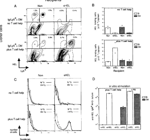
Figure 2. Ag and bm12 Th ceils are both required for expansion and differentiation of nontolerant iysozyme-binding B cells, but remain unable to trigger tolerant B cells. (A-C) Equal numbers of Ig/Ly5 =+ and Dbl/Ly5=- splenocytes were mixed and transferred with B6-strain splenocytes (no T cell help) or bin12 splenocytes (plus T cell help) into irradiated nontransgenic or sHEl.-transgenic recipients. (A) 5 d after transfer, splenocytes were stained for HEL-binding and LyP expression to distinguish nontolerant (Ly5 ~+) from tolerant (Ly5,-) B cells. (B) The number of HEL-binding Ig/Ly5 -+ (open bars) or Dbl/Ly5'- (sappled bars) cells per spleen was determined for all the recipients. (C) Differentiation of B cells in the recipients was mea- sured in parallel by a third-color immunofluorescent stain for the plasma cell marker, syndecan, on HEL-binding Ig/Ly5 ,+ (solid line) or Dbl/Ly5,-
(dashed line) B cells. Histograms were derived from threecolor analysis after gating on the HEL-binding Ly5 ~+ or Ly5'- cells shown in B. The data shown are from one experiment and are representative of three independent experiments involving a total of six separate Ig- and six separate Dbl- transgenic donors. (/9) Numbers of anti-HEL IgM~-secreting cells generated after 4 d in vitro culture of Ig- or Dbl-transgenic splenocytes with an excess of B6-strain and bm12-strain spleen cells (plus T cell help) or with lipopolysaccharide (LPS). Cultures were performed in the presence (+sHEL)
or absence (-sHEL) of 100 ng/ml HEL. The experiment shown is representative of six independent in vitro culture experiments.
cells, tolerant lysozyme-binding B cells from Dbl-transgenic mice showed little proliferation or Ab production in the pres- ence of both bm12 T cells and lysozyme (Fig. 1, B and C). To confirm that activated I-Ab-reactive Th cells were in- deed being generated in recipients of tolerant B cells, Dbl- and Ig-transgenic B cells were mixed and cotransferred with
bm12 T cells into recipient animals. Nontolerant lysozyme- binding cells from the Ig-transgenic mice were in this case distinguished from tolerant Dbl-transgenic cells by the pres- ence of the Ly5 a allelic marker only on the former (e.g., Fig.
2 A, top). Differentiation of lysozyme-binding Ly5 '+ (Ig- transgenic) or Ly5'- (Dbl-transgenic) cells into Ab-secreting
[image:5.612.59.547.63.519.2]cells was measured in parallel by staining with an Ab to the plasma cell marker, syndecan (26 and Fig. 2 C). Nontolerant Ly5 "+ B ceils proliferated and differentiated efficiently in the presence of lysozyme and bml2 T cells (Fig. 2,
A-C, bottom
righ O,
confirming that activated I-Ab-spedfic Th cells wereindeed generated in these recipients. By contrast, the tolerant Ly5'- B cells in the same hosts fared to undergo blastogen- esis, increase in cell number or differentiate into plasma cells (Fig. 2,
A-C).
The possibility that the tolerant B cells were capable of interacting with T ceils but failed to migrate to the appro- priate in vivo microenvironment was tested by performing equivalent experiments with bin12 T cells in suspension cul- tures in vitro. Small numbers of Ly5'-marked spleen cells from B6 Ig- or Dbl-transgenic mice were added to two-way mixed lymphocyte reactions between nontransgenic B6 and bml2 spleen cells. Again, lysozyme-binding B cells from Ig- transgenic mice proliferated, as detected by blastogenesis and
BrdU incorporation (data not shown) and differentiated into Ab-secreting ceils (Fig. 2/9) only when lysozyme was added to the cultures. Dbl-transgenic ceils, by contrast, showed little proliferation and generated 50-100-fold fewer Ab-secreting cells (Fig. 2 D and data not shown). Parallel cultures stimu- lated with the nonspecific B cell mitogen, LPS, neverthdess confirmed that the tolerant B cells were potentially capable of efficient proliferation and differentiation into Ab-secreting cells (Fig. 2 D,
right columns)
as previously described (32).Tolerant B Cells Respond to T Cell-derived Mediators.
Twosignals appeared necessary for efficient proliferation and dif- ferentiation of nontolerant lysozyme-binding B cells under the conditions of I-Ab-specific T cell help used above: one provided by the bin12 T cells, and one resulting from the binding of lysozyme to the B cell (see Fig. 1 A). Unrespon- siveness in the tolerant B cells could therefore have reflected a defective response to either of these signals. To test the re- sponse to T cell-derived signals, membrane fractions from
Freshly
Isolated
A
B
mom - / /
o. 40ooo ()
O/O~O
No / * ~ *
9 o ~ . : . , . . m ~ e ...
1 10 100
,=~
_
dilution antl-CD40
(xlO
"s)
C
8oooo---//+ IL-4
~
~ P '
plus/IL-4
60000
9
"'" ~176
o I o /
+
antl-CD40
~
, . ~ - , ~2O00O
t . . . .
[image:6.612.60.476.299.654.2]number ... 1 ... I,' ... I,o'"'"' ... 1 ... Io ... ~ ... ' o " ...., . . .
o, ce,s | C l a s s II = C T L A 4 - 1 g - * o dilution ,
antbCD40
lo(xlO
"s)
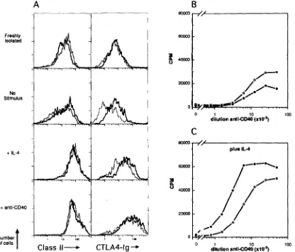
meFigure 3. Response of tolerant and nonto[erant B cells to Th cells signals. (/I) +Immunonuorescent staining with CTLA4-[g to detect B7 and B7- related molecules (right) and class II M H C molecules (left) on Ig-transgenic (solid line) and Dbl-transgenic
(~/m~
line) splenic B cells immediately after removal from the animal (Freshly/sohted) or after 24 h culture with medium alone (No samulv, s) or medium comaiaiag I b 4 or and-CD40 Abs (bottom). The histosrams displayed are gated on B220 + ceils after e~cluding dead cells by staining with propidium iodide. (B and C) Proliferation of Ig-tramgenic (open circles) or Dbl-transgenic (filled circles) B ceils, assessed by pH]thymidine incorporation after stimulation with anti-CD40 (B) or anti-CD40 and IL-4 (C). Data displayed represent means of triplicate cultures and are representative of four independent experiments.activated T cell hybridomas that contain CD40L (14, 33) were initially tested and found to trigger equivalent proliferation in tolerant and nontolerant B cells, and staining with Abs to CD40 showed equivalent expression on both types of B cells (data not shown). More specifically, treatment with a polyclonal antiserum to recombinant mouse CD40 that mimics the action of membrane-bound CD40L (Heath, A. et al., manuscript submitted for publication), or stimulation with IL-4, each induced tolerant and nontolerant B cells to in- crease cell surface expression ofdass II MHC (Fig. 3 A). Ad- ditionally, these stimuli also increased the immunofluores- cent staining of both cell types with CTLA4-Ig fusion protein (Fig. 3 A) which recognizes B7 and B7-related molecules ex- pressed on activated B cells (hereafter referred to CTLA4-Ig ligand; 34; see Discussion). Vigorous proliferation was in- duced in both cell types by antisera to CD40 (Fig. 3 B), and this was augmented in both tolerant and nontolerant B cells by addition of IL4 (Fig. 3 C). Taken together, these findings indicate that the tolerant B cells were fully responsive to key signals mediating T cell help.
Tolerant B Cells Do Not Respond to Ag. In contrast to the
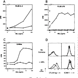
normal response to T cell-derived signals, many cellular re- sponses normally triggered by Ag binding to slg (for review see references 35 and 36) were absent in tolerant B cells. Binding of lysozyme at concentrations as low as 1 ng/ml, which engages <5 % of the slg receptors (22), triggered vig- orous proliferation of nontolerant Ig-transgenic B cells in the presence of either IL-4 (Fig. 4 A), submitogenic concen- trations of LPS (Fig. 4 B), or suboptimal concentrations of
anti-CD40 antiserum (data not shown). However, binding of lysozyme to tolerant B cells had no mitogenic effects in any of these assays (Figs. 4, A and B and not shown). Simi- larly, whereas binding of lysozyme alone was not mitogenic, it promoted activation of nontolerant Ig-transgenic B cells from Go to G: of the cell cycle, as assessed by increased RNA synthesis (Fig. 4 C) or by cell enlargement detected flow cytomerrically (dam not shown). In contrast, lysozyme binding failed to induce cell enlargement or increased RNA synthesis in tolerant B cells (Fig. 4 C, and data not shown). Finally, lysozyrne binding triggered increased expression of CTLA4-Ig ligand and ICAM-1 molecules on nontolerant Ig-transgenic cells, but induced little increase in the expression of these molecules on tolerant Dbl-transgenic cells (Fig. 4 D).
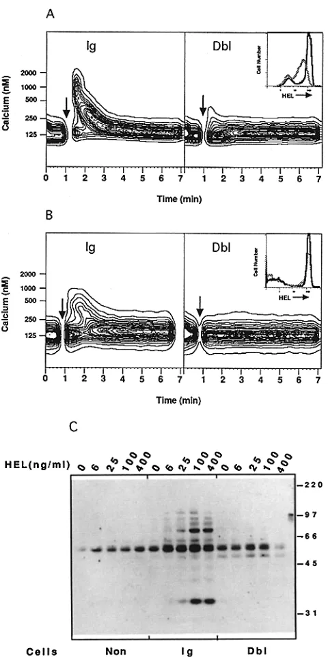
The absence of many cellular responses to Ag binding in the tolerant cells was further explored by following early signal- transduction events normally triggered by sIg engagement. One of the earliest biochemical events, an increase in the level of intracellular calcium ([Ca]i; 35, 36), was measured by four-color flow cytometry after loading spleen cells with the calcium indicator, Indo-1. Binding of lysozyme to receptors on nontolerant Ig-transgenic B cells induced a rapid rise in the level of [Ca]i that peaked within 1 rain and included nearly all of the HEL-specific B cells (Fig. 5 A). By contrast, stimulation of tolerant Dbl-transgenic B cells with a range of concentrations of lysozyme from 20 to 500 ng/ml failed to induce a detectable increase in [Ca]i (Fig. 5 A, and data not shown). Activation of protein tyrosine kinases is an inte- gral event in signal transduction by slg that lies immediately
A
P L U S I L . 4
0 ,,i~111 . - . , . . . ~ . . . . ,
1 10 100
H E L ( n g / m l )
C
Iooo
9oo
7oo
6oo
400
3OO
2OO
IO0
o
Uridk~
/
o
~,,t/ ...
0 0 1 1 10 tOO
H E L ( n g , ~ n t )
B
5 o o o o - t ' /
4OOO0 - r3
3OOOO
==
u 2OOOO
10o0o
o
D
N o
Stimulus
+ sHEL
number
P L U S LPS
f
~ 1 7 6o
~ _ ... ~ --:....~.~..~ ..-...~...o~....~
0 0 1 I 10 100
HEL ( n g / m l )
CTLA4-1w ICAM- 1 ,,
Figure 4. Early cellular responses after Ag binding to tolerant and nontolerant B cells. (A) Proliferation of 13 cells from Ig-rtansgenic (open symbo/~) or Dbl-ttansgenic (fil/ed s,/mbo/s) mice as measured by [3H]thymidine incor- potation after 48 h of culture with the indicated amounts of HEL in the presence of IL-4. (B) Proliferation in re- sponse to HEL in the presence of submitogenic concen- ttafions of LPS (1/~g/znl). For comparison, square symbols show proliferation to optimal concentrations of LPS (20 ~tg/ml). ((3) Transition from Go to G1 of the cell cycle as measured by incorpotarion of [3H]uridine into cellular RNA in response to the indicated concentrations of HEL. (D) Expression of CTLA4-Ig ligand and ICAM-1 after Ag stimulation of Ig-ttansgenic
(solid line)
or Dbl- transgenic(dotted line)
B cells after 12 h(CTLA4-Ig)
or 24 h(ICAM-1)
of culture with 100 ng/ml lysozyme (bottom) or medium alone (to/,). [image:7.612.57.370.420.732.2]Figure 5. Signaling events triggered by HEL-binding in tolerant and nontolerant B cells. (,4) Intracellular calcium levds as a function of time in freshly isolated Ig- or Dbi-transgt'nic splenic B cells before and after the addition of lysozyme (arrows). (Inset) Rehtive number of free HEL binding sites on Ig (solid line)- or Dbl (dotted line)- transgenic B cells before stimulation, as detected using fluorescently hbded HEL. (B) Intracellular calcium levels of Ig- or Dbl-tramgenic B cells that had been parked in nontransgenic mice for 36 h before harvesting and in vitro simulation with HEL (arrows). Note (inset) that the number of available HEbbinding sites is nearly identical on parked Ig- and Dbl-transgenic B cells. (C) Anti- phosphotyrosine immunoblottlng, showing induction of phosphotyrosine- containing proteins 5 min after addition of lysozyme to nontransgenic, Ig-tnusgenic, and Dbl-trausgenic spleen cells. Note that the reduced level of phosphotyrosine-containing proteins in unstimulatcd nontransg~c cells (lane I) and in Dbl-transgenic cells stimulate~i with 400 ng/ml HEL (lane 15) results from poor transfer and is not representative (see Fig. 6 A).
upstream to the initiation of increased [Ca]i (37-40). To measure tyrosine kinase activation, Dbl- and Ig-transgenic spleen cells were stimulated with lysozyme in vitro, and cell lysates were then prepared and probed by immunoblot using Abs specific for phosphotyrosine. Lysates from unstimulated tolerant or nontolerant cells had few phosphotyrosine-con- tainiug proteins (Fig. 5 C). Stimulation with concentrations of lysozyme as low as 6 ug/ml resulted in rapid appearance of many phosphotyrosine-containing proteins within the non- tolerant Ig-transgenic B cells. By contrast, even saturating concentrations of lysozyme failed to induce a detectable in- crease in phosphotyrosine species in the tolerant B cells (Fig.
5C).
Because of modulation of IgM Ag receptors and in vivo occupancy of many IgD receptors with lysozyme, B cells from the Dbl-transgenic mice display 3-4-fold fewer available receptors for binding additional exogenous lysozyme mole- cules than nontolerant B cells from Ig-transgenic mice (Fig.
5 A inset).
To test whether the signaling defects in the Dbl-transgenic B cells were simply a result of fewer receptors, or alternatively, a short-term refractory phenomenon due to recent in vivo exposure to lysozyme, B ceils were removed from the Dbl-transgenic mice and "parked" in irradiated non- transgenic mice to allow bound lysozyme to dissociate. 36 h after transfer, the parked Dbl-transgenic B calls displayed a nearly equivalent capacity to bind fluorescently labeled lyso- zyme as did parked Ig-transgenic B ceils (Fig. 5 B,
inset).
Despite this, the tolerant ceils remained unable to increase [Ca]i after stimulation with lysozyme (Fig. 5 B), indicating that the defect in signaling was not due simply to receptor modulation. Whereas [Ca]i increase is an immediate conse- quence of tyrosine kinase activation (37-40), parked B calls could not be recovered in suflfident numbers or purity to mea- sure protein tyrosine phosphoryhtion directly.
Defective Response of Tolerant B Cells to anti-IgD Abs.
Theabsence of intracellular signaling events after HEL binding to tolerant B cells was fimher egplored by stimulating tolerant and nontolerant B ceils with affinity-purified Abs to the C regions of IgD. Stimulation by anti-IgD differed from lyso- zyme stimulation in that IgD is expressed at identical levels on tolerant and nontolerant B cells, is equally accessible to anti-C region Abs, and thexefore should be cross-linked equally weU on the two cell types. Additionally, the polydonal anti- IgD Abs should promote extensive receptor cross-linking and therefore would test whether tolerant B ceils retained any capacity to signal through sIg. In contrast to stimulation with lysozyme, extensive cross-l/nking of sIgD receptors elicited some early signaling events in the tolerant B cells, although the magnitude and kinetics of the response remained altered compared with nontolerant B cells. Thus, treatment with anti-IgD antisera induced an identical pattern of phospho- tyrosine-containiug proteins in both Dbl- and Ig-transgenic B cells, although the rdative abundance of labeled proteins was markedly reduced in tolerant cells (Fig. 6 A). Similarly, anti-IgD stimulation triggered a prolonged increase in [Ca]i in nontolerant Ig-transgenic ceils, with the fraction of B ceils containing micromolar concentrations of [Call remaining at
[image:8.612.55.295.52.540.2]B
Ig
4000 - - 2 0 0 0 - ~ l g 1 0 0 0 - -
500 - -
1 2 5 - -
. . . . t - t " ; t . . . . L I
0 1 2 3 4 5 6
Dbl
( x l g ~t . . . . i i . . . . l . . . . t " 1 . . . . I . . . . [ 7 1 2 3 4 $ 6 7
Time (min)
Figure 6. IL~ponse of tolerant and nontolerant B cells to receptor cross- linking by polyclonal anti-IgD Abs. (A) Phosphotyrosine-containing cel- lular proteins 5 min after stimulation oflg-, Dbl-, or non-transgenic spleno- cytes with media (0), 10/~g/ml goat anti-IgM Abs (IgM), 10/~g/ml goat
anti-IgD Abs (IgD), or 100 ng/ml HEL
(HEL).
Note that the prominent32- and 33-kD phosphotyrosine species induced by anti-IgM and anti- IgD, respectively, are likely to represent the Ig-associated molecules IgM(x and IgDol (77). (B) Intracellular calcium levels after stimulation of Ig- and Dbl-transgenic B cells with goat anti-IgD Abs (10/~g/ml). (C) Acti- vation from Go to Gt of the cell cycle, as measured by [3H]uridine incor-
poration induced by goat anti-IgD stimulation of Ig (open
symbols)-
andDbl
(filled syrabols)-transgenic
splenocytes. (D) Proliferation of Ig(open
symbols)-
and Dbl(filled symbols)-transgenic
B cells induced by goat anti-IgD in the presence of submitogenic concentrations of LPS (1 /~g/ml).
(E) Mitogenic response of Ig (open bors)- and Dbl
(filled
bars)-transgenicB cells after stimulation with media alone (0), Ionomycin (1 #g/ml), and
PMA (1 ng/ml)
(I/P)
or LPS (20/~g/ml). (F) Induction of CTLA4-Igligand on Ig
(solid line)-
and Dbl(dotted llne)-
transgenic B cells 12 h afterstimulation with goat anti-IgD (10/~g/ml).
C
l o o o
900 - - . o - - i 9 II Obl
800
+
' 0 0 ~
~ 4 ~ z s ~ zoo
s n t l - l o D ( u g / m l )
s o o o o
i+
2OOOOD
P l u s LPS
a n t l - l g O ( u o / m l )
3oooo (J
ZcQoo
10o00
[ ] I g 9 O b l
_ ,IL
0 I / P L P S No S t i m u l u sF
+ a n t i - l g D
t
~ V W l 1 io ioo ot r I CTLA4-1g
10-15% for up to 4 min (Fig. 6 B,
left).
In contrast, stimula-tion of tolerant B cells with anti-IgD induced a more rapid initial [Ca]i response, but this response diminished much more quickly and lacked the sustained phase observed in the
nontolerant B cells (Fig.
6 B, right).
Whereas the ability of anti-IgD antisera to evoke prox- imal signaling events suggested that the necessary receptor- associated components were at least partially present and ac- tivatable in the tolerant B cells, more distal events such as cell cycle entry still failed to occur. Thus, stimulation with the same antiolgD antiserum failed to trigger G0-G1 transi- tion in Dbl-transgenic cells as measured by induction of R N A
synthesis (Fig. 6 C), and promoted little D N A synthesis in the tolerant cells in combination with submitogenic concen- trations of LPS (Fig. 6 D). Similar results were obtained with mAbs to IgD coupled to dextran (41), which also cross-link Ig receptors extensively and promoted vigorous proliferation in the nontolerant Ig-transgenic B cells, but induced little or no mitogenic response in tolerant B cells (data not shown). Proliferation in response to calcium ionophore and phorbol ester, which bypass the requirement for slg signaling, remained intact in the tolerant cells (Fig. 6 E). Whereas extensive receptor cross-linking with anti-IgD Abs was unable to pro- mote cell cycle entry, this stimulus was nevertheless sufficient
[image:9.612.54.492.72.539.2]to induce high expression ofCTLA4-Ig ligand on the tolerant calls (Fig. 6 F).
Tolerant B Cells Respond to Membrane-bound Lysozyma Since
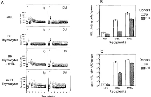
early signaling events could be partially restored and CTLA4-Ig ligand induced on the tolerant B cells after extensive receptor cross-linking with Abs to IgD, we tested whether a highly multivalent form of lysozyme expressed on ceU surfaces as an integral membrane protein (mHEL) would also restore sIg signal transduction. Thymocytes from transgenic mice expressing mHEL on cell surfaces (21) were therefore mixed with Indo-1 loaded B cells and changes in [Call measured as previously. In contrast to stimulation with soluble lyso- zyme which remained unable to dicit a [Call response in tolerant B calls (Fig. 7 A, 2), stimulation with membrane lysozyme triggered a dear [Call response in both tolerant- and nontolerant-transgenic B cells although the response in the latter was somewhat weaker (Fig. 7 A, 7 and 8). Control B6 thymocytes lacking mHEL elicited no response in either cell type (Fig. 7 A, 3 and 4). Combined stimulation with
control B6 thymocytes and soluble HEL remained unable to dicit a calcium response in tolerant B cells (Fig. 7 R, 6), in- dicating that Ag display on membranes was required and that costimulatory molecules on thymocytes were not sufficient. To test whether the partial restoration of sIg signaling in- duced by displaying lysozyme in multimeric cell-bound form was sufficient to restore collaboration with I-Ab-specific Th cells, transgenic B cells, and bm12 T cells were transferred into recipient mice expressing mHEL. Tolerant B cells from Dbl-transgenic mice remained poorly responsive to soluble lysozyme (sHEL) and bm12 helper calls (Fig. 7, B and C,
middle columns),
but their helper ceil-induced proliferationand differentiation into Ab-secreting plasma cells increased 100-fold in recipients expressing mHEL (Fig. 7, B and C,
right columns).
The magnitude of the response mounted bytolerant B cells in the presence of mHEL was equivalent to that mounted by nontolerant Ig-transgenic B cells in the pres- ence of sHEL, and only fivefold less than the response of the latter in mHEL recipients (Fig. 7, B and C). Thus, display
A
~ o o o -
sHEL ~ ,|
J 2 ~ -
7r - 8 6 ~ . . . .
Thymocytes ~ '~-
B6 2OOO -
T h y m o c y t e s ~ ,| + sHEL ~ ,,,
Ig
Ig
s Ig
7 Ig
2 Dbl
" Dbl
o Dbl
8 Dbl
B
c" I 0 e-~ D o n o r s
~fl 1 0 ?
" [ Z ] I g
~[~ Dbl
._
" 0 c - " ~ 10 s
1 0 4
Non
sHEL
mHEL
R e c i p i e n t s
C
C" 10 e ]
-~ ~o', ~ D o n o r s
- ~ - - h
1~
~
[~]Ig!.J- I
I
< to'-
~Dbl
--~T lo'-
d
~ o ~ 10 3.
m F, o . . . .
~ ~ , ~ . . - - 1 0 2 .
T h y m o c y t e s ~ ,~_ ~-
~ ~ 101
Non
sHEL
mHEL
0 , 2 3 , , , 1 2 3 , , ~ R e c i p i e n t s
Time (minutes)
Figure 7. Membrane-bound lysozyme can trigger slg signaling in tolerant B cells and promote effective collaboration with I-Ab-specific helper T cells. (A) Intracellular calcium levels in Ig (/eft)- and Dbl (r~ghO-transgenic B cells after stimulation with soluble HEL (1 and 2), B6 thymocytes (3 and 4), B6 thymocytes plus soluble HEL (5 and 6), or thymocytes bearing membrane-bound lysozyme (mHEL; 7 and 8). At the arrow, B calls were removed and stimulated as indicated (see Materials and Methods for details). (B and C) In vivo collaboration between Ig- and Dbl-transgenic B cells and bm12 Th ceils. (B) Enumeration of HEbbinding B cells and (C) numbers of anti-HEL-specific plasma cells was determined as in Fig. 1, after transfer of Ig-transgenic (open Mrs) or Dbbtransgenic (filled bars) splenocytes with bm12 splenocytes into irradiated B6 strain mice (Non), or into trans- genie mice expressing sohble lysozyme (sHEL) or membrane-bound lysozyme (mHEL). Shown are the arithmetic averages (bars) and individual values
(dots) for cells from three separate Ig- or Dbl-transgenic donors. The experiment shown is representative of three independent experiments involving seven separate Ig- and seven separate Dbl-transgenic donors.
[image:10.612.55.552.303.625.2]of membrane-bound Ag could partially restore slg signaling in tolerant B cells and effective collaboration with I-A b- specific Th cells.
Discussion
The studies above localize the molecular mechanism ac- counting for B cell clonal anergy to a proximal step in the sIg signal transduction pathway. Moreover, the findings in- dicate that Ag-specific B cells require signals from T cells and from Ag binding to sIg for efficient proliferation and differentiation into Ab-secreting cells, as originally proposed by Bretscher and Cohn (42). The conclusion that Ag-derived signals through sIg are required was based on three observa- tions: (a) in the presence of activated I-Ab-reactive T cells, nontolerant, lysozyme-specific B cells expressing bA h made no Ab response unless they were also exposed to antigen; (b) under the same conditions, tolerant, lysozyme-spedfic B cells failed to respond even when exposed to lysozyme, and these cells lacked lysozyme-induced sIg signaling; (c) a more potent multivalent form of lysozyrrle, membrane lysozyme, was able to trigger some signaling through sIg in tolerant cells, and this markedly restored their ability to mount an Ab response in the presence of activated Th cells.
Basis for Blocked Signaling in Tolerant B Cells. The absence of slg signaling after Ag binding to the tolerant B cells could in principle reflect either a biochemical block in signal trans- duction or simply the presence of fewer available receptors on the Dbl-transgenic B cells. Three findings establish that the latter cannot account for the signaling defect. Firstly, the tolerant B cells display 30% of the number of available HEL- binding receptors on nontolerant B cells, yet failed to respond to any concentration of HEL in assays where nontolerant B cells responded optimally to concentrations that engage <5% of available receptors (Figs. 3, B and C, and 4 C). Second, Dbl-transgenic B cells that had been parked in nontransgenic mice and that had recovered comparable HEL-binding ca- pacity remained unable to increase [Call after HEL binding (Fig. 4 B). Third, IgD receptors are expressed at identical levels on tolerant and nontolerant B cells and should be cross- linked identically after treatment with polydonal antisera to IgD or anti-IgD coupled to dextran. These stimuli neverthe- less elicited markedly reduced proximal signaling and failed to induce mitogenesis in tolerant B cells (Fig. 5 C and D, and data not shown).
Given the evidence for a signaling block, the failure of anti- IgD stimulation to induce G0-G1 transition or cell cycle entry in the tolerant B cells despite activating some tyrosine phosphorylation and calcium influx (Fig. 5), could have im- plied a block downstream of these initial events. The fact that the tolerant B cells proliferated normally after stimula- tion with calcium ionophore and phorbol ester (Fig. 6 E) nevertheless argues against a downstream defect. Moreover, the short-lived calcium response induced by anti-IgD in the tolerant cells (Fig. 6 A) is most consistent with a receptor- proximal defect, since coupling of [Call stores to cellular influx, as assessed by exposure to thapsigargin occurs nor- mally in the tolerant B cells (Cooke, M.P., unpublished ob-
435 Cooke et al.
servations). Sustained Ag receptor signaling is necessary for activation of both B (43) and T cells. Removal or blocking of TCR stimuli any time during the first 2 h of stimulation blocks early cellular responses such as IL-2 gene induction (44) and similarly, removal of anti-IgM Abs any time during the first 16 h of B cell stimulation effectively halts cell cycle progression (45). Indeed, the lack of response to weak stimuli (sHEL) and transient responses to strong stimuli (polyclonal anti-IgD) resembles the response to different anti-TCR Abs in mutant T cell lymphomas that lack a single component of the TCR signal-transduction complex, such as the tyro- sine kinase p56 ~k (46).
Exactly how Ag binding induces slg signaling remains to be fully elucidated, although Ag-driven receptor dimeriza- tion or multimerization is thought to play an important role (for a review see reference 47). The ability of sHEL to trigger slg signaling is in this respect surprising, since sHEL is known to bind the transgene-encoded Ig as a monomer (48). Many of the signaling events induced by sHEL (Figs. 4 and 5) nevertheless appear equivalent to those described for nonmito- genic monoclonal IgG Abs to IgM or IgD (for reviews see references 35 and 36). We therefore favor the view that sHEL, like anti-Ig Abs, induces slg signaling by receptor dimeriza- tion. HEL-mediated dimerization could occur via preformed HEL dimers in solution or be driven by high local concen- trations of receptor-bound HEL on the cell surface, since HEL dimerizes in solution at higher concentrations (49). By com- parison, the ability of polyvaleut ligands such as polyclonal anti-IgD and membrane-bound HEL to partially overcome the sHEL-induced signaling blockade may simply reflect more extensive slg multimerization induced by these ligands. Con- sistent with this notion, increasing the valency of initially oligovalent Ags or divalent anti-Ig Abs has been found to markedly increase their potency for B cell activation (39, 50). Since other B cell surface proteins such as CD19, CD21 (for reviews see references 51 and 52), and CD22 (53) may also participate in signaling by slg, it will be important to determine whether differences in their activity may contribute to the sIg signaling defect found in tolerant B cells. It will also be important to relate the regulation of sIg signaling in tolerant B cells to the interruption of sIg signaling in- duced in mature B cells by acute sIg cross-linking (54, 55) or by coligation with the Fc~ receptor (56, 57), particularly since the latter is thought to provide an important negative feedback during Ab responses to foreign Ags (for a review see reference 58).
Role of slg Signaling in B-T Cell Interaction. Whereas the role of slg in mediating Ag uptake for subsequent processing and presentation to Th cells is well established (9-12), the role of slg signal transduction in T cell-dependent Ab re- sponses has remained unclear (59-62). In particular, it is difficult to reconcile the need for both MHC-restricted B cell Ag presentation and the need for slg signaling with recent studies demonstrating that, in the absence of both events, membrane CD40L together with T cell-derived lymphokines can nevertheless trigger resting B cells to proliferate and pro- duce Ab efficiently (for a review see reference 7).
T cell:
B cell:
C D 4 O L ~
f
Useless Useful Harmful
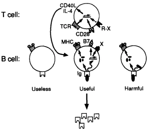
Figure 8. A model to explain the requirement for slg signaling and MHC-restricted antigen presentation during B-T cell interactions. The model assumes that CD40L and Ib4 are transiently expressed during Th cell activation, and must be teinduced during each B-T cell interaction. Reinduction of these growth and differentiation factors during B-T cell synapsis is hypothesized to require the T cell m be stimulated both by antigen peptide-MHC complexes engaging the TCK and by B cell activa- tion molecules such as B7 engaging CD28, or other activation molecules (X) engaging receptor (R-X). T cell help is thus delivered sdectively to B cells that make Abs specific for foreign Ags, since only in these cells does the binding of Ag to slg allow both eificient uptake of Ag for presen- tation and trigger expression of B7 and other activation markers.
MHC-restricted Ag presentation in T cell-dependent Ab production is outlined in Hg. 8. The model assumes that T cell expression of CD40L after activation is transient, as has been shown in vitro (2), requiring that CD40L be rein- duced during T-B cell synapsis. Surface Ig on Ag-specific B cells may thus play two roles in reinducing CD40L, firstly by capturing Ag for effident processing and presentation to the TCK and second by triggering a marked increase in B ceil activation molecules such as B7 on the cell surface (63, and Fig. 3). Other activation molecules triggered by slg sig-
haling after Ag binding, simply denoted "X" in Fig. 8, in- dude ICAM-1, (64, and Fig. 4 D), CD44 (65, 66), and LFA-1 (64). The combined presence of specific peptide-MHC com- plexes and certain activation molecules on the B cell may be required to reinduce CD40L expression and other Th ceil functions. In support of this notion, B7 signaling through CD28 on the T cell is strongly synergistic with TCK sig- naling for restimulating T cell production of IL-2 (27, 6%71) and for stimulating re-expression of CD40L (Clark, E., per- sonal communication).
While the importance of CD28 costimulation during in- teraction between B ceils and T calls remains to be estab- lished, several lines of evidence suggest a pivotal role. In ex- periments here, induction of B7 or B7-related molecules on Ag-binding B cells correlated with their ability to mount an Ab response in the presence of activated I-Ab-specific T cells. Conversely, primary T ceil-dependent Ab responses in mice have been shown to be suppressed by blocking B7 or B7-rdated molecules with soluble CTLA4-Ig (72, 73). Simi- larly, gene knockout mice lacking the CD28 receptor for B7 have greatly diminished T cell-dependent Ab responses but apparently normal cytotoxic and delayed-type hypersensitivity T cell responses (74).
Based on the above, it willbe important to determine what effect is induced in Th ceils when bound Ag is presented by tolerant B cells that fail to signal through sIg and lack costimulatory molecules such as B7. We have recently estab- lished that tolerant Dbl-transgenic B cells do indeed process and present lysozyme efficiently to lysozyme-speciiic T ceils from a TCR gene transgenic mouse (Ho, W., M. M. Davis, M. P. Cooke, and C. C. Goodnow, unpublished observa- tions). In contrast to presentation of Ag by nontolerant Ig- transgenic B cells, however, the tolerant B cells fail to trigger T cell effector functions in vitro unless e~ogenous anti-CD28 Ab is provided, and the tolerant B cells still fail to be induced into proliferation or Ab production in vitro and in vivo. Since TCR signaling in the absence of costimulation through CD28 results in T cell anergy under some conditions (for reviews see references 75 and 76), it will be important to determine whether antigen presentation by tolerant B cells is neutral or has an active negative effect on Th cell function.
We thank our co-workers especially Drs Sarah BeU, Jason Cyster, and Suzanne Hartley, for helpful discus- sions and comments on the manuscript; the staff of the Beckman Center Shared FACS | facility for as- sistance with flow cytometry, and the staff of the Division of Laboratory Animal Medicine at Stanford for excellent animal husbandry.
Address correspondence to Dr. Christopher C. Goodnow, Howard Hughes Medical Institute Research Laboratories, Beckman Center for Molecular and Genetic Medicine, Stanford University Medical Center, Stanford, CA 94305'-5428.
Received for publication 17 August 1993 and in revised.form 18 October 1993.
1. Blackman, M., J. Kappler, and P. Marrack. 1990. The role of nation by T cells. Science (Wash. DC,). 248:1369.
the T cell receptor in positive and negative selection of de- 3. Nossal, G.J.V. 1992. Cellular and molecular mechanisms of B veloping T cells. Science (Wash. DC). 248:1335. lymphocyte tolerance. Adv. Immunol. 52:283.
[image:12.612.55.294.52.259.2]body production using the concept of aloha1 selection. The Australian Journal of Science 20:67.
5. Armitage, R..J., W.C. Fanslow, L. Strockbine, T.A. Sato, K.N. Clifford, B.M. Macduff, D.M. Anderson, S.D. Gimpel, S.T. Davis, and C.R. Maliszewski. 1992. Molecular and biological
characterization of a murine ligand for CD40. Nature (Lond.).
357:80.
6. Armitage, R.J., T.A. Sato, B.M. Macduff, K.N. Clifford, A.R. Alpert, C.A. Smith, and W.C. Fanslow. 1992. Identification
of a source of biologically active CD40 ligand. Eur.J. lramunol.
22:2071.
7. Noelle, R., and E.C. Snow. 1992. T hdper cells. Cu~ Opin.
Immunol. 4:333.
8. Rajewsky, K., V. Schirrmacher, S. Nase, and N.K. Jerne. 1969. The requirement of more than one antigenic determinant for
immunogenicity. J. Ex F Med. 129:1131.
9. Mitchison, N.A. 1971. The carrier effect in the secondary re- sponse to hapten-protein conjugates. II. Cellular cooperation.
Eur. J. Iramunol. 1:18.
10. Katz, D.H., T. Hamaoka, M.E. Doff, and B. Benacerraf. 1973. Cell interactions between histoincompatible T and B lympho- cytes. The H-2 gene complex determines successful physiologic
lymphocyte interactions. Proc Natl. Acad. Sci. USA. 70:2624.
11. Sprent, J. 1978. Restricted helper function of F1 hybrid T cells positively selected to heterologous erythrocytes in irradiated parental strain mice. II. Evidence for restrictions affecting helper cell induction and T-B collaboration, both mapping to the
K-end of the 1-1-2 complex. J. Ex F Med. 147:1159.
12. Lanzavecchia, A. 1990. Receptor-mediated antigen uptake and its effect on antigen presentation to class II-restricted T lym-
phocytes. Annu. Rev. Iramunol. 8:773.
13. Sanders, V.M., J.M. Snyder, J.W. Uhr, and E.S. Vitetta. 1986. Characterization of the physical interaction between antigen-
specific B and T cells. J. Immunol. 137:2395.
14. Noelle, R.J., M. Roy, D.M. Shepherd, I. Stamenkovic, J.A. Ledbetter, and A. Aruffo. 1992. A 39-kDa protein on activated helper T cells binds CD40 and transduces the signal for cog-
nate activation of B cells. Proc Natl. Acad. Sci. USA. 89:6550.
15. Kupfer, A., S.L. Swain, C.A. Janeway, and S.J. Singer. 1986. The specific direct interaction of helper T cells and antigen
presenting B cells. Proc Natl. Acad. Sci. USA. 83:6080.
16. Poo, W.-J., L. Conrad, and C.A. Janeway. 1988. Receptor-
directed focusing of lymphokine release by hdper T cells. Na-
ture (Land.). 332:378.
17. Springer, T. 1990. Adhesion receptors of the immune system.
Nature (Lond.). 346:425.
18. Sanders, V.M., and E.S. Vitetta. 1991. B cell-associated LFA-1 and T cell-associated ICAM-1 transiently cluster in the area
of contact between interacting cells. Cell. Iramunol. 132:45.
19. Linsley, P.S., E.A. Clark, and J.A. Ledbetter. 1990. T-cell an- tigen CD28 mediates adhesion with B cells by interacting with
activation antigen B7/BB-1. Pr0c Natl. Aca~ Sci. USA. 87:5031.
20. Goodnow, C.C.,J. Crosbie, S. Adelstein, T.B. Lavoie, S.J. Smith- Gill, K.A. Brink, H. Pritchard-Briscoe, J.S. Wotherspoon, lk.H. Loblay, K. Raphael, et al. 1988. Altered immunoglob- ulin expression and functional silencing of self-reactive B lym-
phocytes in transgenic mice. Nature (Land.). 334:676.
21. Hartley, S.B., J. Crosbie, R. Brink, A.A. Kantor, A. Basten, and C.C. Goodnow. 1992. Elimination from peripheral lymphoid tissues of self-reactive B lymphocytes recognizing membrane-
bound antigens. Nature (Land.). 353:765.
22. Goodnow, C.C., J. Crosbie, H. Jorgensen, R.A. Brink, and A. Basten. 1989. Induction of self-tolerance in mature periph-
eral B lymphocytes. Nature (Land.). 342:385.
23. Goroff, D.K., A. Stall, J.J. blond, and F.D. Finkelman. 1986. In vitro and in vivo B lymphocyte-activating properties of
monoclonal anti-IgD antibodies. J. Irarauaol. 136:2382.
24. Cooke, M.P., K.M. Abraham, K.A. Forbush, and K.M. Perlmutter. 1991. Regnlation of T cell receptor signaling by
a src family protein-tyrosine kinase p59fyn. Cell. 65:281.
25. Hartley, S.B., M.P. Cooke, D.A. Fulcher, A.W. Harris, S. Cory, A. Basten, and C.C. Goodnow. 1993. Elimination of self- reactive B lymphocytes proceeds in two stages: arrested devel-
opment and cell death. Cell. 72:325.
26. Sanderson, R., P. Lalor, and M. Bernfield. 1989. B lympho- cytes express and lose syndecan at specific stages of differentia-
tion. Cell Regul. 1:27.
27. Linsley, P.S., W. Brady, L. Grosmaire, A. Aruffo, N.K. Damle, and J.A. Ledbetter. 1991. Binding of the B cell activation an- tigen B7 to CD28 costimulates T cell proliferation and inter-
lenkin 2 mRNA accumulation. J. Ex F Med. 173:721.
28. Rabinovitch, P.S., C.H. June, A. Grossmann, and J.A. Led- better. 1986. Heterogeneity among T cells in intracellular free calcium responses after mitogen stimulation with PHA or anti- CD3. Simultaneous use ofindo-1 and immunofluorescence with
flow cytometry. J. Imraunol. 137:952.
29. June, C.H., and P.S. Rabinovitch. 1990. Flow cytometric mea- surement of intracellular ionized calcium in single cells with
indo-1 and fluo-3. Methods Cell Biol. 33:37.
30. Adams, E., A. Basten, and C.C. Goodnow. 1990. Intrinsic B-cell hyporesponsiveness accounts for self-tolerance in lysozyme/anti-
lysozyme double-transgenic mice. Proc Natl. Acad. Sci. USA.
87:5687.
31. Mengh-Gaw, L., S. Conner, H.O. McDevitt, and C.G. Fathman. 1984. Gene conversion between murine class II major histocompatibility complex loci. Functional and molecular evi-
dence from the bm12 mutant. J. Exl~ Med. 160:1184.
32. Goodnow, C.C., R. Brink, and E. Adams. 1991. Breakdown
of self-tolerance in anergic B lymphocytes. Nature (Land.). (R
352:532.
33. Hodgkin, P., L. Yamashita, K. Coffman, and M. Kehery. 1990. Separation of evdnts mediating B-cell proliferation and Ig
production by using T-cell membranes and lymphokines.J, lm-
munoL 145:2025.
34. Linslcy, P.S., W. Brady, M. Umes, L.S. Grosmaire, N.K. Damle, andJ.A. Ledbetter. 1991. CTLA-4 is a second receptor for the
B cell activation antigen B7. J. EXla Med. 174:561.
35. Defranco, A.L. 1987. Molecular aspects of B-lymphocyte acti-
vation. Annu. Rev. Cell Biol. 3:143.
36. Cambier, J., L. Justement, M. Newelt, Z. Chen, L. HarKs, V. Sandoval, M. Kdemsz, and J. Ransom. 1987. Transmem- brahe signals and intracellular 'second messengers' in the regu-
lation of quiescent B-lymphocyte activation. Iramunol. Rev.
95:37.
37. Cambell, M., and B. Sefton. 1990. Protein tyrosine phosphory- lation is induced in murine B lymphocytes in response to stim-
ulation with anti-immunoglobulin. EMBO (Eur. Mol. Biol.
Organ.) f 9:2125.
38. Gold, M.K., D.A. Law, and A.L. DeFranco. 1990. Stimula- tion of protein tyrosine phosphorylation by the B-lymphocyte
antigen receptor. Nature (Land.). 345:810.
39. Brunswick, M., L. Samelson, and J. Monod. 1991. Surface immunoglobulin crosslinking activates a tyrosine kinase path- way in B cells that is independent of protein kinase C. Proc
Natl. A.cad. Sci. USA. 88:1311.
40. Lane, P.J.L., J.A. Ledbetter, F.M. McConnell, K. Draves, J.