This is a repository copy of
Alpha-1 antitrypsin deficiency
.
White Rose Research Online URL for this paper:
http://eprints.whiterose.ac.uk/551/
Article:
Primhak, R.A. and Tanner, M.S. (2001) Alpha-1 antitrypsin deficiency. Archives of Disease
in Childhood, 85 (1). pp. 2-5. ISSN 1468-2044
https://doi.org/10.1136/adc.85.1.2
[email protected]
https://eprints.whiterose.ac.uk/
Reuse
Unless indicated otherwise, fulltext items are protected by copyright with all rights reserved. The copyright
exception in section 29 of the Copyright, Designs and Patents Act 1988 allows the making of a single copy
solely for the purpose of non-commercial research or private study within the limits of fair dealing. The
publisher or other rights-holder may allow further reproduction and re-use of this version - refer to the White
Rose Research Online record for this item. Where records identify the publisher as the copyright holder,
users can verify any specific terms of use on the publisher’s website.
Takedown
If you consider content in White Rose Research Online to be in breach of UK law, please notify us by
Leading article
Alpha-1 antitrypsin deficiency
á-1 antitrypsin is synthesised in the liver and protects lung alveolar tissues from destruction by neutrophil elastase.1
á-1 antitrypsin deficiency is a common autosomal recessive condition (1:1600 to 1:1800) in which liver disease results from retention of abnormal polymerisedá-1 antitrypsin in the endoplasmic reticulum of hepatocytes, and emphysema results from alveolar wall damage. The clinical
conse-quences of á-1 antitrypsin deficiency in childhood are
haemorrhagic disease in infancy, cholestasis in infancy, or chronic liver disease. Lung disease attributable toá-1 anti-trypsin deficiency does not occur in childhood, but is closely linked to smoking in adults. Membranoproliferative glomerulonephritis, panniculitis, and necrotising vasculitis are associations with á-1 antitrypsin deficiency in adult life.2–4
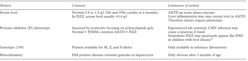
Diagnostic methods are summarised in table 1.
Phenotypes
á-1 antitrypsin is a protease inhibitor, and common Pi
variants have been named by their electrophoretic mobility. PiM, of which there are several minor variants, is the nor-mal protein. PiZ, the mutant responsible for more than 95% of cases of pulmonary and hepatic disease associated withá-1 antititrypsin deficiency, is most frequent in Scan-dinavia and progressively less common as one travels south in Europe. PiS, by contrast, is most common in the Iberian peninsula.5
Emphysema is associated both with null mutations (no protein produced) and with mutations producing defective or non-exported protein. Liver disease is associated only with those mutations that produce a peptide which forms loop sheet polymers that are retained in the liver, namely homozygotes for PiZ, PiM (Malton), and the compound heterozygotes PiZ−, PiSZ, and PiZI.6
The nomenclature of genetic variants is slightly confusing: presumed homo-zygous abnormalities such as PiZZ are conventionally referred to as PiZ unless the null gene has been excluded from the phenotype; thus PiZ might actually be PiZ− (Z
plus null), or PiZZ. The infant with á-1 antitrypsin
deficiency is considered to be PiZZ if both parents carry the Z allele; this may be confirmed by genotype.
Molecular pathology
There are several recent reviews of the molecular pathology of á-1 antitrypsin deficiency.7–9
Newly formed á-1
anti-trypsin peptide enters the endoplasmic reticulum, where a series of molecular chaperones superintends an orderly
process of glycosylation, disulphide bridge formation, and
folding. Having achieved its tertiary structure, á-1
antitrypsin leaves the endoplasmic reticulum and travels by Golgi and secretory vesicles to the plasma membrane, where it is secreted as a 55 kDa glycoprotein at a rate of approximately 34 mg/kg/day. Newly synthesisedá-1 anti-trypsin is subject to a quality control process—protein that fails to achieve the correct tertiary structure enters a
degradative pathway in which misfolded á-1 antitrypsin
remains bound to calnexin in the endoplasmic reticulum, induces conjugation with ubiquitin, and leaves the endoplasmic reticulum to be degraded in proteasomes.
The mutant peptide found iná-1 antitrypsin deficiency
of the common PiZZ phenotype (á-1 antitrypsin Z,
AAT-Z) diVers fromá-1 antitrypsin M (AAT-M) only in a single amino acid change, gly 342→lys. The eVect of this is a severe reduction in the rate at which the peptide folds.10
Slow folding allows peptide monomers to come together by a loop sheet insertion mechanism to form an AAT-Z poly-mer which is retained within the endoplasmic reticulum.12
In liver biopsies from patients with á-1 antitrypsin
deficiency, polymerised AAT-Z may be demonstrated in the endoplasmic reticulum by electron microscopy and is apparent histochemically as characteristic PAS positive, diastase resistant globules.
There are many genetic disorders in which a mutated peptide fails to achieve correct conformation and is retained in the endoplasmic reticulum. As with AAT-Z, the mutated protein may be functionally active but disease results from failure of traYcking to its correct location, whether that be the plasma membrane (for example, cystic fibrosis or sucrase-isomaltase deficiency), other organelle membranes (Wilson’s disease), or the extracellular fluid (fibrinogen, protein C, or thyroglobulin deficiency).7 8
Why is it thatá-1 antitrypsin deficiency diVers from all these disorders in that the retained mutant peptide
damages the liver cell? It might simply be that á-1
antitrypsin is synthesised in large amounts, but probably more important is the fact that polymerised AAT-Z resists degradation. It may fancifully be imagined as doing to the endoplasmic reticulum what haemoglobin S does to the erythrocyte in sickle cell anaemia.
[image:2.612.73.543.617.733.2]Why then do only 10% of PiZZ infants develop liver dis-ease? Among the suggested acquired contributory factors are the following:
Table 1 Diagnostic methods for identification ofá-1 antitrypsin deficiency
Method Comment Limitations of method
Serum level Normal 0.8 to 1.8 g/l (5th and 95th centiles at 6 months). In PiZZ, serum level usually <0.6 g/l
AATD an acute phase reactant
Liver inflammation may raise serum level in AATD. Therefore always request phenotype
Protease inhibitor (Pi) phenotype Assessed by isoelectric focusing on polyacylamide gels. Normal = PiMM; common AATD = PiZZ
Experienced lab essential; CMV infection may cause a spurious Z band
Sometimes PiZZ may spuriously appear like PiSZ in children with liver disease49
+ intrauterine infection, because PiZ babies with liver disease tend to have been small for gestational age;
+ gut derived proteases, suggesting that breast feeding might be protective;
+ autoimmunity, but liver directed antibodies are likely to be secondary to liver damage rather than primary;
+ fever, because a raised temperature increases both the
production and the polymerisation rate of á-1
anti-trypsin. Therefore, transient elevations of plasma transaminases do indeed occur during infections iná-1 antitrypsin deficient children14
;
+ cryptic hepatitis B or C infection,12
but this has not been substantiated.13
None of these acquired factors seems suYcient to
explain fully the enormous variation in hepatic phenotype. A more attractive hypothesis addresses possible genetic
diVerences in AAT-Z degradation. In transfected skin
fibroblasts from PiZZ patients who had developed liver disease, AAT-Z was degraded less rapidly than in fibroblasts from PiZZ individuals who had not developed liver disease.15
This suggests that PiZZ patients who develop liver disease have one of a number of possible inborn errors of AAT-Z clearance. It is tempting to extrapolate this concept to other traYcking disorders.
Epidemiology ofá-1 antitrypsin deficiency liver disease in childhood
In a classic piece of prospective epidemiology,14 16–20
Sveger studied 200 000 infants in 1972–74, identifying 127 PiZ infants among these and following them to the age of 18 years. Only 14 developed cholestatic jaundice in infancy. Eight more had hepatosplenomegaly and mildly disturbed liver function tests.
PROGNOSIS OF THEá-1 ANTITRYPSIN DEFICIENT CHILD WHO DOES NOT DEVELOP INFANTILE CHOLESTASIS
The frequency of abnormal liver function tests in PiZZ
individuals with á-1 antitrypsin deficiency who do not
develop jaundice or hepatomegaly in infancy falls from 60% at 6 months to a plateau of approximately 15% at 12–18 years, with no clinical evidence of liver disease (table 2). Abnormal liver function tests may be transient—most of those with abnormal liver function tests at 16 years were normal at 18 and vice versa. There are occasional reports of PiZZ children who developed new or worsening liver dysfunction in childhood, often in association with other severe illnesses: one case each of appendicitis and pneumonia in Sveger’s series, and one of pancreatitis in another report. These cases seem to be rare, and justify
excluding á-1 antitrypsin deficiency in the work up of
undiagnosed liver disease in childhood.
The prognosis of PiSZ infants is even better. None in Sveger’s series developed jaundice in infancy, and the fre-quency of abnormal liver function tests was lower through-out childhood (table 2).
The risk of cirrhosis in adults is much more diYcult to quantitate because the available data are derived either from patients known to haveá-1 antitrypsin deficient lung disease or from studies of á-1 antitrypsin deficiency in cases of cirrhosis. Thus 12% of aVected Swedish adults
identified through hospital admission had cirrhosis.21
Whether alcohol has anything to do with liver disease iná-1 antitrypsin deficiency is unknown; we are not therefore justified in insisting on abstinence but do advise modera-tion.
Whereas the heterozygous state, PiMZ, carries no risk in childhood, it may be associated with liver disease in adults. The PiMZ phenotype was found in 8% of adult transplant candidates with chronic liver failure, including 27% of those with cryptogenic cirrhosis, compared with 2–4% in
the general population.22
Inverting that statistic, however, shows that the risk of liver disease in a particular PiMZ individual must be small.
PROGNOSIS OF THEá-1 ANTITRYPSIN DEFICIENT CHILD WHO DOES DEVELOP INFANTILE CHOLESTASIS
Of the 22 clinically aVected Swedish babies withá-1 anti-trypsin deficiency, two died of cirrhosis at around 7 years, while one died of aplastic anaemia and had cirrhosis at necropsy.14 16–20
Of 74 children referred to King’s College Hospital and followed to 17 years, 20 died, 20 had cirrho-sis, 19 had persistently abnormal liver function tests, and only 15 made a complete recovery.23
However, these were patients whose infantile cholestasis was suYciently severe for tertiary referral.
A more recent review24
has shown, perhaps unsurpris-ingly, that those children who progressed to end stage liver disease had more severe abnormalities in infancy. They were more likely to have remained jaundiced for more than 6 weeks, to have had higher aspartate aminotransferase at presentation, and to have had more severe changes on the initial biopsy (comprising severe bile duct reduplication, severe fibrosis with bridging septa, and established cirrho-sis). Nevertheless, one should be cautious about the
outlook in an individual jaundiced infant with á-1
antitrypsin deficiency. Volpertet alpoint out that among aVected children with established liver disease in whom transplant was not immediately indicated is a group that remains clinically stable for a prolonged period after the presence of cirrhosis or portal hypertension is established.13
These are not initially distinguishable from those children whose liver function declines more rapidly.
Three diagnostic points in the cholestatic infant merit emphasis. First, the early liver biopsy appearances, with prominent portal tract changes and with PAS positive, dia-stase negative granules not yet apparent, may mimic those of biliary atresia. Giant cell transformation of hepatocytes
is uncommon in á-1 antitrypsin deficiency. Second, the
plasma concentration of á-1 antitrypsin in a deficient
patient may be raised as an acute phase reactant, so theá-1 antitrypsin phenotype must be obtained. Third,
determin-ing theá-1 antitrypsin phenotype may be the most time
consuming laboratory investigation, so must (like the coagulation screen) be requested promptly.
The indications for liver transplantation are the same as for other hepatic disorders. After biliary atresia,á-1 anti-trypsin deficiency is the most frequent reason for liver transplantation in childhood. The lung does not appear to pose any particular problem, and the perioperative and postoperative care does not diVer from the usual routines. Outcomes are good.25 26
Orthotopic liver transplantation
from a donor parent has been successfully performed.27
After transplant the recipient manifests the phenotype of the donor and is expected not to be at risk of emphysema. What is the risk of severe liver disease in the subsequent PiZ sibling of a severely aVected proband? Psacharopoulos
et alreported that the risk was 78%,23
[image:3.612.315.541.656.729.2]while others have reported less pessimistic figures. Parents who have had a
Table 2 Frequency (%) of abnormality of alanine transaminase and/orã
glutamate transferase in prospectively identified Swedishá-1 antitrypsin
deficient children who did not develop infantile cholestasis or hepatomegaly
Age PiZZ PiSZ
3 months 52 24
6 months 60 5
4 years 41 2
8 years 36 2
12 years 15 2
16 years 17 8
18 years 12 15
No clinical features of liver disease found at any age group.4 16–20
severely aVected child are more likely to opt for termination of a subsequent PiZ fetus, so it may be diYcult to acquire further data to refine this risk.
Late haemorrhagic disease in infancy
Whereas early neonatal haemorrhagic disease in breast fed infants is prevented by oral vitamin K at birth, cases of late vitamin K dependent bleeding continue to occur, usually in
babies with undiagnosed cholestasis, of which á-1
antitrypsin deficiency and biliary atresia are the most com-mon. Of 182 000 babies in the United Kingdom North East region given 1 mg of oral vitamin K at birth and, for the breast fed babies, recommended to receive three further 1 mg doses at fortnightly intervals, four developed late haemorrhagic disease.28
Two of these had not received vitamin K and two hadá-1 antitrypsin deficiency. Given that the breast feeding rate was approximately 30% (Wari-yar U, personal communication) and the incidence ofá-1 antitrypsin deficiency is approximately 1:1800, it seems that haemorrhagic disease occurred in 2/30 breast fed infants withá-1 antitrypsin deficiency.
Of 332 686 Swedish babies, about 80% of whom received 1 mg or 2 mg of oral vitamin K at birth, and among whom breast feeding rates were reportedly high, 17 developed late haemorrhagic disease.29
Fifteen of these had
cholestatic liver disease, comprising three with á-1
antitrypsin deficiency, five with biliary atresia, and seven with other conditions. Thus 1/35á-1 antitrypsin deficient infants bled.á-1 Antitrypsin deficiency was also responsi-ble for cases of late haemorrhagic disease in other reports.30 31
This tragic consequence of á-1 antitrypsin
deficiency and other infantile cholestases is prevented by the more physiological Dutch protocol of giving breast fed babies 1 mg of vitamin K at birth and 25 µg daily from 2–13 weeks of life, though late haemorrhagic disease may still occur because of failure of compliance.32
Despite the liver disease, the grossly prolonged pro-thrombin time dramatically improves within hours of giving parenteral vitamin K.
Subsequent aVected siblings of an á-1 antitrypsin
deficient proband should always receive intramuscular vitamin K at birth.
Lung disease iná-1 antitrypsin deficiency
The damage wrought by uninhibited neutrophil elastase in the lung takes many years to manifest itself clinically.33
The characteristic pathology seen iná-1 antitrypsin deficiency is emphysema, caused by loss of elastic recoil. Children and adolescents withá-1 antitrypsin deficiency have not been shown to have significant lung function abnormalities.34 35
Although a study of aVected children with liver disease suggested a tendency to hyperinflation,36
this was not found in Sveger’s subsequent study of 150 adolescents.34
After the age of 30–35 years there is an accelerated decline in forced expiratory volume in one second (FEV1), which is
considerably worsened by cigarette smoking. In a non-smoker, symptoms are generally seen at around 50 years of age, while smokers will be symptomatic by 30–40 years. Although life expectancy is more diYcult to estimate with accuracy, a combination of three studies gives a mean age of death of 50 years in smokers, compared with 66 years in non-smokers.37–39
Interestingly, only 3% of adolescents with á-1 antitrypsin deficiency smoked in Sveger’s study,34
sug-gesting that health education may be eVective in this group of children.
Paediatricians tend to include aná-1 antitrypsin pheno-type in the panel of tests for unexplained pulmonary symptoms. There is little evidence in support of this, although it is theoretically possible that a coexisting inflammatory disease might be worsened byá-1 antitrypsin
deficiency, even in childhood. In a study of adults with bronchiectasis there was no increase in the prevalence of á-1 antitrypsin deficiency alleles, but more emphysema if both diseases coexisted.40
If á-1 antitrypsin deficiency is found in a child with lung symptoms it should not therefore be accepted as the underlying cause of the problem, but it might be an exacerbating factor in disease progression.
Prospects for pharmacological treatment ofá-1 antitrypsin deficiency
A logical therapeutic ambition is to devise a way of moving AAT-Z from the endoplasmic reticulum of the liver, where it causes damage, to the plasma, where its antiprotease activity—though less than the wild type AAT-M—would benefit the lung. This might be achieved either by inhibit-ing the polymerisation of AAT-Z41
or by chaperoning the misfolded AAT-Z from endoplasmic reticulum to the secretory pathway.2
The latter concept is common to all the endoplasmic reticulum retention diseases. Unlike cystic fibrosisÄF508, incubation of cells at lower temperature does not improve the secretory defect in á-1 antitrypsin deficiency, but there are promising results from chemical chaperones. In cultured mouse hepatocytes or transfected skin fibroblasts, glycerol and 4-phenylbutyric acid achieved increases of secretion of AAT-Z, from 3% in controls to 25% and 17%, respectively. 4-Phenylbutyric acid also achieved increased plasma levels, reaching 20–50% of the levels present in PiM mice with transgenicá-1 antitrypsin deficiency.42
Sodium phenylbutyrate is already in clinical use in urea cycle defects and is a potential form of treatment. Its mode of action, however, remains to be defined. Both the protein translation inhibitor cyclohex-imide and the specific inhibitor of proteasome function, lactacystin, prevented intracellular degradation of AAT-Z and partially restored its vesicular transport in transfected
CHO cells and human alveolar macrophages.43
In other cell culture work, glucosidase and mannosidase inhibitors also achieved increased secretion.44
Augmentation therapy with á-1 antitrypsin given
intravenously has been shown to restore serum and sputum antiprotease levels. Obviously this will be of no benefit to infants with liver disease, but oVers the prospect of preven-tion or treatment of lung disease. In non-randomised observational studies, intravenous augmentation therapy
has been asociated with slower declines in FEV1
4
and improved survival, but as yet there is no evidence of long term benefit from a randomised controlled trial. If benefits are seen, they appear to be confined to a subgroup of patients with FEV1below 65% of predicted. The one small
randomised trial which compared 28 treated patients and 28 controls showed marginal benefits in the appearances on computed tomography, but no significant diVerences in
lung function after three years of treatment.46
The treatment is not currently licensed in the United Kingdom.
Recombinant á-1 antitrypsin is now becoming available,
and trials of its administration by nebuliser are planned.
However, the child with á-1 antitrypsin deficiency may
never develop significant lung disease if other damaging factors are avoided, and replacement therapy (where avail-able) is currently only advised for significant or rapidly progressive emphysema rather than as pre-emptive treat-ment.
There are promising early results with gene therapy. A
normal á-1 antitrypsin gene in a plasmid–cationic
liposome complex was delivered to one nostril of each of five aVected patients. á-1 Antitrypsin protein increased, and interleukin 8 (as a marker of inflammation) decreased, in nasal lavage fluid, with a peak eVect on day 5.47
AAT-Z mouse liver,48
that clearly will not correct the cellu-lar defect.
Conclusions
Late haemorrhagic disease remains a hazard for breast fed á-1 antitrypsin deficient babies with the current vitamin K administration protocol. Continued vigilance is necessary if cholestasis in infancy is to be promptly detected. We have
good evidence about the hepatic prognosis of á-1
antitrypsin deficiency, and recent knowledge of the molecular pathology provides hope for newer therapeutic approaches to this and other endoplasmic reticulum reten-tion disorders. The indicareten-tions for, and outcomes of, liver transplantation have been delineated.
What advice should we give to young people withá-1
antitrypsin deficiency? The core message has not changed since it was baldly stated by Januset alin 198539
: “If they smoke, they will develop crippling emphysema by middle age; if they do not smoke, they have a reasonable likelihood of a full life span.” Until a simple and safe treatment is available, this remains the only way to ensure respiratory health iná-1 antitrypsin deficiency.
R A PRIMHAK M S TANNER
Institute of Child Health, University of SheYeld, SheYeld Children’s Hospital, Western Bank, SheYeld S10 2TH, UK
Correspondence to: Dr Primhak email: [email protected]
Accepted 8 March 2001
1 Parmar JS, Lomas DA. Alpha-1-antitrypsin deficiency, the serpinopathies and conformational disease.J R Coll Physicians Lond2000;34:295–300. 2 Elzouki AN, Lindgren S, Nilsson S, et al. Severe alpha1-antitrypsin
deficiency (PiZ homozygosity) with membranoproliferative glomerulone-phritis and nephrotic syndrome, reversible after orthotopic liver transplan-tation.J Hepatol1997;26:1403–7.
3 O’Riordan K, Blei A, Rao MS,et al. Alpha 1-antitrypsin deficiency-associated panniculitis: resolution with intravenous alpha 1-antitrypsin administration and liver transplantation.Transplantation1997;63:480–2. 4 Mazodier P, Elzouki AN, Segelmark M, et al. Systemic necrotizing
vasculitides in severe alpha1-antitrypsin deficiency.Q J Med1996;89:599– 611.
5 Hutchison DC. Alpha 1-antitrypsin deficiency in Europe: geographical dis-tribution of Pi types S and Z.Respir Med1998 Mar;92:367–77. 6 Mahadeva R, Chang WS, DaVorn TR,et al.Heteropolymerization of S, I,
and Z alpha1-antitrypsin and liver cirrhosis.J Clin Invest1999;103:999– 1006.
7 Kuznetsov G, Nigam SK. Folding of secretory and membrane proteins.N Engl J Med1998;339:1688–95.
8 Marcus N, Teckman JH, Perlmutter DH. Alpha1-antitrypsin deficiency: from genotype to childhood disease. J Pediatr Gastroenterol Nutr
1998;27:65–74.
9 Bross P, Corydon TJ, Andresen BS, et al. Protein misfolding and degradation in genetic diseases.Hum Mutat1999;14:186–98.
10 DaVorn TR, Mahadeva R, Elliott PR,et al.A kinetic mechanism for the polymerization of alpha1-antitrypsin.J Biol Chem1999;274:9548–55. 11 Sivasothy P, DaVorn TR, Gettins PG,et al.Pathogenicá1-antitrypsin
poly-mers are formed by a reactive loop-{beta}-sheet A linkage.J Biol Chem
2000;275:33663–8.
12 Propst T, Propst A, Dietze O,et al.High prevalence of viral infection in adults with homozygous and heterozygous alpha 1-antitrypsin deficiency and chronic liver disease.Ann Intern Med1992;117:641–5.
13 Volpert D, Molleston JP, Perlmutter DH. Alpha1-antitrypsin deficiency-associated liver disease progresses slowly in some children.J Pediatr Gastro-enterol Nutr2000;31:258–63.
14 Sveger T. Prospective study of children with alpha 1-antitrypsin deficiency: eight-year-old follow-up.J Pediatr1984;104:91–4.
15 Wu Y, Whitman I, Molmenti E,et al.A lag in intracellular degradation of mutant alpha 1-antitrypsin correlates with the liver disease phenotype in homozygous PiZZ alpha 1-antitrypsin deficiency.Proc Natl Acad Sci USA
1994;91:9014–18.
16 Sveger T. Liver disease in alpha1-antitrypsin deficiency detected by screen-ing of 200,000 infants.N Engl J Med1976;294:1316–21.
17 Sveger T. Alpha 1-antitrypsin deficiency in early childhood.Pediatrics1978;
62:22–5.
18 Sveger T, Thelin T. Four-year-old children with alpha 1-antitrypsin deficiency. Clinical follow-up and parental attitudes towards neonatal screening.Acta Paediatr Scand1981;70:171–7.
19 Sveger T. The natural history of liver disease in alpha 1-antitrypsin deficient children.Acta Paediatr Scand1988;77:847–51.
20 Sveger T, Eriksson S. The liver in adolescents with alpha 1-antitrypsin defi-ciency.Hepatology1995;22:514–17.
21 Larsson C. Natural history and life expectancy in severe alpha1-antitrypsin deficiency, PiZ.Acta Med Scand1978;204:345–51.
22 Graziadei IW, Joseph JJ, Wiesner RH,et al.Increased risk of chronic liver failure in adults with heterozygous alpha1-antitrypsin deficiency. Hepatol-ogy1998;28:1058–63.
23 Psacharopoulos HT, Mowat AP, Cook PJ,et al.Outcome of liver disease associated with alpha 1 antitrypsin deficiency (PiZ). Implications for genetic counselling and antenatal diagnosis.Arch Dis Child1983;58:882–7. 24 Francavilla R, Castellaneta SP, Hadzic N,et al.Prognosis of alpha-1-antitrypsin deficiency-related liver disease in the era of paediatric liver transplantion.J Hepatol2000;32:986–92.
25 Nemeth A. Liver transplantation in alpha(1)-antitrypsin deficiency.Eur J Pediatr1999;158(suppl 2):S85–8.
26 Prachalias AA, Kalife M, Francavilla R,et al. Liver transplantation for alpha-1-antitrypsin deficiency in children.Transpl Int2000;13:207–10. 27 Colombani PM, Lau H, Prabhakaran K,et al.Cumulative experience with
pediatric living related liver transplantation.J Pediatr Surg2000;35:9–12. 28 Wariyar U, Hilton S, Pagan J,et al.Six years’ experience of prophylactic oral
vitamin K.Arch Dis Child Fetal Neonatal Ed2000;82:F64–8.
29 Ekelund H. Late haemorrhagic disease in Sweden 1987–89.Acta Paediatr Scand1991;80:966–8.
30 McNinch AW, Tripp JH. Haemorrhagic disease of the newborn in the Brit-ish Isles: two year prospective study.BMJ1991;303:1105–9.
31 von Kries R, Göbel U. Vitamin K prophylaxis and vitamin K deficiency bleeding (VKDB) in early infancy.Acta Paediatr1992;81:655–7. 32 Cornelissen M, von Kries R, Loughnan P,et al.Prevention of vitamin K
deficiency bleeding: eYcacy of diVerent multiple oral dose schedules of vitamin K.Eur J Pediatr1997;156:126–30.
33 Brantly M, Paul L, Miller B,et al.Clinical features and history of the destructive lung disease associated with alpha1 antitrypsin deficiency of adults with pulmonary symptoms.Am Rev Respir Dis1988;138:327–36. 34 Sveger T, Piitulainen E, Arborelius MJ. Lung function in adolescents with
alpha1-antitrypsin deficiency.Acta Paediatr1994;83:1170–3.
35 Wall M, MoeE, Eisenberg J,et al.Long-term follow-up of a cohort of chil-dren with alpha-1-antitrypsin deficiency.J Pediatr1990;116:248–51. 36 Hird MF, Greenough A, Mieli-Vergani G,et al.Hyperinflation in children
with liver disease due to alpha-1-antitrypsin deficiency.Pediatr Pulmonol
2000;11:212–16.
37 Black L, Kueppers F. Alpha1 antitrypsin deficiency in nonsmokers.Am Rev Resp Dis1978;117:421–8.
38 Tobin M, Cook P, Hutchison D. Alpha1-antitrypsin deficiency. the clinical and physiological features of subjects homozygous for Pi type Z. Br J Dis Chest1983;77:12–27.
39 Janus E, Phillips N, Carrell R. Smoking, lung function andá-1 antitrypsin deficiency.Lancet1985;i:152–4.
40 Cuvelier A, Muir JF, Hellot MF,et al.Distribution of alpha(1)-antitrypsin alleles in patients with bronchiectasis.Chest2000;117:415–19.
41 Elliott PR, Pei XY, DaVorn TR,et al.Topography of a 2.0 A structure of alpha1-antitrypsin reveals targets for rational drug design to prevent conformational disease.Protein Sci2000;9:1274–81.
42 Burrows JA, Willis LK, Perlmutter DH. Chemical chaperones mediate increased secretion of mutant alpha 1-antitrypsin (alpha 1-AT) Z: a poten-tial pharmacological strategy for prevention of liver injury and emphysema in alpha 1-AT deficiency.Proc Natl Acad Sci USA2000;97:1796–801. 43 Novoradovskaya N, Lee J, Yu ZX,et al.Inhibition of intracellular
degrada-tion increases secredegrada-tion of a mutant form of alpha1-antitrypsin associated with profound deficiency.J Clin Invest1998;101:2693–701.
44 Marcus NY, Perlmutter DH. Glucosidase and mannosidase inhibitors mediate increased secretion of mutant alpha1 antitrypsin Z.J Biol Chem
2000;275:1987–92.
45 Seersholm N, Wercker M, Barik N,et al.Doesá1-antitrypsin augmentation therapy slow the annual decline in FEV1 in patients with severe
á1-antitrypsin deficiency?Eur Respir J1997;10:2260–3.
46 Dirksen A, Dijkman J, Madsen F. A randomised clinical trial of
á1-antitrypsin augmentation therapy.Am J Resp Crit Care Med1999;160: 1468–72.
47 Brigham KL, Lane KB, Meyrick B,et al.Transfection of nasal mucosa with a normal alpha1-antitrypsin gene in alpha1-antitrypsin-deficient subjects: comparison with protein therapy.Hum Gene Ther2000;11:1023–32. 48 Zhang G, Song YK, Liu D. Long-term expression of human
alpha1-antitrypsin gene in mouse liver achieved by intravenous administration of plasmid DNA using a hydrodynamics-based procedure.Gene Ther2000;7: 1344–9.
49 Whitehouse DB, Lovegrove JU, Mieli-Vergani G,et al.“SZ like” alpha 1-antitrypsin phenotypes in PI ZZ children with liver disease.Ann Hum Genet1994;58:1–7.