Int. J. Electrochem. Sci., 12 (2017) 7103 – 7120, doi: 10.20964/2017.08.24
International Journal of
ELECTROCHEMICAL
SCIENCE
www.electrochemsci.org
Direct Electron Transfer of Glucose Oxidase in Carbon Paper
for Biofuel Cells and Biosensors
Zongqian Hu1, Zepeng Kang2, Chao Yu1, Bing Wang3, Shuqiang Jiao2,*, Ruiyun Peng1,* 1
Beijing Institute of Radiation Medicine, Beijing, 100850, P.R. China. 2
State Key Laboratory of Advanced Metallurgy, University of Science and Technology Beijing, Beijing, 100083, P.R. China.
3
Beijing Institute of System Engineering, Beijing, 100101, P.R. China. *
E-mail: pengry@bmi.ac.cn, sjiao@ustb.edu.cn
Received: 17 February 2017 / Accepted: 27 May 2017 / Published: 12 July 2017
Advanced bioelectronic devices, such as high-power biofuel cells (BFCs) and highly efficient biosensors, are limited by the difficulty of electron transfer between enzymes and electrodes. Previously reported methods for achieving electron transfer from enzymes to electrodes have relied on the use of complex biomolecule immobilization procedures, complicated matrix materials, or enzyme engineering, resulting in potential relative toxicity, high cost, as well as limited stability. Here, we report a facile method for the rapid preparation of a glucose oxidase (GOx) anode with direct electron transfer (DET) for glucose BFCs and biosensors. GOx is directly incorporated into pretreated carbon paper (CP) by adjusting the pH of the incubation medium during the immobilization process. Eexcellent bioelectrocatalytic activity is obtained when GOx is incorporated into CP near the pI of GOx. The electron transfer rate constant ( ) and the apparent Michaelis-Menten constant ( ) are estimated to be 12.08 ± 1.0 s-1 and 0.13 ± 0.01 mM, respectively. These findings may be extended to the development of highly conductive nanomaterials and the immobilization of other enzymes or biomolecules, providing a promising platform for the development of BFCs, biosensors, and other bioelectrochemical devices.
Keywords: direct electron transfer; electrostatic adsorption; glucose oxidase; biofuel cells
1. INTRODUCTION
practical value, findings from such studies could aid in understanding the fundamental mechanisms of biological redox reactions [1-5]. Owing to its high sensitivity to glucose, glucose oxidase (GOx) has received considerable attention as a potential enzymatic component of BFCs, and for use in real-time glucose monitoring related applications. However, establishing direct electron transfer (DET) between the immobilized GOx and conventional electrodes is a challenge due to the inaccessibility of GOx active centers, which are deeply embedded within a thick insulating protein shell [6,7]. Many attempts at immobilization have been employed to reduce the distance and to improve electron transfer between the active center of GOx and the electrodes [8-11]. These attempts have incorporated various immobilization methods (e.g., physical adsorption [12], cross-linking [13], covalent binding entrapment in gels [14] or membranes [15], self-assembly [16] and layer-by-layer [17] processes), and inorganic/organic matrixes (e.g., metal nanoparticles [18], nanostructured metal oxides [19], carbon nanotubes [10, 20-24], grapheme [25-28], semiconductor nanoparticles [29-32] and conducting polymer nanowires [33]).
Although DET can be achieved on enzyme-modified bioelectrodes, the high cost and complex immobilization procedures for these materials are barriers to their mass commercialization. Therefore, there is great interest in developing a facile procedure enabling enhanced DET to electrodes from immobilized GOx in its natural conformation. Characteristics of the immobilization process, such as pH and temperature, affect the biological activity of the immobilized enzyme and the affinity between the enzyme and the support matrix [34-36]. Differences in the surface charge distribution between an enzyme and a support matrix lead to spatial conformation changes of the enzyme molecule during immobilization [36]. Stanciuc and colleagues recently explored the behavior of enzymes under different pH and temperature conditions, in terms of the relationships among processes, structures, and functions. The enzyme molecule adopted a more flexible conformation under acidic conditions, with the active center being exposed on the molecular surface at optimum pH. Thus, pH treatment can affect the activity and stability of the enzyme molecule [37].
Inspired by previous studies on the behavior of enzymes under different conditions, we developed a facile method for the rapid preparation of a GOx anode with DET for glucose BFCs and biosensors. This method involves adjusting the pH of the incubation medium during the immobilization process. For the support matrix, we used carbon paper (CP) pretreated with a mixed acid solution, which provided good physical and electrical properties and facilitated easy specimen preparation. Using this method, we successfully realized DET from the immobilized GOx to the modified electrode, as well as good bioelectrocatalytic activity for glucose oxidation. The proposed method is highly recommended as a promising platform for advanced bioelectronic device applications.
2. EXPERIMENTAL 2.1. Reagents and materials
activity assay kit, paraformaldehyde, glutaraldehyde, hexamethyldisilazane, 0.1 M cacodylate buffer (pH 7.4), were supplied from Sigma-Aldrich (St. Louis, MO, USA) and used without further purification. Sodium hydrogen phosphate, sodium dihydrogen phosphate, phosphoric acid (85.11%), nitric acid (65%), sulfuric (98.3%), alcohol(99.8%), calcium choride (Sinopharm Chemical Reagent Co., Ltd., China) were of analytical grade or better quality and used without further purification. Milli-Q ultrapure water (Millipore, 18.2 MΩ cm) was used throughout the study.
2.2. Preparation of GOxpH=x/CP
CP was used as the support matrix to prepare the modified electrodes. First, CP was cut into a square with an area of 0.5 cm2. To obtain a highly specific surface with a three-dimensional (3D) architecture, the CP square was treated under air for 10 min with a mixed solution of concentrated sulfuric and nitric acids at 3:1 v/v. The resulting pretreated carbon paper sample was washed with abundant Milli-Q ultrapure water until the pH of the wash fluid was neutral. The washed CP was placed in an oven at 120 for 1h.
Next, GOx was attached to the pretreated CP (GOxpH=x/CP, where x is the pH of the incubation medium). Attachment was achieved by adding 20 mg mL-1 GOx at various pH values (2, 3, 4, 5, and 7) to the CP and incubating the mixture in a refrigerator overnight. Then, the GOxpH=x/CP samples were washed with 0.1 M PBS (pH 7.2). The CP sample with immobilized GOx was stored in PBS (0.1 M, pH 7.2) at 4 , and used as the anodic electrode for enzymatic BFCs.
2.3. Instruments and Characterizations
The distribution of GOx on the CP surface was studied using confocal laser scanning fluorescence microscopy (PerkinElmer UltraVIEW VoX) and field emission scanning electron microscopy (JSM-6701F, JEOL). In this instance a fluorescent conjugate was used for labeling the GOxpH=x/CP (x=2, 3, 7) electrode surface using the covalent bond between the amino groups of GOx and the carboxyl groups of biotin. The background control was performed using the biotinylated fluorescein surface of the CP. The GOxpH=x/CP electrode surface was labeled in two steps. An incubation step with 100 μL of biotin/streptavidin-Cy5 solution (10 mM of biotin and 0.02 mg mL-1
of streptavidin-Cy5 fluorescent dye) in PBS (0.1 M, pH 7.2) at 25 ± 2 during 60 min was performed. After that, three washing steps with PBS (0.1 M, pH 7.2) at 25 ± 2 during 5 min were performed [38].
with gold (ISMSCD-2, sputter coater device). As a control, the bare CP and pretreated CP were treated using the same procedures.
Zeta potentials of pretreated CP in PBS at various pH values were characterized by dynamic light scattering on a Zeta Sizer Nano ZS instrument (Powereach, JS94H, Shanghai). A CHI 660E workstation was used to collect electrochemical data. All experiments were conducted at 25 ± 2 with a conventional three-electrode system under the cyclic voltammetry (CV) cycle, electrochemical impedance spectroscopy (EIS) and chronoamperometry (CA). Electrodes included the prepared electrode as the working electrode, a platinum sheet with a surface area of 2 cm2 as the counter electrode, and Ag/AgCl (3 M KCl) as the reference electrode. Except in the pH-dependent experiments, 0.1 M PBS (pH 7.2) was employed as the supporting electrolyte.
Only β-D-glucose acts as a substrate for the enzymatic reaction with GOx. Therefore, the glucose stock solution (prepared in PBS) was subjected to mutarotation at 4 for at least 24 h before use. Before the CV experiments were performed, the electrolyte was deoxygenated by bubbling highly purified argon through the solution at least 30 min. An argon atmosphere was maintained over the solution during the electrochemical measurements. The apparent Michaelis-Menten constant ( ) of GOx was measured by the chronoamperometric i-t response. Aliquots of β-D-glucose stock solution were added into 30 mL of stirred PBS in an electrochemical cell. The mean value of the anodic current (in μA) was plotted against the β-D-glucose concentration (in mM).
2.4. Design and testing of the BFC
To maximize the bioelectrocatalytic activity of the prepared bioanode, a hybrid BFC design was employed. This design consisted of the GOxpH=x/CP anode and an air-breathing Pt cathode. The anode fuel reservoir and the cathode holder of the cell were connected with a salt bridge. The anode fuel reservoir was operated at room temperature and fed with a fuel solution of β-D-glucose stock solution (0.1 M, pH 7.2) under highly purified saturated argon. A 2 cm2 platinum sheet, used as the cathode, was placed in Britton-Robinson buffer solution (B-R buffer solution, pH 3, prepared with equimolar of 0.04 M phosphoric acid, boric acid and acetic acid; the pH was adjusted with 0.2 M NaOH solution) under highly purified saturated oxygen. To minimize the negative impact of the cathode on the overall performance of the BFC, the working temperature of the Pt cathode was increased to 90 ± 10 . The current density and power density of the BFC were calculated using data from the polarization curve.
3. RESULTS AND DISCUSSION
3.1. Surface characteristics of GOxpH=x/CP
with its larger specific area and 3D structure, can potentially be used as a support matrix for GOx immobilization.
Figure 1. (A) Confocal laser scanning fluorescence microphotograph for the GOxpH=2, 3, 7/CP. The laser excitation is at 640 nm and the voltage is 360 V. (B) FESEM images of the surface of the GOxpH=2, 3, 7/CP. (C) Schematic representation on the effect of pH on the adsorption of GOx in CP. The isoelectric point (pI) of GOx is 4.2, where the protein net charge is expected to be neutral [39], above this value, for example, at a pH of 5 and 7, it is negative, and below this value, is positive (i.e. 4, 3 and 2).
[image:5.596.59.547.117.576.2]
fluorescence area corresponds to GOx bonding with the fluorescent marker. It indicates that the distribution of GOx on the CP surface under acidic conditions is much greater than that under neutral conditions. The FESEM images of the GOxpH=x/CP also indicate that GOx adsorbed on the CP surface with different aggregate morphologies depends on the pH of the incubation medium (Figure 1B), and the adsorption of the immobilized GOx increases with decreasing pH of the incubation medium.
These differences in distribution and morphology may be related to the surface charge distribution between the GOx molecules and the CP [39]. The isoelectric point (pI) of GOx is 4.2 whereas the protein net charge is expected to be neutral, while above this value, for example at an pH of 5 and 7, it is negative, and below this value, it is positive (i.e. 4, 3 and 2) [39]. Zeta potentials of the CP surface in the GOx solution at all tested pH values are negative (Table S1). Considering this fact it can be argued that since at a pH of 4, 3 and 2, where the GOx molecules are positively charged, a higher adsorption is expected to occur. This is due to an increase in the hydrophobic interactions and attractive forces between GOx (positive) and the pretreated CP surface (negative), creating a highly packed GOx layer on the CP surface resulting in higher adsorption, see Figure 1C. However, a thick layer of GOx film was formed on the CP surface when the pH was very low (pH 2; Figure 1A and Figure 1B). This thick insulating layer of GOx hinders electron transfer from the GOx active center to the electrode surface. The results from the FESEM are consistent with those from confocal laser scanning fluorescence microscopy.
3.2. DET of GOxpH=x/CP
The direct electrochemical catalysis reaction of flavin adenine dinucleotide (FAD/FADH2) is a crucial symbol of DET between GOx and the electrodes as well as for the cofactors that are the main places to complete the redox reaction where two electrons and two protons are transferred [40]. The reaction mechanism is shown in formula (I):
GOx (FAD) + 2H+ + 2e- GOx (FADH2) (I)
[image:7.596.114.481.77.348.2]
Figure 2. CVs of the GOxpH=2,3,4,5,7/CP in O2-free 0.1 M PBS (pH 7.2) with the potential scan rate is 10 mV s-1.
[image:7.596.114.483.427.701.2]
Figure 4. CVs of the CP and the GOxpH=3/CP electrodes in Ar-saturated 0.1 M PBS.
Electrochemical impedance spectroscopy was used to determine the electron transfer resistance between the GOx and the electrodes [38, 40]. In the Nyquist plots of Figure 3, a semicircle in the high frequency region and a straight line in the low frequency region are displayed. Here, each semicircle diameter is used to estimate the interfacial electron transfer resistance (Rct) of the GOx [3, 21, 40]. Obviously, the Rct of GOxpH=3/CP is estimated to be minimum, indicating significantly improved electrical conductivity of GOxpH=3/CP compared with that of GOxpH=2,4,5,7/CP. The achievement of good electrical coupling and significantly improved electron transfer resistance of GOxpH=3/CP can be attributed to the appropriate surface charge distribution and the one-step facile immobilization method of the bioanode. Therefore, we chose GOxpH=3/CP as the optimal electrode in the following experiments.
The DET of GOxpH=3/CP was investigated and characterized by CVs in 0.1 M PBS (pH 7.2) with a scan rate of 10 mV s-1. As depicted in Figure 4, the GOxpH=3/CP electrode exhibits a pair of well-defined and quasi-reversible redox peaks that originated from the reversible reaction of FAD while the CP does not show any redox peaks. This shows that there is a distinct redox peak associated with FAD within GOx, further indicating the successful achievement of DET between the GOx active center and the electrode.
3.3. Bioelectrocatalytic activity of GOxpH=3/CP
actually used in air state while carrying out the glucose oxidation reaction at the anode of the EBC, it is necessary to investigate the influence of glucose and O2 on the reactivity of GOxpH=3/CP. The following are reactions that are associated with the reaction of GOxpH=3/CP [22, 41, 42]:
Glucose + GOx (FAD) →Gluconolactone + GOx (FADH2) (II) O2 + GOx (FADH2) → H2O2 + GOx (FAD) (III)
Figure 5. CVs of the GOxpH=3/CP in Ar-saturated PBS, O2-saturated PBS, Ar-saturated PBS containing 0.1 M glucose, and O2-saturated PBS containing 0.1 M glucose. Scan rate is 10 mV s-1, and 0.1 M PBS (pH 7.2) is used as the electrolyte.
Figure 5 shows impact of glucose on the redox reaction of FAD in an O2-free state and in air-state . For the test of the O2-free state, 0.1 M glucose was provided and the result was compared with the result of the test performed without the provision of glucose. As shown, the cathodic peak of FAD (forward reaction of reaction I) is decreased, while the anodic peak of FAD (backward reaction of reaction I) remains unchanged. This indicates that when glucose is provided, some immobilized GOx participate in the glucose oxidation (reaction II). This glucose sensitivity therefore proves our DET GOxpH=3/CP bioanode can function as a sensitive glucose sensor without any mediators.
[image:9.596.128.483.134.423.2]
superposition of reaction I, II and III occurs. Glucose oxidation (reaction II) produces GOx (FADH2), and this relieves the need of GOx (FADH2) provided from the electrode for O2 reduction (reaction III). So the anodic peak of FAD (backward reaction of reaction 1) is increased [40, 43].
These results further demonstrate that the GOx molecules immobilized at pH 3 maintain their natural biocatalytic activities toward the substrate (glucose), again indicating that the GOxpH=3/CP prepared by this facile method can be used as a bioanode for glucose-based BFC and sensors [40].
3.4. ks and of GOxpH=3/CP
Figure 6A shows the CVs of the GOxpH=3/CP electrode in 0.1 M Ar-saturated PBS with the pH increasing from 5 to 9 at a scan rate of 10 mV s-1. The value of varies linearly relativity as the pH increases with a slope nearly equal to the theoretical value of 59 mV pH-1 (insert of Figure 6A), suggesting a quasi-reversible and surface-controlled electrochemical reaction with an equal number (n = 2) [23, 25, 27, 29, 42].
Figure 6. (A) CVs of the GOxpH=3/CP in Ar-saturated 0.1 M PBS with pH values of 5, 6, 7, 8 and 9. The inset is the plot of formal potential vs. pH. The scan rate is 10 mV s-1. (B) CVs of the GOxpH=3/CP at different scan rates in Ar-saturated 0.1 M PBS (pH 7.2). Inset is the relationship between the peak current and the scan rate.
Another way to probe the redox reaction of GOxpH=3/CP is to calculate the electron transfer rate constant (ks). As ks increases, the electron transfer rate increases. This promotes the redox reaction of FAD within GOx. ks can be measured by Laviron theory [27, 44],
ks = mnFv/RT (IV)
[image:10.596.83.516.351.511.2][image:11.596.172.426.73.656.2]
CV measurements were used to determine the value of for GOxpH=3/CP (Figure 6B). The calculated ks value, 12.08 ± 1.0 s-1, is larger than the value reported for a modified GOx electrode in a conductive nanomaterial [45-49]. It indicates that the proposed fabrication method for the enzymatic modified electrode promotes DET and an increased electron transfer rate.
The small value of △Ep for GOxpH=3/CP is 40 mV at a scan rate of 1 mV s-1 (Figure S2). This agrees well with the theoretical value, and indicates a fast electron transfer process [27]. All of the results indicate that the DET of GOx could be achieved by reducing the pH of the incubation medium during the immobilization process. The CV of GOxpH=3/CP was scanned continuously in 0.1 M PBS (pH 7.2), the voltammetric response remains almost constant even after 50 cycles (see Figure S3), indicating that the DET of GOx immobilized on the surface of CP has good stability.
As shown in the inset of Figure 6B, the redox peak current increases linearly as the potential scan rate increases from 5 to 50 mV s-1, further demonstrating that GOxpH=3/CP is a surface reaction-confined system [50]. Faraday’s law is used to estimate the average surface concentration of electroactive GOx (Γ*
, in mol cm-2) in the GOxpH=3/CP electrode from CVs [51]. Ip = nFQv/4RT = n2F2AΓ*v/4RT (V) This equation can be transformed as follows [40]:
Γ*
= Q/nFA (VI)
where Q is the charge consumed in CVs, n is the electron transfer number, F is the Faraday constant, and A is the effective surface area of the electrode. The calculated Γ* of the GOxpH=3/CP is 7.80 × 10⁻⁹ mol cm-2, which is one or two orders of magnitude larger than values reported previously [27, 43, 46, 48, 49]. This shows that the immobilized GOx incubated under acidic conditions (pH 3) has a perfect affinity to the CP and can be maintained with high catalytic activity and stability.
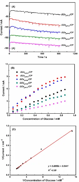
Amperometry was used to determine the relationship between the electrocatalytic reduction current and the glucose concentration. Figure 7A shows the chronoamperometric i-t response of the GOxpH=x/CP electrodes to glucose, where the amperometer was operated at -0.46V in 30 mL of Ar-saturated 0.1 M PBS.
The steady-state current of GOxpH=x/CP (x=2, 3, 4, 5, and 7) increases with the addition of aliquots of glucose (Figure 7B). The electrode responds rapidly to the addition of glucose. Anodic current values of the COxpH=3/CP electrode change linearly as the glucose concentration is increased from 0.01 to 0.9 mM (Figure 7C).The apparent Michaelis-Menten constant ( ) provides an indication of enzyme-substrate kinetics and the biological activity of the immobilized enzyme [23].
is determined from an Eadie-Hofstee plot using Lineweaver-Burk equation [23, 50]. 1/Iss = 1/Imax + /(Imaxc) (VII)
where Iss is the steady-state current after the addition of substrate (glucose), c is the bulk substrate concentration, and Imax is the maximum current measured under saturated substrate conditions.
state current vs. glucose concentration (Figure 7C). The of the immobilized GOx was calculated to be 0.13 ± 0.01 mM, which is much lower than the value of 0.85 mM reported for immobilized GOx on carbon nanodots [49, 51].It implies that the GOx immobilized by our method and the prepared electrode possesses relatively high enzymatic activity and affinity for glucose, respectively.
The apparent rate of catalysis (Kcat, or turnover rate constant), which represents the maximum number of substrate molecules that can be converted into products per catalytic site, is calculated from the Lineweaver-Burk plot according to the equation [45],Imax = nFAKcat, where Imax is the maximum current. When and Kcat values for the immobilized GOx are determined for different pH values (Table S2), the optimal values are obtained at pH 3.
The electrocatalytic characteristics (such as ks, Γ*
and ) of GOx-modified bioanode toward the oxidation of glucose are compared to some of the bioanodes previously reported for the electrocatalytic oxidation of glucose (Table 1).
Table 1. Comparison of electrocatalytic characteristics between GOxpH=3/CP electrode and other modified electrodes.
Electrode ks (s-1) Γ* (mol cm-2)
(mM)
Reference
GOx-CNT mat 1.71 (3.27 ± 0.2) × 10-13 2.18 [46]
Nafion/GOx/Ag-Pdop@CNT/GCE 3.6 n.d. 5.46 [47]
GOx/AP/GCE 4.25 1.23 × 10⁻¹² 2.95 [48]
GOx-CNDs/GCE 6.28 ± 0.05 4.08 × 10⁻¹¹ 0.85 ± 0.03 [49]
GOxpH=3/CP 12.08 ± 1.00 7.80 × 10⁻⁹ 0.13 ± 0.01 This work
3.5. BFC tests
[image:13.596.56.548.380.482.2]
Figure 8. (A) The polarization curves and power outputs of the glucose BFCs: GOxpH=3/CP bioanode vs. platinum foil biocathode in 30 mL 0.1 M PBS containing 0.1 M glucose and 30 mL O2 -saturated 0.1 M B-R solution, respectively. (B) Power density curves as a function of the pH. (C) Stability test of the biofuel cell (GOx, glucose//Pt, O2). The value of power density control (100%) corresponds to 65 μW cm-2
[image:14.596.172.426.72.648.2]
Comparing our data with previous literature is difficult due to the many experimental differences. Nevertheless, the maximum power density value for the GOxpH=3/CP DET system are comparable to values from recent studies employing similar GOx modified anodes and Pt cathodes [27, 52]. The maximum power density generated by the GOxpH=3/CP system remained constant for 2 days at 65 ± 3 W cm-2 and retained 91% of its initial value after 10 days (Figure 8C). These results demonstrate good stability of the bioelectrode over this period.
Considering all of the electrochemical data and BFCs test results, we conclude that the GOxpH=3/CP bioanode is very easy to prepare using our method, and that the electrocatalyst permits efficient DET between GOx and the electrode. The proposed method is highly recommended as a potential platform for use in advanced bioelectronic device applications.
4. CONCLUSIONS
A simple and convenient biomolecule immobilization approach, which does not employ specific reagents or involve a complex multistep process, has been proposed for the fabrication of a GOx modified electrode with DET for glucose BFCs or biosensors. In this approach, GOx is incorporated directly into CP by adjusting the incubation medium to a lower pH (i.e., pH 3) during the immobilization process. The immobilized GOx was capable of electrons transfer with the electrode directly and efficiently due to the electron relaying function of CP. In addition to describing a versatile platform for biomolecule immobilization, this work provides a new insight into the preparation of other enzymes or biomolecules for potential applications in many fields, such as BFCs, biosensing, and other bioelectrochemical devices.
SUPPORTING INFORMATION:
[image:15.596.95.508.503.681.2](1) The morphology of the bare CP and pretreated CP.
Figure S1. SEM images of (a) bare CP and (b) pretreated CP.
Table S1. Zeta potentials of the pretreated CP in the GOx solution at various pH values.
Incubation
pH of GOx 2 3 4 5 7
Zeta potential
/mV -8.5 ± 5 -6.7 ± 2 -5.2 ± 2 -4.3 ± 4 -2.2 ± 2 (3) The peak-peak separation of GOxpH=3/CP.
Figure S2. CVs obtained at GOxpH=3/CP operated in 20 mL Ar-saturated 0.1 M PBS contained 0.1 M β-D-glucose stock solution, scan rate: 1 mV s-1
. (4) The stability of GOxpH=3/CP electrode.
Figure S3. CVs obtained at GOxpH=3/CP operated in 20 ml Ar-saturated 0.1 M PBS contained 0.1 M β-D-glucose stock solution, scan rate: 10 mV s-1
, circles: 50.
[image:16.596.172.424.421.637.2]
Table S2. Apparent Steady-State Michaelis-Menten constant and catalytic efficiencies of immobilized GOx at different pH values.
pH 2 3 4 5 7
/ mM 0.70±0.04 0.13±0.01 0.39±0.03 0.54±0.05 1.45±0.15
/ ×10-12 M cm-2 s-1 69.4±4.2 117.6±9.4 53.8±4.3 29.1±2.6 9.6±1.0
ACKNOWLEDGMENTS
Financial support for this work was provided by National Natural Science Foundation of China through grant 81301345 and 61402486.
References
1. A. Navaee and A. Salimi, J. Mater. Chem. A, 3 (2015) 7623.
2. D. M. Eby, K. Artyushkova, A. K. Paravastu and G. R. Johnson, J. Mater. Chem., 22 (2012) 9875. 3. X. Kang, J. Wang, H. Wu, I. A. Aksay, J. Liu and Y. Lin, Biosens. Bioelectron., 25 (2009) 901. 4. C. Gutierrez-Sanchez, M. Pita, C. Vaz-Dominguez, S. Shleev and A. L. De Lacey, J. Am. Chem.
Soc., 134 (2012) 17212.
5. A. Zebda, C. Gondran, A. Le Goff, M. Holzinger, P. Cinquin and S. Cosnier, Nat. Commun., 2 (2011) 370.
6. C. Cai and J. Chen, Anal. Biochem., 332 (2004) 75.
7. M. R. Karim, Y. Ikeda, T. Ide, S. Sugimoto, K. Toda, Y. Kitamura, T. Ihara, T. Matsui, T. Taniguchi and M. Koinuma, New J. Chem., 38 (2014) 2120.
8. D. Brugger, I. Krondorfer, C. Shelswell, B. Huber-Dittes, D. Haltrich and C. K. Peterbauer, Plos One, 9 (2014) e109242.
9. X. Du, Z. Miao, D. Zhang, Y. Fang, M. Ma and Q. Chen, Biosens. Bioelectron., 62 (2014) 73. 10.O. Yehezkeli, R. Tel-Vered, S. Raichlin and I. Willner, ACS Nano, 5 (2011) 2385.
11.J. T. Holland, C. Lau, S. Brozik, P. Atanassov and S. Banta, J. Am. Chem. Soc., 133 (2011) 19262. 12.M. Ahmad, C. F. Pan, Z. X. Luo and J. Zhu, J. Phys. Chem. C, 114 (2010) 9308.
13.K. Tao, C. Yang, Y. Yiping, Z. Kun, W. Zhenxing and W. Xiaoping, Sensor Actuat. B-Chem., 38 (2009) 344.
14.M. Magro, G. Sinigaglia, L. Nodari, J. Tucek, K. Polakova, Z. Marusak, S. Cardillo, G. Salviulo, U. Russo, R. Stevanato, R. Zboril and F. Vianello, Acta Biomater., 8 (2012) 2068.
15.H. W. Yang, M. Y. Hua, S. L. Chen and R. Y. Tsai, Biosens. Bioelectron., 41 (2013) 172.
16.L. Sasso, S. Suei, L. Domigan, J. Healy, V. Nock, M. A. K. Williams and J. A. Gerrard, Nanoscale, 6 (2014) 1629.
17.Y. Y. Yu, Z. G. Chen, S. J. He, B. B. Zhang, X. C. Li, M and C. Yao, Biosens. Bioelectron., 52 (2014) 147.
18.D. Y. Zhai, B. R. Liu, Y. Shi, L. J. Pan, Y. Q. Wang, W. B. Li, R. Zhang and G. H. Yu, ACS Nano, 7 (2013) 3540.
19.C. Charan and V. K. Shahi, J. Appl. Electrochem., 44 (2014) 953.
20.Y. L. Wang, L. Liu, M. G. Li, S. D. Xu and F. Gao, Biosens. Bioelectron., 30 (2011) 107. 21.M. Zhang, A. Smith and W. Gorski, Anal. Chem. 76 (2004) 5045.
22.F. Giroud and S. D. Minteer, Electrochem. Commun., 34 (2013) 157. 23.M. Zhao, Y. Gao, J. Sun and F. Gao, Anal. Chem., 87 (2015) 2615.
25.Y. Zhang, M. Chu, L Yang, Y. Tan, W. Deng, M. Ma, X. Su and Q. Xie, ACS Appl. Mater. Inte., 6 (2014) 12808.
26.H. U. Lee, H. Y. Yoo, T. Lkhagvasuren, Y. S. Song, C. Park, J. Kim and S. W. Kim, Biosens. Bioelectron., 42 (2013) 342.
27.K. P. Prasad, Y. Chen and P. Chen, ACS Appl. Mater. Inte., 6 (2014) 3387.
28.K. Zhang, X. Duan, X. Zhu, D. Hu, J. Xu, L. Lu, H. Sun and L. Dong, Synthetic Met., 195 (2014) 36.
29.Q. Liu, X. Lu, J. Li, X. Yao and J. Li, Biosens. Bioelectron., 2 (2007) 3203.
30.E. Rozniecka, M. –N. Jonsson, J. W. Sobczak and M. Opallo, Electrochim. Acta, 56 (2011) 8739. 31.B. Astinchap, R. Moradian, A. Ardu, C. Cannas and G. Varvaro, Chem. Mater., 24 (2012) 3393. 32.N. S. Aquino, T. S. Almeida, L. M. Palma, S. D. Minteer and A. R. de Andrade. J. Power Sources,
259 (2014) 25.
33.A. D. Chowdhury, R. Gangopadhyay and A. De, Sensor Actuat. B-Chem., 190 (2014) 348. 34.R. Tosaka, H. Yamamoto, I. Ohdomari and T. Watanabe, Langmuir, 26 (2010) 9950. 35.S. Hudson, J. Cooney and E. Magner, Angewandte Chemie, 47 (2008) 8582.
36.C. Thörn, D. B. R. K. G. Udatha, H. Zhou, P. Christakopoulos, E. Topakas and L. Olsson, J. Mol. Catal. B-Enzym., 93 (2013) 65.
37.N. Stǎnciuc, I. Aprodu, E. Ionitǎ, G. Bahrim and G. Rǎpeanu, Spectrochim. Acta A, 147 (2015) 43. 38.R. Montes, J. Bartrolí, M. Baeza and F. Céspedes, Microchem. J., 119 (2015) 66.
39.S. B. Bankar, M. V. Bule, R. S. Singhal and L. Ananthanarayan, Biotechnol. Adv., 27 (2009) 489. 40.J. Chen, R. Zhu, J. Huang, M. Zhang, H. Liu, M. Sun, L. Wang and Y. Song, Analyst, 140 (2015)
5578.
41.K. P. Prasad, Y. Chen and P. Chen, ACS Appl. Mater. Inte., 6 (2014) 3387.
42.A. A. Sehat, A. A. Khodadadi, F. Shemirani and Y. Mortazavi, Int. J. Electrochem. Sc., 10 (2015) 272.
43.Y. F. Gao, T. Yang, X. L. Yang, Y. S. Zhang, B. L. Xiao, J. Hong, N. Sheibani, H. Ghourchian, T. Hong and -M. A. A. Moosavi, Biosens. Bioelectron., 60 (2014) 30.
44.S. Liu and H. Ju, Biosens. Bioelectron., 19 (2003) 177.
45.M. Mathew and N. Sandhyarani, Anal. Biochem., 459 (2014) 31.
46. J. Ryu, H. Kim, S. Lee, H. T. Hahn and D. Lashmore, J. Nanosci. Nanotechno., 10 (2010) 941. 47. Y. L. Wang, L. Liu, M. G. Li, S. D. Xu and F. Gao, Biosens. Bioelectron., 30 (2011) 107. 48. Z. Nasri and E. Shams, Electrochim. Acta, 112 (2012) 640.
49. M. Zhao, Y. Gao, J. Y. Sun and F. Gao, Anal. Chem., 87 (2015) 2615.
50. X. Kang, J. Wang, H. Wu, I. A. Aksay, J. Liu and Y. Lin, Biosens. Bioelectron., 25 (2009) 901. 51. S. Zhu, H. Li, W. Niu and G. Xu, Biosens. Bioelectron., 25 (2009) 940.
52. C. Hou, D. Yang, B. Liang and A. Liu, Anal. Chem., 86 (2014) 6057.