
Int. J. Electrochem. Sci., 7 (2012) 4692 - 4701
International Journal of
ELECTROCHEMICAL
SCIENCE
www.electrochemsci.org
Electrochemical Studies of Cu(phen)edda Interaction with DNA
C.W.Yaw1, W.T.Tan1, W.S.Tan2, C.H.Ng3, W.B. Yap2, N.H. Rahman1, Mohamed Zidan1
1
Department of Chemistry, Faculty of Science, Universiti Putra Malaysia, 43400 UPM, Serdang, Selangor D.E., Malaysia.
2
Department of Microbiology, Faculty of Biotechnology and Biomolecular Sciences, Universiti Putra Malaysia, 43400 UPM, Serdang, Selangor D.E., Malaysia.
3
Department of Chemical Science, Faculty of Science, Universiti Tunku Abdul Rahman, 53300, Kuala Lumpur, Malaysia.
*
E-mail: [email protected]
Received: 21 December 2011 / Accepted: 28 March 2012 / Published: 1 May 2012
Copper phenanthroline ethylenediaminediacetic acid, [Cu(phen)(edda)]·5H2O, has been synthesized
and characterized using cyclic voltammetry (CV), chronocoulometry (CC), chronoamperometry (CA), and rotating disk electrode (RDE). The copper(II) complex is found to be electroactive as shown by its well defined redox waveforms during cyclic voltammetry at room temperature and at pH 6.8. The interaction between [Cu(phen)(edda)]·5H2O and DNA was also investigated using cyclic voltammetry
at room temperature and at pH 6.8. The above electrochemical interaction was evident as their CV results revealing a slight shift in formal potential (at a range of 5 to 23mV dependant on scan rat, and a significant changes in redox peak currents (at a range of 0.05 to 0.4 times dependant on scan rate) to that of Cu(II) complexes in the presence of DNA. Copper (II) complex dissociates more easily as solution acidity increases as is evident by its larger current and decreases in peaks separation. Based on hydrodynamic voltammetric studies, the diffusion coefficients copper complex is found to be of the order of 10-7cm2/s and kinetic rate constant of 10-5cm/s.
Keywords: [Cu(phen)(edda)]·5H2O, voltammetry, Interaction, DNA
1. INTRODUCTION
property to the biological molecule (Bannister, 1992; Sadler, 1979). Over the years, many studies on the interaction of metal complexes with DNA were reported. A variety of d Ru(II) and Rh(III) complexes of polypyridine or 1,10-phenanthroline (phen) ligand with DNA has been extensively studied. They showed that transition metal complexes can interact noncovalently with DNA by intercalation, groove-face binding or external electrostatic binding (J. Liu et al., 2002). Copper is an essential trace element for the human diet, required for enzymes and occurs in human and animal tissues in biological systems in both the +1 and +2 valence states (Phipps, 1976). Bucholz in 1816 reported that copper occurs in plant and animal tissues. Later, in 1981, it was reported that copper-deficient rats developed anemia (Dawson, 1984). The major functions of copper–biological compounds involve redox reactions in which copper containing biological molecules react directly with molecular oxygen to produce free radicals (Aust, Morehouse, & Thomas, 1985; JM, 1990). Copper was found to bind DNA with high affinity (Bar-Or et al., 2001). A crystal structure was published of a complex formed between CuCl2 and DNA giving a copper-binding to N7 of guanine
residue and forming a pseudo-octahedral geometry in which the other sites are occupied with water molecules (Kagawa, Geierstanger, Wang, & Ho, 1991). Copper mediated DNA damage induced by metabolites of carcinogenic 2-nitropropane have been published depending on the concentration of Cu(II) and exposure to metabolites (Sakano et al., 2001). Macrocyclic copper (II) complexes have been found to react with DNA by different binding modes and to exhibit effective nuclease activity (Chand et al., 2001; C. Liu et al., 1999). In these studies, copper phenanthroline ethylenediaminediacetic acid was synthesized and was used to study its interaction with DNA and with hydrogen peroxide (H2O2). Electrochemical studies have been done on these interaction studies of
copper phenanthroline ethylenediaminediacetic acid and DNA. Electrochemical studies include cyclic voltammetry (CV), chronocoulometry (CC), chronoamperometry (CA) and Osteryoung square-wave voltammetry (OSWV).
2. EXPERIMENTAL
Copper phenanthroline ethylenediaminediacetic acid, Cu(phen)(edda)]·5H2O, is synthesized
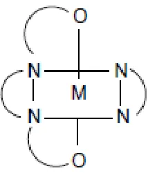
[image:2.596.239.346.610.735.2](Ng et al., 2008). The molecular structure of this metal complex is shown as below (Figure 1):
The electrochemical experiments were performed with a BAS (Bioanalytical Systems, West Lafayette, IN, USA): CV-50W electrochemical workstation. 1mM of [Cu(phen)(edda)].5H2O is
characterized using electrochemical techniques using 0.1M of potassium dihydrogen orthophosphate (KH2PO4) as supporting electrolyte. Copper complex is dissolved into the supporting electrolyte and
placed into an electrochemical cell. The cell was purged with nitrogen gas for 10 minutes in order to remove the dissolved oxygen in the solution. A conventional three electrode potentiostated system was used with 3 mm diameter glassy carbon (GC) as working electrode, Ag/AgCl (3 M NaCl) as reference electrode, and 1 mm diameter platinum wire as counter electrode. The three electrodes were immersed into the solution. Unless otherwise mentioned, the temperature was (25 ± 2)ºC.
Hydrodynamic voltammetry using rotating (carbon) disk electrode (RDE) and model: BAS-100A voltammetric workstation was also employed in the study.
3. RESULTS
3.1. Cyclic Voltammetry (CV) of Cu(phen)edda solution
Figure 2 showed a cyclic voltammogram of the copper complex, [Cu(phen)(edda)]·5H2O,
which reveals a quasi-reversible redox reaction involving the Cu(II)/Cu(0) couple with a peak potential separation of 128 mV. The peak currents for scan rate of 100m/s are 3.28µA (oxidation) and 1.63µA (reduction) vs. Ag/AgCl (3M NaCl) respectively.
Figure 2. Cyclic voltammogram of 1mM [Cu(phen)(edda)]·5H2O in 0.1M KH2PO4, using glassy
carbon as a working electrode, pH 6.8 and 100 mV/s..
3.2. Effect of Varying Scan Rate of Cu(phen)edda solution
The figure 3 is cyclic voltammogram of different scan rates (from lowest scan rate of 5mV/s to highest scan rate of 1000mV/s). As the scan rate increases the peak separation (ΔEp) increases indicating non-reversible process of the redox reaction of the Cu(II)/Cu(0). Therefore, the ΔEp peak is dependent on the scan rate.
[image:3.596.209.389.437.569.2]
Figure 3. Cyclic voltammograms at different scan rates for solution of 1mM [Cu(phen)(edda)]·5H2O
with glassy carbon electrode as working electrode and 0.1M KH2PO4 , and pH 6.8.
Log I versus log υ plot is plotted and two linear graphs were obtained. Both of the linear graphs have correlation value of at least 0.95. Each of this linear graph has its own equation;
y = -0.53x + 6.85 (oxidation)
y = 0.57x + 6.60 (reduction)
This copper complex is a diffusion controlled process (0.53 for oxidation and 0.57 for reduction) where the theoretical value of diffusion controlled process is 0.5. At zero current, the potential for oxidation and reduction are 100mV and 150mV respectively (Figure 4). The zero current potentials are independent of IR and morphological effect of the electrode interacting sites.
Figure 4. Plot of peak current, Ip, versus Peak Potential, Ep, for 1mM [Cu(phen)(edda)]·5H2O
solution with glassy carbon electrode as working electrode and 0.1M KH2PO4 as supporting
electrolyte at room temperature and at pH 6.8.
Scan rate increases Scan rate increases
[image:4.596.136.463.518.700.2][image:5.596.171.429.159.367.2]
3.3. Chronoamperometry of Cu(phen)edda solution
[image:5.596.176.424.477.657.2]Figure 5 shows the monotonic rise in current transient of the redox couple of Cu(II)/Cu(0) of [Cu(phen)(edda)]·5H2O, typical of a diffusion controlled process, derived from the Cottrell equation..
Figure 5. Chronoamperogram of 1mM [Cu(phen)(edda)]·5H2O solution with glassy carbon electrode
as working electrode and 0.1M KH2PO4 as supporting electrolyte at room temperature and at
pH 6.8.
3.4. Chronocoulometry of Cu(phen)edda solution
Figure 6. Plot of chronocoulomogram for 1mM [Cu(phen)(edda)]·5H2O solution with glassy carbon
electrode as working electrode and 0.1M KH2PO4 as supporting electrolyte at room
temperature and at pH 6.8.
Figure 6 is a chronocoulomogram of [Cu(phen)(edda)]·5H2O in 0.1M KH2PO4 as supporting
indicating the presence of sufficient flux of Cu(II)/Cu(0) couple. However, during the oxidation process, there is a rapid increase in the oxidation of Cu(0) in the first 2 msec-1/2 and the process appeared to be nearly complete after 4 msec-1/2. The slope of the forward and the reverse steps are 0.93µC/msec1/2 and -2.16µC/msec1/2 respectively. The intercept of forward and reverse steps are 18.1µC and -2.24µC respectively. The value for surface charge is 288µC/cm2 using Cottrell equation and corresponds to 53.6ng of 1mM [Cu(phen)(edda)]·5H2O electrolyzed on the electrode surface of
3mm diameter.
3.5. Hydrodynamic Voltammetry using Rotating Disk Electrode (HVRDE) of Cu(phen)edda solution
RDE is used to determine the diffusion coefficient and the rate constant of the copper complex. According to the scan rate studies, [Cu(phen)(edda)]·5H2O undergoes diffusion-controlled process and
redox is a quasi-reversible reaction. Therefore, RDE is employed to determine the diffusion coefficient and the rate constant for [Cu(phen)(edda)]·5H2O in aqueous medium. Glassy carbon (GC) and a
solution contained of 1mM of [Cu(phen)(edda)]·5H2O as analyte is dissolved in 0.1M KH2PO4 which
act as supporting electrolyte were tested using RDE technique. Referring to RDE voltammogram (Figure 7), the excitation of current is observed between 50 to 57mV in potential in the scan rate of 50mV/s. This has to be done under lower scan rate in order to allow more time for the solution to diffuse into the GC electrode.
Based on Koutecky-Levich equation, 1/il = 1/ik + 1/(0.62nFAω½Do⅔v-⅙Co), a reciprocal graph
of limiting current, il versus square root of angular velocity, ω. A slope of 45111 is obtained and the
R-squared value is 0.9. The diffusion coefficient of the [Cu(phen)(edda)]·5H2O is 0.24µcm2/s is obtained
using the Koutecky-Levich equation. Kinetic rate constant, kf, of the system is calculated using the
[image:6.596.174.420.509.695.2]Koutecky-Levech equation; experimentally (Figure 8), the kinetic rate constant of 26µcm/s is obtained.
Figure 7. Hydrodynamic voltammogram at scan rate of 50mV/msec of 1mM [Cu(phen)(edda)]·5H2O
solution using glassy electrode as working electrode and 0.1M KH2PO4 as supporting
Figure 8. Linear graph of HVRDE using Koutecky-Levich equation.
3.6. Effect of DNA on the CV of [Cu(phen)(edda)]·5H2O
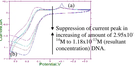
From the cyclic voltamogram, the lowest scan rate (5mV/s) gives a significant change in peak current on the addition of DNA. The current peak is suppressed when 2.95x10⁻¹⁴M (resultant concentration) of DNA is spiked in and the current peak is remained unchanged at the subsequent addition of DNA (region a). A significant extent of current suppression is observed at reduction process (region a in Figure 8). It may be due to a slow diffusion of an equilibrium mixture of the free and DNA-bound complex to the electrode surface (Hirohama et al., 2005).This showed us that DNA of DNA bound species undergo adsorption process as the concentration of DNA is increased.
Figure 9. Cyclic voltammogram at scan rate of 5mV/sec of 1mM [Cu(phen)(edda)]·5H2O solution
with 2.95x10⁻¹⁴M to 1.18x10⁻¹³M (resultant concentration) DNA using glassy electrode as working electrode and 0.1 M KH2PO4 as supporting electrolyte at room temperature and at pH
6.8.
(Figure 9) The anodic peak potential (Epa) and the cathodic peak potential (Epc) in the absence
of DNA are -106mV and -219mV respectively. The separation of the anodic and cathodic peak potential (ΔEp) is -113mV. The average of Epa and Epc in CV (E1/2) is -56.5 mV in the absence of
(a)
(b)
Suppression of current peak in increasing of amount of 2.95x10
-14
[image:7.596.205.481.478.614.2]
DNA. While in the presence of DNA in the same solution at the same conditions causes positive shift of 22.5mV in E1/2. It was reported that a E1/2 shifts in the positive direction when a small molecule
binds to DNA by intercalation and the potential shifts in the negative direction when the interaction with DNA occurs by electrostatic attraction (Carter, Rodriguez, & Bard, 1989). Since the current works shows the presence of positive potential shift of 22.5mV, it is therefore evident that this copper complex, [Cu(phen)(edda)]·5H2O, binds with DNA through intercalation.
However, at higher scan rate of 100m/s and 1000m/s, peak shift appears smaller and rather insignificant (Table 1) as DNA was added in increasing amount from 2.95x10⁻¹⁴M to 1.18x10⁻¹³M into fixed amount of Cu complex. It is therefore evident that the electrochemical interaction of DNA and the copper complex appears to be scan rate dependant, and the interaction studies are best carried out at a low scan rate (eg.5mV/s.). It also implies that under these conditions, the redox peaks of [Cu(phen)(edda)]·5H2O, can be used as an indicator peaks for the presence of DNA during cyclic
voltammetry.
Table 1. Peak potential shifting and current changes of 1mM [Cu(phen)(edda)]·5H2O solution, as a
function scan rate, and in the presence of increasing addition of DNA solution, ranging from 1st addition of 2.95x10⁻¹⁴M, 2nd addition of 5.9x10⁻¹⁴M, and 3rd addition of 1.18x10⁻¹³M.
Scan rate Addition of DNA Reduction Oxidation ΔEp E1/2 Shifting of E ½ / mV
(mV/s) Epc (mV) Ipc (µA) Epc (mV) Ipc (µA) Without and with DNA
5 no addition -219 1.79 -106 0.6 113 56.5 -
1st Addition
-209 1.51 - - - - -
2nd Addition
-202 1.34 -125 0.39 77 38.5 18
3rd Addition
-200 1.09 -132 0.41 68 34 22.5
100 no addition -191 3.56 -94 5.34 97 48.5 -
1st Addition
-166 3.75 -92 4.99 74 37 11.5
2nd Addition
-172 3.76 -94 4.7 78 39 9.5
3rd Addition
-165 3.73 -91 5.11 74 37 11.5
1000 no addition -136 16.76 -2 43.41 134 67 -
1st Addition
-154 13.29 -27 33.8 127 63.5 3.5
2nd Addition
-150 13.87 -24 35.55 126 63 4
3rd Addition
-149 14.19 -25 36 124 62 5
3.7. Chronocoulommetry of Cu(phen)edda solution in the presence of DNA
[image:8.596.19.578.380.666.2]
the DNA, only correspond to 46.7ng of 1mM [Cu(phen)(edda)]·5H2O electrolyzed on the surface of
[image:9.596.144.467.162.393.2]the 3mm diameter working electrode. The decrease in charge and hence the decrease in the amount of Cu(II) complex undergoing electrolysis is also evident of interaction between copper(II) complex and DNA.
Figure 10. Chronocoloumetry of 1mM [Cu(phen)(edda)]·5H2Osolution with 2.95x10⁻¹⁴M to 1.18x10 -13
M (resultant concentration) DNA using glassy electrode as working electrode and 0.1M KH2PO4 as supporting electrolyte.
Based on the CV and CC results, it is suggested that the copper(II) complex (such as [Cu(phen)(edda)]·5H2O can also be used as a possible marker/indicator redox component (such as
Methylene Blue) (A. Erdem, Kerman, Meric, Akarca, & Ozsoz, 2000; Arzum Erdem, Kerman, Meric, & Ozsoz, 2001; Tani, Thomson, & Butt, 2001) for detecting the presence of DNA.
3.8. Chronoamperometry of Cu(phen)edda solution in the presence of DNA
The chronoamperograms of [Cu(phen)(edda)]·5H2O and DNA is also associated with diffusion
controlled process as is for [Cu(phen)(edda)]·5H2O without DNA since monotonic current transient
were observed.
4. CONCLUSION
[Cu(phen)(edda)]·5H2O is successfully studied and characterized using electrochemical
technique. Interaction studies of copper complex with DNA showed that [Cu(phen)(edda)]·5H2O
interact with DNA through intercalation while hydrogen peroxide appears to be a stronger reduction Absence of
DNA
agent for the copper complex. The [Cu(phen)(edda)]·5H2O undergoes a greater interaction with
hydrogen peroxide than that of DNA as shown in the voltammetric results. [Cu(phen)(edda)]·5H2O
like Methylene Blue solution, can also be used as an DNA marker under CV conditions at low and moderate scan rates.
ACKNOWLEDGEMENT
The authors wish to thank MOSTI and Universiti Putra Malaysia for a RU research fund and facilities, and one of us (YCW) is also grateful for the fellowship awarded by MOSTI/UPM.
References
1. S.D. Aust, L.A. Morehouse, & C.E. Thomas. Journal of Free Radicals in Biology & Medicine, 1(1),(1985). 3-25.
2. W.H. Bannister. Biochemical Education, 20(1),(1992). 62-63.
3. D. Bar-Or, L.T. Rael, E.P. Lau, N.K.R. Rao, G.W. Thomas, J.V. Winkler, et al. Biochemical and Biophysical Research Communications, 284(3),(2001). 856-862.
4. M.T. Carter, M. Rodriguez, & A.J. Bard. Journal of the American Chemical Society, 111(24),(1989). 8901-8911.
5. D.K. Chand, H.-J. Schneider*, J.A. Aguilar, F. Escartí, E. García-España*, & S.V. Luis*. Inorganica Chimica Acta, 316(1-2),(2001). 71-78.
6. J.H. Dawson. Synthesis and Reactivity in Inorganic and Metal-Organic Chemistry, 14(3),(1984). 431 - 432.
7. A. Erdem, K. Kerman, B. Meric, U.S. Akarca, & M. Ozsoz. Anal. Chim. Acta, 422(2),(2000). 139-149.
8. A. Erdem, K. Kerman, B. Meric, & M. Ozsoz. Electroanalysis, 13(3),(2001). 219-223.
9. T. Hirohama, Y. Kuranuki, E. Ebina, T. Sugizaki, H. Arii, M. Chikira, et al. Journal of Inorganic Biochemistry, 99(5),(2005). 1205-1219.
10.H.B.G. JM. Methods in Enzymology, 186,(1990). 1-85.
11.T.F. Kagawa, B.H. Geierstanger, A.H. Wang, & P.S. Ho. Journal of Biological Chemistry, 266(30),(1991). 20175-20184.
12.C. Liu, J. Zhou, Q. Li, L. Wang, Z. Liao, & H. Xu. Journal of Inorganic Biochemistry, 75(3),(1999). 233-240.
13.J. Liu, T. Zhang, T. Lu, L. Qu, H. Zhou, Q. Zhang, et al. Journal of Inorganic Biochemistry, 91(1),(2002). 269-276.
14.C.H. Ng, K.C. Kong, S.T. Von, P. Balraj, P. Jensen, E. Thirthagiri, et al. Dalton Transactions, 8(4),(2008). 447-454.
15.D. Phipps. [London: Oxford University Press].(1976). 16.P.J. Sadler. Biochemical Education, 7(3),(1979). 75.
17.K. Sakano, S. Oikawa, M. Murata, Y. Hiraku, N. Kojima, & S. Kawanishi. Mutation
Research/Fundamental and Molecular Mechanisms of Mutagenesis, 479(1-2),(2001). 101-111. 18.A. Tani, A.J. Thomson, & J.N. Butt. Analyst, 126(10),(2001). 1756-1759.
19.T. Theophanides, & J. Anastassopoulou. Critical Reviews in Oncology/Hematology, 42(1),(2002). 57-64
![Figure 2 showed a cyclic voltammogram of the copper complex, [Cu(phen)(edda)]·5H2which reveals a quasi-reversible redox reaction involving the Cu(II)/Cu(0) couple with a peak potential separation of 128 mV](https://thumb-us.123doks.com/thumbv2/123dok_us/1923683.151547/3.596.209.389.437.569/figure-voltammogram-complex-reversible-reaction-involving-potential-separation.webp)
![Figure 3. Cyclic voltammograms at different scan rates for solution of 1mM [Cu(phen)(edda)]·5H2Owith glassy carbon electrode as working electrode and 0.1M KH2PO4 , and pH 6.8](https://thumb-us.123doks.com/thumbv2/123dok_us/1923683.151547/4.596.136.463.518.700/figure-cyclic-voltammograms-different-solution-electrode-working-electrode.webp)
![Figure 5 shows the monotonic rise in current transient of the redox couple of Cu(II)/Cu(0) of [Cu(phen)(edda)]·5H2O, typical of a diffusion controlled process, derived from the Cottrell equation.](https://thumb-us.123doks.com/thumbv2/123dok_us/1923683.151547/5.596.171.429.159.367/figure-monotonic-transient-typical-diffusion-controlled-cottrell-equation.webp)
![Figure 7. Hydrodynamic voltammogram at scan rate of 50mV/msec of 1mM [Cu(phen)(edda)]·5H2O solution using glassy electrode as working electrode and 0.1M KH2PO4 as supporting electrolyte at room temperature and at pH 6.8](https://thumb-us.123doks.com/thumbv2/123dok_us/1923683.151547/6.596.174.420.509.695/hydrodynamic-voltammogram-solution-electrode-electrode-supporting-electrolyte-temperature.webp)
![Table 1. Peak potential shifting and current changes of 1mM [Cu(phen)(edda)]·5H2O solution, as a function scan rate, and in the presence of increasing addition of DNA solution, ranging from 1staddition of 2.95x10⁻¹⁴M, 2nd addition of 5.9x10⁻¹⁴M, and 3rd ad](https://thumb-us.123doks.com/thumbv2/123dok_us/1923683.151547/8.596.19.578.380.666/potential-shifting-presence-increasing-addition-solution-staddition-addition.webp)
![Figure 10. Chronocoloumetry of 1mM [Cu(phen)(edda)]·5H2O solution with 2.95x10⁻¹⁴M to 1.18x10⁻¹³M (resultant concentration) DNA using glassy electrode as working electrode and 0.1M KH2PO4 as supporting electrolyte](https://thumb-us.123doks.com/thumbv2/123dok_us/1923683.151547/9.596.144.467.162.393/chronocoloumetry-solution-resultant-concentration-electrode-electrode-supporting-electrolyte.webp)