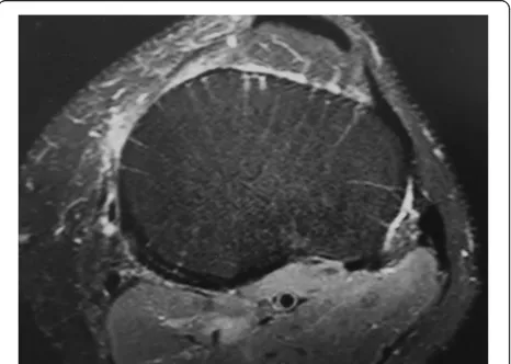
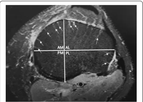
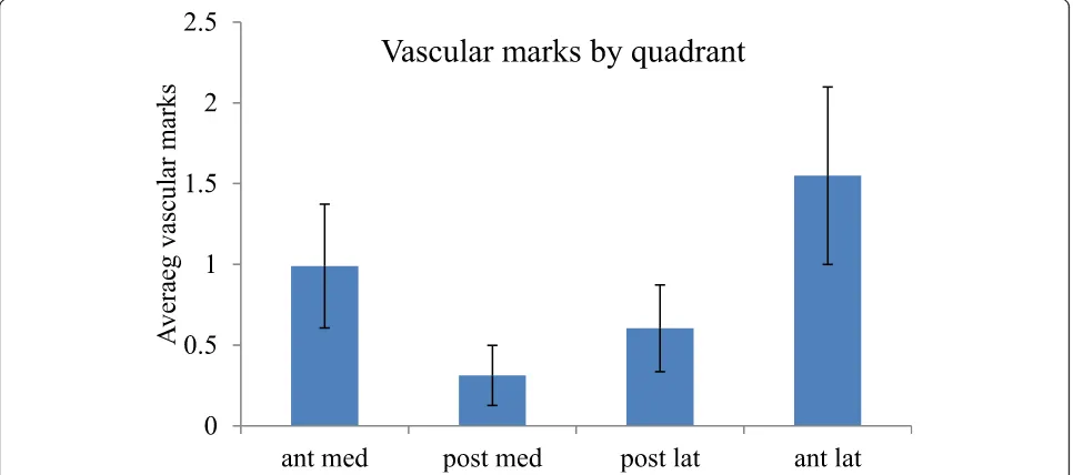
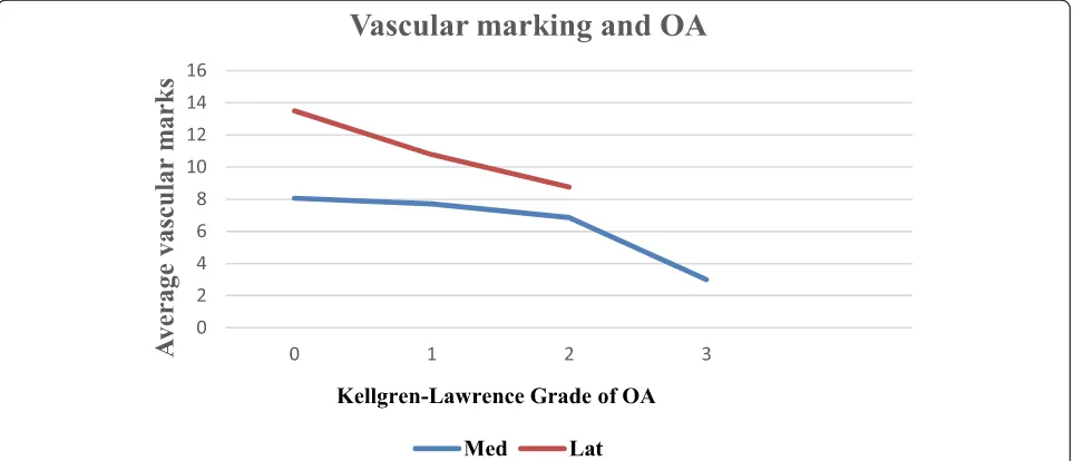
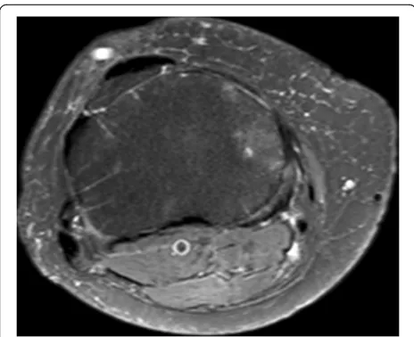
Upper tibial MRI vascular marks lost in early knee osteoarthritis
7
0
0
Full text
Figure
+2
Related documents
