The Harvard community has made this
article openly available.
Please share
how
this access benefits you. Your story matters
Citation
Yang, Shang-Bin, Xiao-Bo Zhou, Hong-Xia Zhu, Lan-Ping Quan,
Jin-Feng Bai, Jie He, Yan-Ning Gao, Shu-Jun Cheng, and Ning-Zhi Xu.
2007. Amplification and Overexpression of Aurora-A in Esophageal
Squamous Cell Carcinoma. Oncol Rep. doi:10.3892/or.17.5.1083.
Published Version
doi:10.3892/or.17.5.1083
Citable link
http://nrs.harvard.edu/urn-3:HUL.InstRepos:32684124
Terms of Use
This article was downloaded from Harvard University’s DASH
repository, and is made available under the terms and conditions
applicable to Other Posted Material, as set forth at
http://
Abstract. Aurora-A/BTAK/STK15 gene which encodes a centrosome-associated kinase is located on chromosome 20q13.2, a highly amplified region in various human tumors. Recent studies have demonstrated the overexpression and amplification of Aurora-A in many malignant human cancers. The purpose of this study was to investigate the amplification and expression of Aurora-A in esophageal squamous cell carcinoma. Amplification of Aurora-A was determined by fluorescence in situ hybridization in 7 eso-phageal cancer cell lines and real-time PCR in 29 esoeso-phageal cancer samples. We detected Aurora-A expression in 7 eso-phageal cancer cell lines and 38 esoeso-phageal cancers samples by semi-quantitative reverse transcription-PCR and Western blot hybridization. The amplification of Aurora-Awas detected in 27 of 29 (93.1%) esophageal cancer samples and 6 of 7 (85.7%) cancer cell lines. Aurora-Awas overexpressed in 27 of 38 (71.1%) esophageal cancer samples and all 7 esophageal cancer cell lines. We conclude that Aurora-Ais amplified and overexpressed in esophageal squamous cancer.
Introduction
The centrosomes are important for cells to maintain genomic stability through establishment of bipolar spindles during cell division, which could allow equal segregation of replicated chromosome to split into two daughter cells. The centro-somal function is strictly regulated by various proteins. Aurora
kinase family, a conserved mitotic serine/threonine kinase family, played a vital role in regulating the centrosomal and microtubule function, ensuring accurate chromosome segregation and efficient completion of cytokinesis (1,2). Among the three members of this family, Aurora-A, attracted most attention because Aurora-A gene is mapped to chromosome 20q13.2, a region amplified commonly in epithelial cancers (3-6).
Amplification and/or overexpression of Aurora-A is detected in several types of tumors, including gastric cancer, bladder cancer, gliomas and hepatocellular cancers (7-12). Combining with the studies that overexpression of Aurora-A
can transform NIH3T3 and Rat1 cells (13,14) and increase the incidence of breast cancer in transgene mouse (15), most researchers would consider Aurora-Aas an oncogene.
ESCC is the fourth most prevalent malignancy in China, but the molecular mechanism for the tumorigenesis of eso-phageal cancer remains to be elucidated. Gain of chromosome 20q was often observed in ESCC (16-20). There are no studies investigating DNA status of oncogene Aurora-A
located on the 20q in ESCC despite recent reports showing the overexpression of Aurora-Ain ESCC (12,21). Therefore, to further reveal the possible role of Aurora-A in esophageal cancer, we detected the DNA copies and expression level of
Aurora-A in both esophageal cancer cells lines and primary tumor samples.
Materials and methods
Cell culture. Seven types of esophageal cancer cell lines KYSE150, 180, 410, 510, 70 (kindly provided by Professor Yutaka Shimada), ESCC109, ESCC9706 (kindly provided by Professor Mingrong Wang) and HLF (human lung fibroblast cell, kindly provided by Dr Youyong Lv, Peking University School of Oncology and Beijing Institute for Cancer Research, Beijing, P.R. China) were cultured in RPMI-1640 medium supplemented by 10% fetal bovine serum at 5% CO2.
Clinical tissue sample collection. We obtained 38 esophageal squamous cell carcinoma tissues and corresponding normal tissues from surgically resected esophageal carcinoma at the Cancer Hospital. None of the patients investigated received
Amplification and overexpression of
Aurora-A
in esophageal squamous cell carcinoma
SHANG-BIN YANG1*, XIAO-BO ZHOU1*, HONG-XIA ZHU1, LAN-PING QUAN1, JIN-FENG BAI1, JIE HE2, YAN-NING GAO3, SHU-JUN CHENG3 and NING-ZHI XU1
1Laboratory of Cell and Molecular Biology, Departments of 2Thoracic Surgical Oncology, 3Etiology and Carcinogenesis, Cancer Institute and Cancer Hospital, Chinese Academy
of Medical Sciences and Peking Union Medical College, 100021 Beijing, P.R. China
Received November 15, 2006; Accepted December 21, 2006
_________________________________________
Correspondence to: Dr Ning-Zhi Xu, Laboratory of Cell and Molecular Biology, Cancer Institute and Cancer Hospital, Chinese Academy of Medical Sciences and Peking Union Medical College, Panjiayuan, Chaoyang District, P.O. Box 2258, 100021 Beijing, P.R. China
E-mail: xningzhi@public.bta.net.cn
*Contributed equally
samples were histopathologically diagnosed as esophageal squamous cell carcinoma.
Fresh samples were dissected manually to remove con-nective tissues and stored immediately at -80˚C until analyzed. The corresponding normal tissues were obtained from the distant edge of dissected esophagus without carcinoma cell invasion by pathological diagnosis. Every tissue sample contained over 80% of normal or tumor epithelial cells.
Fluorescence in situ hybridization (FISH). Cells were treated with 1 μg/ml colchicines overnight. Then the metaphase cell slides were made according to standard protocol.
Dual-color FISH was undertaken to detect 20q13.2 amplification in esophageal cancer cell lines. We co-hybridized an Aurora-Aspecific BAC probe (kindly provided by Dr Subrata Sen and Ms. Hongyi Zhou, University of Texas, M.D. Anderson Cancer Center) with a chromosome 20 specific centromere probe. The probes were labeled with biotin or digoxin by nick translation using Nick Translation Kit (Gibco/ BRL, Rockville, MD, USA). Hybridization was performed at 37˚C for 48 h in a moist chamber. After hybridization, the slides were washed in 50% formamide/2X SSC three times for 5 min each, followed by washes in 2X SSC three times for 5 min each time. Then the slides were blocked by 4X SSCB (4X SSC with 0.5% Blocking Regent bought from Roche 0.02% NaN3) at room temperature for 30 min. The
slides were incubated with Avidin-FITC in wet chamber at 37˚C for 60 min followed by three washings at room temperature for 5 min in 4X SSC. Amplification of the signals was performed by biotinylated anti-avidin antibody (Vector Laboratories, Burlington, Canada). At 37˚C for 60 min in a moist chamber followed by three washings at room temperature with 5 min wash in 4X SSC. A final incubation with Avidin-FITC and Rhodamine-anti-digoxigenin (Roche, Mannheim, Germany) (working solution: 5 μg Avidin-FITC/ml in 4X SSC, 0.5% blocking reagent, 0.2% NaN3)
was done at 37˚C for 60 min in a moist chamber followed by three washings at room temperature for 5 min in 4X SSCB. The nuclei were counterstained with DAPI.
The 20q13.2 amplification was recognized by comparison of the numbers of red and green signals in cell nuclei. Approximately 100-200 cells were numbered for each type of cells when summarizing the 20q13.2 amplification rate.
RNA extraction and semi-quantitative RT-PCR. Total-RNA was extracted from paired specimen of primary esophageal cancer and non-cancerous esophageal epithelium with TRIzol Reagent (Invitrogen, Carlsbad, CA, USA) according to the manufacturer's protocol.
Five microgram of total-RNA extracted from paired eso-phageal carcinoma was used as template respectively to synthesize cDNA in 25 μl reaction mixture with 2.5 mM oligo(dT) primers and M-MLV Reverse Transcriptase (Promega, Madison, WI, USA) at 37˚C for 1 h followed by 85˚C for 10 min. PCR was performed in 20 μl reaction mixture [1X AmpliTaq buffer, 100 ng template cDNA, 200 μM of each dNTPs, 0.5 μM of each primers and 2 U AmpliTaq (Roche)] as follows: 95˚C for 5 min followed by 24 cycles of 95˚C for 1 min, 56˚C for 1 min, 72˚C for 1 min, and the final
PCR primers for Aurora-Aare as follows: upper primer: 5'-GCCTCCTGTGAAGACACCAT-3'; lower primer: 5'-ACTT TGTAACAGAGGGAGCC-3' with an expected product of 471 bp. ß-actin was used as internal control (upper: 5'-GGCG GCACCACCATGTACCCT-3' and downstream: 5'-AGGGG CCGGACTCGTCATACT-3') at 202 bp. Negative control was the amplification omitting the template from the reaction. The reaction products were visualized by electro-phoresis of 5 μl reaction mixture at 70 V for 40 min in 2% agarose gel containing 0.5 μg/ml ethidium bromide, and quantitated by densitometry using Gel-Pro Analyzer version 3.1 (Media Cybernetics, Silver Spring, MD, USA).
Real-time polymerase chain reaction. To detect the amplifi-cation in cancer samples, real-time PCR was performed in the Rotor-Gene RG-3000 Real-Time Thermal Cycler (Corbett Research, Sydney, Australia), which detects the signal from the fluorogenic probe during PCR. As both the precise amount of genomic DNA and its quality are difficult to assess, we also quantified a control gene glyceraldehyde-3-phosphate dehydrogenase (GAPDH). All data were normalized to GAPDH.
The ratio defining the level of amplification is termed ‘Am’, and is determined as follows:
Tthe ratio of gene copy number of Aurora-Ato GAPDH
Am = ––––––––––––––––––––––––––––––––– Nthe ratio of gene copy number of Aurora-Ato GAPDH
(T, tumor tissues; N, corresponding normal tissues).
The kinetic method requires a standard curve which was constructed with serial 10-fold dilutions of specific PCR products. And in this study, we constructed two standard curves with two different pairs of primers used later for real-time quantitative PCR to quantify Aurora-Aand GAPDH gene copy number separately.
PCR amplifications were done with SYBR Premix Ex Taq (Perfect Real-Time) Kit (Takara Biotechnology, Dalian, P.R. China) according to the manufacturer's protocol. The amplifi-cation mixes (25 μl) contains SYBR Premix Ex Taq 12.5 μl, ROX Reference Dye 0.5 μl, each primer 0.5 μl (200 nM) and template 2 μl (around 20 ng). The fragment of Aurora-Awas amplified with the primers 5'-CCTTCGAATGTTGGCAGG AT-3' and 5'-TCCAACACTAACAGACCGCA-3'. Internal control GAPDHused the primers 5'-TGAAGGTCGGAGTC AACGGA-3' and 5'-CATGTGGGCCATGAGGTCCA-3'. PCR was performed as follows: 94˚C for 10 sec followed with 40 cycles of 94˚C for 5 sec and 60˚C for 30 sec. The software Rotor-Gene 5.0 calculated the parameter Ct
(threshold cycle) and determined the starting copy number in the samples.
We investigated 29 cancer samples and their cor-responding normal tissues. The ratio of Aurora-Acopy number to the copy number of GAPDH indicated the gene copy number level of Aurora-A.
were lysed in RIPA lysis buffer containing 1X PBS (137 mM NaCl, 2.7 mM KCl, 10 mM Na2HPO4, 2 mM KH2PO4, pH 7.4),
1% NP40, 0.5% sodium deoxycholate, 0.1% SDS, 1 mM PMSF, 1 μg/ml aprotinin and 5 μg/ml leupeptin. Lysates were centrifuged at 12,000 g for 15 min at 4˚C. The protein concentration was determined using BCA Protein Assay Kit (Pierce, Rockford, IL, USA). Equal amounts of total proteins were then separated on 12% acrylamide gels using standard sodium dodecyl sulfate-polyacrylamide gel electrophoresis (SDS-PAGE) techniques. After transfer to nitrocellulose membranes (Optitran, Schleicher&Schuell, Keene N.H., USA), the proteins were detected with anti-Aurora-A antibody (N20; 1:1000; Santa Cruz, CA, USA), followed by anti-goat IgG-horseradish peroxide-conjugated secondary antibody (Zhongshan Co., Beijing, P.R. China) and chemiluminescence luminol detection kit (Santa Cruz). The same membrane was reprobed with anti-ß-actin (AC-15; 1:5000; Sigma, St Louis, MO, USA) as a control for equivalent protein loading.
Results
Amplification of Aurora-A in esophageal cancer cell lines. Using an Aurora-Agene specific probe and centromere probe
for chromosome 20, we detected the Aurora-A gene copy increments in esophageal cancer cell lines by FISH (Fig. 1). Each numbering included 100-200 metaphase cells, and total amplification rate in each cell type was calculated. Then the cells were assorted into amplification and high amplification of Aurora-Abased on the ratio of 20q13.2 to 20q signals. Six out of seven esophageal cancer cell lines have amplified
Aurora-A, three of which showed high amplification of
Aurora-A(Table I).
We also found many isochromosomes of 20q13.2, which is a common feature of aneuploidy and gene amplification in cancer.
Amplification of Aurora-A in esophageal cancers. Quanti-tative real-time PCR was done to detect the amplification of
Aurora-A in esophageal cancer tissues. Twenty-nine paired cancer samples and distant normal esophageal epithelium were investigated to evaluate the gene copy level of Aurora-Aby using GAPDHas internal control.
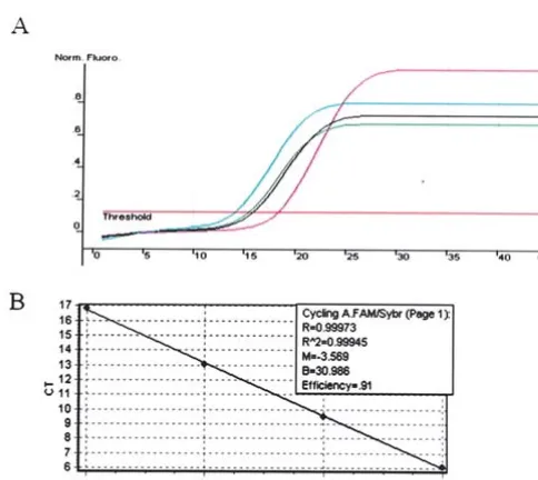
GAPDHstandard curve was constructed (Fig. 2) and then quantitative real-time PCR was done to detect the GAPDH
[image:4.652.129.469.75.222.2]gene copy number (Fig. 3). The same experiment was done to detect the Aurora-A gene copy number. The starting copy numbers were determined by the software Rotor-Gene 5.0,
Figure 1. Amplification of Aurora-Ain esophageal cancer cell lines. Fluorescence in situhybridization was used to detect the amplification of Aurora-Ain metaphase cells by using Aurora-Agene specific probe (green signal) and centromere probe for chromosome 20 (red signal). There are more green signals than red signals in esophageal cancer cells KYSE410 (A) and KYSE510 (B).
Table I. Amplification of Aurora-Ain esophageal cancer cell lines.
––––––––––––––––––––––––––––––––––––––––––––––––––––––––––––––––––––––––––––––––––––––––––––––––––––– Ratioa
Esophageal cancer Numbered cell –––––––––––––––– Amplification rate High amplification
cell lines 1-1.5 ≥1.5 (%) (%)
–––––––––––––––––––––––––––––––––––––––––––––––––––––––––––––––––––––––––––––––––––––––––––––––––––––
KYSE150 113 6 93 87.6 82.3
KYSE410 144 18 125 99.3 86.8
KYSE510 152 121 3 81.6 1.97
KYSE180 131 24 107 100 81.7
KYSE70 172 4 0 2.32 0
ESCC109 106 93 2 89.6 1.89
ESCC9706 144 82 11 64.6 7.64
–––––––––––––––––––––––––––––––––––––––––––––––––––––––––––––––––––––––––––––––––––––––––––––––––––––
aRatio between 20q13.2 and 20q centromere signal number in each cell line. For each cell type, 100-200 cells were counted.
[image:4.652.42.553.308.460.2]and all the data were normalized to GAPDH. As showed in Table II, 27 of 29 (93.1%) cancer samples possessed higher
Aurora-A gene copy level than their corresponding normal tissues. Therefore Aurora-Ais amplified in ESCC.
Overexpression of Aurora-A in esophageal cancers and cancer cell lines. We tested the expression level of Aurora-Agene in 7 esophageal cancer cell lines by semi-quantitative reverse transcription PCR analysis and Western blot assay. We used HLF cell (human lung fibroblast cell) as a normal control. All esophageal cancer cell lines showed overexpression of
Aurora-A compared with HLF cells at mRNA level, KYSE70, 410 and ESCC9706 also have higher expression of Aurora-A from protein level detection (Fig. 4).
We also detected mRNA expression level of Aurora-Ain esophageal cancer samples (Fig. 5). Comparison of band densitometry between Aurora-A and ß-actin by software showed 71.1% (27/38) esophageal cancer samples had more
than 1.5-fold increase of Aurora-AmRNA level in contrast to paired normal adjacent tissues (Table III).
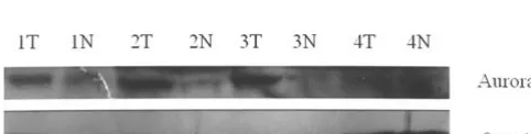
[image:5.652.306.551.94.157.2]In addition to RT-PCR analysis, ten cancer specimens were collected and assayed for Aurora-A expression by Western immunoblot analysis. Six Cancer samples showed increased
Figure 2. GAPDHstandard curve by real-time PCR. Four real-time poly-merase chain reactions were done with serial 10-fold dilutions of specific PCR products of GAPDHas template (A). The parameter Ct(threshold cycle)
was determined by software Rotor-Gene 5.0, and the standard curve was constructed based on the Ctand the template concentration of the four
samples (B).
Figure 3. GAPDHgene copy number was detected by quantitative real-time PCR. Real-time PCR was performed to evaluate the GAPDHgene levels in esophageal cancers, the different color curves represent different samples.
––––––––––––––––––––––––––––––––––––––––––––––––– Amplification level (Am) ––––––––––––––––––––––––– <1 (%) >1 (%) ––––––––––––––––––––––––––––––––––––––––––––––––– No. of patients 2 (6.9) 27 (93.1) –––––––––––––––––––––––––––––––––––––––––––––––––
[image:5.652.307.553.139.318.2]Figure 4. Overexpression of Aurora-Ain esophageal cancer cell lines. Reverse transcription PCR showed increased Aurora-AmRNA level in 6 out of 7 esophageal squamous cell carcinomas (upper panel). HLF cell (human lung fibroblast cell) is the normal control. Protein level of Aurora-A was assayed by immunoblotting in 7 esophageal cancer cell lines by using HLF cells as normal control (lower panel). Six of seven esophageal cancer cell lines showed overexpression of Aurora-Aat protein level. The membranes were reprobed with ß-actin as a loading control.
[image:5.652.304.552.437.484.2] [image:5.652.128.468.658.765.2]protein level of Aurora-A in human esophageal squamous cell carcinoma (Fig. 6).
Discussion
Esophageal cancer is the seventh leading cause of cancer death worldwide. In China, more than 90% of esophageal cancer is the squamous cell carcinoma type, and the area of the high incidence of ESCC is in the north. According to epidemio-logical studies, there are multiple etioepidemio-logical factors, such as nutritional deficiency, toxic stimuli, hot food ingestion and genetic background, contributing to the development of eso-phageal cancer. Because esoeso-phageal cancer usually is not diagnosed until the late stage and the disease has spread, prognosis of esophageal cancer is very poor. Fewer than 20% of people survive more than 5 years (22), therefore novel approaches to the early diagnosis and treatment of this malignant tumor are urgently needed, and understanding the molecular mechanism of human esophageal squamous cell carcinoma is the key to any such approach.
In our present study, we demonstrated amplification and overexpression of Aurora-A/STK15in esophageal cancer cell lines and esophageal cancer tissues.
Previous comparative genomic hybridization studies in esophageal cancer cell lines and tissues revealed that 20q amplification is common in esophageal cancers, but the genes involved in 20q gain are poorly identified. Given that the frequent 20q amplification in breast cancer and bladder cancer is often accompanied by the overexpression of
Aurora-A (23,24), a mitosis regulating gene located on 20q13.2, we proposed Aurora-Aas the possible target gene on amplified 20q in the esophageal cancer. In our present
study, we identified frequent amplification of Aurora-Alocus at 20q13.2 in seven esophageal cancer cell lines by dual-color FISH assay and in cancer samples by real-time PCR assay. Semi-quantitative RT-PCR was used to detect the expression of Aurora-A in esophageal cancer tissues. We found 27/38 (71.1%) esophageal cancers with elevated expression of
Aurora-Awhen compared with corresponding normal tissues, which is consistent with other recent reports that Aurora-Ais overexpressed in human ESCC (12,21). In addition, there are recent reports showing that the Aurora-A polymorphism is associated with ESCC risk (25,26). Although the relationship between the overexpression and polymorphism of Aurora-A
needs to be further revealed, the polymorphism in Aurora-A
gene was demonstrated to be associated with an increased risk in ESCC, as well as in breast cancer (25-27). Therefore, the studies, including ours, strongly suggested Aurora-A involve-ment in the Chinese esophageal cancer developinvolve-ment and progression.
Aurora-A/BTAK/STK15gene, located in 20q13, frequently amplified chromosome region in many tumors including ESCC shown in our present study, belongs to a serine/threonine kinase family (Aurora kinase family) implicated in equal segregation of chromosomes between daughter cells. Aurora-A is important to centrosome duplication, maturation and accurate separation as well as to bipolar spindle assembly and stability. Its crucial role in the normal cell division suggested that change of Aurora-A expression might contribute to the aberrant cell growth in cancer. In fact, ectopic overexpression of Aurora-A in mammalian cells has been demonstrated to cause centrosome amplification, leading to aneuploidy. More importantly, overexpression of
Aurora-Acan lead to genetic instability and cause mammary tumor development in transgene mouse, which indicated that overexpressed Aurora-A could function as a potential oncogene causing carcinogenesis in many solid tumors.
Taken together, our present studies on the overexpression and amplification of Aurora-A in ESCC have provided indicative clues to revealing the mechanism of esophageal carcinogenesis and to find regimens to improve survival of esophageal cancer patients. Although the cause which triggers Aurora-A amplification and the mechanism involved during carcinogenesis in human solid tumors still need to be deeply investigated, the efficient therapy targeting Aurora-A in other malignant tumors (28-31) could also bring hope to esophageal cancer patients if the vital role that Aurora-A plays in esophageal cancer could be addressed further.
Acknowledgements
We thank Professor Subrata Sen for 20q BAC clones; Professor Yutaka Shimada for KYSE cell lines and Professor Mingrong Wang for EC9706 cells. This study was supported by National Natural Science Foundation (39925020, 30271451) and National Basic Research Program (G1998051204, 2004CB518701), P.R. China.
References
[image:6.652.45.288.92.189.2]1. Marumoto T, Hirota T, Morisaki T, et al: Roles of aurora-A kinase in mitotic entry and G2 checkpoint in mammalian cells. Genes Cells 7: 1173-1182, 2002.
Table III. Overexpression of Aurora-Ain esophageal cancers. –––––––––––––––––––––––––––––––––––––––––––––––––
Elevated STK15 No change of
expressiona STK15 expression Total
(%) (%) (%)
––––––––––––––––––––––––––––––––––––––––––––––––– No. of 27 (71.1) 11 (28.9) 38 (100) patients
–––––––––––––––––––––––––––––––––––––––––––––––––
aThe samples had >1.5-fold increase of Aurora-A mRNA level
compared to that of the normal control.
–––––––––––––––––––––––––––––––––––––––––––––––––
[image:6.652.45.286.261.322.2]in centrosome function. Oncogene 21: 6175-6183, 2002. 3. Schlegel J, Stumm G, Scherthan H, Bocker T, Zirngibl H,
Ruschoff J and Hofstadter F: Comparative genomic in situ
hybridization of colon carcinomas with replication error. Cancer Res 55: 6002-6005, 1995.
4. Reznikoff CA, Belair CD, Yeager TR, Savelieva E, Blelloch RH, Puthenveettil JA and Cuthill S: A molecular genetic model of human bladder cancer pathogenesis. Semin Oncol 23: 571-584, 1996.
5. Forozan F, Mahlamaki EH, Monni O, et al: Comparative genomic hybridization analysis of 38 breast cancer cell lines: a basis for interpreting complementary DNA microarray data. Cancer Res 60: 4519-4525, 2000.
6. Jazaeri AA, Lu K, Schmandt R, et al: Molecular determinants of tumor differentiation in papillary serous ovarian carcinoma. Mol Carcinog 36: 53-59, 2003.
7. Klein A, Reichardt W, Jung V, Zang KD, Meese E and Urbschat S: Overexpression and amplification of STK15 in human gliomas. Int J Oncol 25: 1789-1794, 2004.
8. Jeng YM, Peng SY, Lin CY and Hsu HC: Overexpression and amplification of Aurora-A in hepatocellular carcinoma. Clin Cancer Res 10: 2065-2071, 2004.
9. Li D, Zhu J, Firozi PF, et al: Overexpression of oncogenic STK15/BTAK/Aurora A kinase in human pancreatic cancer. Clin Cancer Res 9: 991-997, 2003.
10. Sen S, Zhou H, Zhang RD, et al: Amplification/overexpression of a mitotic kinase gene in human bladder cancer. J Natl Cancer Inst 94: 1320-1329, 2002.
11. Kamada K, Yamada Y, Hirao T, et al: Amplification/over-expression of Aurora-A in human gastric carcinoma: potential role in differentiated type gastric carcinogenesis. Oncol Rep 12: 593-599, 2004.
12. Tanaka E, Hashimoto Y, Ito T, et al: The clinical significance of Aurora-A/STK15/BTAK expression in human esophageal squamous cell carcinoma. Clin Cancer Res 11: 1827-1834, 2005.
13. Zhou H, Kuang J, Zhong L, et al: Tumour amplified kinase STK15/BTAK induces centrosome amplification, aneuploidy and transformation. Nat Genet 20: 189-193, 1998.
14. Ewart-Toland A, Briassouli P, De Koning JP, et al: Identifi-cation of Stk6/STK15 as a candidate low-penetrance tumor-susceptibility gene in mouse and human. Nat Genet 34: 403-412, 2003.
15. Wang X, Zhou YX, Qiao W, Tominaga Y, Ouchi M, Ouchi T and Deng CX: Overexpression of Aurora kinase A in mouse mammary epithelium induces genetic instability preceding mammary tumor formation. Oncogene (In press).
16. Wei F, Ni J, Wu SS, et al: Cytogenetic studies of esophageal squamous cell carcinomas in the northern Chinese population by comparative genomic hybridization. Cancer Genet Cytogenet 138: 38-43, 2002.
17. Fujita Y, Sakakura C, Shimomura K, et al: Chromosome arm 20q gains and other genomic alterations in esophageal squamous cell carcinoma, as analyzed by comparative genomic hybridization and fluorescence in situ hybridization. Hepatogastroenterology 50: 1857-1863, 2003.
hybridization of esophageal and gastroesophageal adeno-carcinomas shows consensus areas of DNA gain and loss. Genes Chromosomes Cancer 22: 305-311, 1998.
19. Yen CC, Chen YJ, Chen JT, et al: Comparative genomic hybri-dization of esophageal squamous cell carcinoma: correlations between chromosomal aberrations and disease progression/ prognosis. Cancer 92: 2769-2777, 2001.
20. Yen CC, Chen YJ, Lu KH, et al: Genotypic analysis of eso-phageal squamous cell carcinoma by molecular cytogenetics and real-time quantitative polymerase chain reaction. Int J Oncol 23: 871-881, 2003.
21. Tong T, Zhong Y, Kong J, et al: Overexpression of Aurora-A contributes to malignant development of human esophageal squamous cell carcinoma. Clin Cancer Res 10: 7304-7310, 2004.
22. Parkin DM, Bray F, Ferlay J and Pisani P: Global cancer statistics, 2002. CA Cancer J Clin 55: 74-108, 2005.
23. Klein A, Jung V, Zang KD, et al: Detailed chromosomal characterization of the breast cancer cell line MCF7 with special focus on the expression of the serine-threonine kinase 15. Oncol Rep 14: 23-31, 2005.
24. Fraizer GC, Diaz MF, Lee IL, Grossman HB and Sen S: Aurora-A/STK15/BTAK enhances chromosomal instability in bladder cancer cells. Int J Oncol 25: 1631-1639, 2004.
25. Miao X, Sun T, Wang Y, Zhang X, Tan W and Lin D: Functional STK15 Phe31Ile polymorphism is associated with the occurrence and advanced disease status of esophageal squamous cell carcinoma. Cancer Res 64: 2680-2683, 2004.
26. Kimura MT, Mori T, Conroy J, Nowak NJ, Satomi S, Tamai K and Nagase H: Two functional coding single nucleotide poly-morphisms in STK15 (Aurora-A) coordinately increase esophageal cancer risk. Cancer Res 65: 3548-3554, 2005. 27. Sun T, Miao X, Wang J, Tan W, Zhou Y, Yu C and Lin D:
Functional Phe31Ile polymorphism in Aurora A and risk of breast carcinoma. Carcinogenesis 25: 2225-2230, 2004. 28. Mahadevan D, Bearss DJ and Vankayalapati H: Structure-based
design of novel anti-cancer agents targeting aurora kinases. Curr Med Chem Anti-Cancer Agents 3: 25-34, 2003.
29. Vankayalapati H, Bearss DJ, Saldanha JW, Munoz RM, Rojanala S, von Hoff DD and Mahadevan D: Targeting aurora2 kinase in oncogenesis: a structural bioinformatics approach to target validation and rational drug design. Mol Cancer Ther 2: 283-294, 2003.
30. Hata T, Furukawa T, Sunamura M, et al: RNA interference targeting aurora kinase a suppresses tumor growth and enhances the taxane chemosensitivity inhuman pancreatic cancer cells. Cancer Res 65: 2899-2905, 2005.