Rochester Institute of Technology
RIT Scholar Works
Theses Thesis/Dissertation Collections
5-2016
Implicating E. coli PAL in the pathogenesis of
Gram-Negative sepsis
Bethany R. Novick [email protected]
Follow this and additional works at:http://scholarworks.rit.edu/theses
Recommended Citation
Implicating
E. coli
PAL in the
pathogenesis of Gram-Negative sepsis
Bethany R. Novick
A thesis submitted in partial fulfillment of the requirements for the
degree of Master of Science in Chemistry in the School of Chemistry
and Materials Science,
College of Science
Rochester Institute of Technology
May 2016
School of Chemistry and Materials Science
College of Science
Rochester Institute of Technology
Rochester, NY 14623-5603
CERTIFICATE OF APPROVAL
M.S. DEGREE THESIS
The M.S. Degree Thesis of Bethany R. Novick has been examined
and approved by the dissertation committee as satisfactory for the
thesis required for the M.S. degree in Chemistry.
Dr. Lea Michel, Thesis Advisor
___________________________________________________________________________
Dr. Suzanne O’Handley
Dr. Thomas Kim
Dr. Ravinder Kaur
Abstract
Acknowledgements
Firstly, I would like to acknowledge the School of Chemistry and Materials Science at RIT, for the constant direction and allowing me to pursue this project, especially my committee, Dr. Thomas Kim, Dr. Ravinder Kaur, and Dr. Suzanne O’Handley for providing me with the feedback and their vast knowledge of Biochemistry over the past two years. I also would like to thank Dr. Michael Pichichiero, Dr. Roberto Vargas and Dr. Robert Zagursky of Rochester General Hospital for collaborating on this project.
I could not complete this thesis without recognizing my wonderful team: Brooke D’Arcy, Kara Farquharson and Emma Snyder, who have gone above and beyond the expectations of
undergraduate researchers with this project and have not only performed experiments but, have impressed me beyond belief with their dedication and problem-solving skills throughout the ups and downs this project experienced. I would like to acknowledge the rest of the Michel Lab for creating not only a supportive work atmosphere, but also a fun environment of appreciation for science and the musical makings of Katy Perry, and always providing me with someone to get a coffee, Diet Coke or snack with.
None of this work would have happened without the love and support of my family. I would like to thank my siblings: Stephen, Lindsey and Lauren, my grandma and my godparents Aunt Kathy and Uncle Mike for supporting my mental sanity over the past two years and reminding me that “everything is going to be ok”. I would like to thank my parents for investing, not only in my education, but also in all my life’s ambitions and for the constant support to allow me to become whom I have today.
Abbreviations
Abbreviation Meaning
GN Gram-negative
OMP Outer Membrane Protein
LPS Lipopolysaccharide
E. coli Escherichia coli
GNS Gram-negative sepsis
PAL Peptidoglycan-associated Lipoprotein
SIRS Systemic Immune Response Syndrome
IgG Immunoglobulin G
OmpA Outer Membrane Protein A
MLP Murein Lipoprotein
Lpp Braun’s Lipoprotein
CLP Caecal Ligation and Puncture
IL-6 Interleukin-6
IL-1β Interleukin-1β
TNF Tumor Necrosis Factor
kDa KiloDaltons
SDS Sodiumdodcylsulfate
WT Wild Type
TLR Toll-like Receptor
TLR4 Toll-like Receptor 4
TLR2 Toll-like Receptor 2
LB Lysogeny broth
KAN Kanamycin
AMP Ampicillin
IPTG Isopropyl β-D-1-thiogalactopyranoside
NaPi Sodium phosphate
NaCl Sodium chloride
SDS-PAGE Sodium Dodecyl Polyacrylamide gel
electrophoresis
APS Ammonium persulfate
TEMED Tetramethylethylenediamine
TBS Tris Buffered Saline
KCl Potassium Chloride
TBST Tris Buffered Saline: Tween20
HRP Horse Radish peroxidase
MS Mass Spectrometry
LC-MS Liquid Chromatography Mass Spectrometry
UTI Urinary Tract Infection
Table of Contents
Certificate of Approval………..ii
Abstract………...iii
Acknowledgements………...iv
Abbreviations………. ………..v
Table of Contents………...vi-vii 1. Introduction………...1-11 a. Sepsis in the United State………...…...1
b. Categorization and physiopathology of sepsis infection ………….…………..1-2 c. Lipopolysaccharide as a target for sepsis treatment……….……..3-4 d. Outer Membrane Protein Release in Gram-negative sepsis……….……….4
e. Peptidoglycan associated Lipoprotein………..……..4-5 f. Pal Release and the Pathogenesis of Pal in GNS models………...……6-8 g. Thesis Project Proposal……….………9
2. Over-expression and Purification of Recombinant Pal protein……….…...10-23 a. Introduction/ Methods i. DNA transformations of TOPO151/PET28A……….…10
ii. Pal Expression in BL-21 E. coli……….……….……...….11
iii. Protein Purification………...…11
b. Results and Discussion………..…14-22
c. Chapter 2 Conclusions……….……..23
3. Detection of Pal in Human Serum/Plasma……….…………...…24-47
a. Introduction/Methods……….…………..24-27
i. Pierce IgG Melon Bead Purification………...24
ii. Millipore PureProteome NHS FlexiBind Magnetic Bead Kit…….24-25
iii. Pierce Albumin/IgG Removal Kit……….…...25-26
iv. Pierce Protein G Antibody Purification……….……...26
v. E. coli Pal Release Study in Human serum……….…….26-27
b. Results and Discussion………....….28-47
i. Magnetic Bead Purification……….….29-34
ii. Melon Bead Purification………...35-40
iii. Alternative Methods………...41-42
iv. Analyzing non-E. coli Sera Samples……….…43-45
c. Chapter 3 Conclusions……….…..46-47
4. Detection of Pal in Human Urine……….……..48-62
a. Introduction/Methods………48-49
i. Low Level detection sample preparation………...…48
ii. E. coli sepsis patient urine sample preparation………..…48
iii. Albumin/IgG depletion of urine samples………...49
b. Results and Discussion………...50-60
c. Chapter 4 Conclusions………..….61-62
5. Final Conclusions and Future Ideas……….………..63-65
1. Introduction
Sepsis in the United States
Sepsis is a disease caused by the effects of a systemic infection accompanied by host hyper-immune response. In the United States, there are an estimated 751,000 cases of sepsis annually, with a mortality rate of about 30% [1]. Approximately 51% of sepsis cases will receive intensive care treatment, and an additional 17% will receive ventilator support in an intermediate care unit or a coronary care unit. The burden of sepsis-related costs to healthcare comes to $17 billion each year [1]. The number of sepsis cases has increased over the last decade, making sepsis the tenth leading cause of death in the United States [2]. Older patients and those with chronic diseases or immune deficiencies are most susceptible to sepsis and are most likely to develop severe sepsis conditions [1-3].
Incidence of sepsis continues to increase due to the persistent use of antibiotics to treat infection and immunosuppressive drugs to treat cancer, HIV/AIDS and autoimmune/auto inflammatory disease, as well as the emergence of antibiotic resistant microbes [3]. Sepsis can also occur through burns, appendicitis, and pneumonia, but more than half of all sepsis infections are nosocomial, resulting from infiltrated intravascular catheters, non-sterile surgical equipment, and other hospital sources [4]. Historically, Gram-positive bacteria have caused the great majority of sepsis cases; however, in more recent years it has been shown that the number of sepsis cases caused by Gram-negative bacteria is increasing [5]. Of the Gram-negative bacteria, the two most common in causing sepsis infections are Escherichia coli (E. coli) and Pseudomonas aeruginosa [5].
Categorization and physiopathology of sepsis infection
thus requiring ventilator support and sometimes resulting in death [8, 7]. The renal, central nervous, respiratory, and cardiovascular systems are most commonly affected by sepsis. [5].
Upon onset of infection, bacteria release toxins into the bloodstream of the host, which interact with the host cell pattern recognition receptors, including the toll-like-receptors. Those receptors then trigger the innate immune response [5]. In Gram-negative sepsis (GNS), the endotoxin lipopolysaccharide (LPS) is released into the plasma initiating the host immune response [9,10]. The innate immune response, usually localized to one area within the body, causes release of pro-inflammatory cytokines, including tumor necrosis factors and interleukins, in an attempt to control the level of infection [5]. When the pro-inflammatory mediators are introduced into the bloodstream, full body (systemic) and/or hyper-inflammation can occur, which can result in tissue damage, necrotic cell death, and multiple organ failures [11]. The immune system will react to the hyper-inflammation by releasing anti-inflammatory mediators, including serotonin and histamine, to control the overwhelming inflammatory response [8]. This period of immunosuppression following the state of hyper-inflammation results in hypotension and can lead the patient into septic shock.
LPS as a target for sepsis treatment
LPS, the highly conserved endotoxin that is released by Gram-negative bacteria during infection, has been extensively studied as a mediator of GNS [14]. LPS has been shown to activate host immune cells through Toll-like receptor 4 [9]. The structural core of LPS is highly conserved, while the O-polysaccharide side-chain is varied among different Gram-negative bacteria [14]. Because of its known ability to activate cytokines, LPS has been a leading candidate for anti-sepsis therapies using monoclonal antibodies.
In one pivotal 1982 clinical study, scientists showed that treatment with human antiserum raised against the heat-killed rough mutant strain of E. coli J5, which features the LPS core without the O-polysaccharide chains, reduces death by GNS [14]. Over the next several decades, multiple investigators tested the concept of treating GNS with antibody directed towards the endotoxin core by passively immunizing with antisera against the E. coli J5 rough mutant. In 1997, immunoblot analysis using the E. coli J5 antiserum showed that, surprisingly, the J5 antiserum did not bind LPS, as expected. [15]. However, Hellman and coworkers did observe antiserum binding to three major OMP’s present in both whole E. coli O18 and the purified OMPs from E. coli O18 [15], suggesting that antibodies to these OMP’s may have also contributed to the therapeutic nature of the J5 antisera. In addition, Hellman and coworkers hypothesized that these OMP’s may also contribute to the pathogenesis of GNS.
administered prior to inducing sepsis in the mice; a more clinically appropriate method would consist of multiple antibody doses and a post-treatment regiment. Furthermore, the models employed in the study were acute and the majority of deaths were observed in the first 72 hours of the study. While the model is similar to that of human sepsis, it is not fully replicative. Patients usually survive the acute systemic inflammatory response and die later due to organ dysfunction. The combination of these limitations makes it impossible to determine if passive immunization against Lpp and Pal would prove therapeutic in humans with sepsis. Further studies are needed, using better sepsis models with post treatment strategies to deliver IgG. It is also crucial that the antibodies against Lpp and Pal be analyzed for their ability to neutralize the inflammatory response caused by these lipoproteins prior to introducing sepsis.
Outer Membrane Protein Release in GNS
In 2000, scientists showed that the three OMP’s that interacted with J5 antisera were also released into human serum in vitro as an OMP-LPS complex [16]. This study also showed the individual release of at least one OMP protein with an estimated molecular mass of 18 kDa [16]. The three OMPs were estimated to have molecular masses of approximately 35, 18, and 5 to 9 kDa [16].
Through immunoblot analysis using protein deficient bacterial strains, the three OMP’s were identified as muerin lipoprotein (MLP), also known as Braun’s lipoprotein (LPP) (5 to 9kDa), outer membrane protein A (OmpA) (35-kDa), and peptidoglycan-associated lipoprotein (Pal) (18kDa) [16]. MLP, OmpA, and Pal proteins have been well studied in their native E. coli, and have been shown to be involved in the maintenance of cell-wall integrity via an OMP-peptidoglycan web-like interaction [14].
Peptidoglycan-associated lipoprotein
cell with structural support [17]. E. coli Pal has been well studied for its role in the Tol-Pal complex, interacting with the peptidoglycan layer [18, 19], LPP [20] and OmpA in the outer membrane [21], TolB in the periplasm [22,19], and TolA of the inner membrane [23, 24]. While known for years as the periplasmic protein that interacts with members of the Tol-Pal complex, Pal was more recently shown to exist in an alternate, surface exposed form, as well.
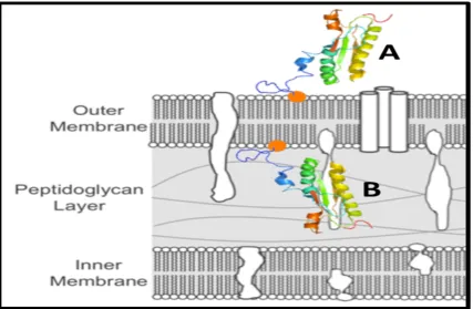
In 2013, the Michel group demonstrated that Pal’s homologue in nontypeable Haemophilus influenzae (NTHi), P6, is dual-oriented: in other words, one subpopulation of P6 was anchored into the outer membrane and faced out into the extracellular space (i.e., surface exposed), while another subpopulation of P6 was anchored into the inner leaflet of the outer membrane and faced into the periplasm, thus allowing it to interact with peptidoglycan [25]. Two years later, the Michel group showed that E. coli Pal was also dual oriented in a very similar manner to P6 [25]. Further, a biotinylation labeling experiment showed that the majority of Pal was oriented facing into the cell, and only ~5-25% of the Pal population was surface exposed (Figure 1).
Figure 1: Schematic of the dual orientation of Pal, in which there are two subpopulations of PAL in the outer membrane of E. coli: one in which Pal faces out toward the extracellular space (A) and one in which PAL faces in toward the periplasm and interacts with peptidoglycan (B).
Pal release and the pathogenesis of Pal in GNS models
In order to study the pathogenic nature of Pal, scientists employed the use of both in vitro and in vivo sepsis models. Examples of commonly used sepsis models include cecal ligation and puncture, macrophage-cultured supernatants, and live mouse models. Cecal ligation and puncture (CLP) in mice involves puncturing the intestines and allowing the naturally existing E. coli in the gastrointestinal tract to spill into the bloodstream. This model is often used to study sepsis, as it most closely resembles the progression and characteristics of human sepsis [27].
In 2002, a CLP mouse model study showed that Pal was released from E. coli into the bloodstream of the septic mouse. Plasma from two groups of mice (CLP mice and healthy mice) was collected 24 hours after puncture; Pal was detected via immunoblotting in the sera of CLP mice only. Pal was detected in 94% of CLP mice (~28 ng/mL), whereas no Pal was detected in the blood sera of healthy mice [27].
into human macrophage-cultured supernatants and levels of interleukin-6 (IL-6) and tumor necrosis factor (TNF) (two cytokines released by macrophages in GNS) were measured. Data revealed that concentrations of Pal greater than 5ng/mL showed a dose dependent increase in production of both IL-6 and TNF, thus demonstrating that Pal stimulates an immune response in an in-vitro human model [27]. In a similar study, Pal was injected into mice to determine the cytokine response. One hour after Pal was injected, TNF-α, IL-6, and IL-1β levels were expressed at maximum levels, thus demonstrating that Pal is capable of inducing an inflammatory response in an in-vivo mouse model [27].
Another mouse model was implemented to show that Pal was toxic. In the study, mice were treated with D-galactosamine, a chemical that makes mice hyposensitive to the effects of LPS. By using D-galactosamine, scientists were able to study the immune response from Pal without an interfering response from LPS [27]. Mice injected with high concentrations of Pal showed a significantly lower survival rate compared to those injected with lower concentrations of Pal; however, Pal was shown to cause death at all concentrations [27].
Bone marrow-derived macrophages were collected from mice and incubated in cultured supernatants with three different supplements: one with Pal alone, one with LPS alone, and a third with Pal and LPS [10]. The cytokine responses of macrophages in each of these samples were analyzed. In the Pal only samples, IL-6 levels were about 2.5 ng/ml; in the LPS only samples, IL-6 levels were less than 1ng/mL; in the LPS/PAL samples, IL-6 concentration was 7 ng/mL, confirming that synergy between Pal and LPS did exist and suggesting that multiple microbial components could be involved in the clinical onset of sepsis [10].
Thesis Project Proposal
In summary, a significant amount of work has already implicated Pal in the pathogenesis of GNS in vitro in human model and in vivo in mice and rats. The main candidate for GNS immunotherapy has historically been LPS, which has known pathogenic properties; however, the J5 antisera studies demonstrated immunoglobulin in J5 binds Lpp and Pal, not LPS [14,15]. Although anti-Lpp and anti-Pal did not offer the same protection as seen with the J5 antisera, other studies showed that 1) Pal stimulates cytokines in vivo in mice and in vitro in human serum; 2) Pal is released in vivo in mice and in vitro in human serum; 3) Pal is toxic in mouse sepsis models; and 4) Pal and LPS act synergistically to a cytokine response in macrophages from mice.
The number of sepsis cases increases each year with the increase in number of medical procedures and use of immunosuppressant drugs; unfortunately, therapy for sepsis patients is limited and the death rate is close to 30%. Currently, sepsis costs the United States close to $17 billion each year, thus underlining the need to identify new immunotherapeutic targets in humans with sepsis.
2. Over-expression and Purification of Recombinant Pal protein
The overall goal of this project was to develop a technique to detect E. coli-released Pal in the serum and urine of GNS patients. In order to develop and optimize a method for detection of Pal in human serum, a recombinant Pal protein was needed as a control. The recombinant Pal allows us to supplement samples of healthy sera and urine test our methods for detection of Pal at higher concentrations to start before determining the lowest level of detection.
Methods
DNA Transformations of Topo151/PET28A
The DNA sequence that codes for the protein of interest, Pal in E. coli, was edited for purification purposes and ordered from GenScript in an ampicillin resistant DNA storage vector (TOPO151). A start codon and a 6-histidine tag were added to the N-terminal end of the Pal sequence. The DNA plasmid was then transformed into competent XL1-Blue (Agilent) and BL-21 (Invitrogen) E. coli cells, as described by the manufacturers. In summary, the lyophilized DNA received from GenScript was reconstituted with sterile water to a final concentration of 50 ng/µL; 1 µL DNA was added to the competent cells, incubated on ice, and heat shocked at 42°C
(XL1-Blue: 45 seconds, BL-21: 30 seconds). Super Optimal broth with catabolite repression (S.O.C media) was added to the cells, followed by incubation for 1 hour at 37˚C, shaking at 225 rpm. The transformed cells were plated onto Luria broth (LB) supplemented with ampicillin (50
µg/mL) and incubated at 37˚C overnight. The following morning, a single colony was used to
inoculate 5 mL of LB supplemented with ampicillin (50 µg/mL). The small culture was grown
(37˚C, shaking at 225 rpm) until the optical density at 600 nm is reached between 0.8-1.0. A freezer stock was prepared with 900 µL of cell culture and 100 µL of an 80% glycerol solution,
Pal Expression in BL-21 E. coli
Recombinant Pal protein was over-expressed and purified as described for Pal’s homologue, P6, from nontypeable Haemophilus influenzae [25] with the following modifications. BL-21 cells containing the Pal plasmid (described above) were cultured on LB supplemented with Kanamycin (Kan) (50 µg/mL Kan: pET28a plasmid) or ampicillin (50 µg/mL Amp: TOPO151
plasmid). Liquid cultures were grown in 25 mL LB supplemented with Amp or Kan at 30˚C, shaking at 180rpm for ~16 hours. Small growths were used to inoculate 1 L LB (supplemented with appropriate antibiotic), which was then incubated at 30˚C, shaking at 180 rpm until the optical density at 600 nm was reached between 0.6-0.8; over-expression was induced with 1 mM IPTG, and the cultures were allowed to grow an additional 3 hours. The cells were harvested by centrifugation (5000xg, 15 minutes, 4˚C), and the cell pellets were stored at -20 ˚C.
Protein Purification
Each frozen cell pellet (from 1 L of culture) was resuspended in 15 mL of 40 mM Tris pH 8.0 + 0.5% Triton X-100 buffer and sonicated on ice for a total of 15 minutes (15 seconds on and 45 seconds off) at a medium horn setting. The lysed cells were centrifuged (20,000xg, 25 minutes) and the supernatant was kept for further processing.
Sodium Dodecyl Sulfate Polyacrylamide Gel Electrophoresis (SDS-PAGE)
The SDS-PAGE gels were prepared using a standard 10% recipe [resolving gel: 3.27 mL of 30% acrylamide/bis-acrylamide, 3.33 mL Tris/SDS (Tris/SDS solution: 182 g Tris base, 1.5 g SDS, pH 8.0), 1.38 mL nanopure water, 2.12 mL 50% glycerol, 100 µL 10% ammonium persulfate (APS), and 10 µL tetramethylethylenediamine (TEMED); stacking: 405 µL 30% acrylamide/bis-acrylamide, 775 µL Tris/SDS, 1.95 mL nanopure water, 20 µL 10% APS, and 5 µL TEMED]. A BioRad gel system was used to cast the gels, and the gel was run in standard buffers (10X Cathode buffer: 60.6 g Tris base, 89.6 g Tricine, 5 g SDS, 500 mL water; 10X Anode buffer: 121.1g of Tris base, 500 mL water, pH adjusted to 8.9 using 12 M hydrochloric acid). The protein samples were prepared by mixing 1:1 with either 2X-sample buffer: 4 mL of 10% SDS, 2 mL Glycerol, 1.2 mL of 1 M Tris pH adjusted to 6.8, 2.8 mL of H2O, 0.001-0.002 g
bromophenol blue; or 2x-reducing buffer: 900 µL 2X-sample buffer, 100 µL β-mercaptoethanol, and boiled for 10 minutes.
All protein samples were loaded onto the gel (14 µL) alongside a Kaleidoscope protein ladder (BioRad and ThermoScientific) (5 µL). Proteins were separated for between 30-45
minutes (120-155 V). Gels that were not transferred for Western blot analysis were incubated for 15 minutes in 15 mL of InstaBlue gel dye (Fisher).
Semi-Dry Transfer
Western Blot Protocol
Transferred nitrocellulose membranes were blocked in 7 mL 5% (m/v) evaporated milk in 1x Tris-buffered saline (TBS) (50 mL of 10X TBS: 80g NaCl, 2g KCl, 30g Tris Base dissolved in 950 mL H2O pH adjusted to 7.4 with HCl, and sterile filtered; diluted to 500 mL with H2O) for
30 minutes, followed by a one hour room temperature incubation with primary antibody [1 µL of
Monoclonal anti-Pal or 1.5 µL of Polyclonal anti-Pal in 6 mL of 1% (m/v) evaporated milk in
TBST (100 mL 1x TBS, 50 µL TWEEN-20, 1 g powdered milk)]. After two washes (1x TBST, 15 minutes each), the membranes were incubated in secondary antibody [0.5 µL of secondary antibody (2°): Goat anti-mouse IgG-H+L HRP conjugate (BETHYL laboratories) in 6 ml of 1% (m/v) evaporated milk in TBST] for 30 minutes. After three washes (1x TBST, 10 minutes each) and an additional two washes (1x TBS, 5 minutes), Pal was detected using a Lumiglo reserve HRP chemiluminescent substrate and a BioRad BioDoc system (Quantity One software).
Mass Spectrometry Sample Preparation
Protein bands (stained with InstaBlue dye) were excised from gels using a clean razorblade and placed in a microcentrifuge tube. The samples were kept cold, and analyzed via Liquid
Chromatography-Mass spectrometry at the University of Rochester Mass Spectrometry (MS) Center. One sample of recombinant purified Pal was prepared: 5µM of protein dissolved in 0.1%
Results and Discussion:
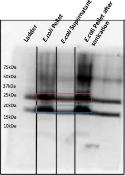
The TOPO151 vector was transformed into E. coli BL-21 cells and the promoter on the Pal gene was activated with IPTG to over-express Pal protein. A Western blot (Figure 2.1) shows the over-expression of Pal in the TOPO151 vector. The harvested cell pellet contains most of the expressed Pal protein, before and after sonication, while the supernatant/lysate shows much less Pal. Purified Pal shows up as two different molecular weight bands: ~25 kDa (non-monomer Pal) and ~17 kDa (monomer Pal).
Figure 2.1. Pal over-expressed in TOPO151 plasmid. Two forms of Pal are detected in the cell pellet (before and after sonication), and in the cell lysate.
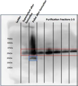
[image:22.612.206.407.226.509.2]Figure 2.2. Pal over-expressed in TOPO151 plasmid, lysed in Tris/TritonX100 buffer. Two forms of Pal are detected in the cell pellet and lysate (after sonication), but only a single higher molecular form of Pal is detected after purification via TALON beads.
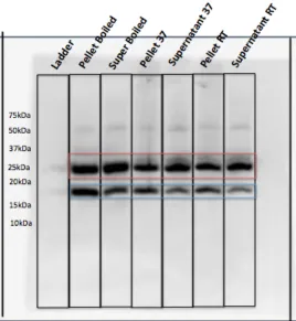
[image:23.612.214.401.75.373.2]Figure 2.3. Pal over-expressed in TOPO151 plasmid, lysed in Tris/TritonX100 buffer; gel samples prepared at 100˚C, 37˚C, or room temperature. Two forms of Pal are detected in the cell pellet and lysate (after sonication) under all conditions, but a greater percentage of the higher molecular weight Pal was detected in samples prepared at lower temperatures.
Figure 2.4. Pal over-expressed in TOPO151 plasmid, lysed in Tris/TritonX-100 buffer; gel samples boiled in reducing sample buffer. Two forms of Pal are detected in the cell pellet after sonication; only Pal-37 is detected in the lysate before and after purification.
The final optimal expression and purification conditions were determined, as follows: E. coli BL-21 (containing TOPO151 Plasmid with Pal gene) cells were grown at 30˚C; a surfactant
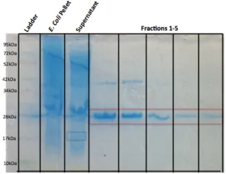
[image:25.612.181.438.75.359.2]Figure 2.5. Optimal expression and purification conditions yield a single population of “heavy Pal.” Two forms of Pal are detected in the cell pellet, but only a single population of “heavy Pal” is detected after purification.
[image:26.612.176.440.75.360.2]Figure 2.6. Pal-25 contains two His-tags at its N-Terminus, yielding a ~25kDa protein.
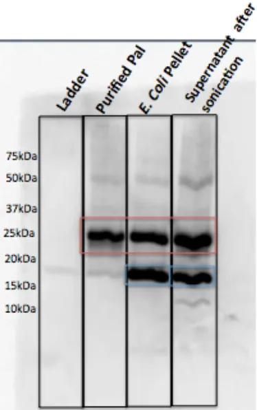
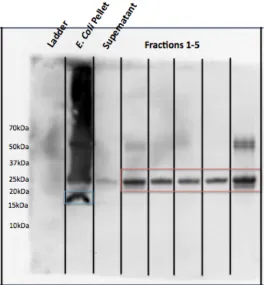
Figure 2.8. Western blot of purified “new” Pal (pET28a), detected with a monoclonal anti-Pal antibody.
Monoclonal anti-Pal detects several versions of “new” Pal as it is eluted off the TALON bead column. Purified Pal runs between 20-25 kDa (red rectangle). A non-His-tagged lower molecular weight version of Pal is detected in the lysed cell pellet and cell lysate (supernatant), prior to purification (blue rectangle).
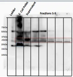
[image:28.612.178.436.71.343.2]Figure 2.9. Western blot of purified “new” Pal (pET28a), detected with a polyclonal anti-Pal antibody.
Polyclonal anti-Pal detects several versions of “new” Pal as it is eluted off the TALON bead column. Purified Pal runs between 20-25 kDa (red rectangle). A non-His-tagged lower molecular weight version of Pal is detected in the lysed cell pellet and cell lysate (supernatant), prior to purification (blue rectangle).
[image:29.612.171.442.74.378.2]PROTEAN® TGXTM pre-cast gel (BioRad), the protein runs at its correct molecular weight (~21 kDa) (Figure 2.10). This finding suggests our recombinant Pal is indeed 21 kDa, but will run as a slightly higher molecular weight protein under certain conditions.
Chapter 2 Conclusions:
The optimal condition for over-expression of the Pal protein in E. coli BL-21 cells containing the PET28A plasmid grown at 30°C, solubilized in equilibration buffer supplemented with Triton X-100, sonicated 20 times and boiling the samples in 2X-sample buffer with β-mercaptoethanol prior to SDS-PAGE analysis. We found that the PET28A plasmid transformed in BL-21 E. coli cells using these conditions, yields approximately 1.5 mg of protein per 1 L of growth with binding affinity to both anti-Pal antibodies.
Adequate yields of purified soluble Pal have been obtained but the purified Pal travels differently based on the type of gel used. Mass spectrometry analysis confirmed the protein obtained has a molecular weight consistent with that expected, 20.6 kDa, based on the gene sequence and the additional mass from the His-Tag. At this time it is not clear as to why the protein has a marker weight of 25 kDa rather than 20 kDa. This could be due to a secondary structure or the protein not unfolding prior to gel analysis.
3. Detection of Pal in Human Serum/Plasma
Pal is one of two dual-oriented OM proteins in E. coli. While the biological relevance and function of lipoprotein dual orientation is not well understood, there is strong evidence linking Pal to the pathogenesis of E. coli sepsis. We propose that Pal’s dual orientation may play a role in its release into serum.
Methods:
PierceTM IgG Melon Bead Purification
To purify IgG from human serum, we used IgG Melon beads (Thermo Scientific Melon Gel IgG Spin Purification Kit 45212), which were swirled to create a slurry. 500 µL bead slurry
was added to each spin column (Pierce), and the storage buffer was discarded by centrifuging at 4000xg for 1 minute. The beads were washed twice with 300 µL Purification Buffer (Thermo
Scientific Melon Gel IgG Spin Purification Kit 45212), centrifuged at 4000xg for 10 seconds. Serum was diluted 1:10 with the Purification Buffer (Thermo Scientific Melon Gel IgG Spin Purification Kit 45212) and a total of 500 µL sera/buffer was loaded onto each spin column and
capped. The column was incubated for 5 minutes at room temperature with end-over-end mixing. The IgG sample was eluted by centrifugation (4000xg, 1 minute), and the column was re-capped. The column was incubated with 500 µL of 2X Sample Buffer for 5 minutes at room temperature
with end-over-end mixing, sealed and boiled for 10 minutes. The “bound” samples (non-IgG proteins) were eluted from the column by centrifuging at 10,000xg for 1 minute.
Millipore PureProteome™NHS FlexiBind Magnetic Bead Kit
beads and remove the buffer. 30 µL of monoclonal anti-Pal antibody was incubated with the
beads for 1 hour (or overnight) at room temperature with end-over-end mixing. The antibody was then removed from the tube (and kept for future use in immunoblotting), and the beads were washed five times with 500 µL Quench buffer (100 mM Tris HCl, 150 mM NaCl, pH 8.0). After
the final wash, 500 µL Quench buffer was incubated with the beads for at least 1 hour at room
temperature with end-over-end mixing. The buffer was removed using the magnet, and the beads were resuspended in 100 µL wash/coupling buffer (PBS) and stored at 2-8ºC for future use (used
within a few months of preparation).
To purify the serum samples, 20 µL anti-Pal conjugated beads were washed with 500 µL
Wash/Coupling buffer (PBS) and vortexed for 20 seconds. The buffer was removed and 100µL
of the sera sample was added to the beads and incubated at room temperature for 2 hours with end-over-end mixing. The “unbound” sample was removed and kept for further analysis (Elution 1). The beads were washed three times with 500 µL Wash/Coupling buffer (PBS), vortexed for
20 seconds, and the buffer was removed from the tube using the magnet to pellet the beads. 50
µL either 2X-sample buffer or reducing buffer was added to the tube and boiled for 10 minutes
to elute the sample (Elution 2).
Pierce Albumin/IgG Removal Kit
To deplete human urine of albumin and IgG, 170 µL of the Immobilized Cibacron
Blue/Protein A Gel (ThermoScientific™Pierce™ Albumin/IgG Removal Kit 89875) was added to a spin column (Pierce). The column was placed in a microcentrifuge tube and centrifuged at 10,000xg for 1 minute to remove the storage buffer. The column plug was placed on the spin column, and the diluted sample (10 µL Urine + 65 µL Binding/wash buffer
microcentrifuge tube that contained the previous elution. The final sample (~150 µL) contained
the urine, which should be depleted of albumin and IgG.
The column with the gel was washed with 100 µL Binding/wash buffer centrifuged at
10,000xg for 1 minute and the filtrate was discarded. The column was plugged and 100 µL
2X-SDS sample buffer was added to the column. The column was capped and incubated with end-over-end mixing for 10 minutes at room temperature. The final sample containing Albumin, IgG and Pal proteins was collected in a microcentrifuge tube by centrifuging the column at 10,000xg for 1 minute.
Pierce Protein G Antibody Purification
The Protein G Antibody purification buffers and columns (Prod. 89949) were brought to room temperature. The columns were uncapped, placed in 2mL collection tubes, and centrifuged for 1 minute at 5000xg to remove storage buffer. The column was washed with 400 µL Binding
Buffer (5000xg), and then incubated with 500µL of sera sample at room temperature with
end-over-end mixing for 10 minutes. The sample was eluted into a clean collection tube (5000xg, 1 minute). The final sample, containing sera depleted of IgG proteins, was concentrated using a VivaSpin Turbo 10,000MWCO filter to 50 µL, and then purified using the anti-Pal conjugated
magnetic beads.
E. coli Pal release study in Human Serum
E. coli cells were grown on 10 mL LB media at 37 oC, shaking at 225 rpm to an OD 600nm
of ~0.85. The culture was then divided into 7-1 mL aliquots labeled A-F. The cells were pelleted by centrifugation at 5000xg for 5 minutes and resuspended in 1 mL LB (Sample A), 1 mL LB supplemented with 200 µg AMP (Sample B), 1mL LB supplemented with 20 µg AMP (Sample
C), 1 mL serum (Sample D), 1 mL serum supplemented with 200 µg AMP (Sample E), 1 mL
serum supplemented with 20 µg Amp (Sample F), or 1 mL serum supplemented with 2 µL
Results and Discussion:
In 2000, scientists showed that Pal, along with 2 other outer membrane proteins, was released into human serum in vitro as an OMP-LPS complex [16]. Pal has been well known for its interactions with peptidoglycan and other OMPs, including LPP and OMPA, in the Tol-Pal complex [18,19, 20]. Scientists have also shown, in vitro and in vivo mouse models, that Pal and Lpp most likely play a role in the pathogenesis of E. coli GNS. In a 2002 study, Pal was shown to be released from E. coli into the sera of 94% of CLP mice (~28ng/mL), while no Pal was detected in the sera of healthy LPS hypo-responsive (C3H/HeJ) mice [27].
Pal is also capable of stimulating an immunological response in macrophage-cultured supernatants. When various concentrations of Pal were injected into supernatants cultured with human macrophages, there was dose dependent production of IL-6 and TNF [27]. Pal was also injected into mice; 1 hour after Pal injection, the TNF and IL-6 levels were expressed at a maximum level [27]. Mice injected with wild-type E. coli (expressing normal Pal) died, while Pal deficient and Pal-nonsense E. coli mutants resulted in improved survival rates in mice.
Taken together, there is strong evidence demonstrating that Pal is released in serum during E. coli infections, has the ability to stimulate an immune response, and can be toxic to
C3H/HeJ mice at any concentration. Based on this evidence, we proposed that we should be able to detect Pal in the serum of humans with E. coli infections/sepsis. We sought to collect sera samples from confirmed E. coli sepsis and other GNS patients at Rochester General Hospital (RGH). We anticipated challenges such as the abundance of other human serum proteins, the predicted low concentration of released Pal (<101 ng/mL), and the ability of Pal to form stable complexes with other OMPs (and potentially with human IgG).
Magnetic bead Purification
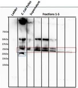
[image:37.612.170.443.367.681.2]Before we switched to other purification techniques, we analyzed a single E. coli sepsis patient sample, obtained from Rochester General Hospital (RGH). The sample was prepared using the Anti-Pal conjugated magnetic beads and eluted in SDS reducing buffer. As seen in Figure 3.2, we detected similar protein bands in the negative (healthy) sera and the E. coli sepsis patient sera. The two bands detected in the blot are likely heavy and light chain IgG.
Figure 3.2: Healthy and E. coli sepsis patient sera, purified using Anti-Pal conjugated Magnetic Beads. Similar bands are detected in healthy and GNS patient sera.
[image:38.612.134.485.220.557.2]3.3). We again observed no significant differences between the negative sera and E. coli sepsis patient sera. If Pal was present in the E. coli sepsis patient sera, we were unable to detect it.
Figure 3.3: Healthy and E. coli sepsis patient sera, purified using Anti-Pal conjugated Magnetic Beads (unbound samples removed). Similar bands are detected in healthy and GNS patient sera.
and patient sera, most likely due to non-specific binding of our anti-Pal antibody to non-Pal proteins.
Figure 3.4: Healthy and E. coli sepsis patient sera, purified using Anti-Pal conjugated Magnetic Beads and eluted in non-reducing buffer. We did not detect proteins in patient sera at the molecular weight predicted for Pal (18 kDa).
Figure 3.5: Healthy and E. coli sepsis patient sera, purified using Anti-Pal conjugated Magnetic Beads and eluted in non-reducing buffer and concentrated in an Amicon concentrator. We did not detect proteins in patient sera at the molecular weight predicted for Pal (18 kDa).
Figure 3.6: Healthy and E. coli sepsis patient sera, purified using Anti-Pal conjugated Magnetic Beads and eluted in non-reducing buffer. We did not detect proteins in patient sera at the molecular weight predicted for Pal (18 kDa). We propose that recombinant Pal formed at least one stable complex (50 kDa) when spiked into healthy sera.
Melon bead Purification
The PierceTM Melon bead kit was used to remove IgG, one of the major proteins that yielded
non-specific bands in our blots. 500 µL beads were loaded onto a column and incubated with 100
µL serum. The beads bound to all non-IgG, resulting in two eluted fractions: unbound sample,
containing IgG, and bound sample, containing non-IgG proteins including Pal. We eluted the bound sample by incubating with reducing SDS sample buffer (β-mercaptoethanol) and boiling for 10 minutes. We used recombinant purified Pal spiked into sera to determine the lowest level of Pal detected using this method. As seen in Figure 3.7, we can detect 750 ng to 5000 ng of Pal spiked into 100 µL of healthy sera (final Pal concentrations: 7,500 ng/mL to 50,000 ng/mL). As
expected, Pal was eluted in the bound fractions. We also were able to see a faint Pal band when as low as 650 ng (6,500 ng/mL), as shown in Figure 3.8. As seen in both figures, a prominent band just below the Pal band (likely light chain IgG) obscured our ability to visualize Pal.
Figure 3.9: Detection of recombinant Pal in sera purified using new Melon beads and non-reducing sample buffer. We were able to detect as low as 1,250 ng/mL of Pal in sera using this purification method.
Figure 3.10: (A) Recombinant purified pal spiked into negative plasma and purified via melon bead purification, eluted with non-reducing SDS buffer. (B) Recombinant Purified pal spiked into negative plasma and purified via melon bead purification, eluted with reducing SDS buffer.
Figure 3.11: Melon Bead Purification of E. coli sepsis Patient #2, eluted in non-reducing SDS buffer. No lower molecular weight bands were detected, but several high molecular weight bands were detected in patient sera that were not detected in healthy sera.
[image:47.612.216.417.72.356.2]Figure 3.12: Melon Bead Purification of E. coli sepsis Patient #2, eluted in reducing SDS buffer. The detected protein band pattern was different in the patient sample compared to the health sera sample.
Figure 3.13: Sera from E. coli sepsis patients 2 and 4, purified using Melon beads, eluted in reducing buffer, and detected using our mouse polyclonal anti-Pal antibody. No major differences between sepsis patient sera and healthy sera were detected.
Alternative methods
[image:49.612.206.416.80.356.2]binding to our primary/secondary antibodies. Unfortunately, even using high concentrations of anti-human IgG did not decrease the intensity of the IgG band in our samples.
We also used an Albumin/IgG purification kit with the goal of removing 80% of the human background proteins in sera. Cibacron beads from the kit were designed to bind Albumin and IgG to the beads and allow all other proteins to flow through. Purification of sera using this method resulted in greater background than the other immunoprecipitation methods, because recombinant Pal eluted in the bound sample, most likely due to its interaction with IgG (data not shown).
We also used Protein G bead purification in tandem with the magnetic beads purification. Protein G beads are designed to bind and remove IgG from sera. A negative sera sample spiked with 750 ng of purified recombinant Pal was incubated on the Protein G column. The eluted sample was concentrated to 50 µL and incubated with the anti-Pal conjugated magnetic beads.
[image:50.612.242.367.385.630.2]After the magnetic bead purification, the sample (eluted in non-reducing buffer) was detected using monoclonal anti-Pal antibody. Figure 3.14 shows the data collected from this experiment; we were successful at detecting a prominent band where we expected to see recombinant Pal.
We employed the same Protein G/magnetic bead approach to purify two GNS patient sera samples. The patients were infected with E. coli and Klebsiella pneumoniae. As seen in Figure 3.15, we were unable to detect differences between either of the GNS patient samples and the negative sample. For reasons unknown, this procedure resulted in higher background than we saw with either the magnetic bead or melon bead preparations. Due to high background and the extensive time involved in this sample preparation method, we decided that this method was not optimal for detecting Pal in patient samples.
Figure 3.15: Sera from E. coli Patient 4 and K. pneumo Patient 3, purified using the Protein G purification kit and anti-Pal conjugated magnetic beads. We observed no significant differences between patient and healthy sera samples.
Analyzing non-E. coli sera samples
[image:51.612.169.439.226.476.2]Two K. pneumoniae patient sera samples were analyzed alongside a sera sample from an
E. coli GNS patient (1) and a Gram-positive sepsis patient (Group A Strep). After Melon beads purification of the sera samples, we were able to detect proteins using our monoclonal anti-Pal antibody (Figure 3.16). We detected a 26-34 kDa protein band, outlined in pink, in every patient sample, including the Gram-positive sample. We cannot conclude the identity of this band, although its presence in the Group A Strep patient suggests it was not Pal.
Figure 3.16: Sera from E. coli sepsis patient 1, K. pneumo patients 2 and 3, and Group A Strep patient 6 was purified using Melon Beads and eluted in reducing buffer. We detected a prominent protein band just above the light chain IgG band in all the patient samples, but we were unable to identify the protein.
[image:52.612.143.480.221.522.2]ampicillin (AMP), sera from healthy donors or sera supplemented with 200 µg/mL AMP, or urine from healthy donors or urine supplemented with 200 µg/mL AMP (Figure 3.17A). E. coli does not release native Pal in LB or urine and only minimally in sera from a healthy donor (as seen in previous studies). AMP was added to the E. coli to kill the cells by breaking up the peptidoglycan. Pal was released at all three growth conditions with AMP, suggesting that either breaking open the cells, disrupting the peptidoglycan, or both lead to release of Pal. In another experiment, E. coli cells were incubated in negative sera supplemented with 2 µL of the
monoclonal anti-Pal antibody. From the blot (Figure 3.17B), we concluded that Pal formed a stable complex with the anti-Pal antibody, which was not dissociated by boiling or separation via SDS PAGE. These results suggest that if there is enough antibody against Pal in sepsis patient serum, Pal could form a stable complex with antibody that could not be dissociated and therefore detected as a complex in the molecular weight range >150 kDa.
Chapter 3 Conclusions:
While the monoclonal primary antibody used in these studies has high fidelity to Pal, the human proteins in sera are in such high abundance, both the primary and secondary antibodies demonstrate non-specific binding to non-Pal proteins. The stable Pal-immunoglobulin complexes formed in human sera have also made the isolation and detection of Pal extremely difficult. We have shown that Pal is capable of forming stable complexes with anti-Pal, which may result in released Pal in patient sera “hiding” among the 150 kDa IgG bands. In summary, we have tried multiple purification, separation, and immunoprecipitation techniques to “clean up” sera to detect Pal in sepsis patient samples. While our blots have improved in cleanliness, background noise, and Pal detection limits, we have still not been able to detect Pal in GNS patient sera.
Another issue that complicates our study is that we don’t know where Pal will appear on a gel. Based on its monomer sequence, Pal is predicted to be between 17-20 kDa, but if it is in a stable complex with itself, another OMP, or immunoglobulin, it would be detected at a higher molecular weight. The release study performed with the spiked anti-Pal in sera suggests that if a patient has a high enough concentration of anti-Pal in their sera, then Pal will form a stable complex with anti-Pal that is not disrupted during sample preparation or gel electrophoresis. We propose that Pal could bind to antibody in patient sera and therefore be detected with a high molecular mass at the top of the gel instead of as a monomer ~18 kDa.
addition of peptidoglycan-targeted antibiotics such as ampicillin). We propose that we have been unsuccessful at detecting Pal in patient sera, because:
A) Pal may be at low ng/mL levels and therefore undetectable using standard immunoblotting (even with our purification techniques);
B) Pal may be forming stable complexes with itself; other OMP’s or anti-Pal antibodies naturally produced by the patients during infection, and therefore is not detectable at its normal monomeric molecular weight;
4. Detection of Pal in Human Urine
The function of the kidney in the body is to filter the blood of excess salts, urea and toxins, that can damage blood cells, then excrete these toxins in the form of urine. Typically the nephrons do not filter cells and protein into the urine, unless the protein is of a small enough molecular weight. We postulated that we might be able to detect Pal in the urine if the kidneys filter the Pal from the blood. Since urine contains far fewer proteins, we anticipated lower background from nonspecific binding and a better signal to noise ratio for Pal detection.
Methods:
Low-level detection sample preparation
Different concentrations of purified Pal (0-2000 ng) were spiked into 20 µL of urine from a healthy young adult donor. The sample was prepared in SDS sample buffer and boiled for 10 minutes. Samples were analyzed on an SDS-PAGE gel and Pal was detected via Western blot.
E. coli Sepsis patient urine sample preparation
Albumin/IgG depletion of Urine samples
A Pierce®
Results and Discussion
We first determined our detection limit of Pal in urine. Purified Pal protein was spiked into urine from a healthy young adult (age 18-45), which was negative for E. coli. Concentrations of purified Pal protein varied from 250 ng to 2000 ng in the first study (Figure 4.1). The purified Pal protein ran to the 26 kDa protein marker and was detectable at all levels in 50 uL of urine, using the monoclonal Anti-Pal antibody and standard Western blot analysis. We prepared lower level Pal samples (10 ng to 250 ng) to determine our lowest level of detection (Figure 4.2). We were able to detect Pal at levels between 10 ng and 25 ng per 50 µL of urine (0.2 ng/µL).
[image:58.612.146.468.322.590.2]According to the Mayo Clinic, normal protein levels in healthy urine are typically ~20 mg/mL, about 105 greater than our lowest limit of detection for Pal in urine. These data suggested that if Pal were present in “normal” concentrations in patient urine, we should be able to detect it.
Figure 4.1:Detection of Purified Pal (2000 ng to 250 ng) in 50 µL of healthy urine. Purified Pal was detected
Figure 4.2:Detection of Purified Pal (250 ng to 10 ng) in 50µL of healthy urine. Purified Pal was detected at
the expected molecular weight at levels as low as 10-25 ng.
Figure 4.3: Western Blot Analysis of E. coli GNS Patient #3 urine developed using monoclonal Anti-Pal antibody. We detected a single prominent band at ~18-20 kDa, the molecular weight of monomer Pal from E. coli.
Figure 4.4: Western Blot Analysis of E. coli GNS Patient #3 urine developed using monoclonal Anti-Pal antibody. To remove any potential intact cells from the patient urine,we filtered the urine (prior to boiling in 2X SDS buffer) or spun down the sample at 5000xg. We detected a prominent band at ~18-20 kDa, the molecular weight of monomer Pal from E. coli, in both prepared urine samples and no proteins in negative/healthy urine.
Figure 4.5: Western Blot of E. coli GNS patient urine (spun down or sterile filtered) using Goat-Anti-Mouse HRP antibody only (no primary). Secondary antibody non-specifically bound to a few proteins in patient urine with higher molecular weights than Pal.
We also used the Albumin/IgG depletion kit to remove more background proteins from the E. coli sepsis patient urine sample. The beads in the kit bind to non-albumin and non-IgG proteins, so we expected to see Pal in the “bound” sample. As seen in Figure 4.6, two bands were
detected in the column-bound sample. A high molecular weight band was detected, as well as the ~19 kDa band we predict to be Pal. The higher molecular weight band may be remnant IgG in the sample and/or a larger Pal complex that remained intact. The same protocol was performed on healthy urine; no proteins were detected in either healthy urine sample. Since 10 µL of the E.
[image:62.612.174.447.77.428.2]Figure 4.6: E. coli sepsis patient urine sample depleted of Albumin and IgG proteins and compared to healthy urine. A higher molecular weight protein and a 19 kDa protein were detected with anti-Pal in the E. coli sepsis patient urine sample (bound to beads). No proteins were detected in the healthy urine.
[image:63.612.165.452.92.419.2]Figure 4.7: Urine from E. coli sepsis patients #1-3 were analyzed via immunoblotting and an anti-Pal antibody. A band at 26 kDa was detected in the urine of patients #1 and #3. The original 19 kDa band in patient #3 was not detected in this blot, which was run X months after the original experiment.
[image:64.612.160.455.71.355.2]Kidney function declines with age, and therefore the majority of elderly people (60 or older) will have more protein in their urine due to decline in kidney function. Here, we used both the monoclonal and polyclonal anti-Pal (Figures 4.8A and 4.8B) to detect Pal in urine from elderly donors. We detected a few proteins at higher molecular weight in donor #2 and #4 samples, but no other significant proteins near the molecular weight of Pal.
[image:65.612.74.505.181.464.2]Figure 4.8:(A) Western Blot of elderly donor urine samples using Polyclonal Anti-Pal Primary (B) Western Blot of elderly donor urine samples using monoclonal Anti-Pal primary. Only a few higher molecular weight proteins were detected in the elderly donor urine samples using anti-Pal.
We also considered the possibility that the patient had a urinary tract infection (UTI). In that case, E. coli cells could be releasing Pal directly into the patient’s urine. A urine sample from a patient with a UTI was analyzed before and after being filtered through a 0.2 µm filter. As seen
As seen in Figure 4.10, a prominent band ~20 kDa can be seen in the unfiltered UTI sample, but is not detected in the filtered UTI sample. Surprisingly, the UTI sample band (unfiltered) is shifted slightly higher than the Pal band from the E. coli pellet. These results suggest the following: 1) Pal (or a homologue to Pal) was present in the bacteria found in the urine sample (our monoclonal anti-Pal antibody interacts with Pal from any enterobacteria, so we cannot determine the source of the bacteria in the sample); 2) Pal (or a homologue to Pal) was not released from the UTI bacteria into urine, as it is not present in the filtered sample; 3) the presumed Pal detected from the UTI sample travels to a slightly higher molecular weight compared to our E. coli Pal control, suggesting that the sequence of the UTI Pal is not identical to E. coli Pal. We are doing further studies to determine whether or not Pal is released from E. coli in the presence of urine.
[image:66.612.177.441.322.592.2]Patients with sepsis often have a plethora of immunological proteins in their system (as a response to their infection). Therefore, comparing a healthy person’s urine to a GNS patient’s urine is not the best comparison. For a better negative control, we collected urine from a donor who was hospitalized for a non-sepsis inflammatory infection. As seen in Figure 4.9, the sick non-sepsis patient’s urine contains no proteins that were detectable using our anti-Pal antibody. In the future, we will collect urine from SIRS (systemic inflammatory response syndrome) patients to be used as negative controls. As long as those SIRS patients do not have E. coli (or other enteric bacterial) infections, we do not anticipate proteins in their urine will be detected using our anti-Pal assay.
Chapter 4 Conclusions:
Our recombinant purified Pal does not run to its true molecular weight of 20.6 kDa, but instead to a molecular weight ~26 kDa. We are unsure of why our recombinant Pal runs to a higher molecular weight, but we propose that the protein exhibits secondary/tertiary structure that is not unfolded via SDS and boiling. Using our recombinant Pal spiked into healthy urine, we determined that we could detect Pal at concentrations as low as 101 ng per 10 µL urine.
Using this method, we were able to detect a protein of the same molecular weight as Pal, using anti-Pal antibody, in the urine of a single E. coli sepsis patient. A similar protein was not detected in any of our negative control urine samples (from healthy young adults, elderly donors, or patients with inflammation, but no E. coli infection). We also detected a higher molecular weight protein in a second patient that aligned with our recombinant Pal. Due to the tendency of Pal to bind other OMPs, it is possible that this higher molecular weight protein is indeed a Pal complex. At this time, without the proper mass spectrometry facilities, we were unable to determine if this band was Pal complexed with itself, Pal complexed with another protein, or a non-Pal protein that nonspecifically bound to anti-Pal. A similar band of the same molecular weight was detected for Patient 3; three months prior, a 19 kDa band was detected in Patient #3’s urine. It is possible that after a long period of storage at 4°C, Pal forms stable complexes and is
therefore detectable at higher molecular weights.
We were able to detect a protein of similar molecular weight to Pal in the urine of a UTI patient who did not receive antibiotics. The band disappeared after filtration of the urine using a 0.2 µm filter, suggesting the Pal was detectable in E. coli whole cells only (and not as released Pal).
liquid chromatography and size exclusion chromatography, we have been unable to definitively identify Pal in GNS patient urine.
5. Final Conclusions and Future Ideas
The recombinant purified Pal does not run to its true molecular weight of 20.6kDa, as indicated by Mass Spectrometry, but instead to a molecular weight ~26 kDa. We are unsure of why our recombinant Pal runs to a higher molecular weight, but we propose that the protein exhibits secondary/tertiary structure that is not unfolded via SDS and boiling. This recombinant protein is needed to study the Pal: Peptidoglycan interaction but is not essential for the detection of Pal in sera/urine studies. Instead of using recombinant Pal as a control in our detection studies, we have been and will continue to use diluted E. coli pellet (which contains native Pal).
For detection of released Pal in sera, the monoclonal anti-Pal antibody we used demonstrated non-specific binding due to the high abundance of human proteins. The Pal-immunoglobulin complexes formed in human sera have made the isolation and detection of Pal extremely difficult. It has also proven difficult to break the interaction between Pal and immunoglobulin; specific immunoprecipitation techniques, Pal purification techniques, and varying buffers were unable to break the Pal-IgG complex. The immunoprecipitation methods we used lowered the levels of human proteins and allowed us to detect purified Pal more easily. In fact, compared to our collaborator’s initial studies to detect Pal in human patient sera, we have been able to optimize the purification protocol and produce cleaner, more sensitive immunoblots. However, even with these improvements, we have not been able to detect Pal in the sera of GNS patients.
Using our recombinant Pal spiked into healthy urine, we determined that we could detect Pal at concentrations as low as 101 ng per 10µL urine. Using the same method, we detected a protein
also must take into account the likelihood that antibodies in the sera will bind to the released Pal protein. Because Pal is a known virulence factor of sepsis, it is likely that immunoproteins will bind to released Pal to neutralize and remove it from the body, thus forming stable complexes that would make Pal appear at higher molecular weights than expected.
Our data suggest that our current methods for detection of Pal in sera are not sensitive or specific enough to detect released Pal, but urine has proven a more sensitive and effective method to detect released Pal from E. coli. The Michel group will continue to analyze urine and sera samples from E. coli sepsis patient samples. We have detected a protein of the same molecular as Pal (using the anti-Pal antibody) in the urine of two E. coli sepsis patients, but we have been unable to detect a similar protein in 4 additional E. coli sepsis patients. Thus, data from more patients are necessary.
Dr. Wei-Jun Qian, Pacific Northwest National Laboratory, has developed a method for targeted quantification of low-level protein concentrations (ng/mL) in human serum using Mass Spectrometry. Dr. Qian has agreed to develop an assay targeted for low level quantifications of Pal in human serum using our recombinant Pal protein. Once the assay is developed, we will use the Mass Spectrometry facilities at Pacific Northwest National Laboratory perform the analysis on our patient samples using this assay.
Our collaborators have demonstrated the release of Pal from E.coli in vivo in mouse/rat models and in vitro in human sera. We have duplicated the in vitro release studies in human sera in our lab, and are currently working on an assay to determine the effect(s) of different antibiotics on Pal’s release. Most sepsis patients will have been given antibiotics while in the hospital, so it is important that we understand how these antibiotics affect the release of Pal from E. coli. We have initiated a similar release study in human urine to determine if E.coli can release Pal into urine in the bladder.
further studied for their effect on Pal release in sera/urine. Our hypothesis is that the Pal:Peptidoglycan noncovalent interaction prevents Pal from release from E. coli, unless peptidoglycan-targeted antibiotics are present.
We postulate that P6, the homolog to Pal in NTHI, may also exhibit characteristics similar to Pal in a GNS model. The Pal release studies will be adapted for P6/NTHI to determine whether or not P6 is released from NTHI in sera. While GNS caused by NTHI is rare in developed countries, many cases of NTHi sepsis are reported each year in developing countries.
References:
(1). Angus, D. C.; Linde-Zwirble, W. T.; Lidicker, J.; Clermont, G.; Carcillo, J.; Pinsky, M. R. Epidemiology of Severe Sepsis in the United States: Analysis of Incidence, Outcome and Associated Costs of Care. LWW2001, 29 (7), 1303–1310.
(2). Martin, G. S.; Mannino, D. M.; Eaton, S.; Moss, M. The Epidemiology of Sepsis in the United States from 1979 through 2000. N Engl J Med2003, 348, 1546–1554.
(3). Lyle, N. H.; Pena, O. M.; Boyd, J. H.; Hancock, R. E. W. Barriers to the Effective Treatment of Sepsis: Antimicrobial Agents, Sepsis Definitions and Host-Directed Therapies. Ann. N.Y. Acad. Sci.2014, No. 1323, 101–114.
(4). Shorr, A. F.; Micek, S. T.; Welch, E. C.; Doherty, J. A.; Reichley, R. M.; Kollef, M. H. Inappropriate Antibiotic Therapy in Gram-Negative Sepsis Increases Hospital Length of Stay*:
Critical Care Medicine2011, 39 (1), 46–51.
(5). Angus, D. C.; Linde-Zwirble, W. T.; Lidicker, J.; Clermont, G.; Carcillo, J.; Pinsky, M. R. Epidemiology of Severe Sepsis in the United States: Analysis of Incidence, Outcome and Associated Costs of Care. LWW2001, 29 (7), 1303–1310.
(6). Bone, R. C. Immunologic Dissonance: A Continuing Evolution in Our Understanding of the Systemic Inflammatory Response Syndrome (SIRS) and the Multiple Organ Dysfunction
Syndrome (MODS). Ann Intern Med1996, 125 (8), 680–687.
(7). Hunter, J. D.; Doddi, M. Sepsis and the Heart. Br. J. Anaesth.2010, 104 (1), 3–11. (8). Porth, C. M. Essentials of Pathophysiology, 3rd ed.; Wolters Kluwer Health, Lippincott Williams & Wilkins: Philadelphia, 2011.
(9). Warren, H. S.; Fitting, C.; Hoff, E.; Adib-Conquy, M.; Beasley-Topliffe, L.; Tesini, B.; Liang, X.; Valentine, C.; Hellman, J.; Hayden, D.; Cavaillon, J.-M. Resilience to Bacterial Infection: Difference between Species Could Be Due to Proteins in Serum. JID2010, 201, 223– 231.
(10). Liang, M. D.; Bagchi, A.; Warren, H. S.; Tehan, M. M.; Trigilio, J. A.; Beasley-Topliffe, L.; Tesini, B.; Lazzaroni, J.-C.; Fenton, M. J.; Hellman, J. Bacterial Peptidoglycan-Associate Lipoprotein: A Naturally Occurring Toll-Like Receptor 2 Agonist That Is Shed into Serum and Has Synergy with Lipopolysaccharide. JID2005, 191, 939–947.
(11). Hotchkiss, R. S.; Karl, I. E. The Pathophysiology and Treatment of Sepsis. New England Journal of Medicine2003, 348 (2), 138–150.