The Role of Subchondral Bone in Osteoarthritis
by
Dawn Doré, BSc(Human Kinetics), BBiotech(Hons)
Submitted in fulfilment of the requirements for the degree of Doctor of Philosophy
(Medical Research)
University of Tasmania, November 2011
Supervisors Professor Graeme Jones
Doctor Tania Winzenberg
Statement of Originality
This thesis contains no material which has been accepted for a degree or diploma by the University or any other institution, except by way of background information and duly acknowledged in the thesis, and to the best of my knowledge and belief no material previously published or written by another person except where due acknowledgement is made in the text of the thesis, nor does the thesis contain any material that infringes copyright.
Statement of Authority of Access and Regarding Published Work
The publishers of the papers comprising Chapters 4 and 5 hold the copyright for that content, and access to the material should be sought from the respective journals. The remaining content of the thesis may be made available for loan and limited copying in accordance with the Copyright Act 1968.
Statement of Co-Authorship
This thesis includes papers for which Dawn Doré (DD) was not the sole author. DD was the lead in the research of each manuscript; however, she was assisted by the co-authors whose contributions are detailed below.
Chapters 4
Doré D, Quinn S, Ding C, Winzenberg T, Jones G. Correlates of subchondral BMD: A cross-sectional study. Journal of Bone and Mineral Research. 2009;24(12):2007-15. The contribution of each author:
DD was responsible for data collection, data management and cleaning, carried out analysis and interpretation of data, prepared the initial manuscript draft, and
completed manuscript revisions.
SQ and TW participated in analysis and interpretation of the data, and critically revised the manuscript.
CD participated in analysis and interpretation of data, and critically revised the manuscript.
GJ designed and carried out the study planning, participated in analysis and interpretation of the data, assisted with the initial manuscript draft, and critically revised the manuscript.
Chapter 5
Doré D, Quinn S, Ding D, Winzenberg T, Cicuttini F, Jones G. Subchondral bone and cartilage damage: a prospective study in older adults. Arthritis & Rheumatism.
2010;62(7):1967-73.
The contribution of each author:
DD was responsible for data collection, data management and cleaning, carried out analysis and interpretation of data, prepared the initial manuscript draft, and
SQ and TW participated in analysis and interpretation of the data, and critically revised the manuscript.
CD participated in analysis and interpretation of data, and critically revised the manuscript.
FC designed and carried out the study planning, participated in analysis and interpretation of data, and critically revised the manuscript.
GJ designed and carried out the study planning, participated in analysis and interpretation of the data, assisted with the initial manuscript draft, and critically revised the manuscript.
Chapter 6
Doré D, Martens A, Quinn S, Ding C, Winzenberg T, Zhai G, Pelletier JP, Martel-Pelletier J, Abram F, Cicuttini F, Jones G. Bone marrow lesions predict site-specific cartilage defect development and volume loss: a prospective study in older adults. Arthritis Research & Therapy. 2010;12(6):R222.
The contribution of each author:
DD was responsible for data management and cleaning, carried out analysis and interpretation of data, prepared the initial manuscript draft, and completed manuscript revisions.
AM participated in data management and cleaning, participated in analysis and interpretation of data, and helped with manuscript preparation.
SQ and TW participated in analysis and interpretation of the data, and critically revised the manuscript.
CD participated in analysis and interpretation of data, and critically revised the manuscript.
JPP and JMP participated in the study planning, carried out data collection, and critically revised the manuscript.
FC designed and carried out the study planning, participated in analysis and interpretation of data, and critically revised the manuscript.
GJ designed and carried out the study planning, participated in analysis and interpretation of the data, assisted with the initial manuscript draft, and critically revised the manuscript.
Chapter 7
Doré D, Quinn S, Ding C, Winzenberg T, Zhai G, Cicuttini F, Jones G. Natural history and clinical significance of MRI-detected bone marrow lesions at the knee: a prospective study in community-dwelling older adults. Arthritis Research & Therapy. 2010;12(6):R223. The contribution of each author:
DD was responsible for data collection, data management and cleaning, carried out analysis and interpretation of data, prepared the initial manuscript draft, and
completed manuscript revisions.
SQ and TW participated in analysis and interpretation of the data, and critically revised the manuscript.
CD participated in analysis and interpretation of data, and critically revised the manuscript.
GZ carried out data collection and critically revised the manuscript.
FC designed and carried out the study planning, participated in analysis and interpretation of data, and critically revised the manuscript.
Chapter 8: Doré D, deHoog J, Giles G, Ding C, Cicuttini F, Jones G. A longitudinal study of the association between dietary factors, serum lipids and bone marrow lesions of the knee. Manuscript submitted to Arthritis Research & Therapy.
The contribution of each author:
DD and JdH are co-first authors on this paper. They were responsible for data management and cleaning, carried out analysis and interpretation of data, prepared the initial manuscript draft, and completed manuscript revisions. DD also collected the data for this paper.
GG contributed to the conception and design of the study and critically revised the manuscript.
CD participated in analysis and interpretation of data, and critically revised the manuscript.
FC designed and carried out the study planning, participated in analysis and interpretation of data, and critically revised the manuscript.
GJ designed and carried out the study planning, participated in analysis and interpretation of the data, assisted with the initial manuscript draft, and critically revised the manuscript.
(Signed) (Date)
Dawn Doré (Candidate)
(Signed) (Date)
Statement of Ethical Conduct
The research associated with this thesis abides by the international and Australian codes on human and animal experimentation, the guidelines by the Australian Government’s Office of the Gene Technology Regulator and the rulings of the Safety, Ethics and Institutional Biosafety Committees of the University.
Abstract
Osteoarthritis (OA) is a complex disease characterised by involvement of multiple tissues in the synovial joint. It is a leading cause of pain and disability in older adults. It has long been hypothesised that subchondral bone plays an important role in the
development and progression of the disease. This thesis aims to investigate how
subchondral bone measures of the knee such as subchondral bone mineral density (sBMD), bone size, and bone marrow lesions (BMLs) are associated with important disease
outcomes in OA.
A population-based sample of older adults aged 50–80 years (51% female; mean age 62 years) participated at baseline and approximately 3.0 years later. sBMD was assessed using dual-energy x-ray absorptiometry (DXA). Cartilage volume, cartilage defects, bone area, and BMLs were determined using magnetic resonance imaging (MRI). X-ray was used to assess radiographic osteoarthritis [joint space narrowing (JSN) and osteophytes]. Blood samples were collected to assess vitamin D and high-density
lipoprotein (HDL) cholesterol. Multiple questionnaires were used to assess pain, function, dietary intake, physical activity, sun exposure, and total knee replacement surgery.
The first study examined the cross-sectional correlates of sBMD and found that many factors were associated with sBMD including age, sex, body mass index (BMI), vitamin D, sun exposure, physical activity, and knee structural measures. The most novel structural measure was cartilage defects and a longitudinal study was required to address causality.
In the second study, bone area at baseline predicted cartilage defect development and cartilage volume loss. Baseline sBMD predicted cartilage defect development, which confirmed the cross-sectional findings above. These associations were independent of each other, indicating there are multiple mechanisms by which subchondral bone may lead to cartilage damage.
In the third and fourth study, 43% of participants presented with a BML at baseline with 25% improving in size and 24% worsening in size over time. Baseline BMLs
pain, only in those participants without radiographic osteoarthritis. Importantly a decrease in BML size was associated with a decrease in pain.
In the final study, baseline energy, carbohydrate and sugar intake (but not fat) were positively associated with a change in BML size. Baseline HDL cholesterol was negatively associated with BML change.
Acknowledgements
I would like to start by thanking my primary supervisor, Professor Graeme Jones. Graeme took me on as an Honour’s student in 2007 and has always showed great belief in me. He has a wealth of knowledge in our field and I am fortunate to have had the
opportunity to do my PhD under him. His teaching of critical thinking, intellectual input, and generosity with financial support to attend significant international conferences has contributed greatly to my success as a student.
I am very grateful to my co-supervisors, Dr Tania Winzenberg and Dr Steve Quinn. Tania is an outstanding mentor to me and has continually provided me with insightful perspective and support. I appreciate her attention to detail and critical review of manuscripts. Tania has also given me valuable advice and feedback on scholarship and fellowship applications and has always been available for future career guidance which I am grateful for. Steve is a wonderful teacher and his knowledge of statistics and
particularly his ability to apply it clinically has benefited me greatly. He was always willing to prioritise my work and went the extra yard to make sure I understood the more complex concepts. Steve also showed interest in my thesis topic and never hesitated to discuss causal pathways with me, which was very helpful.
Thank you to the funding organisations which supported the TASOAC study – National Health and Medical Research Council of Australia, Tasmanian Community Fund, Masonic Centenary Medical Research Foundation, Royal Hobart Hospital Research
Foundation, and Arthritis Foundation of Australia.
During my PhD I have had financial support from a number of sources. I acknowledge the University of Tasmania for awarding me an Endeavour International Postgraduate Research Scholarship (EIPRS) and a Tasmanian Postgraduate Research Scholarship (TPRS). A special mention to Mark Bennett and Kathy Thomson; thank you both for welcoming me to the Menzies Research Institute Tasmania and providing me with assistance in obtaining the funding I received as an international student. I acknowledge the Osteoarthritis Research Society International (OARSI) for awarding me a scholarship which supported a 3 month visit to the University of California, San Francisco (UCSF). Thank you to Professor Michael Nevitt, Susan Rubin, Dr John Lynch, and Felix Liu for welcoming me at UCSF and making me feel at home.
thank you to the 1,099 TASOAC participants who generously gave their time to make this research possible.
I would like to thank the numerous researchers who have given me advice at conferences or assisted with my research in one way or another – Professor Flavia Cicuttini, Dr Anita Wluka, Professor Johanne Martel-Pelletier, and Professor Jean-Pierre Pelletier. Thank you to Dr Ingrid Van der Mei for the engaging casual work and for taking the time to help me learn new STATA commands and statistical approaches. Thank you to Robert and Janine Aitken for welcoming me into your family and giving me considerable encouragement and support. To my fellow PhD students (both current and former) – Dr Stella Just, Dr Dave Scott, Kylie Smith, Laura Laslett, Oliver Stannus, Dr Michele Callisaya, Dr Peta Hitchens, and Dr Kara Martin – thank you for your friendship.
I would like to say a big thank you to my partner Steve Aitken for his love, patience, friendship, and emotional support throughout these years. Thank you for being such a good listener, sitting through numerous presentations with ongoing interest, and always telling me that I am clever.
Publications Arising from the Thesis
Chapter 4: Doré D, Quinn S, Ding C, Winzenberg T, Jones G. Correlates of subchondral BMD: A cross-sectional study. Journal of Bone and Mineral Research. 2009;24(12):2007-15.
Chapter 5: Doré D, Quinn S, Ding D, Winzenberg T, Cicuttini F, Jones G. Subchondral bone and cartilage damage: a prospective study in older adults. Arthritis & Rheumatism. 2010;62(7):1967-73.
Chapter 6: Doré D, Martens A, Quinn S, Ding C, Winzenberg T, Zhai G, Pelletier JP, Martel-Pelletier J, Abram F, Cicuttini F, Jones G. Bone marrow lesions predict
site-specific cartilage defect development and volume loss: a prospective study in older adults. Arthritis Research & Therapy. 2010;12(6):R222.
Chapter 7: Doré D, Quinn S, Ding C, Winzenberg T, Zhai G, Cicuttini F, Jones G.
Natural history and clinical significance of MRI-detected bone marrow lesions at the knee: a prospective study in community-dwelling older adults. Arthritis Research & Therapy. 2010;12(6):R223.
Scientific Presentations Arising from the Thesis
International
2009 Osteoarthritis Research Society International (OARSI) World Congress Montreal, Canada
“Natural history of areal bone marrow lesions” (Moderated poster presentation)
2009 3rd International Osteoarthritis Imaging Workshop York, England
“Which subchondral bone measure is the best predictor of cartilage damage?” (Oral presentation)
2009 2nd Joint Meeting of the International Bone & Mineral Society (IBMS) and the Australian & New Zealand Bone & Mineral Society (ANZBMS)
Sydney, Australia
“Which subchondral bone measure is the best predictor of cartilage damage?” (Poster presentation)
National
2008 Australian & New Zealand Bone & Mineral Society (ANZBMS) Conference Melbourne, Australia
“Determinants of subchondral bone mineral density: a cross-sectional study” (Poster presentation)
2010 Australian Rheumatology Association (ARA) Conference Melbourne, Australia
Local
2008 Sharing Excellence in Research (SEiR), University of Tasmania, Conference Hobart, Australia
“Determinants of subchondral bone mineral density: a cross-sectional study” (Oral presentation)
2010 Sharing Excellence in Research (SEiR), University of Tasmania, Conference Hobart, Australia
“Bone marrow lesions; what are they and how do they relate to osteoarthritis?” (Oral presentation)
Invited Speaker
2010 Australian Physiotherapy Association (APA) State Conference Launceston, Australia
“Natural history and clinical significance of bone marrow lesions in osteoarthritis”
2010 Exercise and Sports Science Australia Seminar Hobart, Australia
Awards Resulting from the Thesis
2008 Endeavour International Postgraduate Research Scholarship (EIPRS)
2008 Tasmanian Postgraduate Research Scholarship (TPRS)
2008 Most outstanding presentation in the ‘Population and Health’ theme area at the Sharing Excellence in Research (SEiR), University of Tasmania, Conference
2008 Travel grant to attend the Australian & New Zealand Bone & Mineral Society (ANZBMS) Conference
2009 Travel grant to attend the 2nd Joint Meeting of the International Bone & Mineral Society (IBMS) and the Australian & New Zealand Bone & Mineral Society (ANZBMS)
2010 Winner of the Australian Society for Medical Research (ASMR) Postgraduate Student Competition
List of Abbreviations
25(OH)D 25-hydroxyvitamin D
2D two-dimensional
3D three-dimensional
ACR American College of Rheumatology
ASU avocado-soybean unsaponifiables
AQoL assessment of quality of life
BMD bone mineral density
BME bone marrow edema
BMI body mass index
BML bone marrow lesion
BMP-7 bone morphogenetic protein 7
CI confidence interval
CTX-II type II collagen C-terminal telopeptide
CV coefficient of variation
dGEMRIC delayed gadolinium-enhanced magnetic resonance imaging of cartilage
DMOAD disease-modifying osteoarthritis drug
DXA dual-energy x-ray absorptiometry
FDA Food and Drug Administration
FFQ food frequency questionnaire
FS fat suppressed
FSE fast spin echo
GAG glycosaminoglycan
GEE generalised estimating equations
GRE gradient-recalled echo
HDL high-density lipoprotein
IL interleukin
JSN joint space narrowing
JSW joint space width
KJ kilojoule
K/L Kellgren and Lawrence
KOOS Knee injury and Osteoarthritis Outcome Score
LDL low-density lipoprotein
LF lateral femoral
LSC least significant criterion
LT lateral tibial
MF medial femoral
MMP matrix metalloproteinase
MOST Multicentre Osteoarthritis Study
MRI magnetic resonance imaging
MT medial tibial
NHMRC The National Health and Medical Research Council
NSAIDS non-steroidal anti-inflammatory drugs
OA osteoarthritis
OR odds ratio
pa per annum
RA rheumatoid arthritis
RCT randomised controlled trial
ROA radiographic osteoarthritis
ROI region of interest
sBMD subchondral bone mineral density
SD standard deviation
TASOAC Tasmanian Older Adult Cohort study
TKR total knee replacement
US United States
VAS visual analog score
WOMAC Western Ontario and McMasters Universities Osteoarthritis Index
WORMS Whole-Organ magnetic resonance imaging score
Table of Contents
Statement of Originality ... II Statement of Authority of Access and Regarding Published Work ... III Statement of Co-Authorship ... IV Statement of Ethical Conduct ... VIII Abstract ... IX Acknowledgements ... XI Publications Arising from the Thesis ... XIII Scientific Presentations Arising from the Thesis ... XIV Awards Resulting from the Thesis ... XVI List of Abbreviations ... XVII Table of Contents ... XX List of Tables ... XXIV List of Figures ... XXVI
Synopsis ... 1
Chapter 1 - Introduction ... 5
1.1 Epidemiology of Osteoarthritis ... 6
1.1.1 History ... 6
1.1.2 Economic impact and disease burden ... 6
1.1.3 Symptoms ... 7
1.1.4 Risk factors ... 7
1.1.5 Treatment and management ... 9
1.2 Knee osteoarthritis ... 11
1.2.1 Radiographic criteria ... 12
1.2.2 Clinical criteria ... 15
1.2.3 X-ray versus MRI ... 15
1.3 Subchondral bone and osteoarthritis ... 17
1.3.1 Definition ... 17
1.3.2 Function ... 18
1.3.3 Subchondral bone changes in osteoarthritis ... 18
1.4 Bone mineral density and osteoarthritis ... 20
1.4.1 Subchondral bone mineral density ... 21
1.5 Tibial bone size and osteoarthritis ... 22
1.7 Summary ... 26
Chapter 2 - Research questions ... 27
2.1 Research Questions ... 28
2.2 Key Hypothesis ... 28
Chapter 3 - Methodology ... 29
3.1 Prelude ... 30
3.2 Study population and design ... 30
3.3 Anthropometrics ... 34
3.4 Dual energy x-ray absorptiometry (DXA) ... 34
3.5 Magnetic Resonance Imaging ... 35
3.5.1 Tibial bone area ... 35
3.5.2 Bone marrow lesions ... 36
3.5.3 Cartilage defects ... 37
3.5.4 Cartilage volume ... 37
3.6 X-ray ... 38
3.7 Physical activity measures ... 38
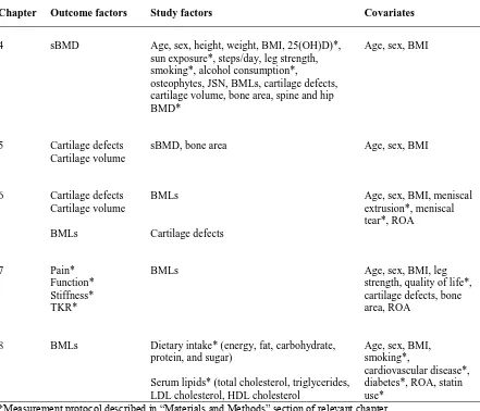
3.8 Summary of outcome factors, study factors, and covariates ... 39
3.9 Sample size and role of the candidate in the TASOAC study ... 40
3.10 Ethical considerations ... 40
3.11 Statistical analysis ... 40
Chapter 4 - Correlates of subchondral bone mineral density: a cross-sectional study ... 41
4.1 Introduction ... 42
4.2 Materials and Methods ... 43
4.2.1 Cartilage defects ... 43
4.2.2 Bone marrow lesions ... 43
4.2.3 Serum 25-hydroxyvitamin D ... 43
4.2.4 Measures related to sun exposure ... 43
4.2.5 Smoking ... 43
4.2.6 Alcohol consumption ... 44
4.2.7 Bone mineral density ... 44
4.2.8 Statistical analysis ... 44
4.3 Results ... 46
4.3.1 Subjects ... 46
4.3.3 Environmental and lifestyle factors ... 48 4.3.4 Medial sBMD and medial structural measures ... 50 4.3.5 Lateral sBMD and lateral structural measures ... 54
4.4 Discussion ... 55
Chapter 5 - Subchondral bone and cartilage damage: a prospective study in older adults ... 59
5.1 Introduction ... 60 5.2 Materials and Methods ... 61
5.2.1 Knee cartilage volume ... 61 5.2.2 Knee cartilage defects ... 61 5.2.3 Tibial subchondral bone mineral density ... 61 5.2.4 Statistical analysis ... 61
5.3 Results ... 63
5.3.1 Subjects ... 63 5.3.2 Tibial cartilage defect increase ... 65 5.3.3 Tibial cartilage volume loss ... 67
5.4 Discussion ... 70
Chapter 6 - Bone marrow lesions predict site-specific cartilage defect development and volume loss: a prospective study in older adults ... 73
6.1 Introduction ... 74 6.2 Materials and Methods ... 76
6.2.1 Knee cartilage volume ... 76 6.2.2 Knee cartilage defects ... 76 6.2.3 Bone marrow lesions ... 76 6.2.4 Meniscal damage evaluation ... 77 6.2.5 Statistical analysis ... 77
6.3 Results ... 79
6.3.1 Subjects ... 79 6.3.2 BMLs and cartilage defects ... 81 6.3.3 Cartilage volume loss ... 84 6.3.4 Additional analysis ... 88
6.4 Discussion ... 89
Chapter 7 - Natural history and clinical significance of MRI-detected bone marrow lesions at the knee: a prospective study in community-dwelling older adults ... 92
7.2 Materials and Methods ... 95
7.2.1 Subjects ... 95 7.2.2 Bone marrow lesions ... 95 7.2.3 WOMAC scores ... 96 7.2.4 Knee replacement surgery ... 96 7.2.5 Quality of life ... 96 7.2.6 Statistical analysis ... 96
7.3 Results ... 98
7.3.1 Subjects ... 98 7.3.2 Natural history and demographic factors ... 99 7.3.3 WOMAC scores and BMLs ... 102 7.3.4 Knee replacement surgery ... 105
7.4 Discussion ... 107
Chapter 8 - A longitudinal study of the association between dietary factors, serum lipids, and bone marrow lesions of the knee ... 112
8.1 Introduction ... 113 8.2 Materials and Methods ... 115
8.2.1 Questionnaire ... 115 8.2.2 Bone marrow lesions ... 115 8.2.3 Dietary factors ... 115 8.2.4 Serum lipids ... 116 8.2.5 Statistical analysis ... 116
8.3 Results ... 117
8.3.1 Subjects ... 117 8.3.2 Cross-sectional ... 119 8.3.3 BML change ... 120 8.3.4 Incident BMLs ... 122 8.3.5 Additional analysis ... 123
8.4 Discussion ... 124
Chapter 9 - Summary and future directions ... 128
9.1 Summary ... 129 9.2 Future directions ... 131
List of Tables
Table 1.1. Summary of evidence based guidelines for the treatment of OA ... 9 Table 1.2. Summary of the current Australian guidelines for the non-pharmacological and pharmacological treatments for knee and hip OA, endorsed by The Royal Australian College of General Practitioners (45) ... 10 Table 1.3. Kellgren and Lawrence (K/L) grading system ... 13 Table 1.4. Atlases developed for the radiographic assessment of osteoarthritis ... 14 Table 1.5. Individual radiographic features measured by the OARSI atlas (77) for
tibiofemoral osteoarthritis ... 14 Table 3.1. Baseline demographic characteristics of those participants who completed the follow-up (n=875) and those which did not (n=224) ... 33 Table 3.2. Demographic and clinical follow-up characteristics of those participants who had an MRI scan at follow-up (n=425) and those which did not (n=450)... 33 Table 3.3. Summary of outcome factors, study factors, and covariates used in this thesis . 39 Table 4.1. Characteristics of participants* ... 47 Table 4.2. Multivariable analysis examining the relationship between anthropometric factors and medial sBMD* ... 48 Table 4.3. Multivariable analysis examining environmental and lifestyle factors associated with medial sBMD* ... 49 Table 4.4. Multivariable analysis examining structural associations with medial sBMD* . 51 Table 5.1. Characteristics of the participants at baseline* ... 64 Table 5.2. Associations of baseline bone measures with increases in cartilage defects during 2.7 years* ... 67 Table 5.3. Associations of baseline bone measures with absolute changes in cartilage volume during 2.7 years* ... 69 Table 6.1. Characteristics of participants according to presence or absence of BMLs at baseline at each site* ... 80 Table 6.2. Association between baseline BMLs (0–3) and site-specific increases in
Table 7.2. Relationship between baseline BMLs and increasing BMLs with baseline
List of Figures
Figure 1.1. Radiographic evidence of knee OA, including medial JSN and osteophytes. .. 13 Figure 1.2. Schematic drawing of the different layers of cartilage and subchondral bone (99), with permission. ... 18 Figure 3.1. Flow chart describing recruitment, participation rates, and withdrawal reasons for TASOAC participants. ... 32 Figure 3.2. Three (A–C) medial and lateral ROIs used to measure sBMD. ... 34 Figure 3.3 (A) Lateral tibial BML. (B) Areal measurement of the BML (15 mm2). ... 37 Figure 4.1. Association between medial sBMD using ROI 1 and steps/day. P for trend was adjusted for age, sex, and BMI. ... 49 Figure 4.2. Mean difference in medial sBMD* between those subjects with and without prevalent cartilage defects using ROIs 1 and 2. Error bars represent standard error. ... 51 Figure 4.3. Interactions between age, sex, and BMI on medial sBMD*. Adjusted for age, sex, and BMI. ... 52 Figure 4.4. Interactions between sex and structural features on medial sBMD* using ROI 1. Adjusted for age, sex, and BMI. ... 53 Figure 5.1. Mean baseline (A) subchondral bone mineral density (sBMD) and (B) bone area in subjects whose cartilage defects decreased or remained stable and subjects whose cartilage defects increased. Error bars represent standard error. ... 66 Figure 5.2. Mean cartilage loss per annum for each quartile of baseline (A) subchondral bone mineral density (sBMD) and (B) bone area. Error bars represent standard error. P
values are for tests of trend. ... 68 Figure 6.1. Baseline BMLs with cartilage defect increases and baseline cartilage defects with BML increases by site. (A) Proportion of participants whose cartilage defects
increased in those with no baseline BML versus those with a baseline BML. (B) Proportion of participants whose BMLs increased in those with baseline cartilage defect grades 0–1 versus those with baseline cartilage defect grades 2–4. ... 82 Figure 6.2. Baseline BMLs and cartilage defects with cartilage volume loss (% per annum). (A) Mean cartilage volume loss of participants with no BML at baseline versus those with a BML at baseline. (B) Mean cartilage volume loss of participants with baseline cartilage defect grades 0–1 versus those with baseline defect grades 2–4. Error bars represent
medial tibial BMLs and medial tibial cartilage defects; and (B) lateral tibial BMLs and lateral tibial cartilage defects, for site-specific cartilage volume loss. ... 87 Figure 7.1. Change in BML size. (A) BML increase from baseline to follow-up. (B) BML decrease from baseline to follow-up. ... 95 Figure 7.2. Natural history of BMLs. *Those with ROA had higher odds of a BML
Synopsis
Osteoarthritis (OA) is the most common form of arthritis and a leading cause of
musculoskeletal pain and disability. By 2020, it is estimated that one in ten Australians will have OA. Although its signature pathologic feature is articular cartilage loss, OA involves many other joint structures including subchondral bone, ligaments, menisci, periarticular muscles, peripheral nerves, and synovium. It was hypothesised many years ago that
subchondral bone plays an important role in OA pathogenesis. There is emerging evidence to support this; however, it remains controversial whether bone changes precede cartilage damage. This thesis examines the role that subchondral bone plays in OA using data from a prospective population-based study of community-dwelling older adults. Specifically, it investigates how different subchondral bone measures such as subchondral bone mineral density (sBMD), bone size, and bone marrow lesions (BMLs) relate to disease progression and disease severity. This Synopsis presents an overview of the content of each chapter.
Chapter 1 provides an overview of OA with a focus on the knee. A working definition is provided and the economic impact, burden of disease, symptoms, risk factors, and
treatment and management options are discussed. A detailed description of the
radiographic and clinical criteria for the diagnosis of knee OA is presented. Lastly, this chapter presents an overview of how subchondral bone relates to OA and provides a rationale for examining sBMD, bone size, and BMLs.
Chapter 2 lists the research questions to be addressed in the thesis.
Chapter 3 describes the Tasmanian Older Adult Cohort (TASOAC) study population and its design, as well as the protocols for measurement of factors which are common to multiple chapters in this thesis. Additional factors which are unique to each chapter are described in more detail within the methodology section of the subsequent chapters.
Chapter 4 describes the cross-sectional relationship between tibial sBMD and
anthropometric, lifestyle, and structural measures in 740 TASOAC participants (mean age 62 years, range 50–80 years, 52% female). Medial tibial sBMD was assessed by
BMLs (by magnetic resonance imaging (MRI)); and hip and spine BMD (by DXA). sBMD using ROI 1 was negatively associated with age and female sex and positively associated with body mass index (BMI). In multivariable analysis, sBMD was positively correlated with steps/day (r = 0.08, P = 0.025), tibial osteophytes (r = 0.08, P = 0.028), JSN (r = 0.11,
P < 0.01), cartilage defects (r = 0.16, P < 0.01), cartilage volume (r = 0.12, P = 0.01), BMLs [r = 0.17, P = 0.013 (tibial); r = 0.16, P = 0.018 (femoral)] and hip and spine BMD (r = 0.36, P < 0.01; r = 0.38, P < 0.01, respectively). Similar associations were observed using ROI 2, with vitamin D also associated with sBMD (r = 0.10, P < 0.01). In
conclusion, this study identified a large number of factors associated with sBMD, of which the most novel is cartilage defects. Longitudinal studies are required to address causality.
Chapter 5 describes the association between baseline tibial bone area and tibial sBMD with tibial cartilage defect development and cartilage volume loss. A total of 341 TASOAC subjects (mean age 63 years, range 52–79 years, 48% female) provided
complete data for baseline bone area and sBMD and changes in tibial cartilage volume and cartilage defects over approximately 2.7 years. In multivariable analysis, baseline bone area positively predicted cartilage defect development at the medial and lateral tibial sites (odds ratio (OR) 1.6 per one standard deviation (SD) increase, P < 0.01; OR 2.4 per one SD increase, P < 0.01, respectively) and cartilage volume loss at the medial tibial site ( = -34.9 per one SD increase, P < 0.01). In contrast, baseline sBMD positively predicted cartilage defect development at the medial tibial site only (OR 1.6 per one SD increase, P = 0.04) and was not associated with cartilage loss. In conclusion, bone area predicted medial and lateral cartilage defect development and medial cartilage volume loss, while sBMD predicted medial defect development but not cartilage loss. These associations were independent of each other indicating there are multiple mechanisms by which subchondral bone may lead to cartilage damage.
and cartilage volume loss (-0.9 to -2.9% difference per annum, all P < 0.05) at the same site. In multivariable analysis, there was a significant relationship between BML severity and cartilage defect progression at all four sites (OR 1.8–3.2, all P < 0.05) and BML severity and cartilage volume loss at the MF, LT, and LF sites ( -22.1 to -42.0, all P < 0.05). Additionally, baseline cartilage defect severity predicted BML progression at the MT and LF sites (OR 3.3–3.7, all P < 0.01). Lastly, there was a greater increase in cartilage volume loss at the MT and LT sites when both larger defects and BMLs were present at baseline (all P < 0.05). In conclusion, baseline BMLs predicted site-specific cartilage defect progression and cartilage volume loss in a dose-response manner suggesting BMLs may have a local effect on cartilage homeostasis. Baseline cartilage defects predicted site-specific BML progression, which may represent increased bone loading adjacent to defects. These results suggest BMLs and cartilage defects are interconnected and play key roles in knee cartilage volume loss; thus, both should be considered targets for intervention.
Chapter 7 describes the natural history of BMLs at the knee using a quantitative measure and examines the association of BMLs with pain, function and stiffness scores, and total knee replacement (TKR) surgery. A total of 395 TASOAC subjects (mean age 63 years, range 52–79 years, 51% female) provided complete data for changes in BMLs, pain, function, and stiffness scores over approximately 2.7 years and total knee replacement (TKR) surgery data at approximately 5 years. BMLs were determined by measuring the maximum area of the lesion. Reproducibility for this method of measurement was
excellent (intraclass correlation coefficient (ICC) 0.97). Pain, function, and stiffness were assessed by Western Ontario and McMaster Universities Osteoarthritis (WOMAC) index scores. At baseline, 43% (n=168/395) had a BML. Of these 25% decreased in size and 24% increased. Of the remaining sample (n=227), 7% developed a new BML. In a multivariable model, a change in BML size was associated with a change in pain and function scores ( = 1.13–2.55 per 1 SD increase, all P < 0.05), only in those participants without radiographic osteoarthritis (ROA). Lastly, baseline BML severity predicted TKR surgery (OR 2.10/unit, P = 0.019). In conclusion, BMLs (assessed by measuring maximal area) were not static, with similar proportions both worsening and improving. A change in BML size was associated with changes in pain in those without established ROA. This finding suggests that fluctuating knee pain may be attributable to BMLs in those
findings suggest therapeutic interventions aimed at altering the natural history of BMLs should be considered.
Chapter 8 describes the association between dietary factors, serum lipids and BMLs. A total of 394 TASOAC subjects (mean age 63 years, range 52–79 years, 50% female) provided complete baseline dietary data, serum lipid measurements and changes in BMLs over approximately 2.7 years later. Nutrient intake (total energy, fat, carbohydrate, protein and sugar) was assessed by food frequency questionnaire (FFQ) and blood samples were collected to assess serum lipids. Cross-sectionally, dietary factors and lipids were not significantly associated with BMLs. Baseline energy, carbohydrate and sugar intake (but not fat) were positively associated with a change in BML size (β = 13.57–19.13 mm2
per 1 SD increase, all P < 0.05). High-density lipoprotein (HDL) cholesterol at baseline was negatively associated with BML change (β = -13.48 mm2
per 1 SD increase, P = 0.045). In conclusion, energy, carbohydrate and sugar intake may be risk factors for BML
development and progression. HDL cholesterol seems protective against BMLs. These results suggest macronutrients and lipids may be important in BML etiology and that dietary modification may alter BML natural history.
1.1 Epidemiology of Osteoarthritis
Osteoarthritis (OA) is the most common form of arthritis and is one of the leading causes of pain and disability among older adults. It is a chronic localised joint disease and commonly involves weight-bearing joints such as the knees, hips, or spine, with hands and neck also being frequently affected sites. In the version 2 estimates for the Global Burden of Disease 2000 study, published in the World Health Report 2002 (1), OA is the
fourth-leading cause of years lost due to disease at the global level.
In 2007, Access Economics estimated that there were 1.62 million Australians with OA (2). Although OA is an incredibly prevalent condition, it is becoming even more prevalent with the combined effects of an ageing and increasingly obese society. By 2050, it is projected there will be 3.1 million Australians or 11% of the population with OA (2).
1.1.1 History
OA was first found in our earliest known ancestors, the Java man (ca 500,000 years ago) (3). It was not clinically separated from other arthritides such as rheumatoid arthritis (3) until 1859. In 1888 OA received its name from John Kent Spender, a physician in Bath. The first radiological description that separated OA from rheumatoid arthritis came from ‘skiagrams’ in 1904 by Goldwaite (4). The definition of OA has evolved over the course of the 20th century and it is increasingly recognised that OA is a disease of the whole joint. Although its signature pathologic feature is articular cartilage loss, it commonly involves many other joint structures including subchondral bone, ligaments, menisci, periarticular muscles, peripheral nerves, and synovium (5). The failure of these joint tissues results in symptoms of pain, stiffness and functional disability (6).
1.1.2 Economic impact and disease burden
While the mortality rate for OA is low, there is also a cost in terms of burden of disease. The pain and disability patients experience can lead to a loss of health and wellbeing, loss of leisure time, and a decreased quality of life. This further contributes to the costs of OA through the loss of production to the economy, increased absenteeism, reduced work capacity and performance, and reduced labour force participation as a result of the related disease morbidity. Arthritis is more common in older people; therefore, a progressively ageing population is likely to further add to the associated disease burden and the cost of providing health services in Australia.
1.1.3 Symptoms
Pain is the most common symptom in OA and the usual reason for seeking medical advice (8). Patients also experience joint stiffness, tenderness, inflammation, crepitus, instability, and muscle weakness (9, 10). These symptoms lead to a limitation of
movement, physical and psychological disability, and impaired quality of life (10). Patients can experience major difficulty with daily activities including walking, stair-climbing, and housekeeping (10).
Late stage OA is often characterised by both structural damage and symptomatic reports of joint pain, stiffness, and disability (11). However there is only a weak correlation between symptoms, such as pain and functional impairments and radiological change, especially in the earlier stages of disease (12, 13). There is also data to suggest that knee pain itself can modify the appearance of joint space width (JSW) in radiographs (14). Unfortunately this has plagued disease modification therapy in OA and treatments for OA largely target symptoms.
1.1.4 Risk factors
For many years OA was thought to be a degenerative joint disease resulting from ‘wear and tear’ and the inevitable effects of aging. It is now widely accepted that OA has a multifactorial aetiology and is the result of many systemic and local factors. Risk factors vary for different joints, for different stages of disease, and for the development versus the progression of the disease (15).
component of OA has been estimated in twin and family studies to be between 50% and 65% with larger genetic influences for hand and hip OA than knee OA (20-22).
The prevalence and patterns of joints affected by OA vary among racial and ethnic groups (15). Chinese participants in the Beijing Osteoarthritis Study reported less hip and hand OA compared to Caucasians in the Framingham Study (23, 24). However, Chinese women had significantly higher prevalence of knee OA (25). The Johnson County Osteoarthritis Project have shown that the prevalence of hip OA in African American women was similar to that in white women, but the prevalence was slightly higher in African American men compared to white men (26).
There is increasing interest to explore the role of diet in OA. Several studies have linked low vitamin D to both hip and knee OA (27-29). In the Framingham Study subjects with a low vitamin D (<34 ng/mL) had a threefold increased risk for knee OA progression compared to those with higher vitamin D levels (≥34 ng/mL) (27). In the Study of
Osteoporotic Fractures, women with a low vitamin D (<30 ng/mL) were 3 times as likely to develop incident hip OA compared to those with higher vitamin D levels (≥30 ng/mL) (29). Randomised, placebo-controlled trials are underway to evaluate vitamin D
supplementation in OA. Vitamin C and vitamin K have also been linked to OA (30, 31). Low vitamin C dietary intake is associated with progressive knee OA (30) and high levels of vitamin K is associated with low prevalence of hand OA (31).
Being overweight or obese are strong risk factors for the development of OA, especially for knee OA (16). Obesity has been shown to be more important than other potential risk factors such as heredity, for knee OA (18, 32). The relationship between being overweight and hip OA is inconsistent and weaker than has been shown for knee OA (33, 34). However there is evidence to suggest that a high BMI is strongly associated with an increased risk of total hip replacement therapy (35).
Previous joint injury is a strong risk factor for OA. Transarticular fracture, meniscal tear requiring meniscectomy or anterior cruciate ligament injury can result in increased risk of knee OA development and musculoskeletal symptoms (36, 37).
Additional risk factors include occupation (e.g. one involving repetitive knee bending), and mechanical factors such as adduction moment, malalignment, and muscle weakness.
As demonstrated there are multiple identified risk factors for OA. The racial and ethnic differences may be related to anatomical or genetic differences. The increase in OA in women suggests that hormonal factors may play a role in the development of OA. Increased load on the joint is most likely the primary mechanism by which being
involved. Risk factor heterogeneity makes OA a difficult disease to treat and new therapies should consider targeted treatment groups in order to maximise treatment benefits.
1.1.5 Treatment and management
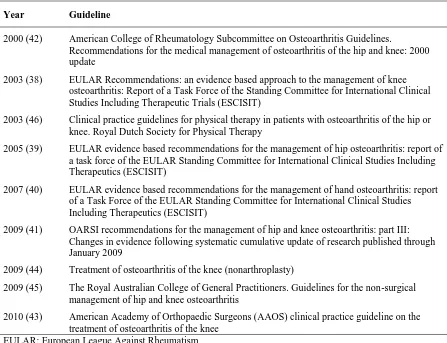
Numerous evidence based guidelines have been developed for the treatment of OA by a number of scientific societies and health care organisations (38-46). Table 1.1
[image:36.595.96.543.344.687.2]displays a summary of these guidelines. Despite some differences in quality, there is a general consistency amongst the recommendations (47) and they mostly cater for specific users, such as clinicians, countries, or health care organizations. Treatments for OA include non-pharmacological, pharmacological, or surgical.
Table 1.1. Summary of evidence based guidelines for the treatment of OA
Year Guideline
2000 (42) American College of Rheumatology Subcommittee on Osteoarthritis Guidelines.
Recommendations for the medical management of osteoarthritis of the hip and knee: 2000 update
2003 (38) EULAR Recommendations: an evidence based approach to the management of knee osteoarthritis: Report of a Task Force of the Standing Committee for International Clinical Studies Including Therapeutic Trials (ESCISIT)
2003 (46) Clinical practice guidelines for physical therapy in patients with osteoarthritis of the hip or knee. Royal Dutch Society for Physical Therapy
2005 (39) EULAR evidence based recommendations for the management of hip osteoarthritis: report of a task force of the EULAR Standing Committee for International Clinical Studies Including Therapeutics (ESCISIT)
2007 (40) EULAR evidence based recommendations for the management of hand osteoarthritis: report of a Task Force of the EULAR Standing Committee for International Clinical Studies Including Therapeutics (ESCISIT)
2009 (41) OARSI recommendations for the management of hip and knee osteoarthritis: part III: Changes in evidence following systematic cumulative update of research published through January 2009
2009 (44) Treatment of osteoarthritis of the knee (nonarthroplasty)
2009 (45) The Royal Australian College of General Practitioners. Guidelines for the non-surgical management of hip and knee osteoarthritis
2010 (43) American Academy of Orthopaedic Surgeons (AAOS) clinical practice guideline on the treatment of osteoarthritis of the knee
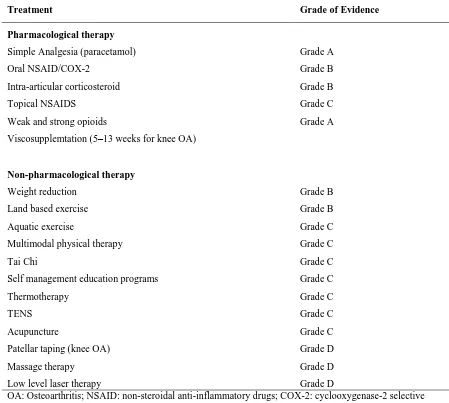
Table 1.2 provides an overview of the current Australian guidelines for the non-pharmacological and pharmacological treatments for knee and hip OA, endorsed by The Royal Australian College of General Practitioners (45). These guidelines are approved and supported by NHMRC (The National Health and Medical Research Council). Each treatment recommended has been rated based on the NHMRC’s body of evidence assessment matrix (48). Grades are given based on a summation of components which include volume of evidence, consistency, clinical impact, generalisability, and
[image:37.595.92.541.297.700.2]applicability.
Table 1.2. Summary of the current Australian guidelines for the non-pharmacological and pharmacological treatments for knee and hip OA, endorsed by The Royal Australian College of General Practitioners (45)
Treatment Grade of Evidence
Pharmacological therapy
Simple Analgesia (paracetamol) Grade A
Oral NSAID/COX-2 Grade B
Intra-articular corticosteroid Grade B
Topical NSAIDS Grade C
Weak and strong opioids Grade A
Viscosupplemtation (5–13 weeks for knee OA)
Non-pharmacological therapy
Weight reduction Grade B
Land based exercise Grade B
Aquatic exercise Grade C
Multimodal physical therapy Grade C
Tai Chi Grade C
Self management education programs Grade C
Thermotherapy Grade C
TENS Grade C
Acupuncture Grade C
Patellar taping (knee OA) Grade D
Massage therapy Grade D
Low level laser therapy Grade D
OA: Osteoarthritis; NSAID: non-steroidal anti-inflammatory drugs; COX-2: cyclooxygenase-2 selective inhibitor; TENS: transcutaneous electrical nerve stimulation.
Body of evidence assessment matrix:
Grade A (Excellent evidence, body of evidence can be trusted to guide practice)
Grade B (Good evidence, body of evidence can be trusted to guide practice in most situations)
Grade C (Satisfactory, some evidence, body of evidence provides some support for recommendation(s) but care should be taken in its application)
In summary, current pharmacological treatments for OA are mostly palliative and are concerned with controlling pain and improving function and quality of life. Analgesic and non-steroidal anti-inflammatory drugs (NSAIDs) are often prescribed in standard medical practice to reduce pain and control joint inflammation. These often prove to be only moderately effective, with >75% of patients reporting need for additional
symptomatic treatment (49). There is an emphasis on non-pharmacological treatments for OA including lifestyle modification, physiotherapy, and exercise (10). Weight reduction and strategies to avoid weight gain are recommended for knee and hip OA (50). There is strong evidence to show that exercise has beneficial effects on pain and function, and a combination of strengthening, aerobic and functional exercise is recommended (10, 51). Exercise helps to strengthen muscles, increase range of motion, maintain joint position, control weight, and improve proprioception, balance, and cardiovascular fitness (52-55). Importantly, exercise has similar effects to analgesic and anti-inflammatory medications, but has fewer contraindications and adverse effects than drugs and surgery (41). Although exercise has proven to be a very effective treatment for OA, it has not been shown to modify disease progression (10). Additional physiotherapy treatments which have shown to be effective in the treatment of OA include taping, bracing, wedge insoles, and manual therapy (51). Surgery is recommended for debilitating pain and major functional
impairments with factors such as walking, working, sleeping (56-58). Total knee and hip replacement are common for advanced disease when conservative treatments are
ineffective (45, 59).
Despite OA’s large disease burden there are no treatments which stop or delay the progression of the disease. Current FDA-approved treatments directed at reducing OA symptoms have not been shown to prevent ongoing joint structural damage (5). For this reason it has been suggested that treatment of the structural changes at a joint level should be assessed separately to patient-reported symptoms. In a review by Lane et al (5) it is stated that future pharmacotherapy for OA should be considered to be ‘structure modifying’, ‘symptom modifying’, or both.
1.2 Knee osteoarthritis
in this thesis focuses on knee OA and unless otherwise stated the remainder of the literature review will discuss OA at this site.
Knee OA is commonly defined by structural pathology, such as on a radiograph, and clinically using joint symptoms (63). Many studies combine both of these for the purpose of epidemiologic investigation, using the term symptomatic radiographic osteoarthritis (ROA) (64-66).
1.2.1 Radiographic criteria
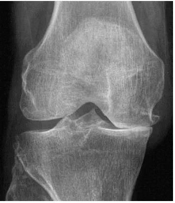
Since 1904, radiographic descriptions of OA have evolved and were first defined in an atlas by Kellgren and Lawrence (K/L) in 1953 (67, 68). Their composite grading system is commonly used today and is displayed in Table 1.3. In epidemiologic and clinical studies the cut-off point of K/L two or more comprises the radiological definition of OA (68). Figure 1.1 highlights tibiofemoral joint space narrowing (JSN) and osteophytes on a radiograph.
Although new techniques have become available, conventional radiographs remain the gold standard for the diagnosis and evaluation of knee OA. Numerous atlases have been developed to be used as guides to evaluate individual radiographic features in OA at many different anatomical sites (69-77). They have great value in screening patients to confirm OA and staging OA. They include both semi-quantitative examination of individual radiographic features, or direct measurement of the interbone distance as an indicator of joint space (i.e. joint space width (JSW)) (15). Key features measured include osteophytes, JSN, subchondral sclerosis, and bony attrition. Table 1.4 summarises these atlases.
The research conducted in this thesis used the atlas from the Osteoarthritis
Research Society International (OARSI) which was first published in 1996, by Altman et al (77). A revised version was published in 2007 (73) and is summarised in Table 1.5. The Altman atlas has many advantages. It provides clinicians and researchers with a
changes, cysts, etc). The scale is also non-linear and therefore insensitive to changes over time (73, 78).
Table 1.3. Kellgren and Lawrence (K/L) grading system
Definition grades Description
Grade 0: No osteoarthritis No osteoarthritis
Grade 1: Doubtful Doubtful narrowing of joint space and possible osteophytic lipping
Grade 2: Mild Definite osteophytes and possible narrowing of joint space
Grade 3: Moderate Multiple osteophytes, definite narrowing of joint space and some sclerosis and possible deformity of bone ends
Grade 4: Severe Large osteophyte, marked narrowing of joint space, severe sclerosis and definite deformity of bone ends
Table 1.4. Atlases developed for the radiographic assessment of osteoarthritis
Author and year Atlas Title
Spector et al, 1992 (69) A Radiographic Atlas of Knee Osteoarthritis
Altman et al, 1987 (70) Radiographic assessment of progression in osteoarthritis
Kallman et al, 1989 (71) New radiographic grading scales of osteoarthritis of the hand
Scott et al, 1993 (72) Reliability of grading scales for individual radiographic features of osteoarthritis of the knee: the Baltimore longitudinal study of aging atlas of knee osteoarthritis
Altman et al, 1995 (77) Atlas of individual radiographic features in osteoarthritis
Altman et al, 2007 (73) Atlas of individual radiographic features in osteoarthritis, revised
Verbruggen et al, 1996 (74) Numerical scoring systems for the anatomic evolution of osteoarthritis of the finger joints
Nagaosa et al, 2000 (75) Development of a logically devised line drawing atlas for grading of knee osteoarthritis
Croft et al, 2005 (76) An introduction to the atlas of standard radiographs of arthritis
Table 1.5. Individual radiographic features measured by the OARSI atlas (77) for tibiofemoral osteoarthritis
Feature Score
Marginal osteophytes
Medial femoral condyle 0–3
Medial tibial plateau 0–3
Lateral femoral condyle 0–3
Lateral tibial plateau 0–3
Joint space narrowing
Medial compartment 0–3
Lateral compartment 0–3
Other
Medial tibial attrition Absent/present
Medial tibial sclerosis Absent/present
[image:41.595.105.543.387.665.2]1.2.2 Clinical criteria
There are many definitions for symptomatic knee OA. One of the most common is regarded as frequent knee symptoms defined as “pain, aching, or stiffness in or around the knee on most days” for at least one month during the past 12 months (79). This definition has been used in many epidemiologic studies (80, 81). Other definitions of symptomatic knee OA may include a particular level of pain reported on a Visual Analog Score (VAS), the Knee injury and Osteoarthritis Outcome Score (KOOS) (82) or the Western Ontario and McMasters Universities Osteoarthritis Index (WOMAC) (83).
1.2.3 X-ray versus MRI
Radiographs continue to be the reference standard in many epidemiologic and clinical studies to define OA and monitor disease progression. This is due to their feasibility and tradition and the fact that scoring and measurement systems have been refined and standardised (84). However, the continued use of radiographs as an outcome measure in OA research has been criticised.
There is widespread belief that a high discordance exists between clinical and radiographic knee OA (12). Many studies have shown that radiographic features such as JSN and osteophytes do not correlate well with clinical symptoms (6, 13, 85-87). Hannan et al (85) found that less than 50% of people with evidence of OA on plain radiographs have symptoms related to these findings. In a systematic review, Bedson et al (12) reported that the proportion of knee pain found to have ROA ranged from 15–76% and in those with ROA the proportion with pain ranged from 15–81%. They reported that considerable variation occurred depending on the x-ray view, pain definition, OA grading scale, and demographic factors.
Radiographs are also insensitive to early disease, given that by the time ROA is detected, 10% of knee cartilage has already been lost (88). Early is a relative term and yet to be defined in OA. However, ‘early’ in this context means changes which occur prior to the development of gross abnormalities (for example, cartilage loss). Early also refers to abnormalities which occur when OA is first becoming established.
Magnetic resonance imaging (MRI) has proven to be an important tool in OA research and has revolutionised the understanding of OA pathology. It allows precise visualisation of joint structures including bone, cartilage, menisci, synovium, and ligaments. While costly, it is free of ionising radiation and has good tissue contrast and anatomical resolution (89) allowing for non-invasive examination of joint structures. MRI is a useful tool to study pre-disease or early stages of disease and there is increasing evidence to demonstrate that structural change can be measured both reliably and with good responsiveness on MRI (90). Although it has not been formally accepted by regulatory authorities, there is great interest to use MRI for assessing diagnostic status, disease severity and monitoring progression in OA (78, 91).
MRIs of OA features can be measured semi-quantitatively or quantitatively with either morphological or compositional measurements (78). Semi-quantitative scoring of MRIs allows for multifeature assessment of the knee. Features which are currently believed to be relevant include articular cartilage integrity, subchondral bone marrow abnormalities, subchondral cysts, subchondral bone attrition, osteophytes, meniscal integrity, cruciate and collateral ligament integrity, synovitis/effusion, intraarticular loose bodies, and periarticular cysts/bursitis (78). Broadly speaking, semi-quantitative
assessment of these features has shown adequate reliability and specificity (78), but the sensitivity to change over time is relatively small (92).
Quantitative measurements using computer-aided image processing allow for assessment of the whole joint. Direct quantification of cartilage volume, bone surface area, cartilage thickness, and bone marrow lesion area and volume are possible (78). Cartilage morphology assessments are the most widely used quantitative measurement. The measurement of cartilage volume from MRI has been shown to correlate well with the ex-vivo assessments of cartilage volume (stripped away from the bone) (93-96). The accurate segmentation of cartilage from surrounding tissue is an important aspect of assessing cartilage on MRI. The ideal segmentation is one which produces accurate and precise results in a reasonable timeframe.
radiographs (97). Using a modified Delphi approach, a panel of experts in the field have developed 11 propositions for a definition of OA on MRI (97). The goal of this exercise was to develop definitions of OA which can be more formally tested in relation to their diagnostic performance.
1.3 Subchondral bone and osteoarthritis
OA was initially considered a disorder of the articular cartilage; however, it is now recognised that OA involves many joint structures including subchondral bone, ligaments, menisci, periarticular muscles, peripheral nerves, and synovium (5). Radin and Rose (98) hypothesised many years ago that subchondral bone plays an important role in OA
pathogenesis and there is emerging evidence to support this. This thesis examines the role that subchondral bone plays in OA using data from a prospective population-based study of community-dwelling older adults. It focuses on different subchondral bone measures, such as bone mineral density (BMD), bone size, and bone marrow lesions (BMLs), and how these measures relate to disease progression and disease severity. A comprehensive introduction specific to each research question will be presented at the start of Chapters 4-8. Therefore, this section presents an overview of how subchondral bone relates to OA and provides a rationale for examining the aforementioned subchondral bone measures.
1.3.1 Definition
Figure 1.2. Schematic drawing of the different layers of cartilage and subchondral bone (99), with permission.
1.3.2 Function
One of subchondral bone’s primary roles is to provide support to the overlying articular cartilage (100). It is believed that the subchondral bone absorbs most of the mechanical force transmitted through the joint (98, 101, 102) and its architecture can adapt during mechanical stress (103). The subchondral bone also plays an important role in articular cartilage metabolism. Canals exist in the subchondral bone which allow blood vessels to reach the cartilage to provide nutrients (99).
1.3.3 Subchondral bone changes in osteoarthritis
Subchondral bone properties are modified through the cell-mediated process of remodelling and modelling (104). Remodelling or bone resorption is mediated by osteoclasts which are responsible for degrading the bone microenvironment (104). Following this is the modelling or bone formation phase in which osteoblasts build up a new extracellular matrix (104). In ideal physiological conditions, the amount of bone removed during the resorption phase is balanced during the formation phase. This cellular process provides a mechanism for repairing damage that occurs to the bone during
mechanical loading and stress (105, 106). During the OA process the breakdown and repair phases are disrupted resulting in subchondral bone structure changes. There is a
and development of subchondral cysts and lesions (104). The tidemark (displayed in Figure 1.2) is a line of separation between the articular cartilage and calcified cartilage. In OA thickening of the calcified cartilage followed by advancement of the tidemark has been observed in the hand (107) and other large joints such as the hip, shoulder, and knee (108). It is suggested that this may be due to vascular invasion of the calcified cartilage from the subchondral bone (109). Tidemark advancement contributes to the overall thinning of articular cartilage (104).
Radin and Rose (98) originally suggested that bone changes occur very early in the course of OA and this has now been confirmed using numerous study approaches. Animal models have been important for studying the chronological order of OA disease pathology. In a shock-load experiment in rabbits, it was found that increased stiffness of subchondral bone reduced its shock-absorbing ability and induced cartilage degeneration (110). In elderly cynomolgus macaques, thickening of the subchondral bone was shown to occur prior to cartilage destruction and the thickness of the subchondral bone was related to the onset of cartilage fibrillation (111). In a more recent study, Muraoka et al (112) have demonstrated using a spontaneous OA guinea pig model that subchondral bone is fragile before the onset of cartilage degeneration.
Studies in humans have also confirmed that subchondral bone changes occur early on in OA. Studies using isotope-labelled bone-seeking agents in patients with hand OA have found that changes in bone turnover precede bony changes detected by standard radiographs such as osteophytes and sclerosis (113, 114). In a scintigraphy study using technetium-labelled bisphosphonate, Dieppe et al (115) studied patients with established knee OA over a 5 year period. They found that elevated subchondral bone cell activity at baseline predicted subsequent loss of joint space on radiographs. Importantly joint space loss did not occur in those patients with normal bone cell activity at baseline. More
recently, numerous studies using MRI have shown that various subchondral bone measures predict disease progression such as quantitative cartilage volume loss. This is covered in more detail in the following sections.
Although it is well recognised that bone changes occur early in OA, whether or not bone changes precede cartilage damage remains controversial. Radin and Rose (98) originally suggested that the health and integrity of the joint cartilage is dependent on the mechanical properties of the underlying subchondral bone. They believed that the initiation of the disease may be partly due to the thickening and increased bone mass of the
subchondral bone. This concept has been supported by surgical, mechanical, and
changes (110-112, 116-119). However these studies may not represent the usual
mechanism of OA development. Furthermore some animal models demonstrate conflicting results. For example, a shock-load experiment in rabbits found that changes in the
mechanical properties of subchondral bone and cartilage degeneration occurred
simultaneously (120). Studies conducted in humans with OA have mostly focused on those with late or end-stage disease, which reveals little about sequential order. Regardless of whether bone changes precede cartilage damage, bone does play an important role in OA disease pathology and there appears to be tight coupling between bone and cartilage damage. The following sections will provide an overview of the different subchondral bone measures used in this thesis.
1.4 Bone mineral density and osteoarthritis
It has been suggested that an inverse relationship exists between osteoporosis and OA. This first arose in the 1970s when surgeons noticed that femoral heads resected for treatment of hip OA were much less likely to have evidence of osteoporosis (121). Since then numerous studies have examined the relationship between OA and BMD.
Cross-sectional studies have demonstrated that the presence of radiographic knee and hip OA is associated with greater BMD at the lumbar spine, femoral neck, and/or total body (122-127). However, longitudinal studies have revealed a more complex relationship between OA and osteoporosis. In the Chingford Study, Hart et al (128) found that a high BMD at the lumbar spine and hip at baseline was associated with the development of knee osteophytes but not JSN. On the contrary, they also reported that low bone density at the hip was weakly associated with OA progression. In the Study of Osteoporotic Fractures, Arden et al (129) found that radiographic hip OA was associated with reduced bone loss in the femoral neck compared with controls over approximately 7 years. However,
associated with the onset of JSN in knees without ROA at baseline. In contrast to previous studies (128, 130), they did not find an association between progression and BMD.
In summary the evidence for an inverse association between OA and osteoporosis mainly comes from cross-sectional studies and is not widely supported by longitudinal studies. The relationship between the two diseases appears to be more complex. A high BMD is associated with the development of incident knee OA; however, studies examining BMD and progression have been inconsistent. This may be because the relationship
between BMD and OA is explored using radiographs which have been taken at sites different to where BMD is measured. There is now increasing interest to study bone density at the site of OA joints.
1.4.1 Subchondral bone mineral density
Our group, as well as others, have shown that dual-energy x-ray absorptiometry (DXA) applied to the subchondral bone of the tibia is a reproducible and valid technique in measuring subchondral bone mineral density (sBMD) (132-134). Cross-sectional studies have produced conflicting data as to whether the density of the subchondral bone is