O R I G I N A L A R T I C L E
Phenotypic and functional analysis of bovine peripheral blood
dendritic cells before parturition by a novel purification method
Tao Zhuang
1,2|
Megumi Urakawa
1,2|
Hidetoshi Sato
3|
Yuko Sato
3|
Teruaki Taguchi
1,2|
Tsuyoshi Umino
1,2|
Shiro Katto
1,2|
Koutaro Tanaka
1,2|
Kozue Yoshimura
1,2|
Naokazu Takada
3|
Hiroko Kobayashi
3|
Megumi Ito
3|
Michael T. Rose
4|
Yoshio Kiku
5|
Yuya Nagasawa
5|
Haruki Kitazawa
6|
Kouichi Watanabe
1,2|
Tomonori Nochi
1,2|
Tomohito Hayashi
5|
Hisashi Aso
1,21
Cellular Biology Laboratory, Graduate School of Agricultural Science, Tohoku University, Sendai, Miyagi, Japan
2
International Education and Research Center for Food and Agricultural Immunology, Graduate School of Agricultural Science, Tohoku University, Sendai, Miyagi, Japan
3
Miyagi Prefecture Animal Industry Experi-ment Station, Iwadeyama, Miyagi, Japan
4
Institute of Biological, Environmental and Rural Sciences, Aberystwyth University, Cardiganshire, UK
5
Hokkaido Research Station, National Institute of Animal Health, NARO, Sapporo, Hokkaido, Japan
6
Food and Feed Immunology Group, Graduate School of Agricultural Science, Tohoku University, Sendai, Miyagi, Japan
Correspondence
Hisashi Aso, Graduate School of Agricultural Science, Tohoku University, Sendai, Miyagi, Japan.
Email: asosan@tohoku.ac.jp
Funding information
Ministry of Education, Culture, Sports, Science and Technology, Grant/Award Number: 24658224, 26660217; Ministry of Agriculture, Forestry and Fisheries, Grant/
Abstract
Dendritic cells (DCs) are specialized antigen presenting cells specializing in antigen
uptake and processing, and play an important role in the innate and adaptive
immune response. A subset of bovine peripheral blood DCs was identified as
CD172a
+/CD11c
+/MHC (major histocompatibility complex) class II
+cells. Although
DCs are identified at 0.1%
–
0.7% of peripheral blood mononuclear cells (PBMC), the
phenotype and function of DCs remain poorly understood with regard to
maintain-ing tolerance durmaintain-ing the pregnancy. All cattle used in this study were 1 month
before parturition. We have established a novel method for the purification of DCs
from PBMC using magnetic-activated cell sorting, and purified the CD172a
+/
CD11c
+DCs, with high expression of MHC class II and CD40, at 84.8% purity.
There were individual differences in the expressions of CD205 and co-stimulatory
molecules CD80 and CD86 on DCs. There were positive correlations between
expression of cytokine and co-stimulatory molecules in DCs, and the DCs
main-tained their immune tolerance, evidenced by their low expressions of the
co-stimu-latory molecules and cytokine production. These results suggest that before
parturition a half of DCs may be immature and tend to maintain tolerance based on
the low cytokine production, and the other DCs with high co-stimulatory molecules
may already have the ability of modulating the T-cell linage.
K E Y W O R D S
cattle, cytokine, dendritic cell, phenotype, positive-selection
Award Number: J120001170; Science and Technology Research Promotion Program for Agriculture, Forestry, Fisheries and Food Industry, Grant/Award Number: J160000725, J170001750; Japan Society for the Promotion of Science (JSPS)
Tao Zhuang and Megumi Urakawa contributed equally to this work.
-This is an open access article under the terms of the Creative Commons Attribution-NonCommercial-NoDerivs License, which permits use and distribution in any medium, provided the original work is properly cited, the use is non-commercial and no modifications or adaptations are made.
©2018 The Authors.Animal Science Journalpublished by John Wiley & Sons Australia, Ltd on behalf of Japanese Society of Animal Science
1
|
I N T R O D U C T I O N
Dendritic cells (DCs) were first identified in the peripheral lymphoid
organs of mice (Steinman & Cohn, 1973), specializing in antigen
uptake and processing as an antigen-presenting cell (APC). DCs also
play an important role in the innate and adaptive immune response
(Banchereau & Steinman, 1998). The phenotypic and functional
char-acterizations of peripheral blood DCs in humans have been
described in several studies (MacDonald et al., 2002; Odoherty et al.,
1994; Thomas, Davis, & Lipsky, 1993). However, the phenotype and
function of peripheral blood DCs in cattle remain poorly understood.
A subset of bovine peripheral blood DCs was identified as
CD172a+/CD11c+/MHC (major histocompatibility complex) class II+
cells in the CD3/B-B2/CD14population (Miyazawa et al., 2006)
and expressed a CD205 molecule on the cell surface
(Gonzalez-Cano, Arsic, Popowych, & Griebel, 2014). CD205, as an
antigen-uptake receptor, was also expressed on DCs in lymphoid tissue
(Gliddon, Hope, Brooke, & Howard, 2004). In addition, it has
previ-ously been reported that the surface molecules of CD40, CD80 and
CD86 in DCs provided co-stimulate signals in T cell activation
(Van-Gool, Vandenberghe, DeBoer, & Ceuppens, 1996).
In order to prevent fetal rejection caused by the recognition of
paternal antigens, the maternal immune system has to be mobilized
toward tolerance (Zenclussen, 2013). T helper (Th) cells play a
cen-tral role in immune responses. However, the expression of Th1 and
Th17-related genes is inhibited in bovine late gestation (Maeda,
Oht-suka, Tomioka, & Oikawa, 2013). The previous report showed the
characterization of higher Th2/regulatory immunity by the increases
ofIFN-coccurring after parturition andIL-4production before calv-ing (Paibomesai, Hussey, Nino-Soto, & Mallard, 2013).
Among periparturient Jersey cows during the 2 weeks before
and 2 weeks after parturition, the percentage of T cells with CD3,
CD4 and gamma delta T-cell receptors reduced substantially in blood
(Kimura, Goff, Kehrli, & Harp, 1999). During the periparturient period
there is a decline in T-lymphocyte cell subsets, which parallels a
reduction in functional capacities of blood lymphocytes (Kimura,
Goff, Kehrli, Harp, & Nonnecke, 2002). Paternal T cells are aware of
the presence of paternal antigens during pregnancy, where they
acquire a transient state of tolerance specific for paternal antigens
(Tafuri, Alferink, Moller, Hammerling, & Arnold, 1995). Regulatory T
cells (Treg), the main function of which is to prevent autoimmunity,
emerged as important players in regulating tolerance toward paternal
and fetal antigens (Sakaguchi, Sakaguchi, Asano, Itoh, & Toda, 1995).
Treg must first encounter antigens presented by antigen-presenting
cells, as for example, DCs in an appropriate cytokine environment, to
proliferate and function. In addition, DCs represent the first event
leading to a protective adaptive immune response (Robertson, Mau,
Tremellen, & Seamark, 1996), and contribute to the expansion of the
peripheral Treg population (Schumacher et al., 2012). Immature DCs
expressed a low level of MHC molecules and co-stimulatory
mole-cules such as CD40, CD80 and CD86, and showed the reduced
pro-duction of pro-inflammatory cytokines (IL-12, TNFa, IL-6) (Lutz & Schuler, 2002). These data are compatible with the hypothesis that
declining T-cell populations may contribute to the
immunosuppres-sion reported for dairy cows at calving, and that DCs may regulate
the population and functions of T cells during the days and weeks
before and after parturition. However, the function for maintaining
the tolerance during the pregnancy has not been clearly described in
DCs in bovine blood. Previous works showed that in late gestation,
the cows had a heightened susceptibility to persistent infections
caused by mastitis and abortion-causing pathogens (Green, Green,
Medley, Schukken, & Bradley, 2002; Williams et al., 2000).
There-fore, we studied cattle which were 1 month before parturition.
In this study, we investigated the phenotypic and functional
characterization of bovine peripheral blood DCs before parturition.
As the population of DCs is less than 5% in bovine peripheral blood
mononuclear cells (PBMC), there is a need to isolate highly purified
DC subpopulations in sufficient numbers. Therefore, we have
estab-lished a novel method of two-step magnetic-activated cell sorting
(MACS) for bovine peripheral DCs, and were able to obtain DCs at a
purity of more than 85% from PBMC. After the purification, we
determined the expressions of surface markers (MHC II, CD205,
CD40, CD80 and CD86) on DCs using flow cytometry and analyzed
the expression of a number of cytokines (IL-12a,IL-4,IFN-c, and IL-6). This study provides the evidence for immune regulation of bovine
DC populations before parturition.
2
|
M A T E R I A L S A N D M E T H O D S
2.1
|
Animals
Sixteen Holstein Friesian cows (average age at 5.22.2 years,
calv-ing number at 2.31.8), housed at the Miyagi Prefecture Animal
Industry Experiment Station, were used in this study. All animal
handing and experimental protocols were conducted in compliance
with guidance approved by the Tohoku University Environmental
and Safety Committee on Experimental Animal Care and Use, and
the Environmental and Safety Committee on Miyagi prefecture
ani-mal industry experiment station. These aniani-mals were clinically
healthy and kept in the same conditions.
2.2
|
Blood sampling
Jugular venous blood (200 ml) was obtained from the cows at
1 month prior to parturition, into tubes containing sodium heparin,
and was diluted 1:1 with phosphate-buffered saline (PBS). PBMC
were separated from the buffy coat using Lympholyteâ-H (1.077 g/
ml; CEDARLANE, Burlington, Ontario, Canada) gradient centrifuged
at 6009gfor 30 min at 18°C. PBMC were washed once with lys-ing buffer (Tris-HCl buffer containlys-ing 0.83% ammonium chloride)
and twice with PBS at 4509geach for 10 min at 4°C.
2.3
|
Purification of peripheral blood DCs
The anti-bovine antibodies in this study were purchased from WSU
(Birmingham, AL, USA), BD Biosciences (Franklin Lakes, NJ, USA) and
Miltenyi Biotec (Bergisch Gladbach, Germany) (Table 1). For the
sort-ing of CD3/sIgM/CD14/granulocytescells, PBMC were washed
with PBS containing 0.2% bovine serum albumen (BSA), and incubated
with the mixture of mouse anti-bovine CD3 (diluted 1/50), mouse
anti-bovine sIgM (diluted 1/100), mouse anti-bovine CD14 (diluted 1/
50), and mouse anti-bovine granulocytes (diluted 1/1000) antibodies
for 30 min on ice, followed by the incubation with rat anti-mouse
IgG1 Micro Beads and rat anti-mouse IgM Micro Beads for 30 min on
ice, respectively. CD3/sIgM/CD14/granulocytescells containing
DCs were negatively selected using Auto MACS magnetic columns
(Miltenyi Biotec, Bergisch Gladbach, Germany). After negative
selec-tion, CD3/sIgM/CD14/granulocytescells were incubated with
mouse bovine CD172a antibody (diluted 1/200) and rat
anti-mouse IgG1 Micro Beads for 30 min on ice, respectively. CD172a+
cells were positively selected from CD3/sIgM/CD14
/granulo-cytescells using Auto MACS magnetic columns.
2.4
|
Flow cytometry
In order to detect bovine DCs, PBMC, CD3/sIgM/CD14
/granu-locytes negative-selected cells in MACS step 1 (negative-selected
cells) and CD172a+positive-selected cells in MACS step 2
(positive-selected cells) were stained with mouse bovine CD172a
anti-body and co-stained with mouse anti-bovine CD11c (diluted 1/500)
and MHC class II (diluted 1/250) antibodies. PBMC and
negative-selected cells were incubated with anti-bovine CD3, sIgM, CD14 or
granulocytes antibody in order to confirm the deletion of T cells, B
cells, monocytes and granulocytes. Negative-selected cells were
incubated with mouse anti-bovine MHC class II, CD40, CD205,
CD80 or CD86 antibody, and treated with secondary fluorescent
antibodies for 30 min on ice in the dark. After the treatment of
sec-ondary fluorescent antibodies as shown in Table 1, each cell was
subjected to the flow cytometry analysis using the Accuri C6 flow
cytometer (BD Biosciences) and the BD Accuri C6 software, Version
1.0.264.21 (BD Biosciences). In each experiment, cells incubated
with isotype-matched antibodies and secondary fluorescent
antibod-ies were selected as controls.
2.5
|
Immunocytochemical staining
Negative- and positive-selected cells were stained with mouse
anti-bovine CD172a antibody and co-stained with mouse anti-anti-bovine
CD11c and MHC class II antibodies, and then stained with peridinin
chlorophyll protein complex conjugated rat anti-mouse IgG1,
Phyco-erythrin (PE) conjugated goat anti-mouse IgM and fluorescein
isothio-cyanate conjugated goat anti-mouse IgG2a fluorescent antibodies
(Table 1). Cells were then centrifuged onto glass slides (Cytospin 2
Thermo Shandon, Pittsburgh, PA, USA) at 6009gfor 5 min. After air drying for 5 min, cells were counterstained with 40
,6-diamidino-2-phe-nylindole (DAPI) for 5 min at room temperature in the dark, and were
washed three times with PBS. Slide images were viewed using a Laser
Scanning Microscope 700 (Carl Zeiss, Jena, German), and
pho-tographed at 4009with LSM software ZEN 2012, Version 8.0.0.273.
T A B L E 1 Antibodies used in this study
Antibodies Specificity Isotype Clone Supplier
CD3 Pan T cells IgG1 MM1A WSU
Surface IgM Pan B cells IgG1 IL-A30 Bio-Rad
CD14 Mø, monocytes IgG1 CAM36A WSU
Granulocytes Granulocytes IgM CH138A WSU
CD172a Mø, monocytes, DCs IgG1 DH59B WSU
CD11c Mø, monocytes, DCs
T cell subset, B cell subset
IgM BAQ153A WSU
MHC II MHC class II IgG2a TH14B WSU
CD205 Mø, DCs IgG2a ILA53A WSU
CD40 FITC Co-stimulatory molecule IgG1 IL-A156 Bio-Rad
CD80 FITC Co-stimulatory molecule IgG1 IL-A159 Bio-Rad
CD86 FITC Co-stimulatory molecule IgG1 IL-A190 Bio-Rad
Control Mouse IgG1 COLIS69A WSU
Control Mouse IgM COLIS52A2 WSU
Control Mouse IgG2a COLIS205C WSU
FITC IgG2a-secondary ab Goat anti-mouse SouthernBiotech
PE IgM-secondary ab Goat anti-mouse SouthernBiotech
PerCP IgG1-secondary ab Rat anti-mouse BD Biosciences
IgG1 Micro Beads ab Rat anti-mouse Miltenyi Biotec
[image:3.595.47.553.430.719.2]2.6
|
Quantitative real-time polymerase chain
reaction (qPCR) analysis
After the negative and positive selections, the purified bovine
peripheral blood DCs were stored at 80°C. Total RNA was
extracted from them using ISOGEN II reagent (Takara Bio Inc.,
Siga, Japan) following the manufacturer’s instructions, and its
concentration was determined by spectrophotometry at 260 nm.
The reverse transcription and complementary DNA (cDNA)
syn-thesis are described below. In brief, 2lg of total RNA was mixed with 500 ng oligo (DT)12–18 and 1ll of 10 mmol/L
deoxynu-cleotide triphosphates (dNTPs) (Invitrogen, Carlsbad, CA, USA).
The mixture was heated to 65°C for 10 min in order to prepare
for cDNA synthesis. Then the first-strand cDNA was incubated
with 200 units of Superscript RT III, 0.1M DTT and 59 First-Strand Buffer (Invitrogen) at 50°C for 1 hr, and then at 70°C for
15 min.
Onell cDNA sample, 7ll SYBR Green Premix Taq (Takara Bio Inc.), 1lof 5pM corresponding primer pair, and RNase-free water were added in a 20ll final volume per well in a 96-well plate. The primer sets of bovine cytokines are listed in Table 2 (Takara Bio
Inc.). The transcripts using the bovine peripheral blood DC cDNA
were amplified with the Thermal Cycler Dice Real Time System
Sin-gle (Takara Bio Inc.): one cycle at 95°C for 30 s; 40 cycles at 95°C
for 5 s, 60°C for 30 s, then 95°C for 15 s, 60°C for 30 s and finally
95°C for 15 s. From template DNA, SYBR green fluorescence was
T A B L E 2 Primer information for quantitative real-time PCR in this
study
Primer Sequence Size (bp)
IL-12a FW GGCAGCTATTGCTGAGCTGATG 136
RV ACGAATTCTGAAGGCGTGAAG
IFN-c FW CATAACACAGGAGCTACCGATTTCA 197
RV CCCTTAGCTACATCTGGGCTACTTG
IL-4 FW CTTAGGCGTATCTACAGGAGCCACA 112
RV TCGTCTTGGCTTCATTCACAGAAC
IL-6 FW ATGCTTCCAATCTGGGTTCAATC 98
RV ATGCTTCCAATCTGGGTTCAATC
GAPDH FW GATGGTGAAGGTCGGAGTGAAC 100
RV GTCATTGATGGCGACGATGT
FW, forward primer; RV, reverse primer
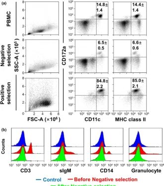
F I G U R E 1 Purification of bovine
[image:4.595.46.290.74.229.2] [image:4.595.46.378.349.725.2]detected for the calculation of copy numbers. The specificity and
the integrity of PCR product were confirmed by the dissociation
curve analysis. GAPDH-specific primers were used as the internal
controls, and the reactions without template were used as negative
control experiments. The results of the target gene are presented as
the relative expression level to the expression of house-keeping
GAPDHgene.
2.7
|
Statistical analysis
Values are reported as meansSD. Statistical analysis were
per-formed using the software GraphPad 6.00 program (GraphPad
soft-ware Inc., La Jolla, CA, USA). The correlation between two
parameters was analyzed by Pearson correlation coefficient test
(*p<0.05,**p<0.01).
3
|
R E S U L T S
3.1
|
Purification of bovine peripheral blood DCs
We tried to purify bovine blood DCs from PBMC. Figure 1 shows
the purification process of bovine peripheral blood DC. The
expres-sion of the surface molecules such as CD172a, CD11c and MHC
class II, specific markers of DC, were assessed by three-color flow
cytometry without any gate (Figure 1a). Among the total PBMC,
14.8% of CD172a+CD11c+cells were present and almost expressed
MHC class II molecules. However, it is well known that CD11c is
highly expressed on monocytes, macrophages (Mø) and natural killer
(NK) cells, and that CD172a+/CD11c+cells possibly include a subset
of T cells, B cells, NK cells and monocytes/Mø. Therefore, we
attempted to remove these cell populations from PBMC using each
F I G U R E 2 Photographs of peripheral
[image:5.595.215.552.259.731.2]specific monoclonal antibody. After the negative selection,
CD172a+/CD11c+ cells were found to represent about 6.5% of
the negative-collected cells and also expressed MHC class II on
the cell surface. The negative selection using MACS removed T
cells (CD3+), B cells (surface IgM+), monocytes (CD14+) and
granu-locytes from PBMC, and these populations in negative-selected
cells disappeared (Figure 1b). Therefore, CD172a+/CD11c+cells in
the negative-selected cells were considered as bovine peripheral
blood DCs, which also expressed MHC class II molecules.
How-ever, the negative-selected cells contained a large population of
CD172a/CD11c non-DC cells. Next, we tried to purify
CD172a+/CD11c+cells from the negative-selected cells. The
posi-tive selection with CD172a antibody revealed that the purity of
CD172a+/CD11c+ DCs was 84.8%, and that they also strongly
expressed MHC class II.
3.2
|
Photographs of peripheral blood DCs
Peripheral blood DCs after the negative and positive selections were
stained with anti-bovine CD172a (red), CD11c (green) and MHC class
II (green) antibodies. All samples were counterstained with DAPI (blue)
(Figure 2). After the negative selection, CD172a+/CD11c+ and
CD172a+/MHC class II+DCs were detected as a small population in
the photographs. Indeed, there were plenty of CD172a/CD11c/
MHC class IInon-DC cells which are indicated with arrows. However,
this cell population indicated with arrows decreased after positive
selection with anti-CD172a antibody. Almost all the positive-selected
cells expressed CD172a, CD11c and MHC class II, which were
consid-ered as the bovine peripheral blood DCs. These data suggest that the
two-step MACS method can highly purify DCs from bovine blood.
3.3
|
Phenotypic analysis and cytokine expression
of bovine peripheral blood CD172a
+/CD11c
+DCs
before parturition
Next, the surface expression of MHC class II, CD40, CD205, CD80
or CD86 was analyzed on CD172a+/CD11c+cells after the negative
selection (Figure 3a). The results demonstrated that almost all the
CD172a+/CD11c+ DCs expressed the molecules of MHC class II
(98.480.54%) and CD40 (94.980.88%). However, there were
individual differences in the expression of CD205, CD80 or CD86 in
the CD172a+/CD11c+ DCs. The percentages of CD205, CD80 and
CD86 positive cells were 17.083.97, 29.684.23, and
23.506.02 of CD172a+/CD11c+DCs, respectively. Before
parturi-tion, there were significant correlations between the percentage of
CD86 and the percentages of CD80 or CD205 on CD172a+/CD11c+
DCs (Figure 3b).
As the purity of bovine peripheral blood DC was more than 85%
after positive selection, it became available for the examination of the
expression of T cell-modulation cytokines in DCs (Figure 4). There
were significant correlations in bovine peripheral DCs with the
acti-vated molecule of CD205 and the messenger RNA expressions of
IFN-candIL-6. In addition, there were significant correlations between the
co-stimulatory molecule CD80 and the expressions ofIL-12a,IL-4and
IFN-c, and between CD86 and the expressions ofIL-4,IFN-candIL-6.
4
|
D I S C U S S I O N
In this study, we have established a novel purification method for
bovine peripheral blood DCs. We have also characterized the
pheno-type and function of the DCs. A previous study revealed that DCs
were identified at 0.1%–0.7% of PBMC (Renjifo et al., 1997).
Because of the low percentage of DCs in the PBMC, it was
neces-sary to deplete the non-DC from bovine PBMC (Gibson, Miah,
Grie-bel, Brownlie, & Werling, 2012; Miyazawa et al., 2006; Renjifo et al.,
1997; Sei, Ochoa, Bishop, Barlow, & Golde, 2014). In this study, T
cells, B cells, monocytes and granulocytes were depleted from PBMC
by negative selection. However, CD172a+/CD11c+ cells with MHC
class II molecules were detected at 6.5% of the negative-selected
cells. This cell fraction was revealed as DCs (Gonzalez-Cano et al.,
2014; Miyazawa et al., 2006); however, it was very difficult to
inves-tigate the functional and the genetic analysis of bovine blood DCs
using it. Using positive selection with anti-bovine CD172a antibody
and immunomagnetic microbeads, we were able to purify the
CD172a+/CD11c+DCs with MHC class II molecules at 84.8% purity,
F I G U R E 3 Phenotypic characterization of bovine peripheral
blood CD172a+CD11c+dendritic cells (DCs) before parturition. After
the negative selection, the surface expression of major
and also confirm the purified cells as DCs using the
immunofluores-cence photographs (Figure 2).
DCs are specialized antigen-presenting cells that regulate both
immunity and tolerance. DCs in the periphery play a key role in
induction of T cell immunity, as well as tolerance. DCs are
phenotyp-ically and functionally heterogeneous, and further classified into
sev-eral subsets depending on distinct marker expression and their
location. Co-stimulatory molecules were necessary to the T-cell
responses and were up-regulated during DC activation (Cools,
Pon-saerts, Van Tendeloo, & Berneman, 2007). The program of
matura-tion of DCs brings about the up-regulamatura-tion of MHC II (Lanzavecchia
& Sallusto, 2001) and co-stimulatory molecules CD80 and CD86
(Mellman & Steinman, 2001). Bovine DCs are characterized by the
increased expression of MHC II, CD11c, CD80/CD86 and the
decreased expression of CD14 and CD21 surface markers (Denis &
Buddle, 2008). CD80 and CD86 on DCs interact with the CD28
(stimulatory) and CTLA-4 (inhibitory) receptors of the T cells. The
absence of CD80 and CD86 results in lack of co-stimulatory signal
delivery to T cells and leads to clonal anergy and lack of proper T
cell response (Schwartz, 1990). The signaling molecule CD40 is
required to induce immunogenic DCs and for the induction ofIFNa (Le Bon et al., 2006; Martin, O’Sullivan, Low, & Thomas, 2003).
The purified DCs from peripheral blood not only expressed
CD172a, CD11c and MHC class II on the surface, but also expressed
CD40, CD205, CD80 and CD86 (Figure 3). The majority of the DCs
expressed the molecules of MHC class II and CD40. It is well known
that CD205 has been expressed on many DCs in the T cell areas of
lymphoid tissues (Gliddon et al., 2004). It has been reported that
CD205 can lead to tolerance in the steady-state immunity after DC
maturation (Bonifaz et al., 2002). Therefore, a part of bovine
periph-eral blood DCs before parturition might have been differentiated
into activated DCs with high CD205. In this study, before parturition
there were strong correlations in CD172a+/CD11c+ DCs between
the CD86 expression and the expressions of CD80, as well as
CD205. Therefore, our phenotype analysis of DCs revealed that
there were both immature DCs and activated DCs in the peripheral
blood, and that the peripheral blood DCs might have the potential of
regulation for T cell lineage.
DCs collect and process antigens for presentation to T cells, and
differ in the regulatory signals they transmit, directing T cells to
dif-ferent types of immune response or to tolerance (Shortman & Liu,
2002; Steinman, 1991). The priming with DCs was strictly dependent
on CD80⁄CD86, and CD86 was well known to induce naive T cells
to become IL-4 producers (Debecker et al., 1994). DCs may
F I G U R E 4 Relationship between
expression of cytokines and surface molecule positivity in bovine peripheral blood dendritic cells (DCs) before parturition. The correlations between the expressions ofIL-12a,IL-4,IFN-candIL-6 and the percentages of surface molecules CD205, CD80 or CD86 are shown in DCs after the negative and positive selections.
[image:7.595.214.548.50.431.2]determine the specificity, the amplitude and the character (Th1 ⁄
Th2) of the immune response. Therefore, we also investigated the
cytokine production of the DCs and the correlations between
expression of cytokine and co-stimulatory molecules. As the
secre-tion ofIL-2,IFN-candIL-4from DCs induced the development of T lymphocytes (Debecker et al., 1994), there were great positive
corre-lations between CD80/CD86 positivity and the expressions ofIL-6,
IFN-candIL-4(Figure 4).IL-12 from DCs appeared as a potent and obligatory inducer of Th1 priming (De Becker et al., 1998). In
addition, IL-12 is produced by DCs and is able to increase their
stimulatory capacity of DCs (Kelleher & Knight, 1998). As CD80
high-positive DCs well inducedIL-12a, there might be an autocrine
effect ofIL-12aon DC maturation (Figure 4). In contrast, a half of
cattle in this study showed low expressions of CD205, CD80 and
CD86 with low expressions ofIL-12a,IL-4,IFN-cand IL-6. A previ-ous study indicates that bovine DCs in late gestation have reduced
Th1-promoting cytokine production compared with regulatory
cyto-kine production (Pomeroy, Sipka, Klaessig, & Schukken, 2015).
Therefore, a half of bovine peripheral DCs before parturition may be
immature and tend to maintain tolerance based on the low cytokine
production. In addition, the other DCs with high CD205 and CD80/
CD86 may already have the ability of modulating the T-cell linage.
Our purification method in this study was considered as a useful tool
to identify the capacity of DCs for activating T cells in vitro. Further
research should explore into the similar phenotype DCs in bovines
after parturition during the lactation period.
A C K N O W L E D G M E N T S
This research was supported by Grants-in-Aid for Scientific Research
(24658224, 26660217) from the Ministry of Education, Culture,
Sports, Science and Technology, a grant (J120001170) from the
Min-istry of Agriculture, Forestry and Fisheries, and two grants
(J160000725, J170001750) from the Science and Technology
Research Promotion Program for Agriculture, Forestry, Fisheries and
Food Industry. This work was also financially supported by the Japan
Society for the Promotion of Science (JSPS) through JSPS
Core-to-Core Program (Advanced Research Networks) entitled“Establishment
of international agricultural immunology research-core for a quantum
improvement in food safety”.
O R C I D
Tao Zhuang http://orcid.org/0000-0003-1475-7138
Yuya Nagasawa http://orcid.org/0000-0003-1392-6454
R E F E R E N C E S
Banchereau, J., & Steinman, R. M. (1998). Dendritic cells and the control of immunity.Nature,392, 245–252. https://doi.org/10.1038/32588 Bonifaz, L., Bonnyay, D., Mahnke, K., Rivera, M., Nussenzweig, M. C., &
Steinman, R. M. (2002). Efficient targeting of protein antigen to the dendritic cell receptor DEC-205 in the steady state leads to antigen
presentation on major histocompatibility complex class I products and peripheral CD8(+) T cell tolerance.Journal of Experimental Medi-cine,196, 1627–1638. https://doi.org/10.1084/jem.20021598 Cools, N., Ponsaerts, P., Van Tendeloo, V. F. I., & Berneman, Z. N. (2007).
Balancing between immunity and tolerance: An interplay between dendritic cells, regulatory T cells, and effector T cells. Journal of Leukocyte Biology, 82, 1365–1374. https://doi.org/10.1189/jlb. 0307166
De Becker, G., Moulin, V., Tielemans, F., De Mattia, F., Urbain, J., Leo, O., & Moser, M. (1998). Regulation of T helper cell differentiation in vivo by soluble and membrane proteins provided by antigen-presenting cells. European Journal of Immunology, 28, 3161–3171. https://doi. org/10.1002/(issn)1521-4141
Debecker, G., Sornasse, T., Nabavi, N., Bazin, H., Tielemans, F., Urbain, J., . . .Moser, M. (1994). Immunoglobulin isotype regulation by antigen-presenting cells in-vivo. European Journal of Immunology, 24, 1523– 1528. https://doi.org/10.1002/(issn)1521-4141
Denis, M., & Buddle, B. M. (2008). Bovine dendritic cells are more per-missive for Mycobacterium bovis replication than macrophages, but release more IL-12 and induce better immune T-cell proliferation. Immunology and Cell Biology,86, 185–191. https://doi.org/10.1038/sj. icb.7100124
Gibson, A., Miah, S., Griebel, P., Brownlie, J., & Werling, D. (2012). Identi-fication of a lineage negative cell population in bovine peripheral blood with the ability to mount a strong type I interferon response. Developmental and Comparative Immunology, 36, 332–341. https:// doi.org/10.1016/j.dci.2011.05.002
Gliddon, D. R., Hope, J. C., Brooke, G. P., & Howard, C. J. (2004). DEC-205 expression on migrating dendritic cells in afferent lymph. Immunology, 111, 262–272. https://doi.org/10.1111/j.0019-2805. 2004.01820.x
Gonzalez-Cano, P., Arsic, N., Popowych, Y. I., & Griebel, P. J. (2014). Two functionally distinct myeloid dendritic cell subpopulations are present in bovine blood. Developmental and Comparative Immunology, 44, 378–388. https://doi.org/10.1016/j.dci.2014.01.014
Green, M. J., Green, L. E., Medley, G. F., Schukken, Y. H., & Bradley, A. J. (2002). Influenceof dry period bacterial intramammary infection on clinical mastitis in dairy cows. Journal of Dairy Science, 85, 2589– 2599. https://doi.org/10.3168/jds.s0022-0302(02)74343-9
Kelleher, P., & Knight, S. C. (1998). IL-12 increases CD80 expression and the stimulatory capacity of bone marrow-derived dendritic cells. Inter-national Immunology, 10, 749–755. https://doi.org/10.1093/intimm/ 10.6.749
Kimura, K., Goff, J. P., Kehrli, M. E., & Harp, J. A. (1999). Phenotype anal-ysis of peripheral blood mononuclear cells in periparturient dairy cows.Journal of Dairy Science,82, 315–319. https://doi.org/10.3168/ jds.s0022-0302(99)75238-0
Kimura, K., Goff, J. P., Kehrli, M. E., Harp, J. A., & Nonnecke, B. J. (2002). Effects of mastectomy on composition of peripheral blood mononu-clear cell populations in periparturient dairy cows. Journal of Dairy Science, 85, 1437–1444. https://doi.org/10.3168/jds.s0022-0302(02) 74211-2
Lanzavecchia, A., & Sallusto, F. (2001). The instructive role of dendritic cells on T cell responses: Lineages, plasticity and kinetics. Current Opinion in Immunology,13, 291–298. https://doi.org/10.1016/s0952-7915(00)00218-1
Le Bon, A., Montoya, M., Edwards, M. J., Thompson, C., Burke, S. A., Ashton, M.,. . .Borrow, P. (2006). A role for the transcription factor RelB in IFN-alpha production and in IFN-alpha-stimulated cross-prim-ing.European Journal of Immunology,36, 2085–2093. https://doi.org/ 10.1002/(issn)1521-4141
MacDonald, K. P. A., Munster, D. J., Clark, G. J., Dzionek, A., Schmitz, J., & Hart, D. N. J. (2002). Characterization of human blood dendritic cell subsets. Blood,100, 4512–4520. https://doi.org/10.1182/blood-2001-11-0097
Maeda, Y., Ohtsuka, H., Tomioka, M., & Oikawa, M. (2013). Effect of pro-gesterone on Th1/Th2/Th17 and regulatory T cell-related genes in peripheral blood mononuclear cells during pregnancy in cows. Veteri-nary Research Communications, 37, 43–49. https://doi.org/10.1007/ s11259-012-9545-7
Martin, E., O’Sullivan, B., Low, P., & Thomas, R. (2003). Antigen-specific suppression of a primed immune response by dendritic cells mediated by regulatory T cells secreting interleukin-10.Immunity,18, 155–167. https://doi.org/10.1016/s1074-7613(02)00503-4
Mellman, I., & Steinman, R. M. (2001). Dendritic cells: Specialized and regulated antigen processing machines. Cell, 106, 255–258. https:// doi.org/10.1016/s0092-8674(01)00449-4
Miyazawa, K., Aso, H., Honda, M., Kido, T., Minashima, T., Kanaya, T.,. . . Yamaguchi, T. (2006). Identification of bovine dendritic cell pheno-type from bovine peripheral blood.Research in Veterinary Science,81, 40–45. https://doi.org/10.1016/j.rvsc.2005.09.003
Odoherty, U., Peng, M., Gezelter, S., Swiggard, W. J., Betjes, M., Bhard-waj, N., & Steinman, R. M. (1994). Human blood contains 2 subsets of dendritic cells, one immunologically mature and the other imma-ture.Immunology,82, 487–493.
Paibomesai, M., Hussey, B., Nino-Soto, M., & Mallard, B. A. (2013). Effects of parturition and dexamethasone on DNA methylation pat-terns of IFN-gamma and IL-4 promoters in CD4+ T-lymphocytes of Holstein dairy cows. Canadian Journal of Veterinary Research-Revue Canadienne De Recherche Veterinaire,77, 54–62.
Pomeroy, B., Sipka, A., Klaessig, S., & Schukken, Y. (2015). Monocyte-derived dendritic cells from late gestation cows have an impaired ability to mature in response toE-colistimulation in a receptor and cytokine-mediated fashion.Veterinary Immunology and Immunopathol-ogy,167, 22–29. https://doi.org/10.1016/j.vetimm.2015.06.016 Renjifo, X., Howard, C., Kerkhofs, P., Denis, M., Urbain, J., Moser, M., &
Pas-toret, P. P. (1997). Purification and characterization of bovine dendritic cells from peripheral blood.Veterinary Immunology and Immunopathol-ogy,60, 77–88. https://doi.org/10.1016/s0165-2427(97)00092-5 Robertson, S. A., Mau, V. J., Tremellen, K. P., & Seamark, R. F. (1996). Role
of high molecular weight seminal vesicle proteins in eliciting the uter-ine inflammatory response to semen in mice.Journal of Reproduction and Fertility,107, 265–277. https://doi.org/10.1530/jrf.0.1070265 Sakaguchi, S., Sakaguchi, N., Asano, M., Itoh, M., & Toda, M. (1995).
Immunological self-tolerance maintained by activated T-cells express-ing IL-2 receptor alpha-chains (CD25)–breakdown of a single mech-anism of self-tolerance causes various autoimmune-diseases. Journal of Immunology,155, 1151–1164.
Schumacher, A., Wafula, P. O., Teles, A., El-Mousleh, T., Linzke, N., Zen-clussen, M. L.,. . .Zenclussen, A. C. (2012). Blockage of heme oxyge-nase-1 abrogates the Protective effect of regulatory T cells on murine pregnancy and promotes the maturation of dendritic cells. PLoS ONE,7, 13.
Schwartz, R. H. (1990). A cell-culture model for lymphocyte-T clonal anergy. Science,248, 1349–1356. https://doi.org/10.1126/science.2113314 Sei, J. J., Ochoa, A. S., Bishop, E., Barlow, J. W., & Golde, W. T. (2014).
Phenotypic, ultra-structural, and functional characterization of bovine peripheral blood dendritic cell subsets.PLoS ONE,9, 17.
Shortman, K., & Liu, Y. J. (2002). Mouse and human dendritic cell sub-types. Nature Reviews Immunology, 2, 151–161. https://doi.org/10. 1038/nri746
Steinman, R. M. (1991). The dendritic cell system and its role in immuno-genicity.Annual Review of Immunology, 9, 271–296. https://doi.org/ 10.1146/annurev.iy.09.040191.001415
Steinman, R. M., & Cohn, Z. A. (1973). Identification of a novel cell type in peripheral lymphoid organs of mice.Journal of Experimen-tal Medicine, 137, 1142–1162. https://doi.org/10.1084/jem.137.5. 1142
Tafuri, A., Alferink, J., Moller, P., Hammerling, G. J., & Arnold, B. (1995). T-CELL Awareness of paternal alloantigens during pregnancy.Science, 270, 630–633. https://doi.org/10.1126/science.270.5236.630 Thomas, R., Davis, L. S., & Lipsky, P. E. (1993). Isolation and
characteriza-tion of human peripheral-blood dendritic cells.Journal of Immunology, 150, 821–834.
VanGool, S. W., Vandenberghe, P., DeBoer, M., & Ceuppens, J. L. (1996). CD80, CD86 and CD40 provide accessory signals in a multiple-step T-cell activation model.Immunological Reviews,153, 47–83.
Williams, D. J., Guy, C. S., Mcgarry, J. W., Guy, F., Tasker, L., Smith, R. F., . . .Trees, A. J. (2000). Neospora caninum-associatedabortion in cat-tle: The time of experimentally-induced parasitaemia during gestation determines foetal survival. Parasitology, 121, 347–358. https://doi. org/10.1017/s0031182099006587
Zenclussen, A. C. (2013). Adaptive immune responses during pregnancy. American Journal of Reproductive Immunology, 69, 291–303. https:// doi.org/10.1111/aji.12097
How to cite this article:Zhuang T, Urakawa M, Sato H, et al.
Phenotypic and functional analysis of bovine peripheral blood
dendritic cells before parturition by a novel purification
method.Anim Sci J. 2018;89:1011–1019.https://doi.org/