COMPARISON OF THE EFFECTIVENESS OF OAK GALL OIL WITH
TRIPHALA OIL AS AN ADJUVANT IN THE REVERSAL OF
ORAL LEUKOPLAKIA –
A DOUBLE BLINDED RANDOMIZED CLINICAL TRIAL
Dissertation submitted
in partial fulfillment of the requirements
for the degree of
MASTER OF DENTAL SURGERY
BRANCH VII
PUBLIC HEALTH DENTISTRY
THE TAMILNADU DR. MGR MEDICAL UNIVERSITY
CHENNAI – 600 032
CERTIFICATE
This is to certify that this Dissertation submitted by Dr. S.Sharmila (2014 –2017 Batch), Post Graduate Student, Department of Public Health Dentistry, Tamil Nadu Government Dental College & Hospital, Chennai, titled “Comparison of the effectiveness of Oak gall oil with Triphala oil as an adjuvant in the reversal of oral leukoplakia – A double blinded randomized clinical trial” was carried out under my guidance in partial fulfillment of the regulations laid down by The Tamil Nadu Dr. M.G.R. Medical University, Chennai for M.D.S in Public Health Dentistry (Branch VII) degree examination.
Dr. M. B. Aswath Narayanan B.Sc., MDS.,
Professor & Head
Department of Public Health Dentistry
Dr.B.Saravanan MDS., Ph.D.,
Principal
Tamil Nadu Government Dental College and Hospital
ACKNOWLEDGEMENT
I would like to appreciate and extend my heartfelt gratitude to the following individuals without whom this study have not turned into reality
First of all, I would like to extend my sincere gratitude to my guide and mentor, Dr.M.B.Aswath Narayanan B.Sc., MDS., Professor and Head, Department of Public Health Dentistry, Tamil Nadu Government Dental College and Hospital, Chennai. Without his constant encouragement and extended support this study would have been practically impossible. I thank him from the bottom of my heart for helping and guiding in every step of the trial and giving me this excellent opportunity to learn the complexities in conducting a clinical trial which will be very helpful in my future works.
My sincere thanks to Dr.B.Saravanan MDS., Ph.D., Principal, Tamil Nadu Government Dental College and Hospital, Chennai, for his extended support to conduct the study at the college and lending me all the possible help to utilise the amenities of the institution.
I would also like to show my gratitude to Dr.S.G.Ramesh Kumar MDS., Dr.A.Leena Selvamary MDS., and Dr.A.Sujatha MDS., Assistant Professors of my department for their encouragement, valuable suggestion and keen interest shown to complete this dissertation successfully.
My sincere thanks to my Co-guide, Dr.M.Pitchaich Kumar, MD(S).,PGDCR., State Licensing Authority (IM), Directorate of Indian Medicine & Homeopathy, for his expertise advice and guidance in this study.
I would like to thank Dr.V.Srimathi MDS., Vice principal and Professor and Head, Department of Oral and Maxillo Facial Surgery for providing all the necessary departmental facilities in performing oral punch biopsies at the Department of Oral and Maxillo-facial surgery.
My sincere thanks to Dr.I.Ponniah MDS., Professor and Head, Department of Oral and Maxillofacial Pathology, Tamil Nadu Government Dental College and Hospital for the timely histopathological evaluation for this trial.
I would like to extend my gratitude to my statistician Mr.K Boopathi, M.Sc., M.B.A., Department of Biostatistics, National Institute of Epidemiology, Indian Council of Medical Research, for providing statistical advice for this study.
I would like to render my sincere thanks to my parents for their constant support and encouragement throughout my life and especially during this study.
My heartfelt thanks to my husband for his unconditional support and encouragement in all my academic endeavours and throughout my life. Thanks to my daughter for her unhampered support during this study and course.
Finally and above all I would like to thank thy Almighty for his showers of blessings on me throughout my life and during this study.
DECLARATION
I Dr. S.Sharmila do hereby declare that the Dissertation titled “Comparison of the effectiveness of Oak gall oil with Triphala oil as an adjuvant in the reversal of oral
leukoplakia – A double blinded randomized clinical trial” was done in the Department of
Public Health Dentistry, Tamil Nadu Government Dental College and Hospital, Chennai-600003. I have utilized the facilities provided in the Government Dental College and Hospital for the study in partial fulfillment of the requirements for the degree of Master of Dental Surgery in the speciality of Public Health Dentistry (Branch VII) during the course period 2014-2017 under the conceptualization in guidance of the dissertation guide Professor and Head Dr.M.B.Aswath Narayanan, B.Sc., MDS.
I declare that no part of the dissertation will be utilized for gaining financial assistance, for research or other promotions without obtaining prior permission from the Tamil Nadu Government Dental College and Hospital, Chennai-3.
I also declare that no part of this work will be published either in the print or electronic media except with those who have been actively involved in this dissertation work and I firmly affirm that the right to preserve or publish this work rests solely with the permission of the Principal, Tamil Nadu Government Dental College and Hospital, Chennai- 600003, but with the vested right that I shall be cited as author(s).
Signature of the PG Student Signature of the HOD
TRIPARTITE AGREEMENT
This agreement herein after the “Agreement” is entered into on this day, December, 2016 between the Tamil Nadu Government Dental College and Hospital represented by its Principal having address at TamilNadu Government Dental College and Hospital, Chennai-3, (hereafter referred to as, ‘the College’)
and
Dr. M.B.Aswath Narayanan B.Sc., MDS., aged 51 years working as Professor and Head of the Department of Public Health Dentistry at the college, having residence address at “Mathuram”, Plot No: 161, No: 5, Murugu Nagar, 5th street, Velachery, Chennai – 42 (herein after referred to as the ‘Researcher and Principal investigator’)
and
Dr. S.Sharmila aged 35 years currently studying as Post Graduate student in the Department of Public Health Dentistry (herein after referred to as the ‘PG/Research student and Co- investigator’).
Whereas the ‘PG/Research student as part of her curriculum undertakes to research on the study titled “Comparison of the effectiveness of Oak gall oil with Triphala oil as an adjuvant in the reversal of oral leukoplakia – A double blinded randomized clinical trial”
for which purpose the Researcher and Principal investigator shall act as Principal investigator and the College shall provide the requisite infrastructure based on availability and also provide facility to the PG/Research student as to the extent possible as a Co-investigator
Whereas the parties, by this agreement have mutually agreed to the various issues including in particular the copyright and confidentiality issues that arise in this regard.
Now this agreement witnesseth as follows
1. The parties agree that all the research material and ownership therein shall become the vested right of the college, including in particular all the copyright in the literature including the study, research and all other related papers.
2. To the extent that the College has legal right to do go, shall grant to licence or assign the copyright do vested with it for medical and/or commercial usage of interested persons/entities subject to a reasonable terms/conditions including royalty as deemed by the college.
3. The royalty so received by the college shall be equally by all the parties.
5. All expenses pertaining to the research shall be decided upon by the Principal investigator/Co-investigator or borne sole by the PG/Research student (Co-investigator). 6. The College shall provide all infrastructure and access facilities within and in other institutes to the extent possible. This includes patient interactions, introductory letters, recommendation letters and such other acts required in this regard.
7. The Principal investigator shall suitably guide the student research right from selection of the research topic and area till its completion. However the selection and conduct of research, topic and area research by the student researcher under guidance from the Principal investigator shall be subject to the prior approval, recommendations and comments of the Ethical Committee of the college constituted for this purpose.
8. It is agreed that as regards other aspects not covered under this agreement, but which pertain to the research undertaken by the Student Researcher, under guidance from the Principal Investigator, the decision of the college shall be binding and final.
9. If any dispute arises as to the matters related or connected to this agreement herein, it shall be referred to arbitration in accordance with the provisions of the Arbitration and Conciliation Act, 1996.
In witness whereof the parties herein above mentioned have on this the day month and year herein above mentioned set their hands to this agreement in the presence of the following two witnesses.
Student Researcher Student Guide
College represented by its Principal
Witnesses 1.
TABLE OF CONTENTS
S.NO. TOPICS PAGE NUMBER
1. INTRODUCTION 1
2. REVIEW OF LITERATURE 6
3. AIM AND OBJECTIVES 13
4. MATERIALS AND METHODS 14
5. RESULTS 29
6. DISCUSSION 57
7. CONCLUSION 63
8. REFERENCES 64
LIST OF ABBREVIATIONS
OPMD Oral potentially malignant disorders OCC Oral Cavity cancers
GATS Global Adult Tobacco Survey
QoL Quality Of Life
MCF-7 Michigan Cancer Foundation-7
KB Subline of the ubiquitous KERATIN-forming tumor cell line HeLa. IC 50 Inhibitory concentration
QI Quercus infectoria
DPPH 1,1Diphenyl-2, picrayl hydraxyl assay HeLa Henrietta Lacks
CONSORT Consolidated Standards Of Reporting Trials OG group Oak gall group
TP group Triphala group
ESR Erythrocyte sedimentation rate
ELISA Enzyme Linked Immuno Sorbent Assay HIV Human Immuno deficiency Virus
SNOSE Sequentially Numbered Opaque Sealed Envelopes TCC Tobacco Cessation Counselling
SPSS Statistical Package for Social Sciences ITT Intention to treat
p-value Probability value ANOVA Analysis of Variance GLM General Linear Model
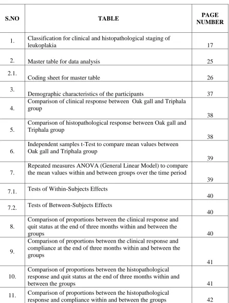
LIST OF TABLES
S.NO TABLE PAGE
NUMBER
1. Classification for clinical and histopathological staging of
leukoplakia 17
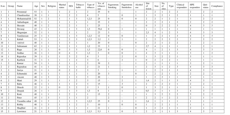
2. Master table for data analysis 25
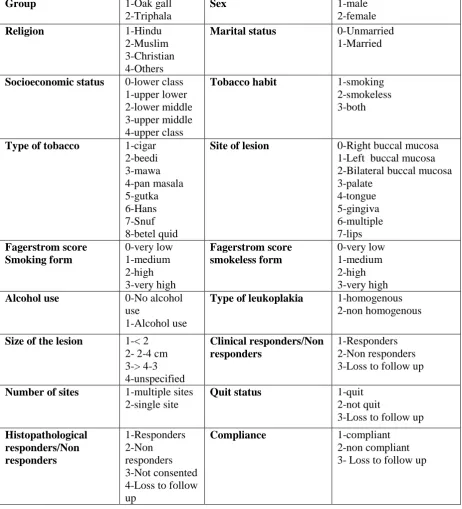
2.1.
Coding sheet for master table 26
3.
Demographic characteristics of the participants 37 4.
Comparison of clinical response between Oak gall and Triphala group
38
5.
Comparison of histopathological response between Oak gall and Triphala group
38
6.
Independent samples t-Test to compare mean values between Oak gall and Triphala group
39 7.
Repeated measures ANOVA (General Linear Model) to compare the mean values within and between groups over the time period
39 7.1. Tests of Within-Subjects Effects
40 7.2. Tests of Between-Subjects Effects
40 8.
Comparison of proportions between the clinical response and quit status at the end of three months within and between the
groups 40
9.
Comparison of proportions between the clinical response and compliance at the end of three months within and between the groups
41 10.
Comparison of proportions between the histopathological response and quit status at the end of three months within and
between the groups 41
11. Comparison of proportions between the histopathological
[image:11.595.73.520.110.702.2]LIST OF DIAGRAMS
S.NO DIAGRAMS PAGE
NUMBER
1. CONSORT Flow Diagram showing inclusion and exclusion criteria, and patient disposition.
30
2. Clinical and histopathological response of Oak gall oil and Triphala oil
43
3. Comparison of mean values between groups by Repeated measures of ANOVA
43
4. Comparison of association between Quit status at 3 months and Clinical responders
44
5. Comparison of association between Compliance at 3 months and Clinical responders
LIST OF PHOTOGRAPHS
S.NO PHOTOGRAPHS PAGE
NUMBER
1. Dried fruits of Emblica officinalis Garten 27
2. Dried fruits of Terminalia chebula Retz. 27
3. Dried fruits of Terminalia bellerica Roxb. 27
4. Dried galls of Quercus infectoria Oliv. (Oak gall) 28
5. Powdered form of Triphala and Oak gall 28
6. Oil preparation of Triphala and Oak gall in a non-transparent plastic bottle
28
7. Comparison of baseline and third month clinical findings of subjects in Oak gall group
45
8. Comparison of baseline and third month clinical findings of subjects in Triphala group
48
9. Histopathological comparison of pre and post intervention among subjects in the Oak gall group
51
10. Histopathological comparison of pre and post intervention among subjects in the Triphala group
54
11. Armamentarium used for clinical examination 79
12. Armamentarium for toluidine blue staining 79
13. Armamentarium for oral punch biopsy used at department of oral surgery
LIST OF ANNEXURES
S.NO ANNEXURE PAGE
NUMBER 1. Annexure I- Information sheet in Tamil version 71
2. Annexure II- Information sheet in English version 74
3. Annexure III -Informed consent in Tamil version 77
4. Annexure IV- Informed consent in English version 78
5. Annexure V -Armamentarium used in the study 79
6. Annexure VI -Case record form 82
7. Annexure VII- Photocopies of Authentication certificates for raw drugs
Introduction
1
INTRODUCTION
Oropharyngeal cancer is the fifteenth most common cancer world-wide and the prevalence is particularly high in men.1 An estimated incidence of 3, 00,373 cases and 145,353 deaths from oral cancer have occurred in 2012 worldwide.1 The highest rates are found in Melanesia, South-Central Asia, and Central and Eastern Europe.2 In India, oral cancer is the third leading site among males and females among all tobacco related cancers and is the second most common cancer among males. It is the third leading cancer for male and seventh leading cancer for females in Chennai.3 Oral cancer accounts for over 30% of all cancers in the country with age-adjusted rates being 20 per 100,000 population.4
Oral cancer may develop from oral potentially malignant disorders (OPMD) or de novo from normal mucosa.5 Potentially malignant disorders of the oral cavity comprises of leukoplakia, erythroplakia, palatal lesions in reverse smokers, sub mucous fibrosis, actinic keratosis, lichen planus, discoid lupus erythematosus, immunodeficiency in relation to cancer predisposition and some inherited cancer syndromes.6,7,8 The clinical significance of oral potentially malignant disorders lies in its association with malignant transformation into Oral Squamous Cell Carcinoma. Oral leukoplakia and erythroplakia has been identified as the one with the highest malignant transformation rates.9
Introduction
2 The annual incidence of oral leukoplakia among subjects greater than 15 years of age was reported as 0.2% to 11.7% in different populations of India.13
Tobacco and alcohol are strong risk factors for both oral cavity cancers (OCC) and oropharyngeal cancers.14,15 Tobacco use is responsible for one-third of all
cancer-related deaths.16
Several studies have shown the role of tobacco products and alcoholic
beverages in the induction of oral precancerous lesions.17 Cross-sectional studies in Chennai regarding prevalence of oral lesions in relation to habits have shown a prevalence rate of 4.1%.18 Presently, India has more than 274.9 million tobacco consumers. According to the Global Adult Tobacco Survey (GATS) 2010 report, 60% of tobacco users in India currently use only smokeless tobacco and an additional 15% are mixed users, (both smokeless tobacco and smoked tobacco).19Detection of asymptomatic cancers could be a problem in dental screening due to poor attendance of high-risk patientsbut oral precancerous lesions provide a long preclinical phase during which high-risk patients could be identified to provide interventions.20 Early detection and management of oral pre-malignant lesions is vital to prevent malignant transformation and thus improve the survival rate and quality of life (QoL).
Review of literature has proven the effectiveness of tobacco cessation counselling on the cessation of habit and reversal of lesions.21-27 However, cessation of tobacco use has deep rooted behavioural pattern that is highly resistant to change and relapse rates are high.28
Introduction
3 agents. As leukoplakias are not morbid or lethal by themselves, have a relatively low risk of transformation and have a long clinical course the proposed treatments should have minimal adverse effects in terms of incidence and severity.29 Surgical resection following long term follow up is also recommended in the literature for the management of leukoplakia but surgical management too has its own demerits and is invasive in nature.30
Exploration of scientific literature reveals that there is no regular treatment protocol for the treatment of oral premalignant lesions. Thus, the need to rely on indigenous health care is warranted for management of oral potentially malignant disorders.
The World Health Organization estimates that 80% of the world's inhabitants rely mainly on traditional medicines for their health care.31 A traditional formulation of herbal medicine usually contains a variety of constituents such as polyphenols, alkaloids, flavonoids, triterpenoids, and other secondary metabolites that have anticancer/ antimutagenic properties.32,33
Triphala is the most commonly used Indian herbal formulation, consisting equal parts of three medicinal dried plant fruits; Emblica officinalis Gaertn., [Indian gooseberry or Aamla], Terminilia bellerica Roxb. [Belleric myrobalan or Baheda]and
Terminilia chebula Retz. [Chebulic Myrobalan or Harad]. The differential effect of Triphala on normal and tumour cells seems to be related to its ability to evoke differential response in intracellular reactive oxygen species generation.34 A study by Deshpande et al., 2014 has proved Triphala to have great potential for reversal of oral precancerous lesions.35
Introduction
4 antibacterial properties.36,37 The galls of Quercus infectoria comprise a large amount of tannins, gallic acid, syringic acid, ellagic acid, β-sitosterol, amentoflavone hexamethyl
ether, isocryptomerin, methyl betulate, and methyl oleanate and hexagalloyl glucose. The plant is reported to possess antidiarrhoeal, anti-amoebic, antibacterial, antifungal, larvicidal, antidiabetic, local anaesthetic, antiviral, anti-inflammatory, hepatoprotective and wound healing properties.38
Sarwat Sultana et al.,2012 has proved Quercus infectoria to be a potent chemopreventive agent as it suppresses Fe-NTA-induced renal carcinogenesis and oxidative and inflammatory response in Wistar rat.39 Kaur et al.,2008 reported Quercus
infectoria is capable of protecting against oxidative damage to lipids and proteins and also chelates metal ions that catalyze the generation of oxidants.40 A study by Wan-Ibrahim et al.,2010 on the anti-oxidant properties of 20 edible plants has shown that Oak gall had the highest radical scavenging activity(86.8%) compared to others.41 However there are very limited in vivo studies till date to evaluate the effectiveness of oak gall on the reversal of oral precancerous lesions.
Introduction
Review of literature
6
REVIEW OF LITERATURE
The purpose of identifying potentially malignant disorders of the oral cavity is to initiate timely and adequate intervention and, where possible to prevent malignant transformation or enable early diagnosis of malignancy.42 In order to conduct treatment for oral leukoplakia the degree of epithelial dysplasia must be assessed. In the presence of moderate or severe epithelial dysplasia, surgical treatment is recommended. An extensive review of surgical management of oral potentially malignant disorders has shown that the evidence for its success is lacking, and in some cases, there have been reports of increased recurrence of malignancy following surgical excision.43 The associated morbidity of surgery also makes it less appealing for extensive lesions.42 Nonsurgical treatment management of oral leukoplakia offers minimal adverse effects to patients, especially for patients with widespread lesions that involves a large area of the oral mucosa or patients with medical problems and consequently high surgical risks. Additionally, potential advantages of the nonsurgical treatment of oral leukoplakia include easy application that does not require treatment at a medical centre and relative low cost.44 Various non-surgical treatments have been reported on the management of oral leukoplakia but currently there is no consensus achieved among them.29 An extensive review of literature on the various non-surgical treatment modalities involved in the management of oral leukoplakia are given below:
Chemo preventive agents in the treatment of leukoplakia
Review of literature
7
Lodi et al [2002] in his systematic review of randomized trials for the treatment of oral leukoplakia have enrolled studies involving Vitamin A, retinoids, bleomycin, mixed tea and beta carotene. It was concluded that although some treatments may be
effective in healing oral leukoplakia, they do not seem to be able to prevent relapses
and malignant change.Topical 13-cis-retinoic acid, 200,000 IU per week of vitamin A,
and mixed tea did not cause any adverse effects. Topical bleomycin, systemic
13-cis-retinoic acid (1 to 2 mg/kg/day), vitamin A (300,000 IU per week), and beta carotene
(360 mg/week) caused adverse effects of various severity in 100 percent, 79 percent,
26 percent, and 9 percent of patients, respectively.46
Lodi et al [2008] in his review on the intervention for treating oral leukoplakia has concluded that none of the treatments studied was shown to be effective in preventing malignant transformation of leukoplakias.29
Ribeiro et al [2010] on his extensive review on the non-surgical treatment modalities of oral leukoplakia has concluded that studies showed a rate higher than 50% of clinical resolution with photodynamic therapy, beta-carotene, lycopene, or vitamin A. Few studies have reported rates of recurrence from 5 to 67% and of malignant transformation from 8 to 23%. The review has highlighted that randomized controlled trials for nonsurgical treatment of oral leukoplakia demonstrate no evidence of effective treatment in preventing malignant transformation and recurrence. It reinforces that even after clinical resolution, oral leukoplakia should be regularly followed.44
Review of literature
8 vitamin C for the chemoprevention of oral cancer when tested on otherwise healthy subjects living in well-nourished countries.47
Tobacco cessation counselling in the treatment of oral leukoplakia
A decrease in the prevalence of oral leukoplakia after tobacco cessation has been observed in many studies, confirming an etiological role.
Banoczy [1971] in a clinicopathologic study of 165 oral leukoplakia cases followed up over a period of 20 years proved that following the elimination of etiological factors, mainly smoking43.2 percent of oral leukoplakias resolved.21
In Roed-Petersen’s study [1982], smoking cessation for at least one year caused resolution of leukoplakia in 58.3 percent of the cases.22
Silverman et al [1984] in his study on two hundred fifty-seven patients with oral leukoplakia followed for an average period of 7.2 years. All lesions were more than one cm in size and had been present and observed for a minimum of 6 months. Of the initial biopsies, 235 revealed a benign hyperkeratosis and 22 others contained some degree of epithelial dysplasia. Seventy-three percent of the patients used tobacco, with cigarette usage being the predominant form. There appeared to be a beneficial effect of decreased risk for malignant transformation when tobacco users discontinued their habits (44%).23
non-Review of literature
9 intervention group thus proving the etiological and beneficial role of tobacco and its cessation. 24
In a study of 3,051 male U.S. military trainees by Martin et al [1999], 302 individuals were using smokeless tobacco, 39.3 percent had leukoplakia compared to 1.5 percent among non-users. After six weeks of tobacco cessation, 97.5 percent of leukoplakia lesions showed complete resolution clinically.25
Shiu et al [2000] studied the effects of betel nut chewing, smoking and alcohol on the occurrence of leukoplakia and its malignant transformation to oral carcinoma and has found that cessation of smoking may reduce by 36% leukoplakia cases, while elimination of betel nuts may prevent 62% of leukoplakia and 26% of malignant transformation to oral carcinoma.26
A case study by Othman Shibly [2008] on the resolution of oral lesions after tobacco cessation with a follow up period of two weeks has shown a complete resolution of oral white lesions and improvement in overall oral health of the patients.27
Studies on anti-oxidant properties of plant phenolic compounds
Newmark, H.L., [1996] in his study has proved that plant products with phenolic compounds have significant antioxidant properties.33
Wahle et al [2010] has extensively reviewed the role of plant phenolics in the
prevention and treatment of cancer.it has been described in the review that plant phenolics appear to have both preventive and treatment potential in combating cancer.48
Review of literature
10
Danaraddi et al. [2014] has reviewed the role ofdietary supplements which are rich in phytochemicals such as spirulina, selenium, green tea, neem, Tomatoes (lycopene), turmeric (curcumin), and some medicinal mushrooms in chemoprevention of oral cancer.50
Triphala as a potent anti-oxidant
Kaur et al [2002] has done a study to evaluate an antimutagenic potential of water, chloroform and acetone extracts of Triphala and has proved the anti-mutagenic activity of Triphala.51 Triphala and its individual components are capable of scavenging free radicals DPPH, superoxide, and nitric oxide (Naik et al., 2005; Jagetia et al., 2004).52
Sandhya et al [2006] has investigated the cytotoxic effects of Triphala on human breast cancer cell line (MCF-7) and transplantable mouse thymic lymphoma (barcl-95) and has concluded that the differential response of normal and tumour cells to Triphala in vitro and the substantial regression of transplanted tumour in mice fed with Triphala points to its potential use as an anticancer drug for clinical treatment.53
Yan Shi et al [2008] has done a study to elucidate the molecular mechanism of Triphala against human pancreatic cancer in the cellular and in vivo model and has proved that Triphala is effective in inhibiting the growth of human pancreatic cancer cells in both cellular and in vivo model.34
Wongnoppavich [2009] in his review on the role of Triphala in cancer treatment has concluded that Triphala has high potentials for inhibition and prevention of mutagenesis and metastasis of cancer cells.54
Review of literature
11 Histological findings after 9 month use of Triphala mouth rinse revealed no changes in cells in 23 (85.2%), hyperkeratinisation in 2 (7.4%). Hyperkeratinization and spongiosis was evident in 1 (3.7%), mild pleomorphism in 1 (3.7%). The authors have concluded that Triphala proved to be effective in reversal of the lesions and confirmed the promising role of Triphala as a potential chemo- preventive drug.35
Oak gall as a potent antioxidant
Kaur et al [2008] has demonstrated that Quercus infectoria (QI) galls possess potent antioxidant activity, when tested both in chemical as well as biological models.38
Umachigi et al [2008] revealed that ethanolic extracts of the galls of QI contains high amount of tannins, presence of gallic acid, ellagic acid, syringic acid, iso-sitosterol and amenotoflavone, implied that tannin is one of the active compounds which may be responsible for the antiproliferative activity.55
Hasmah et al [2010] in a study to evaluateQ.infectoria as an antiproliferative agent towards cervical cancer cells (HeLa) and ovarian cancer cells (Caov-3) has suggested that QI galls extracts have potential to be used as an anticancer agent. In this study it was proved that all the crude extracts of QI galls had IC50 less than 100 μg/ml towards both HeLa and Caov-3 cells.56
Rehman et al [2012] in his study to evaluate the chemo preventive effect of Quercus infectoria against chemically induced renal toxicity and carcinogenesis has concluded that Quercus infectoria is a potent chemo preventive agent and suppresses Fe-NTA-induced renal carcinogenesis and oxidative and inflammatory response in Wistar rat.39
Review of literature
12 galls had high antioxidant activities as measured by DPPH scavenging (30/15±0.83 μg /ml) and β-carotene linolic acid (89/4±1.11/ml).57
Aim and objectives
13
AIM AND OBJECTIVES
AIM:
The aim of the study is to compare the effectiveness of Oak gall oil with Triphala oil as an adjuvant to tobacco cessation counselling in the reversal of oral leukoplakia at a Public Dental hospital in Chennai city.
OBJECTIVES:
Materials and methods
14
MATERIALS AND METHODS
The double blind, parallel, randomized clinical trial conducted on tobacco users diagnosed with oral leukoplakia had an allocation ratio of 1:1. Institutional Ethics Committee of the Tamil Nadu Government Dental College and Hospital, Chennai, Tamil Nadu approved the study (Approval No.R.C.No:0420/DE/2016). The trial was registered in the clinical trial registry of India (CTRI/2016/10/007415) and is reported in accordance with the CONSORT guidelines with extension for herbal interventions. As per the protocol for Ethical approval, the inclusion criteria for the sample comprised of current tobacco users with oral leukoplakia and having mild or moderate dysplasia histologically. The pilot study concluded that the prevalence of dysplasia among oral leukoplakia was minimal. Hence the inclusion criteria for the study has been modified as current tobacco users with pre-diagnosed oral leukoplakia irrespective of presence or absence of dysplasia.
Participants
The trial was conducted under an institutional (hospital) setting among the adult patients pre-diagnosed with oral leukoplakia (by an expert from the Department of Oral medicine and Radiology of Tamil Nadu Government Dental College and Hospital). The recruitment period for the trial was from April 2016 to September 2016. The eligibility criteria for the recruitment of the study were:
Inclusion criteria:
1. Adults aged 18 year and above
Materials and methods
15
Operational definitions:
*Current tobacco users - Current tobacco user is defined as an individual who is using 1 or more tobacco products for the past 30 days from the time of recruitment.
**- Leukoplakia: A white plaque of questionable risk having excluded (other) known disease or disorders that carry no increased risk for cancer.58
Exclusion criteria
1. Subjects with malignant tumours of oral cavity
2. Subjects with any systemic diseases.
3. Subjects who were physically and mentally challenged.
4. Subjects with known hypersensitivity to toluidine blue.
5. Subjects with dental conditions such as orthodontic appliances or prostheses that may interfere with the examination.
6. Subjects who were currently receiving any type of cessation, alcohol or illicit drug treatment at the time of recruitment.
7. Subjects who were under medication for psychiatric illness, cardiovascular diseases,
8. Leukoplakia with p2 and px grading of histopathological evaluation (Vanderwaal et al 2000)9
Sample size
Materials and methods
16 estimated to be 12 patients per group. Assuming a possible drop-out rate of 15%, the required sample size was set at 15 subjects per group.
Patient recruitment and randomization
The study was conducted at the Department of Public Health Dentistry, Tamil Nadu Government Dental College and Hospital, Chennai. Participants who met the inclusion criteria formed the study population. The purpose of the study was clearly explained to the participants. Information sheet was given in Tamil (Annexure I) or English version (Annexure II) and written informed consent was obtained from those who were willing to participate in either Tamil (Annexure III) or English version (Annexure IV).
The case record form (Annexure VI) used for data collection comprised of the demographic data, complete personal and medical history, assessment of tobacco usage (type, number, duration and the frequency of the habit), nicotine addiction (Fagerstrom scale (Todd Heatherton, et al. 1991) for nicotine dependence - smokeless/smoke form), a comprehensive clinical examination of the oral cavity, clinical and histological evaluation of the lesion and the follow up details.
Baseline assessment:
Clinical evaluation of the lesion
The lesion was evaluated in context of shape, size in square milli-meter, consistency, surface texture, induration and ulceration. Measurement was carried out using metal rulers graduated in millimeters. (Annexure V) The area of a lesion was obtained by multiplying the longest length by the greatest perpendicular breadth. In case of multiple lesions, the largest lesion was considered as the index lesion. The lesion
Materials and methods
17 Van der Waal et al., 2000 classification of leukoplakia was adopted for clinical and histopathological staging of leukoplakia (Table 1):
S.No Precancerous lesion
Grading and staging
(Van der Waal et al 2000)9
1 Leukoplakia L (size of the leukoplakia)
L1- Size of single or multiple leukoplakias together <2 cm L2- Size of single or multiple leukoplakias together 2-4 cm L3- Size of single or multiple leukoplakias together >4 cm Lx- Size not specified
P (pathology)
P0 - No epithelial dysplasia (includes "perhaps mild epithelial dysplasia")
P1- Mild or moderate epithelial dysplasia
P2 -Severe epithelial dysplasia (includes "perhaps carcinoma in situ")
Px -Absence or presence of epithelial dysplasia not specified in the pathology report
OL-staging system
Stage 1 L1P0 Stage 2 L2P0
Stage 3 L3P0 or L1L2P1 Stage 4 L3P1 or LP2
As per the clinical requirements of the participants, elimination of local irritating factors such as removal of plaque and calculus, selective grinding of sharp tooth, correction of ill-fitting dentures, correction of faulty restorations, and extraction of grossly decayed were carried out to achieve homogeneity among the study participants.
Laboratory procedure:
Materials and methods
18 After determining the eligibility, baseline clinical evaluation and laboratory investigation, all the participants were subjected to an initial toluidine blue staining and an incisional biopsy of the lesion using a 6 mm punch.
Toluidine blue staining59 (Mashberg, 1980)
Every participant underwent a toluidine blue staining at baseline and at the end of three months to screen for dysplastic changes and to mark the site for biopsy. Instruction was given to rinse the mouth twice with water for 20 seconds to remove debris. Pre rinse with 1% acetic acid for 20 seconds was done to remove ropey saliva followed by 1% toluidine blue application with cotton swab or as a rinse and left for 20 seconds. Post rinse with 1% acetic acid was done twice to reduce the mechanically retained stain. Finally rinsed with water. The stain uptake of the lesion was assessed and categorised. Dark royal blue was considered as positive, light blue as doubtful while no colour as negative. (Annexure V)
Oral Punch biopsy
A histopathological analysis by incisional punch biopsy was performed at baseline and at the end of three months by an expert at the Department of Oral and Maxillofacial Surgery. A 6mm disposable punch was used for obtaining the biopsy specimen for all the participants. The histopathological analysis of the biopsy specimen was carried out at the Department of Oral and Maxillofacial Pathology (Annexure V). Randomization was done after histopathological evaluation into Oak gall (OG) group and Triphala (TP) group.
Randomisation
The trial drugs were dispensed by a research assistant and the allocation was done by computer-generated list of random numbers. Participants were allocated to
Materials and methods
19 concealment was done by sequentially numbered opaque sealed envelopes [SNOSE]. The envelopes were maintained in the possession of chief investigator. The envelopes were opened only prior to intervention after patient enrolment and after receiving the patient’s consent.
The principal investigator recruited the participants and was blinded to the participants’ group allotments. The Oak gall oil and Triphala oil were dispensed in a
non-transparent bottles which were identical in appearance. They were prepacked in bottles and consecutively numbered for each participant according to the randomisation schedule. Each participant was assigned a code number and received the interventions in the corresponding prepacked bottle. The trial drugs were matched in context for taste, colour and quantity. Thus both the investigator and the participants were blinded about the treatment groups.The subjects were allocated to receive the trial intervention (Oak gall or Triphala oil) for a period of three months.
Intervention
The intervention for the study participants comprised of Tobacco cessation counselling followed by systemic administration of multivitamin tablets for both the groups and topical administration of Oak gall oil to OG group and Triphala oil to TP group.
Tobacco cessation counselling (TCC):
Materials and methods
20 The principal investigator attended a three day intensive training programme on counselling for tobacco cessation at the Adyar cancer institute, Chennai before the commencement of the trial. Tobacco cessation counselling with motivation and reinforcement was given at baseline, 15th day (II TCC), 30th day (III TCC), 6th week (IV TCC).
Oak gall and Triphala oil (Fig 1)
The herbal medicine intervention used in this trial was Oak gall (Quercus infectoria) and Triphala oil (equal parts of Emblica officinalis Gaertn., [Indian gooseberry or Aamla], Terminilia bellerica Roxb. [Belleric myrobalan or Baheda]and
Terminilia chebula Retz. [Chebulic Myrobalan or Harad]).
Oak gall and Triphala are registered for use as a natural health care product in India and are thoroughly reviewed in the Indian medicinal literature.62,63 Oak gall and Triphala has been used in the Indian medicinal literature for the treatment of mouth ulcers.64,65,66 It has been recommended for use as both external and internal preparations in Indian medicinal literature. 65,66
Toxicity studies on Oak gall and Triphala has proved that these medicines are relatively safe up to the dose of 2g/kg and 240 mg/kg respectively. 67, 52
Materials and methods
21 The herbal formulations for both the groups were designed by a Siddha physician and the oil preparation of Oak gall and Triphala was done by a qualified pharmacist by using standardized operating procedures (SOP).The dried fruits of equal parts of Emblica officinalis Gaertn., [Indian gooseberry or Aamla], Terminilia bellerica
Roxb. [Belleric myrobalan or Baheda] and Terminilia chebula Retz. [Chebulic Myrobalan or Harad and the dried galls of Oak gall was powdered and mixed with coconut oil in the ratio of 1: 5 (1 part of powder of oak gall or Triphala to five parts of pure coconut oil). Both the oils were dispensed in a non-transparent 100 ml plastic bottles.
Oak gall oil was administered to OG Group and Triphala oil to TP Group. Both the groups were instructed to apply 2 ml of the herbal oil for a period of three months over the lesion three times a day using a cotton applicator and were also instructed not to eat or drink for 30 minutes after the application.
Primary and secondary outcomes
The primary endpoint or outcome with respect to effectiveness of oak gall oil or Triphala oil was the comparison of the clinical and histological reversal of oral leukoplakia. The objective clinical response was evaluated by bi-dimensional measurement of the lesions and colour photography. The objective clinical response was categorized as follows68:
Complete response— disappearance or no evidence of any measurable lesion.
Partial response— decrease in the cross-sectional areas of a measurable leukoplakia
Materials and methods
22
Stable disease—decrease in the cross-sectional area of a measurable leukoplakia lesion
by less than 50% of the product (or sum of products) of measured lesion diameter in the absence of new ‘‘in field’’ lesions;
Progressive disease—increase in the product (or sum of products) of the measured
lesion diameter by greater than 25% or development of a new ‘‘in field’’ leukoplakia lesion.
Clinical response was also graded into a binary response into responders and non-responders (Responders were those who showed a complete or partial clinical response; non-responders were those showed a stable or progression clinical response).
Histologic evaluation of all biopsies were performed independently by oral pathologists who were blinded to treatment groups. Biopsy samples were scored accordingly into a 5-point ordinal scale as Normal epithelium, squamous cell hyperplasia, mild dysplasia, moderate dysplasia, severe dysplasia, and carcinoma in situ and were ranked as 0, 1, 2, 3, and 4 respectively. Histopathological response was also graded into a binary response into responders and non-responders (Responders were those who showed histopathological response; non-responders were those who do not show histopathological response in histopathological grading).
Self-reported abstinence from tobacco habit and compliance of the drug was assessed as secondary outcome at every visit till the end of three months. Self-reported abstinence was grouped into no change in the tobacco habit, reduced use in the tobacco habit, stopped use, lost to follow up and relapse. The quit status of the participants were also graded into a binary response as quitters and non-quitters.
Materials and methods
23 used at every visit was ≥ 80% (i.e., ˂ 20ml of the oil remained at end of each visit). The average compliance of each treatment group was calculated separately.
After the trial commencement no changes to trial outcomes were made. Patients were followed up every 15th day till the end of six weeks and at the end of third month. Clinical measurement of the lesion and colour photography was done at the baseline, 6th week and third month. Oral incisional punch biopsy was performed at baseline and at the end of third month to grade the lesion histopathologically and to assess the reversal of lesion. Self -reported abstinence from the tobacco habit was assessed at the end of six weeks and at the end of third month. The participants were reminded of the follow up sessions through telephone and written instructions in their respective outpatient card.
Interim analyses and stopping guidelines
No interim analysis was made after the commencement of the trial.
Stopping rules:
Stopping rules were set for the trial as follows:
1. Progression of the disease during the trial period despite receiving the intervention 2. Reversal of the lesion prior to trial period
3. Severe adverse effects during the intervention period
Statistical analysis
Materials and methods
24 lesion, number of sites, type of lesion, clinical response, histopathological response, quit status and compliance between the treatment groups.
Outcome analyses was conducted among those participants who completed the
trial. Participants who showed complete or partial response were deemed as clinical
responders and those with stable or progressive disease as non-responders to the
treatment.
To compare the mean values between groups, independent samples t-test was applied. To compare proportions between groups, Chi-Square test was applied and if any expected cell frequency was less than five then Fisher’s exact test was used. Repeated measures of ANOVA (GLM) was used to compare mean values between time points and groups. Significance level was fixed as 5% (α = 0.05) .The collected data was entered into Microsoft excel sheet and master table (Table 2, 2.1) was prepared and subjected to statistical analysis using SPSS version 22.
25
TABLE 2: MASTER TABLE FOR DATA ANALYSIS
S.no Group Name Age Sex Religion Marital status SES Tobacco habit Type of tobacco
Yrs. of tobacco use Fagerstrom Smoking Fagerstrom Smokeless Alcohol use Site of lesion Size No. of sites
Type Clinical responders HPE responders Quit status Compliance
1 1 Perummal 53 1 1 1 2 1 2 35 0 - 1 2 2 1 2 2 2 2 2
2 1 Chandrasekar 38 1 1 1 2 2 4 5 - 0 1 1 3 2 1 2 1 1 1
3 1 MohammadAli 52 1 2 1 1 3 1,2,3 25 0 0 0 2 2 1 2 1 3 2 2
4 1 Anbazhagan 40 1 1 1 1 1 1 35 2 - 1 2 2 2 1 2 2 2 2
5 1 Hussain 40 1 2 1 2 1 1 25 1 - 1 0 3 2 2 3 4 3 3
6 1 Devaraj 43 1 1 1 1 3 1,3 39,4 0 1 1 1 1 1 1 1 2 1 1
7 1 Magarajan 52 1 1 1 1 1 2 23 3 - 1 2,3 4 1 2 3 4 3 3
8 1 Tamilarasan 25 1 1 1 1 3 1,2,3 12 0 0 1 1 3 2 1 3 4 3 3
9 1 Kabali 53 1 1 1 2 3 1,2,3 2,2 3 1 1 2 2 2 2 2 2 2 2
10 1 vetrivel 40 1 1 1 3 1 1 20 0 - 0 2 2 1 2 2 2 2 1
11 1 Subramani 63 1 1 1 1 1 1,2 33 3 - 1 2,7 4 1 1 1 1 2 1
12 1 Ragu 26 1 1 0 3 3 1,3 22,6 0 0 1 4 1 2 1 3 4 3 3
13 1 Sridhar 26 1 1 0 4 3 1,3 3 0 0 1 4 3 1 1 1 3 1 2
14 1 Rajasekar 39 1 1 1 3 2 4 3 - 2 0 1 3 2 2 1 3 1 1
15 1 Karthick 31 1 1 1 1 2 3 6 - 2 1 0 2 2 2 1 1 1 1
1 2 Kumar 54 1 1 1 1 1 2 39 2 - 1 2 1 1 1 1 3 2 1
2 2 Rajendran 41 1 1 1 1 1 2 21 1 - 1 2 3 1 2 3 4 3 3
3 2 Suriya 21 1 1 0 2 2 3 3 - 1 1 1 2 2 1 3 4 3 3
4 2 Ezhumalai 49 1 1 1 1 1 1 20 3 - 0 1 2 2 1 1 2 2 2
5 2 vincent 40 1 3 1 1 2 3 20 - 2 1 1 2 2 1 1 2 1 1
6 2 Mani 29 1 1 1 1 2 3 15 - 2 1 1,4 2 1 1 3 4 3 3
7 2 Babu 46 1 1 1 1 2 3 15 - 3 1 2 2 2 2 1 2 2 1
8 2 Dinesh 22 1 1 0 3 2 3 3 1 0 1 1 3 2 1 1 3 1 2
9 2 Deepak 26 1 1 1 1 3 1,3 8 1 1 1 0,5 3 1 1 3 4 3 3
10 2 Kotti 40 1 1 1 2 1 1 15 2 - 1 1 3 2 2 2 3 2 2
11 2 Peter 45 1 3 1 1 1 2 2 - 1 2 2 1 2 2 2 1 1
12 2 Vasantha sekar 48 1 3 1 1 3 1,2,3 25 0 1 1 2,4 1 1 1 1 1 1 1
13 2 Robby 40 1 3 1 1 2 3 10 - 0 0 1 2 2 1 1 1 1 1
14 2 Maqbhul 41 1 2 1 1 1 1 21 1 - 0 1 2 2 1 1 2 1 1
26 TABLE 2.1: CODING SHEET FOR MASTER TABLE
Group 1-Oak gall
2-Triphala
Sex 1-male
2-female
Religion 1-Hindu
2-Muslim 3-Christian 4-Others
Marital status 0-Unmarried 1-Married
Socioeconomic status 0-lower class 1-upper lower 2-lower middle 3-upper middle 4-upper class
Tobacco habit 1-smoking 2-smokeless 3-both
Type of tobacco 1-cigar 2-beedi 3-mawa 4-pan masala 5-gutka 6-Hans 7-Snuf 8-betel quid
Site of lesion 0-Right buccal mucosa 1-Left buccal mucosa 2-Bilateral buccal mucosa 3-palate 4-tongue 5-gingiva 6-multiple 7-lips Fagerstrom score Smoking form 0-very low 1-medium 2-high 3-very high Fagerstrom score smokeless form 0-very low 1-medium 2-high 3-very high Alcohol use 0-No alcohol
use
1-Alcohol use
Type of leukoplakia 1-homogenous 2-non homogenous
Size of the lesion 1-< 2 2- 2-4 cm 3-> 4-3 4-unspecified Clinical responders/Non responders 1-Responders 2-Non responders 3-Loss to follow up
Number of sites 1-multiple sites 2-single site
Quit status 1-quit
2-not quit
3-Loss to follow up Histopathological responders/Non responders 1-Responders 2-Non responders 3-Not consented 4-Loss to follow up
Compliance 1-compliant
Materials and methods
27
Photographs of dried fruits of Triphala (Emblica officinalis Gaertn., Terminalia chebula Retz. and Terminalia bellerica Roxb.)
Photograph 1. Emblica officinalis Gaertn.
Photograph 2. Terminalia chebula Retz.
Photograph.3.Terminalia bellerica Roxb.
1.1 1.2
2.1 2.2
Materials and methods
28
Photograph.4 Dried galls of Quercus infectoriaOliv. (OAK GALL)
Photograph.5 Powdered form of Triphala and Oak gall
Photograph.6 Oil preparation of Triphala and Oak gall in a non-transparent plastic bottle
4.1 4.2
5.1 5.2
6.1 6.2
Results
29
RESULTS
A total of 1354 individuals with tobacco habit were screened for oral leukoplakia during the recruitment period. Thirty eight individuals with oral leukoplakia were assessed for eligibility criteria. Five subjects were excluded as they did not fulfill the inclusion criteria. Three Subjects did not consented for the trial and were excluded. 30 subjects consented for the trial. Therefore a total number of 30 participants were included at the baseline for evaluation. These thirty participants were randomized into two groups with an allocation ratio of 1:1 with 15 subjects in OG group and 15 subjects in TP group.
The demographic characteristics, tobacco habits and clinical findings were comparable in the two groups. There were 8 subjects in OG and 7 subjects in the TP group who had completed both clinical and histopathological evaluation at the end of three months.
Among the study population 6 subjects (3 in OG group and 3 in TP group) were assessed clinically without histopathological evaluation at the end of three months. The reasons for not undergoing post biopsy at the end of three months were fear, personal reasons and subjective clinical improvement expressed among individuals. 4 Subjects in the OG group and 5 subjects in the TP arm had loss to follow up visits. Therefore, 21 subjects (11 subjects in the OG group and 10 subjects in the TP group) were available at the end of 3 months for primary outcome analyses. The dropout rate in OG and TP group in this trial was found to be 26.7% and 33.3% respectively.
Results
[image:49.595.81.516.81.622.2]30
Figure 1: CONSORT Flow Diagram showing inclusion and exclusion criteria, and patient disposition. E NRO L L M E NT
ASSESSED FOR ELIGIBILITY n= 38
Excluded n= 8 Not meeting the inclusion
criteria n = 5
Not willing to participate n=3
RANDOMIZED N= 30
Allocated to OG group n=15 Received intervention n=15 Did not receive intervention n=0
Allocated to TP group n=15 Received intervention n=15 Did not receive intervention n=0
F O L L O W UP
Lost to follow up n=4 Discontinued intervention n= 0
Adverse events n=0 Withdraw consent n=3
Non-compliance=6 ANAL Y S IS Analyzed n=11 Excluded from analysis n=4
AL L O C AT IO N
Lost to follow up n=5 Discontinued intervention n= 0
Adverse events n=0 Withdraw consent n=2
Non - compliance=4
Results
31 Participant’s demographic characteristics, risk factors, clinical and histological features of the oral leukoplakia are shown in Table 3.
Demographic characteristics
A total number of 30 male participants reported in this study and there were no female participants. The mean age of the study participants was 39 years (SD ±11.18). Among the study participants, 73.3% (n=22) were Hindus, 16.7% (n=5) were Christians and 10% (n=3) were Muslims. There were 88.3% (n=25) married and 16.7% (n=5) unmarried among the study population. Kuppuswamy socioeconomic status scale (July 2016) indicated that 63.3% (n=19) of study participants belonged to upper –lower class, 20% (n=6) belong to lower –middle class 13.3% (n=4) to upper –middle
class and 3.3% (n=1) belonged to upper class and none of the participants belong to lower class. (Table 3)
Tobacco habit
The study revealed that 40% (n=12) of participants used smoking form of tobacco, 30% (n=9) of participants used smokeless form of tobacco and 30% (n=9) used both smoking and smokeless form. Among the smoking form cigar (20%) was found to be more prevalent and mawa (23.3%) was found to be the common form of smokeless tobacco. There is no statistical difference regarding the type of tobacco use between the study groups (p=0.252). (Table 3)
Very low dependence was the predominant level of dependence as assessed by Fagerstrom scale of dependence for smoking form and smokeless form of tobacco in the OG group and was found to be 58.3% and 55.6% respectively.
Results
32 statistical difference (p=0.028) between the level of dependence for smoking form between the OG group and TP group. Alcohol dependence was found among 80% (n=24) of the participants and 20% (n= 6) had no habit of consuming alcohol.
Clinical evaluation of the lesion
There were 30 cases of leukoplakia reported during the study period. Left buccal mucosa (36.7%) was found to be the most common site followed by bilateral buccal mucosa (33.3%), right buccal mucosa (6.7%) among the treatment groups. (Table 3)
Among the trial groups, 13.3% of the lesions were less than 2cm in diameter, 46.7% were of 2-4cm in diameter, 33.3% were greater than 4 cm in diameter and 6.7% were of unspecified size. There is no statistical difference (p= 0.683) in the size of the lesion in both the OG and TP group. 53.3% (n=16) of the participants had lesion at a single site and 46.7% (n=14) patients had multiple sites of lesion. (Table 3)
Non-homogenous type of leukoplakia was found among 53.3% and homogenous in 46.7% of the participants in OG group. Whereas in the TP group the non-homogenous constituted 26.7% and homogenous being 73.3%. (Table 1) At the baseline the mean size of the lesion in the OG group was 441.07mm2 and the mean size in the TP group was 361.93mm2.
Histopathological response revealed that there were 10 participants in the OG group with non-dysplastic lesions and 5 participants with dysplastic lesions. Similarly there were 10 participants with non-dysplastic lesions and 5 participants with dysplastic lesions in the TP group. (Table 3)
Results
33 trial by creating heterogeneity between the treatment groups due to the combination of non-compliant, dropouts and compliant subjects during the evaluation of outcome. Moreover ITT is more prone to type II error.
Therefore primary outcome analyses was performed for those 21 participants who completed the trial and underwent both clinical and histopathological evaluation and only clinical evaluation.
Clinical response within groups:
Clinical response was evaluated for 21 participants (OG group n=11, TP group n =10) with a response rate of 45.5 % in the OG group and 70 % in the TP group. (Fig 2)
Clinical response to OG group:
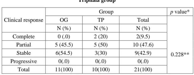
Clinical response was evaluated in 11 participants in the OG group (n=15) who had clinical assessment done at the end of three months. Clinical grading of leukoplakia revealed that none (n=0) showed complete clinical response, 45.5% (n=5) showed partial clinical response, 54.5% (n=6) showed stable clinical response and none (n=0) showed progression of disease. (Table 4)
Clinical response to TP group
Clinical response evaluated for the 10 participants of TP group who completed the trial revealed that 20% (n=2) showed complete clinical response, 50 % (n=5) showed partial clinical response and 30 % (n=3) showed stable clinical response and none showed progression of disease. (Table 4)
Results
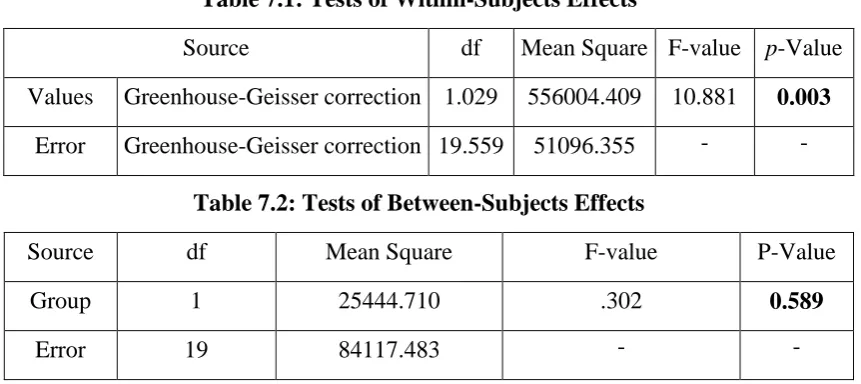
34 statistical significance (p=0.003), which infers that there is a decrease in the size of the lesion at various time points in both the groups. (Table 7, 7.1)
Histopathological response within groups
Histopathological response to OG group:
Histopathological findings of pre-treatment biopsy for eight participants who had completed the trial in the OG group (n=15) revealed orthokeratosis in 62.5% (n=5) of the participants, hyperplasia was found in 50% (n=4) of the participants, mild dysplasia was found in 25% (n=2) participants. The post treatment biopsy done at the end of three months for these participants showed reversal of mild dysplasia to hyperplasia in two participants, stable response in six participants.
Histopathological response to TP group:
Histopathological evaluation for 7 subjects were available at the end of three months for primary outcome analysis in the TP group. There were 71.4% (n=5) participants with orthokeratosis, 42.9% (n=3) participants with hyperplasia and 28.6% (n=2) with mild dysplasia in baseline histopathological evaluation. The post treatment biopsy at the end of three months revealed reversal of mild dysplasia to hyperplasia in two participants. The histopathological response was stable in 4 participants and there was progression of disease seen in one participant from hyperplasia to mild dysplasia.
Comparison of clinical response between Oak gall and Triphala group
Results
35 groups comparison by repeated measures of ANOVA at baseline, sixth week and third month with a Greenhouse-Geisser correction determined that the decrease in the size of the lesion (mean square millimeter) did not differed between time points (p=0.589). (Table7.2) Binary analysis of clinical response into responders and non-responders showed that the difference between the two groups is statistically insignificant (p=0.387).
Comparison of histopathological response between OG and TP group
Chi-square test done to compare the histopathological responses has proved that there is no statistical difference in the histopathological response between the two treatment groups (p=1.000). (Table 5)
Self-reported abstinence to tobacco habit
Self-reported abstinence for tobacco habit at the end of three months for the OG and TP group revealed a quit rate of 45.5% and 50% respectively. In the OG group, 9.1% (n=1) had no change in the tobacco habit, 45.5% (n=5) had partial change in the tobacco habit, 45.5% (n=5) had stopped use. Similarly in the TP group 10% (n=1) had no change in the tobacco habit, 40% (n=4) had partial change in the tobacco habit, 50% (n=5) had stopped use. The difference between the quit rate in OG group and TP group is statistically insignificant. (p=1.000).
Drug compliance
Results
36
Sub-group analyses in OG group
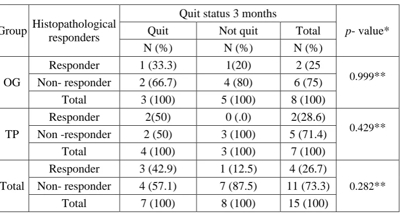
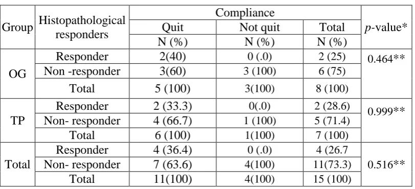
Binary analysis to assess the association between clinical response and quit status in the OG group has exposed that the difference between the clinical response and the quit status of the participants is statistically insignificant (p=0.567). Similarly the association between the clinical response and compliance of drug in the OG group is statistically insignificant (p=0.999). (Table 8, 9, 10, 11. Fig 4, 5).
Sub-group analyses in TP group
Results
37
[image:56.595.79.533.111.600.2]RESULTS IN TABLES
Table 3: Demographic characteristics of the participants
Variables OG
N (%)
TP
N (%) Total
P
-value*
Age Age ≤ 40 Age ≥ 40 9 (60) 6 (40)
8 (53.3) 7 (46.7)
17 (56.3)
13 (43.7) 1.000**
Sex Male
Female
15 (100) 15 (100) 30 (100) - Marital status Unmarried
Married
2 (13.3) 13(86.7)
3 (20) 12 (80)
5 (16.7)
25 (83.3) 0.995**
Socioeconomic status Upper class Upper-middle class Middle--lower-middle class Lower--upper-lower class Lower class
7 (46.7) 4 (26.7) 3 (20) 1 (6.7) 0 (0)
12 (80) 2 (13.3) 1 (6.7) 0 (0) 0 (0)
19 (63.3) 6 (20) 4 (13.3) 1 (3.3) 0 (0)
0.288**
Tobacco habit Smoking form Smokeless form Both 6 (40) 3 (20) 6 (40) 6 (40) 6 (40) 3 (20) 12 (40) 9 (30) 9 (30)
0.433**
Alcohol habit Alcohol user Non-user-alcohol
12 (80) 3 (20)
12 (80) 3 (20)
24 (80)
6 (20) 1.000** Type of leukoplakia Homogenous Non- Homogenous 7 (46.7) 8 (53.3) 11(73.3) 4 (26.7) 18 (60)
12 (40) 0.136
Multiple sites Yes No 7 (46.7) 8 (53.3) 11(73.3) 4 (26.7) 18 (60)
12 (40) 0.136 Site of lesion
Buccal mucosa Tongue Palate Gum/Sulcus 11(73.3) 2(13.3) 1(6.7) 1(6.7) 12 (80) 2 (13.3) 0 (0) 1 (6.7) 23 (76.7) 4 (13.3) 1 (3.3) 2 (6.7)
0.274** Classification by size <2 cm 2-4 cm >4 cm Unspecified 2 (13.3) 6 (40) 5 (33.3) 2 (13.3) 2 (13.3) 8 (53.3) 5 (33.3) 0 (0)
4 (13.3) 14 (46.7) 10 (33.3) 2 (6.7)
0.683** Histopathological evaluation Non-dysplastic Dysplastic 10(66.7) 5 (33.3) 10(66.7) 5(33.3) 21 (70)
09 (30) 0.999
Results
[image:57.595.93.503.117.276.2]38
Table 4: Comparison of clinical response between the Oak gall and Triphala group
Clinical response
Group p value*
OG TP Total
0.228**
N (%) N (%) N (%)
Complete 0 (.0) 2 (20) 2(9.5)
Partial 5 (45.5) 5 (50) 10 (47.6)
Stable 6(54.5) 3(30) 9(42.9)
Progressive 0(.0) 0(.0) 0(.0)
Total 11(100) 10(100) 21(100)
OG- Oak gall group; TP-Triphala group; *- p < 0.005 –statistically significant; ** Fisher exact tests done if expected cell frequency is less than five
Table 5: Comparison of histopathological response between Oak gall and Triphala group
Histopathological responders
Group
p-value*
OG TP Total
N (% ) N (%) N (%)
Responders 2 (25) 2 (28.6) 4 (26.7)
1.000** Non-responders 6 (75) 5 (71.4) 11 (73.3)
Total 8 (100) 7 (100) 15 (100)
[image:57.595.95.505.420.531.2]Results
[image:58.595.89.503.108.304.2]39
Table 6: Independent samples t-Test to compare mean values between Oak gall and Triphala group
Variables Group N Mean S.D t-Value p-value*
Age OG 11 43.45 10.820 0.222 0.826
TP 10 42.50 8.593
No of years of tobacco use
OG 11 18.73 15.199
0.010 0.992
TP 10 18.67 10.062
Baseline OG 11 395.45 285.078 0.194 0.848
TP 10 370.40 305.931
6th week OG 11 213.00 147.694 0.442 0.663
TP 10 182.30 170.598
3rd month OG 11 199.36 139.603 0.782 0.444 TP 10 146.00 172.569
[image:58.595.90.524.430.621.2]OG- Oak gall group; TP-Triphala group; *- p < 0.005 –statistically significant
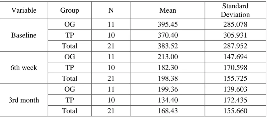
Table 7: Repeated measures ANOVA (General Linear Model) to compare the mean values within and between groups over the time period
Table 6.1: Descriptive statistics
Variable Group N Mean Standard
Deviation
Baseline
OG 11 395.45 285.078
TP 10 370.40 305.931
Total 21 383.52 287.952
6th week
OG 11 213.00 147.694
TP 10 182.30 170.598
Total 21 198.38 155.725
3rd month
OG 11 199.36 139.603
TP 10 134.40 172.435
Results
[image:59.595.88.519.86.278.2]40
Table 7.1: Tests of Within-Subjects Effects
Source df Mean Square F-value p-Value
Values Greenhouse-Geisser correction 1.029 556004.409 10.881 0.003
Error Greenhouse-Geisser correction 19.559 51096.355 - -
Table 7.2: Tests of Between-Subjects Effects
Source df Mean Square F-value P-Value
Group 1 25444.710 .302 0.589
Error 19 84117.483 - -
[image:59.595.88.517.385.601.2]OG- Oak gall group; TP-Triphala group; *- p < 0.005 –statistically significant
Table 8: comparison of proportions between the clinical response and quit status at the end of three months within and between the groups
Group Clinical responders
Quit status 3 months
p-value*
Quit Not quit Total
N (%) N (%) N (%)
OG
Responder 3 (60) 2 (33.3) 5 (45.5)
0.567**
Non- responders 2 (40) 4 (66.7) 6 (54.5)
Total 5(100) 6 (100) 11(100)
TP
Responder 4 (80) 3 (60) 7(70)
0.999**
Non- responders 1 (20) 2 (40) 3 (30)
Total 5 (100) 5 (100) 10 (100)
Total
Responder 7 (70.0) 5(45.5) 12(57.1)
0.387** Non- responder 3 (30.0) 6 (54.5) 9 (42.9)
Total 10 (100) 11(100) 21(100)