J
OURNAL OFV
IROLOGY, Jan. 2006, p. 149–160
Vol. 80, No. 1
0022-538X/06/$08.00
⫹
0
doi:10.1128/JVI.80.1.149–160.2006
Copyright © 2006, American Society for Microbiology. All Rights Reserved.
A Novel, Highly Selective Inhibitor of Pestivirus Replication That
Targets the Viral RNA-Dependent RNA Polymerase
Jan Paeshuyse,
1Pieter Leyssen,
1Eric Mabery,
2Nina Boddeker,
2Robert Vrancken,
3Matheus Froeyen,
1Israrul H. Ansari,
4He
´le
`ne Dutartre,
5Jef Rozenski,
1Laura H. V. G. Gil,
4Carine Letellier,
3Robert Lanford,
6Bruno Canard,
5Frank Koenen,
3Pierre Kerkhofs,
3Ruben O. Donis,
4Piet Herdewijn,
1Julia Watson,
2Erik De Clercq,
1Gerhard Puerstinger,
7and Johan Neyts
1*
Rega Institute for Medical Research, Katholieke Universiteit Leuven, Leuven, Belgium
1; Gilead Sciences, Foster City, California
2;
Veterinary and Agrochemical Research Centre, Ukkel, Belgium
3; Department of Veterinary and Biomedical Sciences,
University of Nebraska—Lincoln, Lincoln, Nebraska
4; Laboratory AFMB-UMR 6098, Marseille, France
5; Department of
Virology and Immunology, Southwest National Primate Research Center, and Southwest Foundation for
Biomedical Research, San Antonio, Texas
6; and Department of Pharmaceutical Chemistry,
Institute of Pharmacy, University of Innsbruck, Austria
7Received 27 June 2005/Accepted 8 October 2005
We report on the highly potent and selective antipestivirus activity of
5-[(4-bromophenyl)methyl]-2-phenyl-5
H
-imidazo[4,5-c]pyridine (BPIP). The 50% effective concentration (EC
50) for inhibition of bovine viral
diarrhea virus (BVDV)-induced cytopathic effect formation was 0.04
ⴞ
0.01
M. Comparable reduction of viral
RNA synthesis (EC
50ⴝ
0.12
ⴞ
0.02
M) and production of infectious virus (EC
50ⴝ
0.074
ⴞ
0.003
M) were
observed. The selectivity index (ratio of 50% cytostatic concentration/EC
50) of BPIP was
⬃
2,000. BPIP was
inactive against the hepatitis C virus subgenomic replicon and yellow fever virus but demonstrated weak
activity against GB virus. Drug-resistant mutants were at least 300-fold less susceptible to BPIP than wild-type
virus; showed cross-resistance to
N
-propyl-
N
-[2-(2
H
-1,2,4-triazino[5,6-b]indol-3-ylthio)ethyl]-1-propanamine
(VP32947), and carried the F224S mutation in the viral RNA-dependent RNA polymerase (RdRp). When the
F224S mutation was introduced into an infectious clone, the drug-resistant phenotype was obtained. BPIP did
not inhibit the in vitro activity of recombinant BVDV RdRp, but did inhibit the activity of replication complexes
(RCs). Computational docking revealed that F224 is located at the top of the finger domain of the polymerase.
Docking of BPIP in the crystal structure of the BVDV RdRp revealed aromatic ring stacking, some hydrophobic
contacts, and a hydrogen bond. Since two structurally unrelated compounds, i.e., BPIP and VP32947, target the
same region of the BVDV RdRp, this position may be expected to be critical in the functioning of the
polymerase or assembly of the RC. The potential of BPIP for the treatment of pestivirus and hepacivirus
infections is discussed.
Pestiviruses cause important diseases of livestock such as
bovine viral diarrhea in cattle, classical swine fever in pigs, and
border disease in sheep. The genus
Pestivirus
is classified along
with the genus
Hepacivirus
and
Flavivirus
in the family
Flavi-viridae
. The members of the family display contrasting host
range specificity (31). Flaviviruses multiply in arthropods and a
large number of vertebrate species; pestiviruses infect
cloven-hoofed animals, and hepaciviruses infect humans naturally and
chimpanzees experimentally. Two biotypes of pestiviruses
ex-ist: those that result in lysis of in vitro infected cells, named
cytopathogenic (CP), and those that do not, termed
noncyto-pathogenic (NCP) (31).
Bovine viral diarrhea virus (BVDV) is a major pathogen of
cattle. In the United States, most estimations of losses caused
by BVDV at the national level range between $10 and $40
million per million calves (21). Losses are projected in reduced
milk production, reduced reproductive performance, growth
retardation, and increased mortality among young stock (21).
Also, the classical swine fever virus (CSFV) can be responsible
for major economic losses, especially in countries with an
in-dustrialized pig production (14). Regardless of the availability
of vaccines against BVDV and CSFV and the implementation
of elaborate eradication or control programs (20, 44), both
viruses remain an agronomical burden. An alternative
ap-proach to combating BVDV and CSFV infections could be the
use of antiviral agents that specifically inhibit the replication of
the virus. Although likely not suited to treat large herds, it may
be important to have selective antipestivirus compounds on
hand. For example, in case of an outbreak of CSFV, an option
could be to prophylactically treat pigs that live in farms located
in close proximity to the infected farm with an antipestivirus
drug. Antiviral treatment may result in almost immediate
pro-tection against infection (propro-tection following vaccination is
obtained only 10 to 14 days later) and hence prevent
transmis-sion of the virus (and avoid large-scale culling of healthy
ani-mals). In the future, as pestivirus eradication programs reach
their final stages, it will be important to have methods to
control the spread of reintroduced viruses. Other possible uses
for antipestivirus drugs could be (i) to treat valuable animals in
zoologic collections, (ii) to treat expensive animals in breeding
* Corresponding author. Mailing address: Rega Institute for Medical
Research, Minderbroedersstraat 10, B-3000 Leuven, Belgium. Phone:
32-16-337341. Fax: 32-16-337340. E-mail: [email protected]
.be.
149
on November 8, 2019 by guest
http://jvi.asm.org/
programs and in vitro embryo production (45), and (iii) to cure
established cell lines from contaminating pestiviruses (13, 19).
Recently, a number of selective anti-BVDV compounds have
been reported. These include polymerase inhibitors, e.g.,
N
-pro-pyl-
N
-[2-(2
H
-1,2,4-triazino[5,6-b]indol-3-ylthio)ethyl]-1-propana-mine (VP32947) (2), a thiazole urea derivative (24), a cyclic urea
derivative (47), and inhibitors of the NS3/NS4A protease, for
example, a boron-modified peptidyl mimetic (7). Aromatic
cat-ionic molecules were also reported to inhibit BVDV replication,
although their mechanism of action remains to be elucidated (18).
Other BVDV inhibitors target cellular enzymes such as
␣
-gluco-sidase (4, 12, 58) and inosinate dehydrogenase (46).
BVDV is considered to be a valuable surrogate virus for
hepatitis C virus (HCV) (6). HCV is a major cause of cirrhosis
and primary hepatocellular carcinoma and the main reason for
liver transplantations among adults in western countries (48).
The current standard therapy for hepatitis C, i.e., the
combi-nation of pegylated alpha interferon and the nucleoside
ana-logue ribavirin, is effective in only about 50 to 60% of patients
who suffer from chronic HCV infection and is associated with
important side effects (28). Consequently, there is an urgent need
for highly effective and selective inhibitors of HCV replication.
In some aspects of viral replication, BVDV is more
advan-tageous in comparison to the currently used HCV replicon
systems (3, 17, 33, 39). The latter do not undergo a complete
replication cycle; hence, early stages (attachment, entry, and
uncoating) or late stages (virion assembly and release) of the
viral replication cycle cannot be studied in the HCV replicon
system. However, very recently, robust HCV cell culture
sys-tems have been reported (32, 51, 56). Yet, insight into the
mechanism of antiviral activity of antipestivirus compounds
may provide valuable information for the design of novel
an-tiviral strategies against HCV (6).
Here, we report on the activity and mechanism of action of
a novel, potent, and highly selective inhibitor of the replication
of pestiviruses, 5-[(4-bromophenyl)methyl]-2-phenyl-5
H
-imi-dazo[4,5-c]pyridine (BPIP). Implications for the development
of HCV inhibitors are discussed.
MATERIALS AND METHODS
Compounds.The synthesis of BPIP (Fig. 1) will be reported elsewhere (J. Paeshuyse, J. Neyts, P. Herdewijn, E. De Clercy, and G. Puerstinger, unpub-lished data). VP32947 was synthesized by standard methods. 3⬘ -Deox-yguanosine-5⬘-triphosphate (3⬘-dGTP), 2⬘-C-methylguanosine-5⬘ -triphos-phate (2⬘-C-Me-GTP), and 2⬘-O-methylguanosine-5⬘triphosphate (2⬘-O -Me-GTP) were purchased from Trilink (San Diego, CA). Solubility of BPIP was determined spectrophotometrically at an optical density at 300 nm (OD300) in minimal essential medium (MEM; Gibco, Merelbeke, Belgium) containing 5% heat-inactivated fetal bovine serum (FBS; Integro, Zaandam, The Neth-erlands) and in RNA-dependent RNA polymerase (RdRp) buffer (50 mM HEPES [pH 8.0], 10 mM KCl, 10 mM dithiothreitol [DTT], 1 mM MgCl2, 2 mM MnCl2, 0.5% igepal [Sigma, Bornem, Belgium]).
Cells and viruses.Madin-Darby bovine kidney (MDBK) cells were grown in MEM (Gibco) supplemented with 5% heat-inactivated FBS (Integro). FBS was shown to be free of BVDV type 1 (BVDV-1) and BVDV-2 by reverse transcrip-tion (RT)-PCR (30). Porcine kidney cells (PK15) were grown in MEM supple-mented with 10% heat-inactivated FBS. First-passage BVDV strain NADL stock was generated from pNADLp15a as previously described (50). The CSFV strain Alfort was obtained from the Institut fu¨r Virologie, Hanover, Germany. BVDV-1 CP strain PE515 was obtained from J. M. Aynaud (INRA, Thiverval, France), and BVDV-1 NCP strain Marloie, BVDV-1 NCP strain L2565, and BVDV-2 CP strain 3435 are field isolates obtained from the Veterinary and Agrochemical Research Center (Ukkel, Belgium). Border disease virus (BDV) NCP strain Aveyron was obtained from E. Thiry, University of Lie`ge, Lie`ge,
Belgium. Human hepatoma cells (Huh 7) containing subgenomic HCV replicons I389luc-ubi-neo/NS3-3⬘/5.1 (Huh 5-2) were kindly provided by R. Bartenschlager (University of Heidelberg, Heidelberg, Germany) and were used to assess activ-ity against HCV. Huh 5-2 cells were grown in Dulbecco’s modified Eagle’s medium (DMEM; Gibco) supplemented with 10% heat-inactivated FBS (Inte-gro), 1⫻nonessential amino acids (Gibco), 100 IU/ml penicillin (Gibco), 100 g/ml streptomycin (Gibco), and 250g/ml Geneticin (Gibco). GB virus strain B (GBV-B) was propagated in primary tamarin (Saguinus mystax) hepatocytes (27). Yellow fever virus (YFV; strain 17D) was the vaccine strain Stamaril from Aventis Pasteur S.A.
Anti-BVDV assay for CP strains (MTS).MDBK cells were seeded at a density of 5⫻103cells per well in 96-well cell culture plates (confluence, 10 to 15%) in MEM-FBS. Following 24 h of incubation at 37°C and 5% CO2, medium was removed and fivefold serial dilutions of the test compounds were added in a total volume of 100l, after which the CP BVDV inoculum (multiplicity of infection⫽ 2) was added to each well. This inoculum resulted in a greater than 90% de-struction of the cell monolayer after 3 days of incubation at 37°C. Uninfected cells and cells receiving virus without compound were included in each assay plate. After 5 days, medium was removed, and 90l of MEM-FBS supplemented with 10l of 3-(4,5-dimethylthiazol-2-yl)-5-(3-carboxymethoxyphenyl)-2-(4-sul-fophenyl)-2H-tetrazolium/phenazinemethosulfate (MTS/PMS) solution (Pro-mega, Leiden, The Netherlands) was added to each well. Following a 2-h incu-bation period at 37°C, the optical density of each well was read at 490 nm in a microplate reader (signal to noise ratio⫽5). The percent CPE was calculated as follows: %CPE⫽[(ODtreated)BVDV⫺(ODcontrol)BVDV]/{[(ODcontrol)mock]⫺ (ODcontrol)BVDV}, in which (ODtreated)BVDVis the OD490of cells infected with BVDV and treated with a certain dilution of compound, (ODcontrol)BVDVis the OD490of cells infected with BVDV and left untreated, and (ODcontrol)mockis the OD490of cells mock infected and left untreated. The 50% effective concentration (EC50) was defined as the concentration of compound that offered 50% protec-tion of the cells against virus-induced cytopathic effect (CPE) and was calculated using logarithmic interpolation.
Anti-BVDV assay for NCP strains (enzyme-linked immunosorbent assay).The antiviral assay was performed as described above, but evaluation of viral replication was done by means of an enzyme-linked immunosorbent assay method. At 3 days postinfection, cell culture fluid was removed and plates were washed once with phosphate-buffered saline (PBS) and dried for 1 h at 37°C. Next, plates were frozen for 20 min at⫺80°C, fixed for 10 min in 4% formaldehyde, and washed twice with PBS, after which they were incubated with an anti-BVDV polyclonal serum (diluted 200-fold in 5% horse serum [Gibco], 500 mM NaCl, 1% Tween 80) for 90 min at 37°C. Plates were then washed three times with wash buffer (150 mM NaCl, 1% Tween 80), and a 500-fold dilution of anti-bovine-coupled peroxidase antibody (Sigma) in dilution buffer was added for 1 h at 37°C. Following three wash steps, plates were incubated with detection buffer (4 mg 3-amino-9-ethylcarbazole dis-solved in 1 mlN,N-dimethylformamide and 14 ml of 0.1 M acetate buffer [pH 5.2] containing 150l of 3% H2O2) for 15 min until a dark red color appeared. EC50 values were calculated using the method of Reed-Muench (41).
Anti-CSFV and BDV assays.Antiviral assays with CSFV and BDV were per-formed in a similar manner as the antiviral assay with the BVDV NCP strains. For CSFV, PK15 cells (ATCC CCL-33) were used. Infected cells were stained with a polyclonal anti-CSFV serum conjugated with biotin, and streptavidin-conjugated horseradish peroxidase was used as secondary antibody. For BDV, MDBK cells were used. Infected cells were stained using an anti-BDV polyclonal serum (bovine origin) as primary antibody and an anti-bovine-coupled peroxidase antibody as secondary antibody. Plates were stained and read as described above.
Anti-GBV-B assay.Antiviral assays of GBV-B were essentially performed as described previously (27).
Anti-HCV assay.Huh 5-2 cells (3; Paeshuyse et al., unpublished) were seeded at a density of 5⫻103
cells per well (confluence, 10 to 15%) in a white view 96-well plate (Perkin-Elmer, Boston, Mass.) in complete DMEM supplemented with 250g/ml G418. Following a 24-h incubation at 37°C and 5% CO2, medium was removed and threefold serial dilutions of compound in complete DMEM were added in a total volume of 100l. After an incubation period of 4 days at 37°C, cell culture fluid was removed and luciferase activity was assessed by means of the Steady-Glo luciferase assay system (Promega) and a Luminoskan Ascent (Thermo, Vantaa, Finland). The EC50 was defined as the concentration of compound that reduced the luciferase signal with 50% and was calculated as described for BVDV.
Cytostatic assay.MDBK or Huh 5-2 cells were seeded at a density of 5⫻103 cells per well of a 96-well plate (confluence 10 to 15%) in MEM-FBS; 24 h later, serial dilutions of the test compounds were added. Cells were allowed to prolif-erate for 3 days at 37°C, after which the cell number was determined by means of the MTS/PMS (Promega) method (signal to noise ratio⫽5). The percent cell
on November 8, 2019 by guest
http://jvi.asm.org/
FIG. 1. (A) Effect of BPIP on BVDV (strain NADL)-induced CPE formation in MDBK cells (bars) and on the proliferation of exponentially
growing MDBK cells (diamonds). BPIP caused, at concentrations above 0.3
M, complete inhibition of BVDV-induced CPE, as assessed
microscopically (although not evident from the MTS assay at these concentrations). (B) Inhibitory effect of BPIP on release of extracellular viral
RNA. (C) Inhibitory effect of BPIP on infectious virus yield.
151
on November 8, 2019 by guest
http://jvi.asm.org/
growth was calculated as (ODtreated)/(ODcontrol), in which (ODtreated) is the OD490of cells treated with a certain dilution of compound and (ODcontrol) is the OD490of cells left untreated. The 50% cytostatic concentration (CC50) was defined as the concentration that inhibited the proliferation of exponentially growing cells by 50% and was calculated using logarithmic interpolation.
Time of drug addition studies.MDBK cells (6.5⫻105cells/well; confluence, 10 to 15%) were seeded in a six-well culture plate (Asahi Technoglass Corpo-ration, Tokyo, Japan). Cultures were inoculated with BVDV (strain NADL; multiplicity of infection⫽2). The inoculum was removed following a 1-h incu-bation period, and cells were washed three times with prewarmed PBS. To obtain precise information on the replication kinetics of BVDV in untreated cultures, supernatant and cells were harvested every 2 h and samples were stored at⫺80°C until further use. In a parallel set of cultures, the test compounds (at 15M) were added at different time points after infection. Cultures were further incu-bated until 24 h postinfection, at which time cell culture supernatant was col-lected and stored at⫺80°C until further use.
Virus yield assay.MDBK cells were seeded at a density of 5⫻103cells per well of a 96-well plate (confluence, 10 to 15%) in MEM-FBS and were, 24 h later, infected with 10-fold serial dilutions of culture supernatant. After 4 days, me-dium was removed and cultures were fixed with 70% ethanol, stained with Giemsa solution, washed, and air dried. Virus-induced CPE was recorded mi-croscopically, and the viral titer was quantified according to the method of Reed and Muench (41). Viral titers were expressed as cell culture 50% infectious dose (per milliliter).
Isolation of BPIP-resistant BVDV.BPIP-resistant (BPIPr
) virus was generated by culturing wild-type BVDV in MDBK cells in the presence of increasing concentrations of the compound. After 3 days of incubation, cultures were freeze-thawed. Lysates of infected and treated cultures that exhibited a cyto-pathic effect under drug selection were used to infect new cell monolayers. These were further incubated in the presence of higher concentration of the compound. The procedure was repeated (12 passages) until drug-resistant virus was selected. The putative drug-resistant viruses were plaque purified twice in the presence of 10g/ml of BPIP.
Introduction of the F224S mutation in a BVDV infectious clone.Introduction of the F224S mutation (TTC to TCC) in the NS5B gene in pNADLp15a and generation of infectious viral particles was carried out essentially as described previously (50).
RNA isolation.Viral RNA was isolated from cell culture supernatant using the QIAamp viral RNA minikit (QIAGEN, Venlo, The Netherlands). Total cellular RNA was isolated from cells using the RNeasy minikit (QIAGEN).
RT-qPCR.A 50-l RT-quantitative PCR (qPCR) reaction contained TaqMan EZ buffer (50 mmol/liter Bicine, 115 mmol/liter potassium acetate, 0.01 mmol/liter EDTA, 60 nmol/liter 6-carboxy-X-rhodamine, and 8% glycerol, pH 8.2; Applied Biosystems, Nieuwerkerk, The Netherlands), 200mol/liter dATP, 200mol/liter dGTP, 200mol/liter dCTP, 500 mol/liter dUTP, 300 nmol/liter forward primer (5⬘-TGA GCT GTC TGA AAT GGT CGA TT-3⬘), 300 nmol/liter re-verse primer (5⬘-AGA AAT ACT GGG TCA TCT GAT GCA A-3⬘), 300 nmol/liter TaqMan probe (6-carboxyfluorescein-CGA AGC AGG TTA CCA AGG AGG CTG TTA GGA-6-carboxytetramethylrhodamine), 3 mmol/liter manganese acetate, 0.5 U AmpErase uracil-N-glycosylase (UNG), 7.5 U rTth DNA polymerase, and template BVDV RNA. Following initial activation by UNG at 50°C for 2 min, the RT step was performed at 60°C for 30 min, followed by inactivation of UNG at 95°C for 5 min. Subsequent PCR amplification con-sisted of 40 cycles of denaturation at 94°C for 20 s and annealing and extension at 62°C for 1 min in an ABI 7000 sequence detector. All samples were analyzed in three replicate reactions.
Sequencing.PCR fragments that cover the entire BVDV genome were gen-erated and analyzed using the cycle-sequencing method (ABI Prism BigDye Terminator Cycle Sequencing Ready Reaction kit). Both DNA strands were sequenced. Sequence data were obtained using an ABI 373 Automated Sequence Analyzer (Applied Biosystems), and sequences were analyzed using the Vector NTI software package (Invitrogen, Merelbeke, Belgium).
RC assay.The RC assay was essentially similar, as published by Sun and colleagues (47). In brief, BVDV-infected MDBK cells were suspended in ice-cold hypotonic buffer A (10 mM Tris-HCl [pH 7.4], 1.5 mM MgCl2) and were incubated for 30 min on ice, after which they were further disrupted by 20 strokes with a Dounce homogenizer. The disrupted cells were pelleted by centrifugation at 1,000⫻gfor 5 min at 4°C. The supernatant fraction, containing cytoplasmic material and plasma membranes, was concentrated by high-speed centrifugation at 200,000⫻gfor 30 min at 4°C. The pellet was resuspended in 120l of buffer B (10 mM Tris-HCl [pH 8.0], 10 mM NaCl, 15% glycerol) and used for an RNA polymerase assay. Replicase reactions were carried out in a total volume of 50l in 50 mM HEPES (pH 8.0), 50 mM potassium acetate, 3 mM MgCl2, 10 mM
dithiothretol, 5 mM creatine phosphate, 25g/ml creatine phosphokinase, 1 mM ATP, 0.5 mM GTP, 0.5 mM CTP, 40M UTP, 10Ci of [␣-33
P]UTP (3,000 mCi/mmol) (Amersham, Uppsala, Sweden), 40 U of RNasin (Promega), and 20 l of the membrane preparation. Following incubation at 30°C for 1 h, the reactions were stopped by adding sodium dodecyl sulfate up to 1% and extracted twice with phenol-chloroform. The RNA products were precipitated in ethanol and analyzed on a 0.6% denaturing glyoxal-agarose gel. Radioactivity incorpo-rated into virus-specific RNA was quantitated using ImageQuant software for the Storm 820 PhosphorImager (Amersham) and expressed as % replication⫽ (density sample/density of untreated control)⫻100.
RNA-dependent RNA polymerase reaction.BVDV (strain NADL) RdRp was expressed and purified as described before (57). The purified BVDV polymerase (100 nM) was mixed with 100M GTP (containing 8.3M of [3H]GTP; Am-ersham) and increasing concentrations of inhibitor (0.1M, 10M, and 96M for BPIP or 100M for the other inhibitors) in 50 mM HEPES, pH 8.0, 10 mM KCl, 10 mM DTT, 1 mM MgCl2, 2 mM MnCl2, and 0.5% igepal (Sigma). Enzyme mix and inhibitors were preincubated (30 min) in order to favor enzyme-inhibitor interaction before RNA binding in case of competition for the RNA-binding site. Reactions were started by the addition of 100 nM poly(C) (about 500 nucleotides in size) template. Reactions were incubated at 30°C and stopped by the addition of 50 mM EDTA after 1, 5, or 15 min. Samples were transferred onto DE-81 filters, washed with 0.3 M ammonium formate, and dried. Radio-activity bound to the filter was determined by liquid scintillation counting. The assay was also carried out as described above except that the poly(C) template was replaced by either oligo(dT)/poly(A), oligo(G)/poly(C), or an artificial hair-pin (this is, an RNA transcript that encompasses nucleotides 1,768 to 2,016 of the sequence with accession number AF305422).
Computational docking.The published X-ray structure of the BVDV RdRp by Protein Data Bank (PDB) entry 1S48 (9) was used in all docking experiments. Selenium atoms in the selenomethionine residues were modified back to S atoms to get methionine residues. Explicit hydrogen atoms were added to the enzyme and inhibitor structures using Reduce (55). The inhibitor BPIP was drawn using Macromodel 5.0 (36). The molecular geometry was optimized in the Amber force field (54) and saved as a PDB file. This file was fed into Gamess for geometry optimization using the AM1 force field (42). The PDB file was then converted to mol2 files using Babel. The position of atom CZ in F224 in the published PDB file 1S48 was used as the center of the docking sphere with a sphere radius of 6.0 Å. Because there is no clear cavity in the neighborhood of F224, the “detect cavity” option was turned off. Default settings were used in Gold for all dockings (22, 23). The structures in the top 50 of the docking scores were retained for visual inspection and analysis. One docked BPIP conformation was retained for further analysis. Criteria to withhold this docked complex were stacking interaction with the F224 phenyl ring, hydrogen bonding, and hydro-phobic interactions. Molecular energy optimization of docked complex was car-ried out using the Amber software. This complex was energy-minimized using the Amber 8.0 software (37). The Amber ff03 force field (11) was used for all polymerase atoms. The BPIP inhibitor parameters and atomic charges were from the general Amber force field by running the Antechamber program (53). The complex was then relaxed by a short energy minimization (500 steps) in the Sander program. The nonbonded cutoff was set to at least 12 Å. Then a further energy minimization of 5,000 steps was performed on the inhibitor and all residues having atoms within a 15 Å distance from an inhibitor atom.
Intracellular metabolism of BPIP.MDBK cell cultures were incubated for 10 h with 55M BPIP. Then cultures were washed three times with PBS. Cells were trypsinized and resuspended in PBS, and equal volumes of cell suspen-sion and ethyl acetate were combined. Samples were vortexed three times for 30 s, and the aqueous phase was snap frozen in an ethanol dry ice mixture. The organic phase was transferred to a new centrifuge tube, and the ethyl acetate was evaporated in a vacuum centrifuge at 40°C for 1 h. Prior to detection, samples were dissolved in 50% methanol. Separation and analysis were carried out using a capillary liquid chromatograph (CapLC; Waters, Milford, MA) connected to a Q-Tof-2 mass spectrometer (Micromass, Manchester, United Kingdom). Sam-ples were separated on a reverse-phase column (XTerra column 0.32⫻50 mm, Waters) with a gradient of 0.1% formic acid and acetonitrile at a flow rate of 5l/min. This setup has a dynamic range of over 3 orders of magni-tude, allowing the detection of the presence of less than 0.1% impurities and metabolites.
RESULTS
Antiviral activity of BPIP.
Following optimization of a lead
compound, BPIP (Table 1) was identified in a multicycle
on November 8, 2019 by guest
http://jvi.asm.org/
growth assay in MDBK cells as a highly selective inhibitor of
BVDV (strain NADL) replication. The EC
50as assessed by
monitoring CPE reduction by the MTS method was 0.04
⫾
0.01
M. The compound inhibited virus-induced CPE
forma-tion in a dose-dependent way (Fig. 1A). To confirm the
anti-BVDV activity of BPIP, the effect of the compound on viral
RNA synthesis (Fig. 1B) and on infectious viral yield was
determined (Fig. 1C). Overall, the pattern of inhibition of viral
RNA synthesis and infectious virus yield were very similar
(Fig. 1A, B, and C). The EC
50for inhibition of viral RNA
production in culture supernatant was 0.12
⫾
0.02
M and for
inhibition of infectious virus yield was 0.074
⫾
0.003
M. BPIP
also inhibited the replication of four other BVDV-1 strains (of
which two were NCP strains and two were CP strains), as well
as a BVDV-2 CP strain (Table 2). Moreover, the compound
inhibited the replication of the CSFV (strain Alfort) and the
BDV (strain Aveyron) (Table 2). The activity of BPIP against
the latter viruses was assessed by an immunohistochemical
method. The resulting EC
50values obtained were three- to
eightfold higher than the EC
50value obtained by means of the
other methods (the reason for which is unclear but may be related
to overstaining and thus underestimating the antiviral activity).
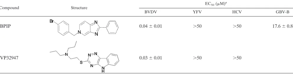
BPIP exhibited an only modest inhibitory effect on GBV-B
rep-lication (EC
50⫽
17.5
⫾
0.7
M) and had no inhibitory effect on
the replication of hepatitis C virus subgenomic replicons
(geno-type 1b) or the yellow fever virus (vaccine strain 17D), a flavivirus
(Table 1). The replication of unrelated RNA or DNA viruses
(such as respiratory syncytial virus, coxsackievirus B3, herpes
sim-plex virus [types 1 and 2], and human immunodeficiency virus
type 1) was not inhibited by BPIP (data not shown). BPIP is
relatively noncytotoxic: the concentration that reduced the
pro-liferation of exponentially growing MDBK cells by 50% (CC
50)
was 80
⫾
11
M. (Fig. 1A). This is almost equal to the solubility
limit of 96
M (data not shown). Hence, a selectivity index
(against BVDV strain NADL) or the ratio of CC
50/EC
50of about
2,000 was calculated.
Intracellular metabolism of BPIP.
To study whether
intra-cellular metabolites of BPIP are formed, that may represent,
rather than BPIP itself, the active antiviral component, the
intracellular metabolism of BPIP in MDBK cells was analyzed.
Cellular extracts from MDBK cells that had been treated for
10 h with 55
M BPIP were analyzed by reverse-phase
high-performance liquid chromatography connected to a mass
spec-trometer. Only intact BPIP, but no metabolites or degradation
products, could be detected.
Time-of-(drug) addition studies.
To understand at what time
during the viral replication cycle BPIP interferes with viral
repli-cation, detailed time-of-(drug) addition experiments were carried
out. The kinetics of one replication cycle of BVDV was first
determined by means of both RT-qPCR and viral yield assay. It
was ascertained that a linear correlation exists between
extracel-lular viral RNA and infectious virus yield (
R
2⫽
0.99). In
[image:5.585.45.541.82.208.2]un-treated cells, release of viral RNA (Fig. 2A) and infectious viral
particles (Fig. 2A) into the supernatant was first detected between
12 and 14 h postinfection; hence, under our experimental
condi-tions, a single cycle of BVDV growth takes an average of 13 h
TABLE 1. Effect of BPIP and VP32947 on the replication of various
Flaviviridae
Compound Structure
EC50(M)a
BVDV YFV HCV GBV-B
BPIP
0.04
⫾
0.01
⬎
50
⬎
50
17.6
⫾
0.8
VP32947
0.03
⫾
0.01
⬎
50
⬎
50
a
EC50of BPIP and VP32947 for inhibition of BVDV (strain NADL) replication in MDBK cells, YFV (strain 17D) replication in Vero cells, HCV subgenomic replicon (genotype 1b) replication in Huh 5-2 cells, and GBV-B replication in primary tamarin hepatocytes. Data are given as mean values⫾standard deviations from at least two independent experiments.
TABLE 2. Effect of BPIP on pestivirus replication
Virus Strain Biotype
EC50(M)a
ELISA MTS RT-qPCR Virus yield
BVDV-1
L2565
NCP
0.6
⫾
0.2
Marloie
NCP
0.5
⫾
0.2
PE515
CP
0.1
⫾
0.2
NADL
CP
0.33
⫾
0.03
0.04
⫾
0.01
b0.12
⫾
0.02
0.074
⫾
0.003
BVDV-2
3435
CP
0.2
⫾
0.1
CSFV
Alfort
2
⫾
1
BDV
Aveyron
1.7
⫾
0.5
aData are given as mean values⫾standard deviations from three independent experiments.
bEC50of VP32947 for the inhibition of BVDV NADL replication in MDBK cells (as assessed in parallel by means of the MTS assay) was 0.03⫾0.01M.
V
OL. 80, 2006
INHIBITION OF BVDV REPLICATION
153
on November 8, 2019 by guest
http://jvi.asm.org/
[image:5.585.43.546.608.708.2]on November 8, 2019 by guest
http://jvi.asm.org/
(Fig. 2A). As of 6 to 8 h postinfection, a gradual increase of
intracellular viral RNA was noted (Fig. 2B). This rise must
coin-cide with the formation of functional RCs which produce viral
RNA. When BPIP was added during the first 8 h postinfection, it
resulted in the complete inhibition of extracellular (Fig. 2C) and
intracellular (Fig. 2C) viral RNA yield, as detected at 24 h
postin-fection. A gradual loss in the antiviral efficacy of BPIP was noted
when the compound was first added at a time point later than 8 h
postinfection.
Isolation and characterization of drug-resistant viruses.
BPIP
rvirus was selected by propagating BVDV (strain NADL)
for 12 passages in increasing concentrations (from 0.2 to 27
M) of the drug. The resulting drug-resistant virus pool was
plaque purified twice in the presence of a 27
M concentration
of the compound. The plaque-purified BPIP
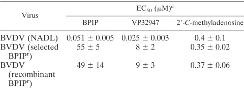
rvirus proved to
be at least a 300-fold less susceptible to the inhibitory effect of
BPIP than the parent wild-type strain (Table 3). The resistant
virus replicated about as efficiently as the parent wild-type virus.
VP32947, which was included as a reference drug, showed
cross-resistance with BPIP. By contrast, 2
⬘
-
C
-methyladenosine, a
nu-cleoside analogue inhibitor of HCV (8, 15) that we also found to
be active against BVDV, was equally active against the wild type
as it was against the BPIP-resistant virus (Table 3).
Molecular characterization of BPIP-resistant virus.
Since
the BPIP
rvirus proved to be cross-resistant with VP32947, the
region in which the mutation was found that resulted in
resis-tance against VP32947 was sequenced. By comparing the
se-quences of this region obtained from six independently
iso-lated BPIP
rclones to the parent wild-type strain (GenBank
accession no. AJ781045), we identified a T-to-C transition at
position 10,863 in the viral RNA. This point mutation resulted
in an amino acid change of phenylalanine (F) to serine (S) at
amino acid residue 224 in the mature NS5B protein.
Subse-quently, the remaining part of the nonstructural (NS) region of
the genome of BPIP
rclones was sequenced. No other
muta-tions were detected throughout the NS region of the genome
of the drug-resistant virus.
To confirm that the F224S change in NS5B alone was
re-sponsible for the BPIP
rphenotype, the mutation was
intro-duced in the infectious clone pNADLp15a of the wild-type
virus. RNA transcripts derived from plasmid pNADLp15a/DR
were transfected into MDBK cells, and the resulting
recombi-nant BPIP
rvirus was isolated. Sequence analysis confirmed
that this virus indeed carried the F224S mutation. Akin to the
original selected BPIP
rvirus, the recombinant BPIP
rvirus was
resistant to the inhibitory effects of BPIP and VP32947 but
remained sensitive to the antiviral activity of 2
⬘
-
C
-methylad-enosine (Table 3).
In vitro RdRp assay.
BPIP, VP32947, and the nucleotide
analogues 3
⬘
-dGTP, 2
⬘
-
C
-Me-GTP, and 2
⬘
-
O
-Me-GTP
(in-cluded as positive controls) were studied for their effects on the
polymerase activity of highly purified BVDV RdRp by using
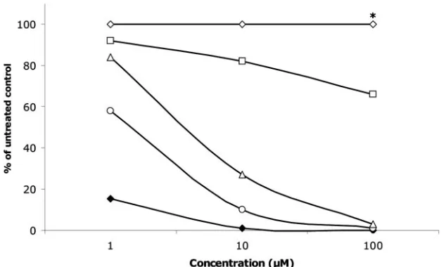
poly(C) as a template. BPIP had no, and VP32947 only a
minor, effect on the activity of the viral polymerase (Fig. 3).
The 50% inhibitory concentrations for BVDV polymerase
ac-tivity were
⬍
1
M for 3
⬘
-dGTP,
⬃
1
M for 2
⬘
-
C
-Me-GTP, and
between 5 and 10
M for 2
⬘
-
O
-Me-GTP. The effect of BPIP on
the purified BVDV RdRp was further assessed using various
RNA templates [oligo(dT)/poly(A), oligo(G)/poly(C), and an
artificial hairpin]. No significant inhibition by BPIP of the
activity of the viral polymerase was noted when these other
templates were used (data not shown).
Viral replicase complex assay.
Because BPIP did not inhibit
the activity of the purified BVDV RdRp, the effect of the
compound on viral RCs, isolated from MDBK cells that had
been infected with the wild-type virus, was studied. BPIP
al-most completely inhibited the activity of the BVDV RCs over
a concentration range of 0.5 to 10
M (Fig. 4A, B). In contrast,
BPIP had only a weak inhibitory effect on the activity
(maxi-mum 50% at concentrations as high as 10
M) of RCs isolated
from MDBK cells that had been infected with the
laboratory-selected BPIP
rvirus (Fig. 4A, B).
Computational docking of BPIP in the BVDV RdRp crystal
structure.
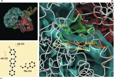
Molecular modeling revealed that F224 is located at
the tip of the finger domain of the BVDV polymerase. Docking
of BPIP to the BVDV RdRp and performing a Ligplot/HBPlus
analysis (34, 52) (Fig. 5C) on the docking complex revealed
possible interactions between the polymerase and BPIP. One
possible interaction was in agreement with our hypothesis that
this inhibitor should bind close to the F224 residue. Through
this analysis (Fig. 5A, C), the following interactions were
sug-gested: (i) a stacking interaction between the
imidazo[4,5-c]pyridine ring system of BPIP and F224, the average distance
between both aromatic systems is about 3.5 Å, (ii) hydrophobic
contacts of the inhibitor with A221, A222, and F224, and (iii)
a hydrogen bond between N3 of the imidazole ring in BPIP
with the backbone oxygen of residue F224 (distance, 3.27 Å).
FIG. 2. Yields of extracellular viral RNA (circles), infectious virus (bars), or intracellular viral RNA (diamonds) during a single replication cycle
of BVDV strain NADL in MDBK cells. Viral RNA levels were monitored at various times postinfection by RT-qPCR. (A) Extracellular viral RNA
and infectious virus yields during a time course of 24 h. (B) Intracellular viral RNA yields over a time course of 24 h (the inset presents the viral
RNA levels during the period from 0 to 12 h on a more detailed scale). CCID
50, cell culture 50% infectious dose. (C) Effect of time-of-(drug)
[image:7.585.43.283.89.179.2]addition on the antiviral activity of BPIP. Extracellular and intracellular viral RNA was monitored by RT-qPCR at 24 h postinfection (in cells
treated with BPIP [at 15
M], starting at different times postinfection) and compared with untreated infected cells (the inset presents the viral
RNA levels during the period 6 to 10 h on a more detailed scale). Values are expressed as percentages of viral RNA of untreated infected cells.
TABLE 3. Susceptibility of wild-type and BPIP
rBVDV to BPIP,
VP32947, and 2
⬘
-
C
-methyladenosine
Virus
EC50(M)a
BPIP VP32947 2⬘-C-methyladenosine
BVDV (NADL)
0.051
⫾
0.005 0.025
⫾
0.003
0.4
⫾
0.1
BVDV (selected
BPIP
r)
55
⫾
5
8
⫾
2
0.35
⫾
0.02
BVDV
(recombinant
BPIP
r)
49
⫾
14
9
⫾
3
0.37
⫾
0.06
aData are given as mean values⫾standard deviations from three independent
experiments.
V
OL. 80, 2006
INHIBITION OF BVDV REPLICATION
155
on November 8, 2019 by guest
http://jvi.asm.org/
No specific interaction of the inhibitor’s Br atom with the
enzyme emerged from this analysis.
DISCUSSION
A small molecule (BPIP) was identified (following lead
op-timization) as a highly selective and potent in vitro inhibitor of
the replication of pestiviruses. The compound proved highly
active against both cytopathic and noncytopathic biotypes of
BVDV-1 and BVDV-2 as well as against other members of the
genus
Pestivirus
, i.e., classical swine fever virus and border
disease virus.
BPIP was inactive against the hepacivirus HCV (as assessed
in the HCV genotype 1b subgenomic replicon system) and
against the flavivirus YFV strain 17D but exhibited moderate
activity against GBV-B. However, when the entire series of
compounds that were synthesized during lead optimization for
anti-BVDV activity were evaluated against HCV, a few
mole-cules were found to inhibit the replication of this virus in the
replicon system. Molecules in this series of imidazopyridines
could be classified in three groups: (i) compounds solely active
against BVDV, (ii) compounds active against both BVDV and
HCV, and (iii) compounds active only against HCV. Studies on
the structure-activity relationship of the BVDV and
anti-HCV series will be reported elsewhere. These data
demon-strate for the first time that distinct changes to a class of small
molecule inhibitors of pestivirus replication may result in
mol-ecules with antihepacivirus activity, despite the fact that the
most potent antipestivirus molecules in this series are devoid of
anti-HCV activity.
We then further focused on the mechanism of anti-BVDV
activity of BPIP. Detailed time-of-(drug) addition studies
re-vealed that the time point at which BPIP exerts its activity
coincided with the onset of viral RNA synthesis at about 8 h
postinfection. Addition of compound at a time point before the
onset of intracellular viral RNA synthesis led to the complete
inhibition of viral RNA production, whereas addition at later
time points resulted in a gradual loss of antiviral activity. These
data suggest that BPIP interferes with the formation or
func-tion of the replicafunc-tion complex of the virus. When the genotype
of in vitro-generated BPIP
rvirus was determined, a
phenylal-anine (F)-to-serine (S) mutation was detected (in six out of six
sequenced clones) at position 224 (F224S) of the NS5B, the
gene that encodes the viral RNA-dependent RNA polymerase.
No other mutations were detected throughout the NS region of
the drug-resistant virus.
Introduction of the F224S mutation in a full-length wild-type
infectious clone of BVDV (strain NADL) again resulted in the
drug-resistant phenotype. This confirms that the F224S
muta-tion indeed is responsible for the resistant phenotype. The
phenylalanine residue at position 224 of the RdRp is almost
strictly conserved within the genus pestivirus. Only BVDV type
2 carries a tyrosine at this position. BVDV-2 is roughly equally
susceptible to BPIP as BVDV-1 and the other pestiviruses.
This may not be surprising, as tyrosine and phenylalanine (but
not serine) share similar aromatic properties. Interestingly, the
antipestivirus compound VP32947, a molecule that has a
chemi-cal structure that is very different from that of BPIP, induced the
same mutation. In line with this observation, VP32947 proved
cross-resistant with BPIP. In contrast, the nucleoside analogue
2
⬘
-
C
-methyladenosine was equally effective against both
wild-type and BPIP-resistant virus.
[image:8.585.135.452.69.259.2]Although the drug-induced mutation in the viral RdRp
strongly suggests interference with the viral polymerase
activ-ity, we were unable to detect inhibition of the highly purified
enzyme with BPIP (or with VP32947), even at relatively high
concentrations. The fact that two very different compounds
induce the same mutation suggests that this amino acid (F224)
and its environment play a crucial role in the normal
function-ing of the BVDV RdRp. The viral polymerase assay was
val-idated by demonstrating that 2
⬘
-
C
-Me-GTP (the 5
⬘
-triphos-phate metabolite of 2
⬘
-
C
-methyl-guanosine, an analogue of
FIG. 3. Effect of BPIP (open diamonds), VP32947 (open squares), 3
⬘
-dGTP (open triangles), 2
⬘
-
C
-Me-GTP (open circles), and 2
⬘
-
O
-Me-GTP
(filled diamonds) on the activity of purified BVDV RdRp using poly(C) as a template and incubated for 5 min. Data are from a typical experiment
and are expressed as the percentage of untreated control. Similar data were obtained using other templates. BPIP was tested at the highest
concentration of 96
M (*).
on November 8, 2019 by guest
http://jvi.asm.org/
2
⬘
-
C
-methyl-adenosine) and related molecules inhibited the
BVDV RdRp activity. The lack of activity of VP32947 against
BVDV polymerase we observed is consistent with results
ob-tained by King and coworkers (24). However, within the cell,
BVDV NS5B functions in the context of membrane-bound
RCs that consist of several virus-encoded proteins, host
pro-teins, and various forms of viral RNA (47, 49). We therefore
evaluated the effect of BPIP on RCs isolated from MDBK cells
that had been infected either with wild-type virus or with the
mutant F224S virus. BPIP effectively inhibited the function of
the wild-type RC but not that of the mutant RC. A possible
explanation for the lack of activity of BPIP on the purified
polymerase may therefore be that the compound, following its
interaction with NS5B, disturbs the
formation/stability/func-tion of the replicaformation/stability/func-tion complex. Although neither we nor King
et al. (24) detected inhibition of the activity of the highly
purified polymerase of BVDV NS5B by VP32947, Baginski
and coworkers did observe inhibition of the viral polymerase
by VP32947 (2). It should be noted, however, that low template
concentrations were used in their assay and that the 50%
inhibitory concentration for the viral polymerase was 35-fold
higher (700 nM) than the EC
50for inhibition of viral
replica-tion (2). These authors also speculated that VP32947 may
disturb an interaction of NS5B within the BVDV replication
complex.
The crystal structure of the RNA-dependent RNA
ase of BVDV was recently reported (9). The BVDV
polymer-ase shares several structural characteristics with the RdRp of
HCV (1, 5, 9, 29). The F224S mutation responsible for
resis-tance to BPIP and VP32947 is located in a turn in the finger
domain between two beta-sheets of the BVDV RdRp. The
corresponding region in the HCV RdRp is believed to be
involved in finger flexibility for template/product translocation
(26), dimerization of the RdRp in the replication complex, or
protein-protein interactions (10, 38), enabling the assembly of
an active replication complex.
Computational docking of BPIP to the BVDV RdRp
sug-gested that there were a limited number of interactions
be-tween the inhibitor and the polymerase. Hypothetical binding
of the inhibitor to the polymerase could result in reduced
finger flexibility or impairment of the ability of the polymerase
to translocate its template/product during polymerization.
An-other possibility could be that the 4-bromophenyl moiety
in-fluences the entrance of the template RNA in the
template-binding channel. It should be mentioned, however, that
FIG. 4. Effect of BPIP on the activity of replication complexes isolated at 14 h postinfection from MDBK cells that had been infected with the
wild type (WT) (strain NADL) or with the selected BPIP
rBVDV strain. (A) Reaction product of the RC assay was separated on a 0.6%
agarose-glyoxal denaturing gel. Replication complexes for this assay were isolated from MDBK cells infected either with WT virus or with the
selected BPIP
rBVDV strain. The arrow indicates the position at which full-length BVDV RNA migrates (confirmed by using molecular weight
markers and RNase treatment). (B) Densiometric analysis of the autoradiograph depicted in panel A. Black bars represent the activity of WT
(strain NADL) RCs, and open bars represent the activity of RCs from cells infected with selected BPIP
rvirus. % replication
⫽
(density
sample/density of untreated control)
⫻
100.
V
OL. 80, 2006
INHIBITION OF BVDV REPLICATION
157
on November 8, 2019 by guest
http://jvi.asm.org/
computational modeling does not provide hard evidence that
the modeled interactions do indeed occur in vivo. If BPIP
binds to the RdRp, there is, however, no evidence that this
results in an allosteric inhibition of the highly purified
poly-merase or a reduction of its template-binding capability.
Re-cently, it was shown that the generation of cocrystals with
VP32947 was hampered by a dimer interface near the putative
binding site of VP32947 in the BVDV RdRp crystal (9). The
authors suggested that this dimer could play a significant role
in the replication complex. Furthermore, it was suggested that
the top of the fingers domain may be a protein-binding site
important for interaction with other proteins of the replicase
complex. BPIP could possibly prevent the interactions between
different proteins of the replication complex. It will now be
important to study whether the BPIP analogues that exhibit
anti-HCV activity result in drug-resistant mutations at a
com-parable/homologous position in the HCV RdRp.
In conclusion, we report here on a highly potent and
selec-tive inhibitor of the replication of pestiviruses. Structural
an-alogues of this molecule also exhibit anti-HCV activity. It is
intriguing that this compound, although very different in
chem-ical structure, appears to possess a similar mechanism of action
as VP32947 (2), suggesting that the site of the polymerase with
which these two compounds interact may be crucial for the
functioning of the polymerase and the viral replication
com-plex. The present study contributes to the understanding on
how the polymerases of
Flaviviridae
can be targeted by specific
inhibitors.
ACKNOWLEDGMENTS
This work was supported by a postdoctoral position of the
Onder-zoeksfonds of the KULeuven to P. Leyssen and by a grant
(S-6146-Sectie1) from the Belgian Government (“Federale Overheidsdienst
Volksgezondheid, Veiligheid van de Voedselketen en Leefmilieu”).
This work is part of the activities of the VIRGIL European Network of
Excellence on Antiviral Drug Resistance supported by a grant
(LSHM-CT-2004-503359) from the Priority 1 “Life Sciences, Genomics and
Biotechnology for Health” Programme in the Sixth Framework
Pro-gramme of the European Union. This work was supported by a Ph.D.
grant from the Institute for the Promotion of Innovation through
Science and Technology in Flanders (IWT Vlaanderen) to J.
Pae-shuyse.
We thank W. Delaney for helpful discussions and R. Bartenschlager
for kindly providing Huh 5-2 cells. We thank Katrien Geerts for
ex-cellent technical assistance and Dominique Brabants, Chantal
Bier-naux, and Christiane Callebaut for dedicated editorial help.
REFERENCES
1.Ago, H., T. Adachi, A. Yoshida, M. Yamamoto, N. Habuka, K. Yatsunami, and M. Miyano.1999. Crystal structure of the RNA-dependent RNA poly-merase of hepatitis C virus. Struct. Fold. Des.7:1417–1426.
[image:10.585.101.485.71.337.2]2.Baginski, S. G., D. C. Pevear, M. Seipel, S. C. Sun, C. A. Benetatos, S. K. Chunduru, C. M. Rice, and M. S. Collett.2000. Mechanism of action of a pestivirus antiviral compound. Proc. Natl. Acad. Sci. USA97:7981–7986.
FIG. 5. Modeling of BPIP near the position of F224 in the RNA-dependent RNA polymerase. (A) Overview of the entire structure of the RdRp
of BVDV with BPIP docked in the vicinity of F224. (B) Detail of the predicted part of the RdRp surface that possibly interacts with BPIP. The
following predicted interactions are shown: (i) a hydrogen bond (presented as a yellow dashed line) and (ii) interacting hydrophobic residues A221,
A222, and F224 (highlighted). The loop that contains F224 is colored in cyan (residues 212 to 232). The BVDV polymerase domains are colored
yellow (N-terminal domain, residues 1 to 138), blue (fingers, residues 139 to 313 and residues 351 to 410), green (palm, residues 314 to 350 and
residues 411 to 500), or red (thumb, residues 501 to 679). Picture was generated using Bobscript, Molscript, and Raster3D (16, 25, 35). (C) Ligplot
depicts a schematic diagram of protein-BPIP interactions. The interactions shown are hydrogen bonds (dashed light blue line) and hydrophobic
contacts (represented by an arc with spokes radiating toward the ligand atoms they contact, and the contacted atoms are shown with spokes
radiating back). Br atom (green circle), carbon atoms (black circles), nitrogen atoms (blue circles), and oxygen atom (red circle) are also illustrated.
on November 8, 2019 by guest
http://jvi.asm.org/
3.Bartenschlager, R., and V. Lohmann.2001. Novel cell culture systems for the hepatitis C virus. Antivir. Res.52:1–17.
4.Branza-Nichita, N., D. Durantel, S. Carroue´e-Durantel, R. A. Dwek, and N. Zitzmann.2001. Antiviral effect ofN-butyldeoxynojirimycin against bovine viral diarrhea virus correlates with misfolding of E2 envelope proteins and impairment of their association into E1-E2 heterodimers. J. Virol.75:3527– 3536.
5.Bressanelli, S., L. Tomei, F. A. Rey, and R. De Francesco.2002. Structural analysis of the hepatitis C virus RNA polymerase in complex with ribonucle-otides. J. Virol.76:3482–3492.
6.Buckwold, V. E., B. E. Beer, and R. O. Donis.2004. Bovine viral diarrhea virus as a surrogate model of hepatitis C virus for the evaluation of antiviral agents. Antivir. Res.60:1–15.
7.Bukhtiyarova, M., C. J. Rizzo, C. A. Kettner, B. D. Korant, H. T. Scarnati, and R. W. King.2001. Inhibition of the bovine viral diarrhoea virus NS3 serine protease by a boron-modified peptidyl mimetic of its natural substrate. Antivir. Chem. Chemother.12:367–373.
8.Carroll, S. S., J. E. Tomassini, M. Bosserman, K. Getty, M. W. Stahlhut, A. B. Eldrup, B. Bhat, D. Hall, A. L. Simcoe, R. LaFemina, C. A. Rutkowski, B. Wolanski, Z. Yang, G. Migliaccio, R. De Francesco, L. C. Kuo, M. MacCoss, and D. B. Olsen.2003. Inhibition of hepatitis C virus RNA rep-lication by 2⬘-modified nucleoside analogs. J. Biol. Chem.278:11979–11984. 9.Choi, K. H., J. M. Groarke, D. C. Young, R. J. Kuhn, J. L. Smith, D. C. Pevear, and M. G. Rossmann.2004. The structure of the RNA-dependent RNA polymerase from bovine viral diarrhea virus establishes the role of GTP in de novo initiation. Proc. Natl. Acad. Sci. USA101:4425–4430. 10.Dimitrova, M., I. Imbert, M. P. Kieny, and C. Schuster.2003.
Protein-protein interactions between hepatitis C virus nonstructural Protein-proteins. J. Vi-rol.77:5401–5414.
11.Duan, Y., C. Wu, S. Chowdhury, M. Lee, G. Xiong, W. Zhang, R. Yang, P. Cieplak, R. Luo, and T. Lee.2003. A point-charge force field for molecular mechanics simulations of proteins. J. Comput. Chem.24:1999–2012. 12.Durantel, D., N. Branza-Nichita, S. Carroue´e-Durantel, T. D. Butters, R. A.
Dwek, and N. Zitzmann.2001. Study of the mechanism of antiviral action of iminosugar derivatives against bovine viral diarrhea virus. J. Virol.75:8987– 8998.
13.Durantel, D., S. Carroue´e-Durantel, N. Branza-Nichita, R. A. Dwek, and N. Zitzmann.2004. Effects of interferon, ribavirin, and iminosugar derivatives on cells persistently infected with noncytopathic bovine viral diarrhea virus. Antimicrob. Agents Chemother.48:497–504.
14.Edwards, S., A. Fukusho, P. C. Lefevre, A. Lipowski, Z. Pejsak, P. Roehe, and J. Westergaard.2000. Classical swine fever: the global situation. Vet. Microbiol.73:103–119.
15.Eldrup, A. B., C. R. Allerson, C. F. Bennett, S. Bera, B. Bhat, N. Bhat, M. R. Bosserman, J. Brooks, C. Burlein, S. S. Carroll, P. D. Cook, K. L. Getty, M. MacCoss, D. R. McMasters, D. B. Olsen, T. P. Prakash, M. Prhavc, Q. Song, J. E. Tomassini, and J. Xia.2004. Structure-activity relationship of purine ribonucleosides for inhibition of hepatitis C virus RNA-dependent RNA polymerase. J. Med. Chem.47:2283–2295.
16.Esnouf, R.1999. Further additions to Molscript 1.4, including reading and contouring of electron-density maps. Acta Crystallogr.277:505–524. 17.Frolov, I., E. Agapov, T.-A. J. Hoffman, B. M. Pragai, M. Lippa, S.
Schlesinger, and C. M. Rice.1999. Selection of RNA replicons capable of persistent noncytopathic replication in mammalian cells. J. Virol.73:3854– 3865.
18.Givens, M. D., C. C. Dykstra, K. V. Brock, D. A. Stringfellow, A. Kumar, C. E. Stephens, H. Goker, and D. W. Boykin.2003. Detection of inhibition of bovine viral diarrhea virus by aromatic cationic molecules. Antimicrob. Agents Chemother.47:2223–2230.
19.Givens, M. D., D. A. Stringfellow, C. C. Dykstra, K. P. Riddell, P. K. Galik, E. Sullivan, J. Robl, P. Kasinathan, A. Kumar, and D. W. Boykin.2004. Prevention and elimination of bovine viral diarrhea virus infections in fetal fibroblast cells. Antivir. Res.64:113–118.
20.Greiser-Wilke, I., B. Grummer, and V. Moennig.2003. Bovine viral diar-rhoea eradication and control programmes in Europe. Biologicals31:113– 118.
21.Houe, H.2003. Economic impact of BVDV infection in dairies. Biologicals
31:137–143.
22.Jones, G., P. Willet, and R. Glen.1995. Molecular recognition of receptor sites using a genetic algorithm with a description of desolvation. J. Mol. Biol.
245:43–53.
23.Jones, G., P. Willet, R. Glen, A. Leach, and R. Taylor.1997. Development and validation of a genetic algorithm for flexible docking. J. Mol. Biol.
267:727–748.
24.King, R. W., H. T. Scarnati, E. S. Priestley, I. De Lucca, A. Bansal, and J. K. Williams.2002. Selection of a thiazole urea-resistant variant of bovine viral diarrhoea virus that maps to the RNA-dependent RNA polymerase. Antivir. Chem. Chemother.13:315–323.
25.Kraulis, P.1991. MOLSCRIPT: a program to produce both detailed and schematic plots of protein structures. J. Appl. Crystallogr.24:946–950. 26.Lai, V. C., C. C. Kao, E. Ferrari, J. Park, A. S. Uss, J. Wright-Minogue, Z.
Hong, and J. Y. Lau.1999. Mutational analysis of bovine viral diarrhea virus RNA-dependent RNA polymerase. J. Virol.73:10129–10136.
27.Lanford, R. E., D. Chavez, B. Guerra, J. Y. Lau, Z. Hong, K. M. Brasky, and B. Beames.2001. Ribavirin induces error-prone replication of GB virus B in primary tamarin hepatocytes. J. Virol.75:8074–8081.
28.Lauer, G. M., and B. D. Walker.2001. Hepatitis C virus infection. N. Engl. J. Med.345:41–52.
29.Lesburg, C. A., M. B. Cable, E. Ferrari, Z. Hong, A. F. Mannarino, and P. C. Weber.1999. Crystal structure of the RNA-dependent RNA polymerase from hepatitis C virus reveals a fully encircled active site. Nat. Struct. Biol.
6:937–943.
30.Letellier, C., P. Kerkhofs, G. Wellemans, and E. Vanopdenbosch.1999. Detection and genotyping of bovine diarrhea virus by reverse transcription-polymerase chain amplification of the 5⬘untranslated region. Vet. Microbiol.
64:155–167.
31.Lindenbach, B. D., and C. M. Rice.2001. Flaviviridae: the viruses and their replication, p. 991–1041.InD. M. Knipe, P. M. Howley, D. E. Griffin, M. A. Martin, R. A. Lamb, B. Roizman, and S. E. Straus (ed.), Fields virology. Lippincott Williams & Wilkins, Philadelphia, Pa.
32.Lindenbach, B. D., M. J. Evans, A. J. Syder, B. Wolk, T. L. Tellinghuisen, C. C. Liu, T. Maruyama, R. O. Hynes, D. R. Burton, J. A. McKeating, and C. M. Rice.2005. Complete replication of hepatitis C virus in cell culture. Science309:623–626.
33.Lohmann, V., F. Korner, J. Koch, U. Herian, L. Theilmann, and R. Bartenschlager.1999. Replication of subgenomic hepatitis C virus RNAs in a hepatoma cell line. Science285:110–113.
34.McDonald, I., and J. Thornston.1994. Satisfying Hydrogen bonding poten-tial in proteins. J. Mol. Biol.238:777–793.
35.Merritt, E.1997. Raster3D photorealistic molecular graphics. Methods En-zymol.277:505–524.
36.Mohamadi, F., N. Richards, W. Guida, R. Liskamp, M. Lipton, C. Caufield, C. Chang, T. Hendrickson, and W. Still.1990. An integrated software system for modeling organic and bioorganic molecules using molecular mechanics. J. Comput. Chem.11:440–467.
37.Pearlman, D., D. Case, J. Caldwell, W. Ross, T. Cheatham, S. DeBolt, D. Ferguson, G. Seibel, and P. Kollman.1995. AMBER, a computer program for applying molecular mechanics, normal mode analysis, molecular dynam-ics and free energy calculations to elucidate the structures and energies of molecules. Comp. Phys. Commun.91:1–41.
38.Piccininni, S., A. Varaklioti, M. Nardelli, B. Dave, K. D. Raney, and J. E. McCarthy.2002. Modulation of the hepatitis C virus RNA-dependent RNA polymerase activity by the non-structural (NS) 3 helicase and the NS4B membrane protein. J. Biol. Chem.277:45670–45679.
39.Pietschmann, T., and R. Bartenschlager.2001. The hepatitis C virus replicon system and its application to molecular studies. Curr. Opin. Drug Discov. Dev.4:657–664.
40. Reference deleted.
41.Reed, L. J., and A. H. Muench.1938. A simple method of estimating fifty percent endpoints. Am. J. Hyg.27:493–497.
42.Schmidt, M., K. Baldridge, J. Boatz, S. Elbert, M. Gordon, J. Jensen, S. Koseki, N. Matsunaga, K. Nguyen, S. Su, T. Windus, M. Dupuis, and J. Montgomery.1993. General atomic and molecular electronic structure. Syst. J. Comput. Chem.14:1347–1363.
43. Reference deleted.
44.Stegeman, A., A. Elbers, H. de Smit, H. Moser, J. Smak, and F. Pluimers.
2000. The 1997–1998 epidemic of classical swine fever in the Netherlands. Vet. Microbiol.73:183–196.
45.Stringfellow, D. A., K. P. Riddell, M. D. Givens, P. K. Galik, E. Sullivan, C. C. Dykstra, J. Robl, and P. Kasinathan.2005. Bovine viral diarrhea virus (BVDV) in cell lines used for somatic cell cloning. Theriogenology63:1004– 1013.
46.Stuyver, L. J., T. Whitaker, T. R. McBrayer, B. I. Hernandez-Santiago, S. Lostia, P. M. Tharnish, M. Ramesh, C. K. Chu, R. Jordan, J. Shi, S. Rachakonda, K. A. Watanabe, M. J. Otto, and R. F. Schinazi.2003. Ribo-nucleoside analogue that blocks replication of bovine viral diarrhea and hepatitis C viruses in culture. Antimicrob. Agents Chemother.47:244–254. 47.Sun, J. H., J. A. Lemm, D. R. O’Boyle II, J. Racela, R. Colonno, and Gao, M.
2003. Specific inhibition of bovine viral diarrhea virus replicase. J. Virol.
77:6753–6760.
48.Thomson, B. J., and R. G. Finch.2005. Hepatitis C virus infection. Clin. Microbiol. Infect.11:86–94.
49.Uchil, P. D., and V. Satchidanandam.2003. Architecture of the flaviviral replication complex. Protease, nuclease, and detergents reveal encasement within double-layered membrane compartments. J. Biol. Chem.278:24388– 24398.
50.Vassilev, V. B., and R. O. Donis.2000. Bovine viral diarrhea virus induced apoptosis correlates with increased intracellular viral RNA accumulation. Virus Res.69:95–107.
51.Wakita, T., T. Pietschmann, T. Kato, T. Date, M. Miyamoto, Z. Zhao, K. Murthy, A. Habermann, H. G. Krausslich, M. Mizokami, R. Bartenschlager, and T. J. Liang.2005. Production of infectious hepatitis C virus in tissue culture from a cloned viral genome. Nat. Med.11:791–796.
V
OL. 80, 2006
INHIBITION OF BVDV REPLICATION
159
on November 8, 2019 by guest
http://jvi.asm.org/
52.Wallace, A., R. Laskowski, and J. Thornton.1995. LIGPLOT: a program to generate schematic diagrams of protein-ligand interactions. Protein Eng.
8:127–134.
53.Wang, J., R. Wolf, J. Caldwell, P. Kollman, and D. Case.2004. Development and testing of a general amber force field. J. Comput. Chem.25:1157– 1174.
54.Weiner, P., P. Kollman, D. Nguyen, and D. Case.1986. An all atom force field for simulations of proteins and nucleic acids J. Comput. Chem.7:230–252. 55.Word, M., S. Lovell, J. Richardson, and D. Richardson.1999. Asparagine
and glutamine: using hydrogen atom contacts in the choice of sidechain amide orientation. J. Mol. Biol.285:1733–1745.
56.Zhong, J., P. Gastaminza, G. Cheng, S. Kapadia, T. Kato, D. R. Burton, S. F.
Wieland, S. L. Uprichard, T. Wakita, and F. V. Chisari.2005. Robust hepatitis C virus infection in vitro. Proc. Natl. Acad. Sci. USA102:9294–9299. 57.Zhong, W., L. L. Gutshall, and A. M. Del-Vecchio.1998. Identification and
characterization of an RNA-dependent RNA polymerase activity within the nonstructural protein 5B region of bovine viral diarrhea virus. J. Virol.
72:9365–9369.
58.Zitzmann, N., A. S. Mehta, S. Carrouee, T. D. Butters, F. M. Platt, J. McCauley, B. S. Blumberg, R. A. Dwek, and T. M. Block.1999. Imino sugars inhibit the formation and secretion of bovine viral diarrhea virus, a pestivirus model of hepatitis C virus: implications for the development of broad spectrum anti-hepatitis virus agents. Proc. Natl. Acad. Sci. USA
96:11878–11882.
on November 8, 2019 by guest
http://jvi.asm.org/