0022-538X/05/$08.00⫹0 doi:10.1128/JVI.79.14.9301–9305.2005
Copyright © 2005, American Society for Microbiology. All Rights Reserved.
Cloning and Identification of a MicroRNA Cluster within the
Latency-Associated Region of Kaposi’s
Sarcoma-Associated Herpesvirus
Mark A. Samols, Jianhong Hu, Rebecca L. Skalsky, and Rolf Renne*
Department of Molecular Genetics and Microbiology and UF Shands Cancer Center, University of Florida, Gainesville, Florida 32610
Received 15 February 2005/Accepted 16 March 2005
MicroRNAs (miRNAs) are small, noncoding regulatory RNA molecules that bind to 3ⴕuntranslated regions (UTRs) of mRNAs to either prevent their translation or induce their degradation. Previously identified in a variety of organisms ranging from plants to mammals, miRNAs are also now known to be produced by viruses. The human gammaherpesvirus Epstein-Barr virus has been shown to encode miRNAs, which potentially regulate both viral and cellular genes. To determine whether Kaposi’s sarcoma-associated herpesvirus (KSHV) encodes miRNAs, we cloned small RNAs from KSHV-positive primary effusion lymphoma-derived cells and endothelial cells. Sequence analysis revealed 11 isolated RNAs of 19 to 23 bases in length that perfectly align with KSHV. Surprisingly, all candidate miRNAs mapped to a single genomic locale within the latency-associated region of KSHV. These data suggest that viral and host cellular gene expression may be regulated by miRNAs during both latent and lytic KSHV replication.
MicroRNAs (miRNAs) range from 19 to 24 nucleotides (nt) in length and are small, noncoding RNA molecules which bind complementary mRNAs to regulate gene expression at the posttranscriptional level. In plants, miRNAs bind precisely to
complementary sequences within the 3⬘ untranslated region
(UTR) of a target gene and induce mRNA degradation through the RNA interference pathway. Most animal miRNAs have limited complementarity to their target sequences within
the 3⬘UTR and either degrade mRNA via the RNA
interfer-ence pathway or down-regulate translation by a mechanism not yet understood. In human cells, over 235 miRNAs have been identified to date (for review, see references 1 and 4). Targets and functions of very few miRNAs have been experimentally determined thus far, yet some molecules, such as human hsa-miR-14 and hsa-miR-181, are known to have roles in funda-mental biological processes like apoptosis, cell proliferation, and hematopoiesis (6, 9).
Most recently, miRNAs have been identified and isolated in the gammaherpesvirus Epstein-Barr virus (EBV) (21) and pre-dicted for the human immunodeficiency virus using in silico methods (5). Kaposi’s sarcoma (KS)-associated herpesvirus (KSHV), also called human herpesvirus type 8 (HHV-8), is another gammaherpesvirus. The virus is associated with KS and two lymphoproliferative diseases: primary effusion lym-phomas (PELs) and a subset of multicentric Castleman’s dis-ease (7, 8, 26). In this report, we demonstrate that KSHV, like EBV, encodes miRNAs.
Cloning of small RNAs from KSHV-infected cells.To deter-mine whether KSHV encodes miRNAs, we generated small RNA libraries by positional cDNA cloning from a primary
effusion lymphoma-derived cell line (BCBL-1) undergoing ei-ther latent or tetradecanoyl phorbol acetate (TPA)-induced lytic KSHV infection (23). Additionally, we cloned small RNAs from a telomerase-immortalized endothelial cell line latently infected with KSHV (TIVE-LTC) (F. Q. An and R. Renne, data to be published elsewhere). Cloning was per-formed as described in reference 16, with minor modifications.
Briefly, 600g of total RNA was size fractionated by
denatur-ing polyacrylamide gel electrophoreses (PAGE). The gel area containing RNA molecules around 24 nt in length was excised, and RNA was recovered by elution and precipitation. RNA
molecules were dephosphorylated, ligated to a 3⬘ adapter
primer (RNA/DNA hybrid), and size fractionated by PAGE again. Following recovery, RNAs were phosphorylated and
ligated to a 5⬘adapter (RNA/DNA) hybrid. Reverse
transcrip-tion was initiated using a primer complementary to the 3⬘
adapter. Differences between 3⬘and 5⬘adapters allowed us to
determine the orientations of the captured RNA inserts. The resulting cDNA pool was amplified by PCR (20 cycles followed by 12 cycles) using a second PCR primer pair which introduced BanI restriction sites. Amplicons were digested with BanI, concatamerized by ligation, and after size fractionation on agarose gels inserted into pCRII-Topo (Invitrogen) for transformation, resulting in thousands of white colonies. One hundred fifty clones each derived from BCBL-1, BCBL-1 with 24 h of TPA treatment, and latently infected TIVE-LTC cells were analyzed by restriction enzyme diges-tion. Sequencing of 260 clones revealed a total of 634 cap-tured small RNA sequences.
Identification of 11 KSHV-encoded candidate miRNAs.To determine the genomic origins of the cloned sequences, three homology searches were performed. First, sequences were aligned to known miRNAs within the miRNA registry (15, 20, 24, 25), which contains 235 human miRNA sequences. Next, all sequences were compared to the human genome and, finally,
* Corresponding author. Mailing address: Department of Molecular Genetics and Microbiology, University of Florida, Gainesville, FL 32610 Phone: (352) 392-9848. Fax: (352) 368-5802. E-mail: rrenne @ufscc.ufl.edu.
9301
on November 8, 2019 by guest
http://jvi.asm.org/
the KSHV genome (U75698, U93872) (20, 24) using NCBI BLAST. Table 1 summarizes our results.
The majority of sequences identified represented rRNA (47 to 57%). Known human miRNAs represented about 14% of all cloned sequences derived from BCBL-1 cells (66 sequences representing 17 miRNA species) and 21% from TIVE-LTC cells (31 sequences representing 15 miRNA species), while smaller fractions represented other small nuclear RNAs (4%). The distribution of identified rRNA sequences and miRNA
sequences correlates well with previously reported miRNA cloning studies (21).
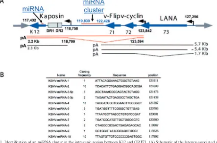
A total of 52 sequences representing 11 unique RNA species between 19 and 24 bases in length matched with 100% comple-mentarity to KSHV. Surprisingly, all sequences aligned with a single region of the KSHV genome: the latency-associated locus (11, 27). During latency, KSHV gene expression is re-stricted to a few viral genes, most of which are located within this region (Fig. 1). These polycistronic latency-associated transcripts encode the latency-associated nuclear antigen (LANA/open reading frame 73 [ORF73]), v-cyclin (ORF72), and v-Flip (ORF71). The kaposin gene is located approxi-mately 4 kbp downstream and encodes at least three proteins that are expressed from alternate start codons upstream of direct repeat 1 (DR1) and DR2 (Fig. 1) (25). Both loci are highly expressed in KS tumors and PEL-derived cell lines and play an important role in regulating viral and cellular gene expression (3, 13, 14, 18, 19, 22, 25). To date, no coding regions or functional elements have been annotated for the 3.6-kbp-long intragenic region between the kaposin gene and ORF71. Strikingly, 10 out of 11 candidate miRNAs are located within this intragenic region, while one is located in the kaposin locus at position nt 117992. The genomic location of this newly identified viral miRNA cluster and their sequences are shown in Fig. 1A and B.
[image:2.585.43.283.82.197.2]miRNAs are believed to be RNA polymerase II transcripts.
TABLE 1. Distribution of cloned small RNA moleculesa
Type
Distribution of cloned small RNA molecules (%)
BCBL-1 (273 sequences)
TPA-induced BCBL-1 (216 sequences)
TIVE-LTC (145 sequences)
rRNA 57.51 47.69 47.59
tRNA 0.37 0.93 0.00
Human miRNA 13.19 13.89 21.38
Other snRNA 4.03 4.63 2.76
mRNA 6.59 4.63 5.52
Viral miRNA 8.42 13.43 0.00
Genomic loci 4.03 7.41 8.28
Not matched 5.86 7.41 14.48
a
A total of 450 clones were analyzed by restriction digestion with 260 clones sequenced resulting in 634 sequences. Genomic loci represent RNA sequences that match annotated loci in the human genome. “Not matched” sequences are very short highly repetitive sequences.
FIG. 1. Identification of an miRNA cluster in the intragenic region between K12 and ORF71. (A) Schematic of the latency-associated region of KSHV. All nucleotide annotations are based on the BC-1 KSHV sequence (24). ORFs, transcripts, DR1 and DR2, and promoters are drawn as described in references 11, 17, 25, and 27. The miRNA cluster, as well as the proposed kaposin transcripts by Li et al. (17), are shown in orange. (B) Sequences of cloned candidate miRNA species, nucleotide position, and cloning frequency are indicated. Preliminary names of candidate miRNAs as submitted to GenBank are given (accession number AY973824).
9302 NOTES J. VIROL.
on November 8, 2019 by guest
http://jvi.asm.org/
[image:2.585.71.514.385.677.2]FIG. 2. Isolated miRNAs fold into hairpin structures of appropriate length. (A) Secondary structure predictions of candidate KSHV miRNA sequences using mFold (28). Shown are only structures with the lowest free energy. (B) Northern blot analysis for the expression of KSHV miRNAs 3-3p, 4, and 10 and the human miRNA hsa-miR-16 as a positive control. Thirty micrograms of total RNA from BCBL-1, TPA-induced BCBL-1, TIVE-LTC, and latently infected SLK cells (SLK KS⫹) was separated on 15% PAGE, blotted to nylon membranes, probed with an end-labeled antisense oligonucleotide, and detected by phosphorimaging as previously described (16).
on November 8, 2019 by guest
http://jvi.asm.org/
While some are expressed from miRNA genes, many
mamma-lian miRNAs are expressed as clusters from introns and 5⬘
UTRs of mRNAs (for review, see reference 10). In our anal-ysis, all isolated candidate KSHV miRNAs are derived from the same strand, suggesting that they are expressed from a single transcript. Within this region, two promoters have been reported. Sadler et al. reported a transcript initiation site at nt 118758 (25), and Li et al. reported on a 2.3-kbp-long cDNA encoding kaposin proteins A and C from a promoter located within ORF73, initiating transcription at nt 123842 (17). The spliced intron (nt 123594 to 118799) has the potential to en-compass the entire KSHV miRNA cluster (Fig. 1). Thus, KSHV encodes a cluster of miRNAs which may be expressed from an intron and encodes one miRNA expressed within an exon (KSHV-miRNA-10) (Fig. 1).
Several miRNA clusters have been reported in animal cells which are expressed from otherwise protein coding mRNA species; likewise, these KSHV miRNAs might be expressed coordinately with the kaposin locus. Interestingly, the expres-sion of kaposin proteins is detectable in latently infected cells but is also induced by TPA (25). Li et al. have also reported TPA responsiveness of the newly described kaposin promoter (17). In congruence with this data, we observed viral miRNA expression in both latent BCBL-1 cells and TPA-induced BCBL-1 cells, which suggests a role for miRNA-dependent gene regulation during both latent and lytic growth of KSHV.
KSHV-encoded candidate miRNAs fold into hairpin struc-tures. In order to annotate miRNAs, evidence for both the expression and the biogenesis of a novel sequence should be provided (2). One strong indicator for biogenesis is the pres-ence of complementary adjacent sequpres-ences that form stable hairpins. miRNAs are excised from precursor RNAs by a mat-uration process that entails cleavage by two cellular RNase III enzymes, namely Drosha and Dicer. These enzymes recognize
specific RNA secondary hairpin structures to process the 5⬘
and 3⬘ends of the mature miRNA (1, 4).
To analyze potential precursor structures for the isolated 11 KSHV candidate miRNAs, each sequence including an area of 60 to 80 bases surrounding the sequence was analyzed by mFold (28), which predicts RNA secondary structure. Figure 2A shows the predicted folding patterns which suggest that all KSHV miRNAs show stable hairpin formation with long paired stems and an appropriate distance between the end of the miRNA and the loop, a feature recognized by Dicer (1, 4). Hence these predictions further support that the cloned se-quences represent miRNAs. To further confirm the expression of KSHV miRNAs, Northern blot analysis using total RNA isolated from BCBL-1, BCBL-1/TPA, TIVE-LTC, and latently infected SLK cells was performed. We detected both the im-mature miRNA precursor and the im-mature miRNA for KSHV miRNAs 3-3p, 4, and 10, while we were unable to detect expression of the remaining miRNAs (Fig. 2B). This further confirms that KSHV-infected cells of both lymphoid and en-dothelial origin express viral miRNAs. In summary, we have identified a microRNA cluster within the latency-associated region of KSHV between nt positions 119839 and 124426 (Fig. 1).
Four criteria for the existence of this newly identified miRNA cluster are addressed by our data. (i) We identified 11 unique miRNA species from 52 clones (Table 1). (ii) Observed
miRNA sequences folded into hairpin structures (Fig. 2A). (iii) All sequences are expressed from a single strand, compat-ible with miRNA maturation but not with small interfering RNA maturation, which often involves the formation of larger double-stranded RNA species generated by bidirectional tran-scription. (iv) Northern blot analysis demonstrates both pre-miRNA and mature pre-miRNA forms in infected lymphoid and endothelial cells. We detected 3 out of the 11 miRNA species, indicating that although these miRNAs may be expressed from a common pre-mRNA, differential processing might explain their relative abundance.
The main and most important question to solve in the future will be to determine the functional consequences of miRNA expression in KSHV-infected cells. Most recent computational predictions have drawn a complicated picture in which each human miRNA can target many different genes. The same is true for the KSHV-encoded miRNA. Utilizing a recently pub-lished algorithm, miRanda, several hundred human and many viral target genes were predicted even under most stringent 95% cutoff conditions (data not shown) (12). Therefore, it will require genetic approaches to decipher whether this fascinat-ing mechanism of posttranscriptional regulation contributes to KSHV latent and/or lytic replication and will ultimately have implications in viral pathogenesis.
This work was supported by start-up funding from the University of Florida Shands Cancer Center. R.R. is supported by grants from the National Institutes of Health (RO1 CA 88763, R21 CA97939).
ADDENDUM IN PROOF
After this work was accepted, the identification of KSHV-encoded miRNAs was independently reported by two other groups (S. Pfeffer, A. Sewer, M. Lagos-Quintana, R. Sheridan, C. Sander, F. A. Grasser, L. F. van Dyk, C. K. Ho, S. Shuman, M. Chien, J. J. Russo, J. Ju, G. Randall, B. D. Lindenbach, C. M. Rice, V. Simon, D. D. Ho, M. Zavolan, and T. Tuschl, Nat.
Methods2:269-276, 2005; and X. Cai, S. Lu, Z. Zhang, C. M.
Gonzalez, B. Damania, and B. R. Cullen, Proc. Natl. Acad. Sci.
USA102:5570-5575, 2005).
REFERENCES
1.Ambros, V.2004. The functions of animal microRNAs. Nature431:350–355. 2.Ambros, V., B. Bartel, D. P. Bartel, C. B. Burge, J. C. Carrington, X. Chen, G. Dreyfuss, S. R. Eddy, S. Griffiths-Jones, M. Marshall, M. Matzke, G. Ruvkun, and T. Tuschl.2003. A uniform system for microRNA annotation. RNA9:277–279.
3.An, F. Q., N. Compitello, E. Horwitz, M. Sramkoski, E. S. Knudsen, and R. Renne.2005. The latency-associated nuclear antigen of Kaposi’s sarcoma-associated herpesvirus modulates cellular gene expression and protects lym-phoid cells from p16 INK4A-induced cell cycle arrest. J. Biol. Chem.280:
3862–3874.
4.Bartel, D. P.2004. MicroRNAs: genomics, biogenesis, mechanism, and func-tion. Cell116:281–297.
5.Bennasser, Y., S. Y. Le, M. L. Yeung, and K. T. Jeang.2004. HIV-1 encoded candidate micro-RNAs and their cellular targets. Retrovirology1:43. 6.Brennecke, J., D. R. Hipfner, A. Stark, R. B. Russell, and S. M. Cohen.2003.
bantam encodes a developmentally regulated microRNA that controls cell proliferation and regulates the proapoptotic gene hid in Drosophila. Cell
113:25–36.
7.Cesarman, E., Y. Chang, P. S. Moore, J. W. Said, and D. M. Knowles.1995. Kaposi’s sarcoma-associated herpesvirus-like DNA sequences in AIDS-re-lated body-cavity-based lymphomas. N. Engl. J. Med.332:1186–1191. 8.Chang, Y., E. Cesarman, M. S. Pessin, F. Lee, J. Culpepper, D. M. Knowles,
and P. S. Moore.1994. Identification of herpesvirus-like DNA sequences in AIDS-associated Kaposi’s sarcoma. Science266:1865–1869.
9.Chen, C. Z., L. Li, H. F. Lodish, and D. P. Bartel.2004. MicroRNAs modulate hematopoietic lineage differentiation. Science303:83–86.
9304 NOTES J. VIROL.
on November 8, 2019 by guest
http://jvi.asm.org/
10.Cullen, B. R.2004. Transcription and processing of human microRNA pre-cursors. Mol. Cell16:861–865.
11.Dittmer, D., M. Lagunoff, R. Renne, K. Staskus, A. Haase, and D. Ganem.
1998. A cluster of latently expressed genes in Kaposi’s sarcoma-associated herpesvirus. J. Virol.72:8309–8315.
12.Enright, A. J., B. John, U. Gaul, T. Tuschl, C. Sander, and D. S. Marks.
2003. MicroRNA targets in Drosophila. Genome Biol.5:R1.
13.Friborg, J., Jr., W. Kong, M. O. Hottiger, and G. J. Nabel.1999. p53 inhibition by the LANA protein of KSHV protects against cell death. Nature
402:889–894.
14.Fujimuro, M., and S. D. Hayward.2003. The latency-associated nuclear antigen of Kaposi’s sarcoma-associated herpesvirus manipulates the activity of glycogen synthase kinase-3. J. Virol.77:8019–8030.
15.Griffiths-Jones, S.2004. The microRNA Registry. Nucleic Acids Res.32:
D109–D111.
16.Lagos-Quintana, E. A.2001. Current protocols in molecular biology. Greene Publishing Associates, J. Wiley, New York, N.Y.
17.Li, H., T. Komatsu, B. J. Dezube, and K. M. Kaye.2002. The Kaposi’s sarcoma-associated herpesvirus K12 transcript from a primary effusion lym-phoma contains complex repeat elements, is spliced, and initiates from a novel promoter. J. Virol.76:11880–11888.
18.McCormick, C., and D. Ganem.2005. The kaposin B protein of KSHV activates the p38/MK2 pathway and stabilizes cytokine mRNAs. Science
307:739–741.
19.Moore, P. S., C. Boshoff, R. A. Weiss, and Y. Chang. 1996. Molecular mimicry of human cytokine and cytokine response pathway genes by KSHV. Science274:1739–1744.
20.Neipel, F., J. C. Albrecht, and B. Fleckenstein.1997. Cell-homologous genes in the Kaposi’s sarcoma-associated rhadinovirus human herpesvirus 8: de-terminants of its pathogenicity? J. Virol.71:4187–4192.
21.Pfeffer, S., M. Zavolan, F. A. Grasser, M. Chien, J. J. Russo, J. Ju, B. John, A. J. Enright, D. Marks, C. Sander, and T. Tuschl.2004. Identification of virus-encoded microRNAs. Science304:734–736.
22.Radkov, S. A., P. Kellam, and C. Boshoff.2000. The latent nuclear antigen of Kaposi sarcoma-associated herpesvirus targets the retinoblastoma-E2F path-way and with the oncogene Hras transforms primary rat cells. Nat. Med.
6:1121–1127.
23.Renne, R., W. Zhong, B. Herndier, M. McGrath, N. Abbey, D. Kedes, and D. Ganem.1996. Lytic growth of Kaposi’s sarcoma-associated herpesvirus (hu-man herpesvirus 8) in culture. Nat. Med.2:342–346.
24.Russo, J. J., R. A. Bohenzky, M. C. Chien, J. Chen, M. Yan, D. Maddalena, J. P. Parry, D. Peruzzi, I. S. Edelman, Y. Chang, and P. S. Moore.1996. Nucleotide sequence of the Kaposi sarcoma-associated herpesvirus (HHV8). Proc. Natl. Acad. Sci. USA93:14862–14867.
25.Sadler, R., L. Wu, B. Forghani, R. Renne, W. Zhong, B. Herndier, and D. Ganem.1999. A complex translational program generates multiple novel proteins from the latently expressed kaposin (K12) locus of Kaposi’s sarco-ma-associated herpesvirus. J. Virol.73:5722–5730.
26.Soulier, J., L. Grollet, E. Oksenhendler, P. Cacoub, D. Cazals-Hatem, P. Babinet, M. F. d’Agay, J. P. Clauvel, M. Raphael, L. Degos et al.1995. Kaposi’s sarcoma-associated herpesvirus-like DNA sequences in multicen-tric Castleman’s disease. Blood86:1276–1280.
27.Talbot, S. J., R. A. Weiss, P. Kellam, and C. Boshoff.1999. Transcriptional analysis of human herpesvirus-8 open reading frames 71, 72, 73, K14, and 74 in a primary effusion lymphoma cell line. Virology257:84–94.
28.Zuker, M., D. H. Mathews, and D. H. Turner.1999. Algorithms and ther-modynamics for RNA secondary structure prediction: a practical guide in RNA biochemistry and biotechnology. Kluwer Academic Publishers, Dor-drecht, The Netherlands.