0022-538X/05/$08.00⫹0 doi:10.1128/JVI.79.13.8182–8188.2005
Copyright © 2005, American Society for Microbiology. All Rights Reserved.
Development of a Novel System To Study Hepatitis Delta Virus
Genome Replication
Jinhong Chang, Severin O. Gudima, Chi Tarn, Xingcao Nie, and John M. Taylor*
Fox Chase Cancer Center, 333 Cottman Avenue, Philadelphia, Pennsylvania 19111-2497
Received 19 November 2004/Accepted 28 February 2005
Hepatitis delta virus (HDV) genome replication requires the virus-encoded small delta protein (␦Ag).
During replication, nucleotide sequence changes accumulate on the HDV RNA, leading to the translation of
␦Ag species that are nonfunctional or even inhibitory. A replication system was devised where all␦Ag was
conditionally provided from a separate and unchanging source. A line of human embryonic kidney cells was
stably transfected with a single copy of cDNA encoding small␦Ag, with expression under tetracycline (TET)
control. Next, HDV genome replication was initiated in these cells by transfection with a mutated RNA unable
to express␦Ag. Thus, replication of this RNA was under control of the TET-inducible␦Ag. In the absence of
TET, there was sufficient␦Ag to allow a low level of HDV replication that could be maintained for at least 1
year. When TET was added, both␦Ag and genomic RNA increased dramatically within 2 days. With clones of
such cells, designated 293-HDV, the burst of HDV RNA replication interfered with cell cycling. Within 2 days,
there was a fivefold enhancement of G1/G0cells relative to both S and G2/M cells, and by 6 days, there was
extensive cell detachment and death. These findings and those of other studies that are under way demonstrate the potential applications of this experimental system.
Natural infections with hepatitis delta virus (HDV) are al-ways associated with the presence of the helper virus, be it the natural helper virus, hepatitis B virus, or, in the case of exper-imental infections of woodchucks, woodchuck hepatitis virus (10). The basis for this dependence is that the envelope pro-teins of the hepadnavirus are required for both HDV entry into hepatocytes and the assembly of new HDV particles. Con-sistent with this interpretation, HDV is also able to infect primary cultures of primate and woodchuck hepatocytes. Once inside a susceptible cell, the small 1,679-nucleotide single-stranded circular RNA genome of HDV can achieve genome replication by RNA-directed RNA synthesis using redirection of a host RNA polymerase, probably PolII (25). During this replication, the antigenome, an exact complement of the ge-nome, is produced as well as a smaller 800-nucleotide 5⬘ -capped and 3⬘-polyadenylated RNA that acts as mRNA for the translation of a 195-amino-acid species, the small delta protein,
␦Ag. This␦Ag is absolutely essential for HDV genome repli-cation (5). It may be multifunctional since several different abilities have already been ascribed to it (20).
It is possible to study HDV genome replication in cultured cell lines in the absence of the helper hepadnavirus. For ex-ample, replication is initiated when cells are transfected with a cDNA version of the HDV sequence (5). Transfection with just HDV RNA sequences is usually not successful, since the replication has an early need for the essential␦Ag (11). How-ever, this need can be met if the cells are already expressing
␦Ag or if␦Ag is cotransfected with the RNA (7, 11). In addi-tion, the need can be met by cotransfecting the HDV RNA along with an mRNA for␦Ag (21).
During both infection and transfections, it has been found
that HDV RNAs with numerous nucleotide changes accumu-late (13, 22). In particular, there is a specific change that arises via RNA editing acting at a site corresponding to the amber termination codon for␦Ag (19). This editing is carried out by ADAR-1, a member of a family ofadenosinedeaminases that act onRNA (2, 27). The specific change on HDV allows the translation of a longer form of␦Ag. This large␦Ag is known to have two functions. First, it acts as a dominant negative inhib-itor of genome replication as supported by the small␦Ag (5). Second, this large␦Ag has the unique ability, in the presence of the hepadnavirus envelope proteins, to drive the assembly of HDV genome RNA and to produce new virus particles (3). In addition to this specific editing change, there are additional changes that accumulate during replication to produce altered
␦Ag species that are not supportive of genome replication and thus contribute to a general decline in replication (1, 13, 22, 25). Therefore, the aim of the present study was to develop a system in which HDV genome replication could be studied under conditions where the small ␦Ag was provided solely from an inducible source. Here, we describe the development of this novel system and some of its properties. It offers unique opportunities for studying important aspects of HDV genome replication.
MATERIALS AND METHODS
Plasmid.Beginning with a sequenced HDV cDNA (15), the ScaI-XbaI frag-ment containing the ␦Ag open reading frame was inserted into pcDNA5/ FRT/TO (Invitrogen). This vector has a hybrid human cytomegalovirus/TetO2 promoter for tetracycline-regulated gene expression. It also has an Flp recom-bination target (FRT) site for Flp recombinase-mediated integration.
Generation of 293-␦Ag, a cell line expressing the small form of␦Ag under TET-on control.Human embryonic kidney cells, Flp-In T-Rex (Invitrogen), were used to generate a␦Ag expression cell line. The genome of this cell line has been stably transfected with two plasmids, one containing the FRT site and the other expressing a TET repressor. Transfection of this cell line was performed with Lipofectamine 2000 (Invitrogen) according to the manufacturer’s instructions. The plasmid containing the HDV sequence was cotransfected with pOG44,
* Corresponding author. Mailing address: Fox Chase Cancer Cen-ter, 333 Cottman Avenue, Philadelphia, PA 19111-2497. Phone: (215) 728-2436. Fax: (215)728-3105. E-mail: [email protected].
8182
on November 8, 2019 by guest
http://jvi.asm.org/
which expresses Flp recombinase. This cotransfection led to the integration of HDV sequence through the FRT site with its expression under TET-on control. Two days after transfection, cells were reseeded at less than 25% confluence. Two to 3 h later, selection antibiotics were added to the medium. Selection antibiotics, hygromycin and blasticidin (Invitrogen), were used to maintain both the␦Ag and TET repressor genes. Two weeks later, separate foci appeared, and the pool of such cells was expanded to generate what we refer to as 293-␦Ag cells. After TET induction, immunoblot and immunocytochemistry were used to con-firm the expression of␦Ag in 100% of these cells. 293-␦Ag cells were maintained in Dulbecco’s modified Eagle’s medium with high glucose and 10% fetal bovine serum along with the selection antibiotics mentioned above.
Transfection of 293-␦Ag cells with HDV RNA sequences to initiate HDV genome replication.To initiate HDV replication, these 293-␦Ag cells under Tet⫺ conditions were transfected with a greater-than-full-length HDV RNA species. This RNA had a 2-nucleotide deletion at the unique EcoRI site, a change that interferes with the translation of a functional␦Ag (5). Using strategies described in the legend of Fig. 3, we consider that this transfection led to the replication and accumulation of HDV RNA in about 15% of cells. As one approach to achieve replication in more of the cells, total RNA was extracted from some of the cells and used to retransfect the culture. This cycle of retransfection was repeated as many as four times. The fraction of transfected cells was thus raised to⬇40%. As an additional approach, we carried out cell cloning under Tet⫺ conditions. We thus obtained clones, designated 293-HDV, with HDV replica-tion occurring in all cells.
Quantitation of␦Ag and genomic RNA.Tri Reagent (Molecular Research Center) was used to extract both protein and RNA.␦Ag was measured in protein samples by immunoblot followed by125I-staph A protein (Perkin-Elmer) and detection by Bio-imager (Fuji). Quantitation was measured relative to a standard of purified recombinant␦Ag provided by Harmon Zuccola and James Hogle (Harvard University), as previously described (13). Genomic HDV RNA was detected by Northern analysis using a32P-labeled specific RNA probe and quantitated relative to a standard of in vitro-transcribed HDV genomic RNA, as previously described (13). Detection and quantitation of radioactivity were done by using a Bio-imager (Fuji).
Immunocytochemistry.The procedure was based on a method described pre-viously (1, 14). As primary antibodies, for␦Ag, we used a 1:1,000 dilution of a rabbit polyclonal antibody; for SC35, we used a 1:2,000 dilution of a mouse monoclonal antibody (Abcam); and for fibrillarin, we used a 1:500 dilution of a mouse monoclonal antibody (Abcam). As secondary antibodies, we used a 1:500 dilution of Cy2-conjugated AffiniPure donkey anti-rabbit immunoglobulin G (heavy plus light chains) and rhodamine red-X-conjugated AffiniPure donkey anti-mouse immunoglobulin G (heavy plus light chains) (Jackson ImmunoRe-search). Cellular DNA was stained with DAPI (4⬘,6⬘-diamidino-2-phenylindole) (Sigma). Samples were analyzed using an Inverted Nikon TE2000-U microscope with 40⫻Plan ELWD, WD 3.7-2.7 objective, and specific filter blocks. Images were acquired with a Cascade 650 monochrome camera (Photometrics) and MetaVue software (Universal Imaging). Subsequently, images were processed with Canvas 9.0 and Adobe Photoshop 7.0 software.
Cell cycle and apoptosis analysis.For 293-␦Ag and 293-HDV cells, both with and without TET induction, we used propidium iodide staining and analytical cell sorting with a Guava PCA apparatus (Guava Technologies) to determine the distribution of cells within the cell cycle. Data were analyzed using Flowjo software. Using the same apparatus together with a Nexin kit to stain for annexin V and 7-amino-actinomycin D (7-AAD) (Guava Technologies), we also assayed for various stages of cell death. Trypan blue was used as recommended by the supplier (Gibco).
RESULTS
Conditional expression of ␦Ag in 293-␦Ag cells. The first
step in our strategy was to create a cell line conditionally expressing the small ␦Ag. We used a commercially available engineered form of 293 cells, human embryonic kidney cells. These cells contain a single target site for the recombinaseflp, into which vector sequences can be efficiently inserted. Fur-thermore, these cells express a protein that in the absence of TET will repress transcription from the transposed sequences. In this way, we transferred cDNA for the small␦Ag to create what we refer to as 293-␦Ag cells.
To test whether all cells were expressing␦Ag, we used
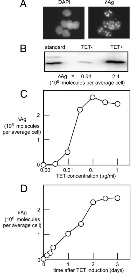
im-munocytochemistry with a␦Ag-specific rabbit polyclonal anti-body. As shown in Fig. 1A, 2 days after TET was added at 1
g/ml, all cells expressed␦Ag, with staining located predomi-nantly in the nucleolus. This distribution was as expected from previous transient transfection studies (1).
[image:2.585.308.530.71.535.2]Next, we used immunoblots to detect and quantitate␦Ag. As shown in Fig. 1B, in the absence of TET, there was a low
FIG. 1. Expression in the 293-␦Ag cell line. (A) Monolayer cultures at 2 days after the addition of TET were examined for staining with DAPI and by immunocytochemistry to detect␦Ag. (B) Immunoblots were used to detect␦Ag in cells before and after TET induction relative to a standard of recombinant ␦Ag. We thus deduced the number of molecules of␦Ag per average cell. (C and D) A similar quantitation was made for cells that were exposed to added TET either for 3 days in the presence of varied concentrations of TET (C) or for varied times but with a single concentration of TET (1g/ml) (D).
on November 8, 2019 by guest
http://jvi.asm.org/
amount of expression, about 40,000 copies per cell. However, after TET addition, the amount increased 60-fold, to 2.4 mil-lion copies. This high number approximates the abundance of host cell ribosomal components and many required translation factors (8).
As further characterization of these cells, Fig. 1C shows that 0.1 to 1g/ml of TET was sufficient to give maximal induction of␦Ag expression. In addition, Fig. 1D shows a time course of the induction for cells treated with 1g/ml of TET. Maximal levels were achieved within 2 days.
Initiation of HDV genome replication in 293-␦Ag cells.The
second step was to transfect 293-␦Ag cells with HDV RNA sequences. We specifically chose an HDV RNA with a 2-nu-cleotide deletion in a region that disrupts expression of␦Ag
(4). That is, we wanted the integrated DNA copy of␦Ag cDNA to be the only and unchangeable source of this protein. At various times after transfection, we analyzed the RNA in these cells for replication and accumulation of HDV genomic RNA. Consider first the situation, when these cells were main-tained in the absence of TET. As shown in Fig. 2, the trans-fected cells achieved a low level of HDV genomic RNA, about 1,000 copies per average cell. As shown, this level was essen-tially stable for at least 1 year.
We next assessed HDV replication when TET was added to the medium. Figure 2 shows examples of when TET was added at 8 and 52 weeks after the initial RNA transfection. In both cases, a burst of HDV RNA accumulation was detected within 3 days. During the following weeks, we then observed
[image:3.585.125.459.68.178.2]signifi-FIG. 2. Accumulation of replicating HDV genomic RNA in the 293-␦Ag cell line after transfection with HDV RNA sequences. To initiate HDV genome replication, cells were transfected with a linear antigenomic HDV RNA that was greater than unit length and contained a 2-nucleotide deletion to disrupt the open reading frame for␦Ag. As indicated, cultures were incubated in the presence (open circles) or absence (filled circles) of TET (1g/ml). Cells were routinely subcultured every 3 to 4 days. After 8 and 52 weeks, some cultures were shifted from the absence to the presence of TET. At the times indicated, total RNA was extracted and examined by Northern analysis to quantitate the amount of HDV genomic RNA per average cell.
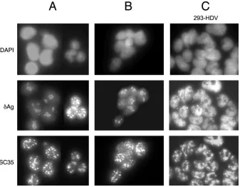
FIG. 3. Immunocytochemistry of 293 cell cultures. The three rows show staining with DAPI, detection of␦Ag, and detection of SC35. Columns A and B represent 293-␦Ag cells that were transfected with HDV RNA maintained under Tet⫺conditions and then TET induced for 2 and 14 days, respectively. Column C shows 293-HDV, a clone of cells that are replicating HDV RNA, as examined at day 2 after TET induction.
on November 8, 2019 by guest
http://jvi.asm.org/
[image:3.585.125.462.435.697.2]cant detachment of cells from the monolayer. Furthermore, for the remaining cells, the amount of HDV RNA per average cell decreased dramatically so that by 2 to 3 weeks, no HDV could be detected. In contrast, for 293-␦Ag cells that had not been transfected with HDV RNA, we did not detect detachment of cells during 3 weeks of TET induction. Our preliminary inter-pretation of these data was that the initial HDV RNA trans-fection efficiency was less than 100%. Furthermore, it was considered that after TET addition, only those transfected cells that were replicating the HDV RNA were the ones that ulti-mately became detached from the monolayer. As now ex-plained, this interpretation was supported by data obtained using immunocytochemistry.
As shown in Fig. 1A, we already knew that the 293-␦Ag cells all expressed ␦Ag and that it was primarily localized to the nucleolus. However, when such cells transfected with the HDV RNA were subsequently induced with TET for 2 days, there were two different patterns of intranuclear distributions, as shown in Fig. 3A. More than half of the cells had the nucleolar
distribution, while about 40% had a dramatic exclusion from the nucleolus together with a nucleoplasmic localization. This nucleoplasmic pattern included but was not limited to speckles, some of which colocalized with those that contain the splicing factor SC35 (16). This distribution is very much like that re-ported for replication of the HDV genome in Huh7 cells, where the staining was nucleoplasmic and included SC35 speckles (1). As shown in Fig. 3B, when we examined cells at 2 weeks after TET addition, the only cells staining for␦Ag had a nuclear pattern that was predominantly nucleolar. These data further support the interpretation that only a fraction of the cells were initially transfected with HDV RNA and that it was these cells that were being detached during the extensive TET induction.
To further establish these interpretations, we subcloned cells that had been RNA transfected and maintained for a long period in the absence of TET. Seventeen such clones were generated and tested by Northern analysis for HDV replica-tion. Only six clones gave extensive HDV RNA accumulation when induced for 2 days with TET (data not shown). The other clones were negative for HDV RNA. Furthermore, when the induced clones were examined by immunocytochemistry, it was only with the HDV RNA-positive clones that we detected a pattern of ␦Ag in the nucleoplasm and excluded from the nucleolus. Data for such a clone, designated 293-HDV, are shown in Fig. 3C. Note that all the cells had this nucleoplasmic distribution of␦Ag after a brief TET induction. (Without the induction, the distribution was the same, except that the signal was less intense [data not shown].)
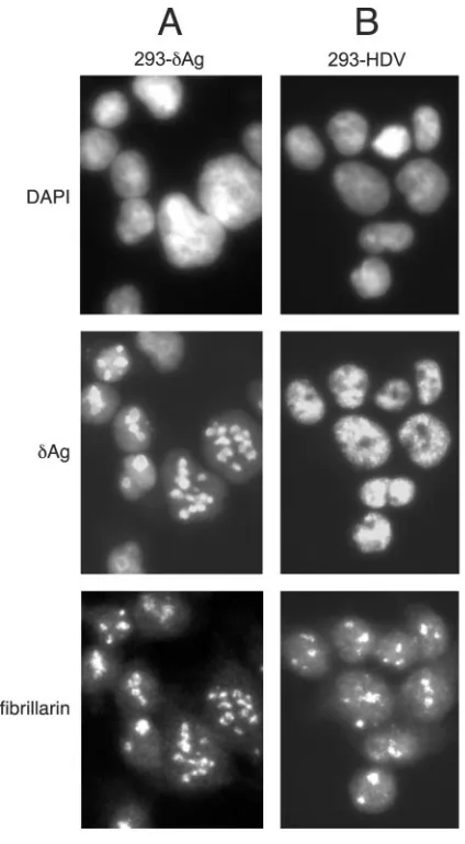
An additional immunostaining experiment was used to com-pare the original 293-␦Ag cells with the clone 293-HDV inter-preted as replicating the HDV. As shown in Fig. 4A, at 1 day after TET induction, all the nuclear␦Ag staining colocalized with that for fibrillarin, a marker for the nucleoli. In contrast, for 293-HDV cells, the ␦Ag staining was nucleoplasmic but excluded from the sites of fibrillarin staining (Fig. 4B).
[image:4.585.55.267.67.452.2]As a further characterization, we examined two of the 293-HDV clones for the accumulation of 293-HDV RNA after TET induction. Figure 5 shows the average HDV RNA values for these two clones as a function of the time after TET induction, with the error bars representing the range of values. In other
[image:4.585.307.537.70.189.2]FIG. 4. Immunocytochemistry of 293-␦Ag and 293-HDV cells. The three rows show staining with DAPI, detection of␦Ag, and detection of fibrillarin. Columns A and B show 293-␦Ag and 293-HDV cells, respectively, at day 1 after TET induction.
FIG. 5. Prompt accumulation of replicating HDV genomic RNA after TET induction of 293-HDV cells. Northern analyses were used to quantitate the average number of genomic RNA molecules accumu-lated per average adherent cell. The data are the averages for two different clones, with the error bars representing the range of the averaged values.
on November 8, 2019 by guest
http://jvi.asm.org/
words, both clones were essentially the same in terms of HDV RNA accumulation. The HDV RNA levels reached 4.3⫻104
copies per average adherent cell, a value about twice that achieved prior to subcloning (Fig. 2). Also, the accumulation
was prompt, reaching half-maximal values in less than 1 day. If we compare these data with the time course of accumulation of
␦Ag during TET induction as presented in Fig. 1D, the results are very similar; that is, the levels of ␦Ag and HDV RNA increase in parallel during TET induction.
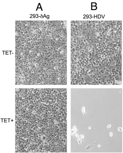
Next, we examined the consequences of TET induction on the viability of 293-HDV cells. As mentioned earlier, prior to subcloning, we did observe detachment of some cells following several days of TET induction (Fig. 2 and 3). Furthermore, we interpreted the 293-HDV cells but not the 293-␦Ag cells as being susceptible to such detachment. We confirmed this in-terpretation by observation of the cloned 293-HDV cells rela-tive to 293-␦Ag at day 6 of culture with or without TET induc-tion. As shown in Fig. 6, extensive detachment occurred for the TET-induced HDV cells (Fig. 6B). In contrast, the
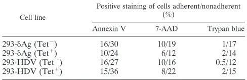
293-␦Ag cells showed no detachment (Fig. 6D). There was no detachment in the absence of TET induction (Fig. 6A and C). Finally, we used several procedures in parallel to better compare the effects of TET induction on both 293-␦Ag and 293-HDV cells. Cultures were studied at days 1 to 4, as well as at day 6, when TET induction led to extensive cell detachment of 293-HDV cells. We counted adherent and nonadherent cells. Trypan blue staining was used to detect dead cells. Stain-ing for annexin V and 7-AAD followed by cell sortStain-ing was used to detect stages of apoptosis. Finally, propidium iodide staining followed by cell sorting was used to detect stages of the cell cycle.
Overall, TET induction of 293-␦Ag cells had no effect de-tectable by these assays. However, for 293-HDV cells, TET induction began the following series of events. (i) Even by day 1, we could detect a reduction in the S- and G2/M-phase cells
relative to G1/G0by cell cycle analysis. (ii) By day 2, these cell
cycle effects were dramatic, as shown in Fig. 7, with a fivefold
[image:5.585.59.266.66.323.2]FIG. 6. Comparison of the effect of TET induction of 293-HDV relative to that of 293-␦Ag. The two cell clones were seeded and then cultured for 6 days in the absence or presence of TET, as indicated. Charge-coupled-device images were taken using regular light micros-copy with a⫻20 objective.
FIG. 7. Cell cycle analyses of 293-␦Ag and 293-HDV cells. Data were obtained using propidium iodide staining followed by analytical cell sorting. This allowed separation of cells by DNA content as G1/G0, S, and G2/M, as indicated. Columns A and B refer to 293-␦Ag and 293-HDV
cells, respectively, with data shown for cells both without and with 2 days of TET induction.
on November 8, 2019 by guest
http://jvi.asm.org/
reduction in S- and G2/M-phase cells relative to G1/G0. There
was also a significant increase in cell debris. (iii) By day 3, the cell cycle blockage continued, and in addition, we could detect a 60% decrease in the total cell number relative to that of uninduced 293-HDV cultures. Also, about 6% of the total cells were no longer adherent. (iv) By day 4, the proportion of nonadherent cells increased to about 20%. This was at least 20 times more than that for uninduced 293-HDV cultures. (v) By day 6, virtually 100% of the cells were no longer adherent.
The adherent and nonadherent cells were also examined by staining for annexin V, 7-AAD, and trypan blue. Typical data for day 4 are summarized in Table 1. The data show that for all four culture conditions, the adherent cells were not signifi-cantly different in terms of these viability markers. Similarly, the nonadherent cells were also not significantly different. Thus, by these particular viability assays, the only specific dif-ference for the TET-induced 293-HDV culture was in the dramatic increase in the fraction of cells that were nonadher-ent.
In summary, from these studies, we conclude that HDV genome replication in TET-induced 293-HDV cells was spe-cifically interfering with cell growth, cell cycling, and finally, cell adherence. It was secondarily leading to cell death, some but not necessarily all of which was via apoptotic mechanisms; that is, HDV genome replication was indirectly cytotoxic. Oth-ers have previously shown that the loss of cell adhesion can lead to the onset of apoptotic cell death (17).
DISCUSSION
We have described here a novel system in which HDV ge-nome replication was initiated by RNA transfection of a stable cell line, 293-␦Ag, that provided fully functional ␦Ag under TET control. Following subcloning of these transfected cells, we were able to obtain populations of cells, designated 293-HDV, all of which were replicating the HDV RNA. An im-portant concept of this system is that the replicating HDV RNA has a mutation so that it does not make any form of␦Ag. That is, this essential protein is provided solely from an inte-grated single-copy gene. Since the expression from this gene is under TET control, the replication of the HDV RNA is as well. It is important that even in the absence of TET, there is a low level of␦Ag expression and a corresponding low level of HDV RNA. This offers the advantage that HDV replication can be maintained in culture for at least 1 year. However, after the addition of TET, the levels of HDV RNA increase
dramati-cally and almost in parallel with the increase of␦Ag (Fig. 1D and 5).
The persistence of replication for 1 year in the absence of TET allows us to conclude that such low levels of HDV rep-lication are not significantly cytotoxic. This “chronic” replica-tion is similar to the long-term replicareplica-tion of viroids in infected plants. In our system, just as for viroid replication, the repli-cating RNA encodes no protein and is totally dependent upon proteins provided by the host cell.
In contrast to the chronic HDV replication, induction of HDV replication with TET soon leads to the loss of cells efficiently replicating the HDV genome (Fig. 2). We show that this loss is associated with significant levels of detachment of cells from the monolayer (Fig. 2 and 6). In addition, from a cell cycle analysis at earlier times, we observed that most of the cells move out of S and G2/M into G1/G0phase. Such effects
are detectable at day 1 and dramatic by days 2 and 3 (Fig. 7 and data not shown). This cell cycle arrest leading to cell detach-ment and death is not dictated by the levels of the␦Ag, since 293-␦Ag cells show no such phenomenon.
For some time, it has been a controversial issue as to whether␦Ag alone or in conjunction with HDV genome rep-lication causes cytotoxic effects in cultured cells (as reviewed in references 12 and 18). The observations of a cytopathic effect associated with HDV replication induced in 293-HDV cells reported here could explain a previous report where transient expression of HDV replication was shown to interfere with cell colony-forming ability (26). More importantly, our observa-tions might provide an explanation of what happens in the liver during acute HDV infections of humans and experimental animals. It is known that during such acute infections, there are real cytopathic effects, ones that are not immunity mediated (12). In fact, as many as 20% of acute HDV infections of humans are associated with fulminant hepatitis and death (10, 24). While there could be multiple factors contributing to this, the death of 293-HDV cells is not due to the expression of␦Ag per se but is a consequence, direct or indirect, of extensive RNA-directed HDV RNA replication.
We consider that this new model has some important addi-tional advantages for the study of HDV replication. (i) Repli-cation no longer needs be initiated by a potentially disruptive procedure such as transfection or electroporation of DNA or RNA species but simply by the addition of TET. (ii) At the time of TET induction, the HDV RNA templates are predom-inantly unit length and circular (data not shown), and this is no doubt a major reason why the response of new HDV synthesis and accumulation can be so fast (Fig. 5). (iii) The levels of HDV genome replication and accumulation are extensive (4.3
⫻ 104 copies per cell) and almost the same as those
deter-mined for the liver during an acute infection (⬇6⫻104copies
per average cell) (6, 13).
This new model has already opened up for us the following three new areas of HDV research. (i) We have shown that in the absence of TET, HDV genome replication can be main-tained for long periods of time (Fig. 2). It is essentially a form of chronic replication. It is worth noting that under these experimental conditions, the HDV genome is replicating in a manner analogous to the replication of viroids, which are min-imal noncoding RNAs demonstrating autonomous replication in plants (9). In other words, for both our system and
viroid-TABLE 1. Viability assays of adherent and nonadherent cellsa
Cell line
Positive staining of cells adherent/nonadherent (%)
Annexin V 7-AAD Trypan blue
293-␦Ag (Tet⫺) 16/30 10/19 1/17
293-␦Ag (Tet⫹) 10/24 6/12 2/14
293-HDV (Tet⫺) 16/27 10/16 0.5/12
293-HDV (Tet⫹) 15/36 8/22 2/15
a
Monolayer cultures of 293-␦Ag and 293-HDV cells were grown for 4 days in the absence or presence of TET, as indicated. The adherent and nonadherent cells were then assayed for the percentage of cells which stained positive for annexin V, 7-AAD, or trypan blue.
on November 8, 2019 by guest
http://jvi.asm.org/
[image:6.585.41.285.82.161.2]infected plants, the replication becomes chronic. We have al-ready begun studies of the nucleotide changes that accumulate during extended HDV replication. We expect that a detailed examination of the time-dependent evolution of the HDV ge-nome sequence will provide new clues as to what are essential sequences and structures.
(ii) Another application of our novel system is suggested by the rapid and extensive burst of HDV RNA-directed RNA transcription that occurs when TET is added (Fig. 2 and 5). In part, this speed is because the cells have not needed to suffer deleterious associated effects typical of other experimental sys-tems where transfection is used to deliver RNA templates. Also, the HDV RNA templates already present in the chronic cells are “ready to go,” that is, unit-length circular RNAs (data not shown). In addition, there is only one form of␦Ag, the one essential for genome replication, and there are no variant forms of␦Ag, including large␦Ag, that might redirect some of the HDV ribonucleoproteins away from the replication path-way. Our system therefore offers unique advantages for study-ing the mechanism of HDV replication. Currently, we are using immunoaffinity procedures to isolate and characterize the HDV transcription complexes from such cells. We should be able to identify the host polymerase(s) and other compo-nents present in such complexes and maybe use such pre-formed complexes to characterize HDV RNA transcription in vitro.
(iii) The rapid and extensive burst of HDV RNA-directed RNA synthesis and accumulation that follows TET induction also allows us to test the action of antiviral agents. We can use periods of treatment so short that overall drug toxicity can be reduced or avoided. We are currently testing nucleoside inhib-itors such as ribavirin.
In summary, we have described here a novel system for studying HDV genome replication, presented some results to support its value, and described examples as to how it will be used in the near future.
ACKNOWLEDGMENTS
This work was supported by grants AI-26522 and CA-06927 from the National Institutes of Health and by an appropriation from the Com-monwealth of Pennsylvania.
We thank Harmon Zuccola and James Hogle for purified delta protein. For the imaging, we thank Irina Shchaveleva and the Fox Chase Imaging Facility (Sandra Jablonski, Manager). For the cell sorting, we thank Maureen Murphy and the Flow Cytometry and Cell Sorting Facility (Richard Hardy, Manager). Constructive comments on the manuscript were given by Glenn Rall, Ju-Tao Guo, Richard Katz, and William Mason.
REFERENCES
1.Bichko, V. V., and J. M. Taylor.1996. Redistribution of the delta antigens in cells replicating the genome of hepatitis delta virus. J. Virol.70:8064–8070. 2.Casey, J. L., and J. L. Gerin.1995. Hepatitis D virus RNA editing: specific modification of adenosine in the antigenomic RNA. J. Virol.69:7593–7700.
3.Chang, F. L., P. J. Chen, S. J. Tu, M. N. Chiu, C. J. Wang, and D. S. Chen. 1991. The large form of hepatitis␦antigen is crucial for the assembly of hepatitis␦virus. Proc. Natl. Acad. Sci. USA88:8490–8494.
4.Chao, M., S.-Y. Hsieh, and J. Taylor.1991. The antigen of hepatitis delta virus: examination of in vitro RNA-binding specificity. J. Virol.65:4057– 4062.
5.Chao, M., S.-Y. Hsieh, and J. Taylor.1990. Role of two forms of the hepatitis delta virus antigen: evidence for a mechanism of self-limiting genome rep-lication. J. Virol.64:5066–5069.
6.Chen, P.-J., G. Kalpana, J. Goldberg, W. Mason, B. Werner, J. Gerin, and J. Taylor.1986. Structure and replication of the genome of hepatitis␦virus. Proc. Natl. Acad. Sci. USA83:8774–8778.
7.Dingle, K., V. Bichko, H. Zuccola, J. Hogle, and J. Taylor.1998. Initiation of hepatitis delta virus genome replication. J. Virol.72:4783–4788.
8.Duncan, R., and J. W. Hershey.1983. Identification and quantitation of levels of protein synthesis initiation factors in crude HeLa cell lysates by two-dimensional polyacrylamide gel electrophoresis. J. Biol. Chem.258: 7228–7235.
9.Flores, R., S. Delgado, M. E. Gas, A. Carbonell, D. Molina, S. Gago, and M. De la Pena.2004. Viroids: the minimal non-coding RNAs with autonomous replication. FEBS Lett.567:42–48.
10.Gerin, J. L., J. L. Casey, and R. H. Purcell.2001. Hepatitis delta virus, p. 3037–3050.InD. M. Knipe, P. M. Howley, et al. (ed.), Fields virology, 4th ed., vol. 2. Lippincott Williams & Wilkins, Philadelphia, Pa.
11.Glenn, J. S., J. M. Taylor, and J. M. White.1990. In vitro-synthesized hepatitis delta virus RNA initiates genome replication in cultured cells. J. Virol.64:3104–3107.
12.Gowans, E. J., and F. Bonino.1993. Hepatitis delta virus pathogenicity. Prog. Clin. Biol. Res.382:125–130.
13.Gudima, S. O., J. Chang, G. Moraleda, A. Azvolinsky, and J. Taylor.2002. Parameters of human hepatitis delta virus replication: the quantity, quality, and intracellular distribution of viral proteins and RNA. J. Virol.76:3709– 3719.
14.Gudima, S. O., J. Chang, and J. M. Taylor.2005. Reconstitution in cultured cells of replicating HDV RNA from pairs of less than full-length RNAs. RNA11:90–98.
15.Kuo, M. Y.-P., J. Goldberg, L. Coates, W. Mason, J. Gerin, and J. Taylor. 1988. Molecular cloning of hepatitis delta virus RNA from an infected woodchuck liver: sequence, structure, and applications. J. Virol.62:1855– 1861.
16.Lamond, A. I., and D. L. Spector.2003. Nuclear speckles: a model for nuclear organelles. Nat. Rev. Mol. Cell Biol.4:605–612.
17.Law, S. F., G. M. O’Neill, S. J. Fashena, M. B. Einarson, and E. A. Golemis. 2000. The docking protein HEF1 is an apoptotic mediator at focal adhesion sites. Mol. Cell. Biol.20:5184–5195.
18.Lazinski, D. W., and J. M. Taylor.1994. Recent developments in hepatitis delta virus research. Adv. Virus Res.43:187–231.
19.Luo, G., M. Chao, S.-Y. Hsieh, C. Sureau, K. Nishikura, and J. Taylor.1990. A specific base transition occurs on replicating hepatitis delta virus RNA. J. Virol.64:1021–1027.
20.Macnaughton, T., and E. Gowans.1995. Hepatitis delta antigen, p. 69–82.In
G. Dinter-Gottlieb (ed.), The unique hepatitis delta virus. R. G. Landes Co., Austin, Tex.
21.Modahl, L. E., and M. M. C. Lai.1998. Transcription of hepatitis delta antigen mRNA continues throughout hepatitis delta virus (HDV) replica-tion: a new model of HDV RNA transcription and regulation. J. Virol. 72:5449–5456.
22.Netter, H. J., T.-T. Wu, M. Bockol, A. Cywinski, W.-S. Ryu, B. C. Tennant, and J. M. Taylor. 1995. Nucleotide sequence stability of the genome of hepatitis delta virus. J. Virol.69:1687–1692.
23.Reference deleted.
24.Rizzetto, M., R. H. Purcell, and J. L. Gerin.1980. Epidemiology of HBV-associated delta agent: geographical distribution of anti-␦and polytransfused HBsAg carriers. Lanceti:1215–1218.
25.Taylor, J. M.2003. Replication of human hepatitis delta virus: recent devel-opments. Trends Microbiol.11:185–190.
26.Wang, D., J. Pearlberg, Y. T. Liu, and D. Ganem.2001. Deleterious effects of hepatitis delta virus replication on host cell proliferation. J. Virol.75: 3600–3604.
27.Wong, S. K., S. Sato, and D. W. Lazinski.2001. Substrate recognition by ADAR1 and ADAR2. RNA7:846–858.