A DISSERTATION ON
COMPARATIVE STUDY OF SUBTROCHANTERIC FRACTURES MANAGED BY PROXIMAL FEMUR NAIL AND
PROXIMAL FEMUR LOCKING PLATE
Dissertation submitted to
THE TAMIL NADU DR.MGR.MEDICAL UNIVERSITY CHENNAI – 600032
In partial fulfilment of the regulations
For the awards of the degree of
M.S. DEGREE BRANCH – II ORTHOPAEDICS
MAY 2018
ACKNLOWDGEMENT
At the outset I thank the Lord, Almighty, for giving me strength to
perform all my duties.
It is indeed a great pleasure to recall the people who have helped me a lot
in completion of my dissertation. Naming all the people who have helped
me in achieving this goal would be impossible, yet I attempt to thank a
few, who have helped me in diverse ways.
First & foremost, I would like to thank Dean, Dr.Kanagaraj M.D.,
Government Mohan Kumaramangalam Medical College Hospital, Salem
for allowing me to use the available clinical resources and materials of
this hospital.
I Acknowledge and express my humble gratitude and sincere thanks to
Prof.Dr.C.KAMALANATHAN M.S.Ortho., DNB Ortho., D.Ortho.,
Professor and HOD, Department of Othopaedics, Government Mohan
Kumaramangalam Medical College Hospital, Salem for his constant
supervision and help for this study.
It gives me immense pleasure to express my deep sense of gratitude and
indebtedness that I feel towards my teacher, inspiration and guide
of Orthopaedics, Government Mohan Kumaramangalam Medical College
Hospital, Salem for his valuable suggestions, guidance, great care,
motivation, constant encouragement and attention to detail that he has so
willingly shown throughout the study which facilitated the completion of
this dissertation.
I owe a great deal of respect and gratitude to Professor and unit
heads Prof.Dr.T.M.MANOHAR M.S Ortho,
Prof.Dr.R.T.PARTHASARATHY, M.S. Ortho for their scholarly suggestions and all around encouragement.
I take this opportunity to express my most humble and sincere
gratitude to Assistant Professors Dr.S.Kumar M.S.Ortho.,
Dr.P.Radhakrishnan M.S.Ortho., Dr.S.Jawahar M.S.Ortho.,
Dr.R.N.Suresh Kumar M.S.Ortho., Dr.T.Karikalan M.S.Ortho.,
Dr.M.Kannan M.S.Ortho., Dr.N.Karthikeyan M.S.Ortho.,
Dr.P.Arunanand M.S.Ortho., Dr.Chinnaudrai D.Ortho.,
Dr.L.Kumar M.S.Ortho., Dr.S.Selvakumar M.S.Ortho.,
Dr.Satish Kumar D.Ortho, DNB Ortho., Dr.T.Senthil Kumar D.Ortho.,
Dr.Syed naser M.S.Ortho., and Dr.P.Sivakumar M.S.Ortho.,
I am also grateful to the Deputy Superintendent and Regional Medical
officer of government Mohan Kumaramangalam Medical College
I also take this opportunity to thank the Department of Anaesthesiology
and Department of radio diagnostics, Operation Theatre Staff nurses, Non
Medical Staff for their continuous support and cooperation.
I would like to make a special mention about
Dr.P.KARTHIKEYAN for his immiscible support for my thesis work. No amount of words can measure up to the deep sense of gratitude and
thankfulness that feel towards my parents and wife for their valuable
help, everlasting support and motivation.
My sincere thanks to all my postgraduate juniors for their
wholehearted support.
From the bottom of my heart I convey my heartful gratitude to all
my parents and their relatives who formed the backbone without whose
CONTENTS
S.No. TITLE Page No
1 INTRODUCTON 1
2 ANATOMY 3
3 BIOMECHANICS 9
4 CLASSIFICATON 14
5 MANAGEMENT 19
6 REVIEW OF LITRATURE 25
7 PROXIMAL FEMUR LCP 29
8 PROXIMAL FEMUR NAIL 33
9 MATERIALS AND METHODS 38
10 OBSERVATION AND RESULTS 51
11 DISCUSSION 85
12 CONCLUSION 99
Appendices
a. Bibliography
b. Proforma
c. Master Chart
LIST OF FIGURES
SL
NO LIST OF PICTURES
PAGE NO
1 SUBTROCHANTERIC REGION 3
2 MUSCULAR ATTACHMENTS OF FEMUR 6
3 DEFORMING FORCES IN SUBTROCHANTERIC
FRACTURES 6
4 ANATOMY OF FEMUR 8
5 STRESS ON SUBTROCHANTERIC AREA 11
6 FIELDING CLASSIFICATION 14
7 RUSSEL TAYLOR CLASSIFICATION 16
8 SEINSHEIMER CLASSIFICATION 17
LIST OF TABLES
SL
NO TABLES
PAGE NO
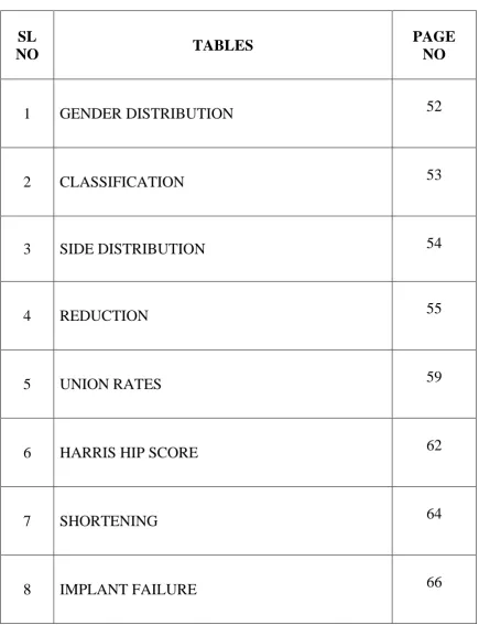
1 GENDER DISTRIBUTION 52
2 CLASSIFICATION 53
3 SIDE DISTRIBUTION 54
4 REDUCTION 55
5 UNION RATES 59
6 HARRIS HIP SCORE 62
7 SHORTENING 64
[image:14.595.102.538.91.659.2]LIST OF CHARTS
SL
NO CHARTS
PAGE NO
1 AGE DISTRIBUTION 51
2 GENDER DISTRIBUTION 51
3 MODE OF INJURY
52
4 RUSSEL TAYLOR CLASSIFICATION 53
5 SIDE DISTRIBUTION 54
6 MODE OF REDUCTION 55
7 BONE GRAFTING 56
8 OPERATING TIME 57
9 BLOOD LOSS 58
10 UNION RATE
58
11 UNION TIME 60
12 FOLLOW UP 60
13 HARRIS HIP SCORE 61
14 VARUS COLLAPSE
62
15 SCREW BREAKAGE 63
16 SHORTENING 63
LIST OF ABBREVIATIONS
1 AO ARBEITSGEMEINSCHAFT FUR
OSTEOSYNTHESEFRAGEN
2 AP ANTEROPOSTERIOR
3 DHS DYNAMIC HIP SCREW
4 DCS DYNAMIC CONDYLAR SCREW
5 ECG ELECTROCARDIOGRAM
6 ECHO ECHOCARDIOGRAPHY
5 HIV HUMAN IMMUNODEFECIENCY VIRUS
6 HCV HEPATITIS C VIRUS
7 HBsAg HEPATITIS B SURFACE ANTIGEN
8 IV INTRAVENOUS
9 IM INTRAMEDULLARY
10 P value PROBABILITY VALUE
11 PFLCP PROXIMAL FEMUR LOCKING
COMPRESSION PLATE
12 PFN PROXIMAL FEMUR NAIL
13 RTA ROAD TRAFFIC ACCIDENT
14 HHS HARRIS HIP SCORE
ABSTRACT
Subtrochanteric fractures are a variant of peritrochanteric fractures
of femur extending 5cm distal to the lesser trochanter.Management of
subtrochanteric fractures is a major challenge and treatment failure is
common for it .
AIM : To compare the functional outcomes of subtrochanteric fractures manged by Proximal femur nail and Proximal femur locking plate.
MATERIALS AND METHODS: This is a prospective study of 20 cases of subtrochanteric fractures admitted in Govt Mohan
Kumarangalam Medical College Hospital, Salem during the period from
December 2015 to September 2017.The cases were classified under
Russel Taylor classification.Out of the 20 cases 10 cases were managed
by Proximal femur nail and 10 cases were managed by Proximal femur
locking plate.
RESULTS: In our study, we observed that there were significant reduction in operating time(p value:0.001) and decrease in blood loss (p
value:0.000) in cases managed by PFN when compared to
PFLCP.Among the cases managed by PFN closed nailing was done in
50% of cases whereas open reduction was required in all cases managed
by PFLCP which was a significant difference (p value:0.033). Among
which went for hypertrophic non union. One case had breakage of the
nail distal to the lag screw and one case had breakage of derotation screw.
Among the cases managed by PFLCP,3 cases went for non union with
implant failure , one among these 3 cases revision surgery was done with
PFN.
CONCLUSION: Even though both PFN and PFLCP are effective in the treatment of subtrochanteric fractures, we observed that PFN was a better
implant than PFLCP, because PFN enables more of a biological fixation
with less disturbance of fracture haematoma, faster than PFLCP and
lesser amount of blood loss.
Keywords:Subtrochanteric fractures,Proximal femur Nail,Proximal femur
AIM OF THE STUDY
The aim of the study is to do a comparative analysis of the functional
outcome of subtrochanteric fractures managed with ‘PROXIMAL
FEMORAL NAIL’and PROXIMAL FEMUR LOCKING
COMPRESSION PLATE at Government Mohan Kumaramangalam
1
INTRODUCTION
Sub-trochanteric fractures have evolved as one of the most important
causes of morbidity and mortality in elderly patients.They account for
approximately 10-30%1 of peritrochanteric fractures.Subtrochanteric
region is area below the inferior border of lesser trochanter extending
distally 5 cm2 to the junction of proximal and middle third of
femur.These fractures have a bimodal distribution 2and are seen in two
main populations, older osteopenic patients following low energy falls
and younger patients with high energy trauma.
3
Early surgical intervention is needed in majority of the patients to avoid
the major complications that can occur due to long term immobilisation
which include deep vein thrombosis, thrombophlebitis, urinary and lung
infections and ulcers. This pattern of fracture is assosciated with higher
rates of maluniuon and non-union than any other femoral fractures
because of the anatomical peculiarity of this area.
A number of modalities of management exists for this pattern of fracture.
However the main modality of treatment can be divided into two groups,
the cephalomedullary hip nails and the lateral plate screw systems.
Fixed nail plate devices were used for the treatment of these fractures
initially. Later sliding hip screw devices became popular in the treatment
2
plates,dynamic condylar screws and cephalomedullary nails. All these
implants had its own advantages and disadvantages.
Traditionally the medial and posteromedial fracture fragments were
considered to be important elements in determining severity of
peritrochanteric hip fractures.4Later GOTFRIED emphasized the
importance of lateral trochanteric wall in stabilizing subtrochanteric
fractures.Locking plates for stabilising subtrochanteric fractures were
developed in 21st century as it can act as a buttress for the lateral
3
ANATOMY
SUBTROCHANTERIC REGION OF THE FEMUR is the region
between lesser trochanter and junction of proximal and middle thirds of
femur. It is defined as a zone extending from the lesser trochanter of the
femur to 5cm distal to the lesser trochanter. This area is subjected to
higher stresses and compressive forces anatomically. Anatomically this
part of the femur is prone for non union and slow healing. Due to the
predominance of cortical bone in this area and decreased vascularity to
the cortical bone,healing capacity is impaired. A large amount of
significant weight transmission occurs to this area even with normal day
to day activities.5About 6 times the body weight of a person is transmitted
to the subtrochanteric area during normal activities of daily life.
4 MUSCULAR ATTACHMENTS
Subtrochanteric region of femur is covered by various muscle groups.
Anteriorly and laterally muscular attachments includes vastus
medialis,vastus intermedius and vastus lateralis. and medially by
adductor brevis and adductor longus . Deforming forces in
subtrochanteric fractures are due to the various muscular attachments to
the proximal and distal fragments.
These include the abductors attached to the greater trochanter. Gluteus
medius orginates from the gluteal surface of illium between the middle
and posterior gluteal lines and gets attached to the greater trochanter.
Gluteus minimus is the other main muscle which gets attached to the
anterior surface of greater trochanter. It arises under the cover of gluteus
medius from the gluteal surface of illium between the middle and inferior
lines. Both these muscles are supplied by superior gluteal nerve and they
abduct the proximal fragment.
Iliopsoas muscle typically causes the proximal fragment to flex.
Illiacus along with the psoas tendon forms a powerful flexor of the hip
joint. Illiacus muscle arises from the iliac fossa it blends alongs with the
rounded psoas tendon ,psoas muscle arises from the lumbar spine and
5
powerful flexor of hip joint.Glutei muscles and illiopsoas will abduct and
flex the proximal fragment in subtrochanteric fractures
Distal fragment because of the unopposed pull from the adductor
magnus, always displaces it medially and further aggravates the
deformity. Adductor magnus is a composite muscle mass formed by the
fusion of adductors along with the hamstring muscles. Hamstrings
orginate from the ischial tuberosity and the fibres vertically downwards
get attached to the adductor tubercle. Adductor group of muscles
constitute the adductor longus and the adductor brevis. Adductor longus
is the most superficial of all the three adductors which arises from a
circular area on the body of pubis by a strong rounded tendon,this tendon
may sometimes get ossified , known as rider’s bone. It gets attached into
the middle third of the linea aspera of femur. Adductor brevis arises from
the body and inferior ramus of the pubic bone and is insertred in a
triangular fashion into the upper part of linea aspera immediately behind
6
7 PARTS OF PROXIMAL FEMUR
GREATER TROCHANTER: It is a type of traction apophysis, a bony quadrangular prominence at the junction of upper part of neck and shaft.
Muscular attachements to the greater trochanter are Gluteus medius,
Gluteus minimus, Pyriformis.
Obturator internus along with Superior and Inferior Gemelli form a
common tendon which gets inserted into the medial aspect of the greater
trochanter. Obturator externus gets inserted into the trochanteric fossa .
PYRIFORMIS FOSSA: It is a depression situsted just medial to the greater trochanter which forms the entry point for intra medullary nailing.
LESSER TROCHANTER: It is a small bony protuberance from the posteromedial aspect of femur.The apex of lesser trochanter has three
borders and provides attachment for Psoas major and illiacus. Psoas
major is inserted into the apex of trochanter and Illiacus is inserted into
8
9
BIOMECHANICS
Forces applied to the hip during ambulation produces stresses in the
proximal femur due to the combined effects of axial, bending and
torsional loads.3Major compressive stresses in the femur are greatest in
the medial cortex 5 -7cm below the lesser trochanter, i.e. the
subtrochanteric region and this region is considered to be one of the
highly stressed areas in the body.4 Tensile stresses of about 25% less
occur at the lateral cortex slightly proximally. Following a
subtrochanteric fracture, deforming muscle forces play a vital role in
causing malunion and produce difficulty in achieving union.Typically the
proximal fragment undergoes flexion, abduction and external rotation due
to the unopposed pull by the glutei muscles which gets attached to the
greater trochanter, the illiopsoas attached to the lesser trochanter will
produce flexion of the proximal fragment. The short external rotators are
responsible for the external rotation of the proximal fragment. Distal
fragment because of the unopposed pull from the adductor magnus,
always displaces it medially and further aggravates the deformity,
hamstring muscles are responsible for shortening of the distal fragment.
In addition comminution of the medial cortex further adds to the injury.
In addition comminution of the medial cortex further adds to the insult of
10
placed devices such as plates and screws as compared to the
centromedullary devices
There are 6,7four main factors affecting the healing process in
subtrochanteric fractures, the first and the foremost important factor is the
high stress in the subtrochanteric region, especially in the posteromedial
cortex.8,9Frankel and Burstein showed that the hip joint reaction forces
reaches almost 3 times the body weight upon muscle contraction. Second
the abundant large amount of cortical bone hinders the healing process
because of the decreased vascularity to the cortical bone when compared
to cancellous bone. Furthermore stripping of the muscles and soft tissue
damage occurring during surgical procedure will further devascularise the
area. Thirdly,7Fromison described the concept of deforming forces in the
subtrochanteric area as a factor for slow healing of subtrochanteric
fractures. Lastly the union can be accelerated by keeping the fractured
bone segments stationary relative to each other and allowing some
11
Cephalomedullary nails are able to provide necessary bending and
torsional stability to combat the displacement of the fracture fragments.
Proximal femoral nail being an intra medullary device is a load sharing
device and has the inherent advantage of shorter lever arm, thereby
decreasing the tensile strain on the implant. The hip screw and the anti
rotational screw proximally provide increased rotational stability of the
head-neck fragment. The two distal locking screws control the rotational
stability of the distal fragment. A biomechanical analysis by10TENCER etal on various implants used for subtrochanteric fracture have found that bending stress, torsional stress, load to axial failure are superior in
cephalomedullary implants than all the other implants. Another
biomechanical evaluation done by11 PAUL R.T. KUZYK etal in 2009,
12
devices were significantly stiffer and had a greater load to failure than the
135 degree and 95 degree constructs, especially with a gap between the
bony fragments. Indirect fracture reduction, preserving the fracture
hematoma, less soft tissue dissection, decreased amount of blood loss add
to the decreased overall morbidity7,12.PAJARINEN etal reported a series
of 103 subtrochanteric fractures treated with PFN and concluded that
those patients treated with PFN could weight bear earlier and take
functional recovery training earlier when compared to other surgical
modalities. It also better prevents the varus collapse of medial cortex of
subtrochanteric region and is highly useful in fractures with medial
comminution.In cases of posteromedial comminution or breakage of the
medial cortex ,extramedullary devices tend to fail and intramedullary
implants form the better option for treatment.
Proximal femur LCP is anatomically pre contoured plate to the
subtrochanteric region,Biomechanical analysis by1 BRETT D CHRIST et
all comparing locking plates with angled blade plates showed on bone
models showed PFLCP has high axial load stiffness,in their study they
concluded that PFLCP with kickstand screw was the stiffest
construct.Precontoured anatomical structure can prevent varus collapse
and malreduction by contouring the tip of the plate with the tip of greater
trochanter.Also the presence of three proximal locking screws into the
13
relationship. The three proximal screws adds to the increased pull out
strength and are most effective in osteoporotic bones. Morover this plate
acts as an internal external fixator and allows some amount of elasticity
across the fracture site which enhances callus formation by secondary
bone healing. Meta analysis by 13Parker and Handoll in their study
concluded that intramedullary nails was not superior to extramedullary
device.3,14The lateral wall is also an important stabilising factor in
subtrochanteric fractures was first reported by Gotfried. It plays a key
role in stabilisation and fixation of subtrochanteric fractures. While using
cephalomedullary hip nails, an intact lateral wall is a must in case of
subtrochanteric fractures.3On reaming fractures with ruptured lateral wall
reaming of proximal femur would cause distraction of the fragments and
peritrochanteric instability, so in cases of ruptured lateral trochanteric
wall, proximal femur locking plates acts as a good alternative method of
fixation. However open reduction of the fractured fragments can cause
increased blood loss, increase in the operating time and devascularisation
14
CLASSIFICATION
Subtrochanteric fractures were initially grouped under comminuted
intertrochanteric factures.15Boyd and Griffin initially considered them as
a variant of intertrochanteric fractures. At least 15 different classification
systems has been devised for subtrochanteric fractures. Out of them most
widely used classification systems are the Russel and Taylor
Classification, Fielding Classification, Seinsheimer and AO
classification.
1. FIELDING AND MAGLIATO Devised a three part anatomical classification in 1966.
TYPE 1: Fracture at the level of lesser trochanter
TYPE 2: Fracture within 1 inch below lesser trochanter
15
2. RUSSEL TAYLOR classification : This classification is based on current techniques and principals of closed intramedullary nailing and
continuity of lesser trochanter and extension of fracture lines into greater
trochanter (or) posteriorly into pyriform fossa. It disregards the degree of
comminution.
TypeI: Fracture does not extend into pyriform fossa.
TypeIA: Comminution and fracture line extend from below lesser trochanter to femoral isthmus
TypeIB: Fracture line and comminution involve area of lesser trochanter to isthmus.
TypeII: Fracture extends into Pyriform fossa.
TypeIIA. No significant comminution (or) fracture of lesser trochanter is seen. TypeIIB.Comminution of medial cortex and loss of continuity
According to the Russel and Taylor Classification,initially for Type I
fractures where the pyriformis fossa is not involved can be treated with
Ist generation intramedullary nails and for Type II fractures
extramedullary implants are used.But with the development of newer
generation nails, this classification system has lost its popularity and
16
3. In 1978 16SEINSHEIMER developed a classification based on
fracture pattern. Significance of this classification is that it identified
fractures with loss of medial cortex stability, which is known to have a
higher rate of implant failure
TYPE I: Undisplaced (or) Less than 2mm displacement
TYPE II: Two Part Fracture.
TYPE IIA. Transverse fracture
17
TYPE IIC. Spiral Fracture with lesser trochanter attached to distal fragment
TYPE III: Three Part Fracture.
TYPE IIIA: Three part spiral fracture with lesser trochanter as a part of third fragment.
TYPE IIIB: Three part spiral fracture with third part a butterfly fragment.
TypeIV: Comminuted fracture with four (or) more fragments
TypeV: Subtrochanteric-Intertrochanteric configuration.
17
Recently a new classification system was proposed by Guyver et al and
18
Type I: Lesser and greater trochanter are preserved.
Type II: Greater trochanter is involved but lesser trochanter is intact.
Type III: Lesser trochanter is involved and is highly unstable.
AO Classification system is now the widely used and accepted universal
classification system.
This classification takes into account the bone,(femur-3),the
location(diaphysis-2),the energy of trauma (A,B,C) and the mechanism
(1,2,or 3).The subtrochanteric fractures are categorised as 1.
Disadvantage of this system is including subtrochanteric fractures under
diaphyseal fractures.
19
MANAGEMENT
At present the management of subtrochanteric fractures in adults is
entirely surgical. Previously many non surgical methods was used. In the
late 1960’s cast bracing was used, traction followed by bracing with hip
spica cast was another modality ,but due to increased morbidity of
prolonged bed rest caused in elderly people non operative treatment
methods have been abandoned.
CONSERVATIVE METHODS: 15Till date there has been no
comprehensive randomised controlled trials comparing the conservative
and operative management.18Parker and colleagues in their study
observed a fixation failure rate of 12% in 103 patients where 93 patients
underwent operative treament
1. SKELETAL TRACTION: In subtrochanteric fractures larger
weights are used in skeletal traction as compared to trochanteric fractures
because of the larger deforming forces.Under radiographic control
adjustments are made in traction until we obtain a satisfactory reduction
in both anteroposterior and lateral views.15Varus or valgus angulation of
less than 5 degrees,at least 25% opposition of fracture fragments in both
20
continued for 8-12 weeks until there is evidence of radiological
union.Patient is then mobilised ,in non weight bearing walking.In a study
conducted, 15Velasco and Comfort found that satisfactory results was
found in only 50% of 32 cases treated conservatively.Perkins traction was
found to be advantageous as it prevents quadriceps atrophy.15,19Wadell
observed that 10 satisfactory and 8 unsatisfactory results and found 90 –
90 traction superior than Thomas splint traction.
2.CAST BRACING: A report on external support by hip spica or cast
brace with a pelvic extension splint has been described.1515
subtrochanteric fractures were managed with a 90-90 traction followed by
traction using bracing with hinged knee single20 hip spica cast
3.PINS IN PLASTERS:15,21In this technique 2 pins are inserted above
the fracture and 2 pins are inserted below the fracture and incorporating
them in plaster.Full weight bearing was allowed.22Seligson and Harman
in 6 patients found problems like shortening, angulation and malrotation
4. EXTERNAL FIXATION:20External fixation was first described for
trochanteric fractures by Anderson,however it was assosciated with
21
modern external fixators full weight bearing is not possible,but it may be
of use in the case of open subtrochanteric fractures.
OPERATIVE MANAGEMENT: Implant used for fixation of
subtrochanteric fractures can be broadly divided into intramedullary and
extramedullary implants.
EXTRAMEDULLARY IMPLANTS:
1. Dynamic Hip Screw: DHS has been widely used in the
management of subtrochanteric fractures.Numerous studies have been
done using DHS as a mode of fixation. 23In a study conducted in King
Saud University of Saudi Arabia,where DHS was used in 24 patients with
comminuted subtrochanteric fractures union was achieved in 19 patient
within a span of 18 weeks.15However we require an anatomic
reduction,stable fixation and reconstruction of medial cortex is important
for a favourable outcome in DHS.20Radford and co-workers reported 64%
good results in 11 patients
2. Dynamic Condylar Screw:24Dynamic condylar screw has been a
favourable implant for subtrochanteric fractures.It exerts a vertical forces
on weight bearing and is a better option as it provides a stronger fixation
in the cancellous bones of head and neck with considerable rotational
22
3. 95 Degrees angled blade plate:It has been the gold standard for the
treatment of subtrochanteric fractures.Many comparative prospective
randomised controlled trials between DCS and angled blade plate has
been conducted.24A research article has recently been published in March
2017 in the International Journal of Research in medical sciences where
DCS and angled blade plate was compared in subtrochanteric fractures
.The study was conducted in Rajindra Hospital Patiala Punjab and in their
study they concludedthat DCS is superior to angled blade plate.The main
limitations of angled blade plate is the extensive lateral approach required
for the plate insertion and devascularisation of fracture fragments due to
extensile exposure.
4. MEDOFF AXIAL COMPRESSION SCREW:It has been recently
used for the management of subtrochateric fractures.25Mainly
recommended for transverse subtrochanteric fractures with or without
reverse obliquity.It is recommened to use uniaxial dynamization in pure
subtrochanteric fractures and we can use staged biaxial dynamization for
intertrochantric fractures with extension into subtrochanteric area.26It is a
highly technically demanding procedure
5.PROXIMAL FEMUR LCP: 27Anatomically pre contored angular
stable plate for the proximal metaphseal region of femur.This plate was
developed as it acts as a stabilising factor for 27,28lateral trochanteric wall
23
fragments.27,29It is an implant of choice for transverse subtrochanteric
fractures with ruptured lateral trochanteric wall
6.DISTAL FEMUR LCP: Opposite side distal femur LCP is a less
invasive locking plate system used recently .30Biomechanically sound
implant its shape fits well with contour of greater trochanter and shaft fits
well with the anterolateral curve of femur.Advantages include
preservation of periosteal blood supply and no need of image intensifier.
INTRAMEDULLARY INPLANTS:
1.INTERLOCKING NAIL: 31Hey-Groves first reported IM nailing of
subtrochanteric fractures .This modality of treatment has been
recommended for simple subtrochanteric fractures where there is no
trochanteric extension.15To date the most effective is Russel Taylor
Reconstruction nail. Various interlocking nail used were:
a) KAMPALA OR HUCKSTEP NAIL
b) KUNTSCHNER NAIL
c) RUSSEL TAYLOR IM NAIL
d) AO FEMORAL NAIL
e) DERBY IM NAIL
f) ZIMMER RECONSTRUCTION NAIL
2.ZICKEL NAIL: One of the first intramedullary implant to give
24
following fracture union is one of the major problems concerned with the
nail and refracture is a recognized complication.
3.ENDERS NAIL: This nail has been mainly used for low energy
fractures with minimal comminution. Usually open reduction and internal
fixation supplemented with cerclage wiring is recommended.31Levy and
colleagues reported a high prevelance of post operative knee pain.
4.GAMMA NAIL: 31This nail has been encountered with many
complications including intra-operative and post-operative femoral shaft
fractures due to three point loading on trochanter and femoral corticres.
5.PROXIMAL FEMUR NAIL: 4Advantages of PFN includes the shorter
lever arm ,load sharing device producing less stress on
implant,introduction without exposing fracture site,transmits weight close
to calcar,distal locking screw provides length and rotational control
permits early weight bearing.It also causes less soft tissue damage and
devascularisation of the fracture fragments.
25
REVIEW OF LITERATURE
Subtrochanteric fractures were treated conservatively in 1902 by
32
HIBBS in a position of flexion, abduction and external rotation by
bringing distal fragment into alignment with the proximal fragment.32In
1960 , Sarimento introduced the concept of femoral cast bracing for the
conservative management of subtrochanteric fractures32. Mooney also did
the same in 1975 and this was regarded as a poor modality of treatment
with respect to varus angulation.33SEINSCHEIMER in 1978 advocated
conservative management of subtrochanteric fractures due to higher rate
of complications assosciated with operative management of
subtrochanteric fractures.3332DE LEE in 1981 reported excellent results
with 90-90 traction followed by hip spica immobilisation and
recommended this for patients with inoperable and open fractures.
Traction as a mode of treatment in subtrochanteric fractures was analysed
by 33WADELL since the deforming forces was well dissipiated in this
modality of treatment.
Operative treatment of subtrochanteric fractures was made as early as in
1910 by 33DELBET with a thick screw with higher pitch that had a better
purchase into the bone.33 In1947 CLEVELAND and in 1951 EVANS
used plate Moore Bount plate ,NEUFLED plate and Lorenzo screw
26
for the treatment of subtrochanteric fractures.BOYD AND GRIFFIN,
KRIK WATSON and CAMPBELL popilarised the use of JEWETT nail
in 1940’s and 1950’s.Due to the high failure rates with Jewett nail as
observed by FELDING and MAGILATO the usage of this nails
decresed.This led to the development of newly designed angled plates
with a “U” profile and fixed angles of 95 degrees and 135 degrees by AO
group.33iIn 1971 Arnoff,in 1972 Distefano in 1974 Cech and Felding
widely used these plates and it led to higher rates of complications like
varus, rotational deformities, non-union, implant failure and medialisation
of distal fragment.34In late 1970’s and in 1978 HANDSON and TULLOS
used the AO blade plates which became a popular device for
subtrochanteric fractures.Sliding hip screw for subtrochanteric fractures
was used by WADELL in 1979.Later in 1992 SCHLEMINGER,
CLAWSON and MASSIE popularised the use of DHS designed by
AO/ASIF group as they noted 32% complications with the AO blade
plate.Zickel in 1966 introduced intramedullary device with an inbuilt
screw and it provided excellent strength with good control of varus and
rotation of proximal fragment, but the implant failed due to rotational
control over distal fragment as there was no facility for distal locking.
From early 1980’s closed nailing techniques started gaining importance
27
infections. Russel Taylor introduced in late 1980’s intramedullary
interlocking nail and in 1990 35HALDER introduced Gamma nail.
Proximal femur nail was introduced in 1997 by AO /ASIF group to
overcome all implant related complications. An increased stability and a
significant reduction of distal stress was observed by HUBER
SM,HEINING SMR,EULER.E. 36SIMMERMACHER RK, BOSCH in
1999 and A HERRERA in their respective studies on PFN showed
relatively low percentage of complications and low incidences of implant
failure as compared to Gamma nail .SUDAN M SADOSWIKI in their
prospective randomized study on 206 patients compared DHS with PFN
and stated the advantages of this intramedullary nail .CHRISTIAN
BOLDIAN 1 year later showed that PFN is suitable for unstable
subtrochanteric fractures .In 2005 DANIEL F.A suggested PFN was a
very good implant for management of subtrochanteric fractures because
of the lower rate of shaft fractures and also low rates of failure in fixation
assosciated with this implant.WOOKIE MIN et al in 2007 had done a
comparative study on PFN and Gamma nail for reverse oblique
trochanteric fractures and observed that results was biomechanically
better with PFN in terms of less liding of lag screw and less changes of
neck shaft angle.MSG BALLAL in 2008 observed that with good
reduction and minimal dissection,use of appropriate length of nail and
28
to decrease the failure rates.Development of LCP and studies on general
principles for clinical uses of LCP start as early as 2003 by WAGNER M
on his studies showed that locking screws in the locking plate minimises
the compressive forces exerted by the plate on the bone.In 2004,Egol
KA,KUBAIKEN et al concluded that locked plates and conventional
plates rely on completely different mechanical principles of fracture
fixation.In 2008 SCHMIDT ANDREW H showed that anatomically
precontoured locking plates revolutionised the treatment of many
fractures.In 2009,Mc GREGORY,BJ LUCAS R conducted a comparative
study demonstrated that proximal femur locking plate was the stiffest
construct37.In 2010,KIMJW,OH,CW,BYUN YS conducted a study where
biomechanical testing of comminuted subtrochanteric fractures proved
29
PROXIMAL FEMUR LOCKING COMPRESSION PLATE
PF-LCP is a part of the LCP periarticular plating system,which merges
locking screw technology with conventional plating techniques.The LCP
has Combi holes in the plate shaft a dynamic compression unit hole with
a threaded locking hole.It is a limited contact stainless steel plate.The
proximal portion of the plate is precontoured for the proximal
femur38.Plate was first developed in 2007 by AO group in West
Chester,USA.
FEATURES
Anatomically contoured to approximate the lateral aspect of the proximal
femur. Plates specifically designed for left or right femurs to
accommodate average femoral neck anteversion.39The three proximal
holes are at the following angles to the plate shaft:
First proximal hole ,95 degrees
Second proximal hole,120 degrees
Third proximal hole,135 degrees
PROXIMAL SCREWS
The 2 proximal plate holes are threaded and accept 7.3mm cannulated
30
locking screws.40Necessity of this screw is fracture configuration
dependent and should be identified during preoperative planning.41The
third screw at 135 degrees is known as2,4243 kickstand screw and it
enhances the stability of the construct .The stability of fixation increases
with the application of40,44 kick stand screw.For better results with PFLCP
all the three proximal screws along with the kickstand screw must be
applied .2,43,45Kickstand screw plays an important role in preventing varus
collapse of the construct.
DISTAL SCREWS
Holes in the shaft of the plate are Combi holes.These holes accept 4.0mm
or 5.0mm locking screws in the threaded portion of the hole and 4.5mm
cortex screws in the DCU portion.Use of the locking screws provide the
option of an angularly stable construct independent of bone quality.
46
PF-LCP acts as a fixed angle internal fixator device and is more stable
when compared to other implants like DHS/DCS./Angled blade plate.The
multiple and various angles at which the screw is inserted enhances the
mechanical stability.The 120-135 degree screw provides calcar stability
and alo maintains neckshaft angle.
Usually in unstable fractures of proximal femur,lateral trochaneric wall is
31
implant for these cases as it can act as a2 buttress for the lateral
trochanteric wall as well as acts as stress shield . 47In cases of fractures
where there is no lateral trochanteric wall no lag screw can be applied and
cephallomedullary devices are contraindicated. 14PFLCP is indicated in
subtrochanteric fractures where use of intramedullary implant is
precluded ,by distal implants,in revision surgeries after corrective
osteotomies of malunions and non unions of proximal femur.
32
PROXIMAL FEMUR LCP INSTRUMENTATION
33
PROXIMAL FEMUR NAIL
48
A proximal femoral nail was designed by AO – ASIF group in 1997 for
the treatment of proximal femoral fractures. PFN being an intramedullary
nail is positioned closer to the mechanical axis of femur and therefore is
subjected to less bending moment when compated to laterally placed
plate and screw devices.36 The short lever arm lowers tensile strain on
the implant there by reducing risk of implant failure. Additional anti
rotational screw will increase the stability of head and neck fragment. The
nail can be inserted percutaneously. It has the facility of static or dynamic
locking distally. The nail is tapered towards the end to minimize the risk
of postoperative fracture at the nail tip and also the distal locking screws
are placed more proximally, to avoid abrupt changes in stiffness of the
construct. 48,49This nail has only 6º mediolateral angle which not only
makes insertion of the nail easier but decreases chances of intraoperative
fracture
COMPONENTS OF PROXIMAL FEMORAL NAIL
The proximal diameter of nail is 15mm which accomodates wide
medullary canal of proximal femur and distal end of the nail is tapered to
9 – 12 mm . The mediolateral inclination is 6 degrees. The proximal part
of nail above the mediolateral angular bend has two holes for insertion of
34
holes for insertion of interlocking screws. The upper hole is a static hole
and lower hole is a dynamic hole which allows dynamization up to 5mm.
The nail is available in angles of 130 degrees 135 degrees to match with
various femoral neck – shaft angles and diameters of 9,10,11,12 mm sizes
and the length of nail varies in sizes from 36cm to 42 cm. The proximal
end of the nail also has threads for insertion of end cap which prevents in
growth of bone into the nail.
FEMORAL NECK SCREW
This is an 8.0mm screw which bears and gives main stability in the
proximal fragment for fracture fixation the screw is available in lengths
from 70-110mm.
ANTI ROTATION HIP SCREW
This is a 6.4 mm stabilization screw, provides the rotational stability for
the proximal fragment and the screw is available in lengths from
70-110mm.
DISTAL LOCKING SCREWS :
35
COMPONENTS OF PROXIMAL FEMORAL NAIL SYSTEM 1.INSERTION HANDLE
It helps in the insertion of nail along with conical locking bolt and
locking nut. The lugs on the handle should engage the positioning notches
at the upper end of nail for insertion. It is used for insertion of proximal
neck screws and distal locking screws. The holes in the insertion handle
position the locking instruments.
2.THREADED CONICAL BOLT
The threaded bolt is screwed by hand into the nail and assembled with
insertion handle. Once the lugs of the handle have engaged in notches,
firm tightening is box spanner
3.DRIVING PIECE AND DRIVING HEAD
These are used for insertion of nail with a hammer. Driving piece is
screwed onto the threaded conical bolt and driving head is screwed onto
the proximal end of the driving piece for insertion with a hammer. The
hole in the neck of the driving head allows insertion of Tommy bar
4.LOCKING INSTRUMENTS a.PROTECTION SLEEVES
These sleeves should be inserted through the zig for proximal neck
screws and distal locking screws to guide for insertion of screws.
b.DRILL SLEEVES
36 c.TROCAR : 8.0mm
This trocar is used with 11mm / 8mm protection sleeves for insertion
through
d.DRILL BITS: 6.5mm, 5.0mm, and 4.0mm.
The 6.5 mm drill bit and 5.0mm drill bit are used to drill holes for 8.0mm
femoral neck screw and 6.4 mm anti rotation hip screw respectively.
These two drill bits are cannulated for drilling over a guide wire and are
marked to know the length of screws to be inserted. The 4.0mm drill bit is
used to drill hole for 4.9mm distal locking bolts.
e.DEPTH GAUZE FOR LOCKING BOLTS
This depth gauze measures up to 115mm. It has a long neck allowing
measuring for locking bolts through distal locking holes in insertion
handle.
f.HEXAGONAL SCREW DRIVER
This large hexagonal screw driver is used for insertion of 8.0mm femoral
neck screw, 6.4mm anti rotational hip screw and 4.9mm distal locking
37
38
MATERIALS AND METHODS
The study was conducted in 20 patients with subtrochanteric fractures
admitted in the emergency department.Out of the 20 cases,10 cases were
treated by Proximal femur nail and 10 cases were treated by Proximal
femur locking compression plate.The duration of study was from
December 2015 to September 2017
INCLUSION CRITERIA
1. Patients admitted in our hospital with subtrochanteric fractures
2. Skeletally mature patients.
3. Injury within 2 weeks.
EXCLUSION CRITERIA
1. Patients with pathological subtrochanteric fractures.
2. Patients in whom surgery was contraindicated due to systemic
diseases.
3. Immature Skeleton.
4. Open fractures.
5. Injury more than 3 weeks
The cases were studied on the basis of the mechanism of
injury,classification and their functional outcomes were assessed with or
39 Emergency Management
Cases were admitted in the emergency department .
Airway , Breathing , Circulation were assessed .
Life threatening injuries immediately assessed.
Blood transfusion was given
Monitoring of all vital parameters
All other vital organs and associated injuries were examined and
managed.
IV analgesics were given.
Patient was immobilised with skeletal traction if there was no
contraindications
PRE OPERATIVE MANAGEMENT
All routine investigations were done
• Random blood sugar level
• Hemoglobin level
• Bleeding time, clotting time
• Blood grouping, Rh typing
• HIV, HCV , HbSAg
• Serum urea , creatinine
• Serum electrolytes
40
• ECG
Cardiology opinion and ECHO was taken for relevant cases.
For all other co morbid conditions physician fitness was obtained.In
our study, one patient had chest wall injury,2 patients had diabetes and
41
OPERATIVE PROCEDURE- PFLCP
POSITIONING
Position of the patient is a crucial factor in subtrochanteric
fractures.Patient is positioned in supine position in fracture table.Traction
is given and satisfactory reduction and alignment is obtained and verified
under C –arm guidance.Patient can also be positioned laterally.
APPROACH
27,44
Lateral approach is widely used approach,if good satisfactory
reduction is obtained and displacement is minimal ,MIPPO technique is
used.Length of the incision varies accrding to the fracture pattern.A
lateral longitudinal incision of about 10-15 cm is made from 2 cm below
the tip of the greater trochanter.After the longitudinal incision of the skin
and subcutaneous tissues,the fascia of the vastus lateralis muscle is split
at its proximal insertion and the muscle is flipped to visualise the lateral
aspect of proximal femur.
REDUCTION
Fracture is successfully reduced mostly by open reduction,using bone
holding forceps and collinear reducion clamps,and we must check for
reduction in both AP and lateral views under fluoroscopy.14After
42
aspect of proximal femur.Plate was temporarily fixed to the shaft using K
wires and both alignment and reduction of plate is checked under AP and
lateral views.Using C arm guidance 3.2mm guide wires were inserted into
the proximal hooded position. The position of guide wires are checked in
both AP and lateral views.The most distal screw of the proximal hooded
portion was first inserted to maintain the femoral neck shaft angle.After
ensuring correct position of the guide wires,they were removed and drill
bit was inserted through the drill sleeve and screws of adequate length
inserted in order to ensure that the screws have a satisfactory subchondral
purchase.46The position and length of all screws is further rechecked
under image intensifier in both AP and lateral views.The plate is then
fixed to the distal shaft with minimum cortical screws of 4.5mm (6
cortical purchases)
43
OPERATIVE TECHNIQUE
TEMPORARY FIXATION
PLATE POSITIONING AND PROXIMAL SCREW
DISTAL SCREW FIXATION WITH
44
OPERATIVE PROCEDURE –PFN
PATIENT POSITIONING
Supine on fracture table will allow good radiological evaluation and
better manipulation of leg with application of traction. The body is
positioned at an angle of 15 degrees inclination towards the normal side.
The normal limb is flexed , abducted and externally rotated for providing
enough space thereby helps in positioning of the image intensifier. 32The
affected lower limb is held in traction and adduction attached to the foot
piece. Reduction is achieved by traction(disengaging fracture fragments)
and internally rotating the limb while maintaining traction and confirmed
with image intensifier.If reduction cannot be achieved by closed means
the fracture site has to be opened using lateral approach,an anaotomic
reduction of the fragments is achieved using bone clamps,K wires and
then the nailing is done.
APPROACH
A 3cm incision made from the proximal tip of greater trochanter slightly
bent dorsally. Subcutaneous tissue and deep fascia is incised along the
lines of skin incision. Gluteus maximus split by blunt dissection. 32The tip
of trochanter is palpated using finger for making the entry point.This
45
open reduction of subtrochanteric fractures ,the lateral approach is used
for reduction of the fracture.
REDUCTION TECHNIQUES: If reduction cannot be achieved by closed methods,then other techniques are attempted,this includes
methods50 like depression of proximal fragment with the help of a mallet
externally.This method not very effective due to the shortness of the
proximal fragment.Insertion of Schanz screw into one of the proximal
fragments,is another method,other methods are usage of a bone hook,use
of collinear clamps and reduction clamps after opening the fracture
site.51Another method of reduction of subtrochanteric fractures is by
making a small incision using lateral approach and a finger is inserted to
reduce the fracture fragments and the nail is then inserted.Clamp assisted
reduction technique was developed by52 Afsari A et al where bone clamps
were inserted through the lateral approach and fracture was well reduced
using clamps and supplemented by cerclage wiring showed better results.
ENTRY POINT
Reduction of the fracture is an essential pre-requisite for determining the
entry point.Once the reduction is found to be satisfactory under C arm
guidance the entry point is made.50The entry point is tip of trochanter or
just medial to the tip of greater trochanter, If the reduction is not obtained
46
and Steinman pin for temporarily holding the reduction in such a way that
it does not interfere in the trajectory of the nail . By confirming the entry
point in AP and lateral view, the awl is driven upto the level of lesser
trochanter
GUIDE WIRE INSERTION AND REAMING
A 3.2mm guide wire is inserted through the entry point and is driven
distally. Proximal reaming is done with the help of a 15mm cannulated
awl by passing along the guide wire to accommodate the proximal part of
the nail which is wider when compared to its distal part. Distal reaming
sequentially done 1mm more than the desired diameter of the nail.During
reaming protection sleeves can be used in order to prevent soft tissue
injuries. After passing guide wire, the position of guide wire is checked
under the fluoroscopic guidance in order to ensure that the position of
guide wire is central,this will avoid unnecessary eccentric reaming and
other deformities.The guide wire is inserted upto 5mm proximal to the
intercondylar notch.The guide wire is then gently tapped into the bone to
obtain a purchase in the bone which will prevent in advertent guide wire
displacement on removal and exchange of the reamers.
NAIL INSERTION The nail which closely matches to the neck shaft angle of the unaffected hip is assembled in the zig. The nail is inserted
47
twisting hand movement.If there is difficulty in negotiating the nail,gentle
blows are given on the nail with a mallet or further reaming is done. The
mounted PFN of appropriate width is further passed distally to the
medullary canal to accommodate the proximal two screws into the neck
of femur
PROXIMAL TARGETING
The nail with the zig is checked for alignment of proximal and distal
targeting guide to the corresponding holes in the nail before insertion .
Through a stab incision made along the lateral aspect of the shaft drill
sleeves are inserted into the proximal targeting guide upto the lateral
cortex,then the trocar is inserted through the drill sleeve. Guide wires for
lag screw and derotation screw is passed through guide pin sleeves upto
5 mm from the articular surface of the femoral head.The position of guide
wires are checked under fluroscopic guidance,the guide wire for lag
screw should be inferior to the neck in AP view and passing through the
central in lateral view. With the help of a cannulated drill bit,dilling is
done and the length of the lag screw and derotation screw are checked
with the depth gauze and appropriate length lag screw and derotation
screw are inserted. The length of derotation screw should be 10 to 15mm
48
DISTAL TARGETING :Distal targeting is done with distal targeting guide and drill sleeves using 4.0mm drill bit in cases of short PFN. In
case of long nail, distal locking is done through free hand technique under
49
OPERATIVE TECHNIQUE
POSITIONING C ARM IMAGE
INCISION ENTRY POINT
REDUCTION NAIL INSERTION
50 POST OPERATIVE MANAGEMENT
Post operatively patient was managed with IV third generation
cephalosporin and aminoglycosides .Oral antibiotics started from 7th day
onwards.Parentral analgesics were given for the first 2 days depending
upon the tolerance level of pain by the patient.Drain was removed on 2nd
day.Static and quadriceps strengthening exercises and physiotherapy
started on 2nd day.Nonweight-bearing walking was started on 3rd day
with walker for cases managed by PFN.In cases of PFLCP weight bearing
was delayed upto 8 weeks depending on the evidence of callus
formation.Sutures removed on 12 th postoperative day.Radiological
evaluation was done on 8th week and then every month until evidence of
union followed by at 6months and 1 year.Further weight bearing and
rehabilitation of the patients were decided based on radiological evidence
of callus formation and union. The patients were evaluated with Harris
Hip Score at the end of 6 months . In our study of 20 patients 1 patient
had chest wall injury,2 patients had diabetes mellitus, one patient in PFN
group and one patient in PFLCP group,1 patients in PFLCP group had
hypertension,none of the patients had major cardiac disorders.
51
OBSERVATION AND RESULTS
In our study the average age of patients where PFN was used was found
to be 47 and average age of patients where PFLCP used was 58.
Among our 20 patients in the study 18 patients were males and 2 patients
were females.
Out of the 10 cases of PFN all patients were males and among the 10
cases of PFLCP only 2 were females
0.00 10.00 20.00 30.00 40.00 50.00 60.00 70.00
Long PFN PFLCP
AGE
GENDER
52
In our study we found that among 20 cases 8 cases were following
accidental fall and 12 cases were due to RTA.
In our study most of the cases were Russel Taylor Type IB.3 cases each
were classified under Russel Taylor type IIB and 4 cases were classified
under type IA.
MODE OF INJURY
53
0% 20% 40% 60% 80% 100%
Long PFN PFLCP
RUSSEL TAYLOR CLASSIFICATION
TYPE IA TYPE IB TYPE IIB
RUSSELTAYLORCLASSIFICATION
54
In our study we observed that 11 out of 20 cases were Lt sided and 9 out
of the 20 cases were Rt sided
In our study among the 20 cases,15 cases were treated by closed
reduction and 5 cases were treated by open reduction.Among the PFN
case 50% of cases were reduced by closed reduction SIDE
55
0% 20% 40% 60% 80% 100%
Long PFN PFLCP
REDUCTION
CLOSED OPEN
REDUCTION
56
P value: 0.033,We observed that when the method of reduction was
compared in PFN and PFLCP groups,50% of cases managed by
PFN,reduction could be achieved by closed method and this is a
significant difference in the method of reduction as compared to PFN and
PFLCP group.
In our study bone grafting was done in a total of 4 cases out of which 3
cases primary bone grafting was done and for one case secondary bone
grafting was done.Out of the 3 cases of primary bone grafting ,2 were
done for PFN patients and one for a acse treated by PFLCP.Secondary
bone grafting was done for a case of PFLCP which had implant failure
and later revision surgery was done with PFN and secondary bone
grafting.
BONE GRAFTING
57
The average operating time in PFN patients was found to be 80 min and
average operating time in PFLCP patients was found to be 104 minutes.
The average blood loss in PFN patients was found to be 78 ml and in
PFLCP patients was found to be 152.50 ml
0% 20% 40% 60% 80% 100%
Long PFN PFLCP
BONE GRAFTING
NIL Secondary PRIMARY
0 20 40 60 80 100 120
Long PFN PFLCP
58
Out of the 20 cases 4 cases went for non union,Among the 4 cases ,3
were treated with PFLCP.Among the three cases for one of the case
revision surgery was done with PFN.,One case which was managed by
PFN went for hypertrophic non-union.
0 20 40 60 80 100 120 140 160 180
Long PFN PFLCP
BLOOD LOSS/ML
0% 20% 40% 60% 80% 100%
Long PFN PFLCP
UNION
59
P value on comparing the union rate of both grops was found to be 0.453
and it means there is not much stastical difference in union rates between
2 implants.
The average time for union in weeks for cases managed with PFN was
found to be 16 weeks and those managed with PFLCP was found to be 18
weeks.
UNION
60
The average follow up of patients with PFN was 10 months for PFN and
12 months for PFLCP
In our study of 20 patients ,25% that is 5 patients had an excellent Harris
Hip Score.Out of this 5 cases with excellent Harris hip score,4 cases were
managed by PFN and 1 case managed by PFLCP.2 cases that is 20% of
cases had a poor outcome and these 2 cases with poor outcome was
managed by PFLCP.
15 16 16 17 17 18 18 19
Long PFN PFLCP
UNION TIME/Weeks 0.0 2.0 4.0 6.0 8.0 10.0 12.0 14.0
Long PFN PFLCP
61
Patients with Harris Hip Score was categorised as follows:
Excellent :90 – 100
Good : 80 – 90
Fair : 70-80
Poor :less than 70
0% 20% 40% 60% 80% 100%
Long PFN PFLCP
Harris Hip Score
62
In our study of 20 patients varus collapse was seen and in 3 cases
manged by PFLCP. HHS
EXCELLENT FAIR GOOD POOR
0% 50% 100%
Long PFN PFLCP
VARUS COLLAPSE
63
In our study screw breakage of proximal locking screws were seen in 2
cases managed by PFLCP and in one case of PFN there was breakage of
derotation screw.
Among the 20 cases in our study, shortening was observed in 8 cases,out
of which 3 cases was seen in PFN group and 5 cases belong to PFLCP
group.3cm shortening was seen in one case ,all other cases had shortening
of less than 3cm.
0% 20% 40% 60% 80% 100%
Long PFN PFLCP
SCREW BREAKAGE
ABSENT PRESENT
0 5 10 15
1 cm 2 cm 3 cm NIL
64
P value: 0.316
In our study no significant assosciation(p 0.316) was observed with
shortening in both the group of patients.
0% 20% 40% 60% 80% 100%
Long PFN PFLCP
SHORTENING
65
0% 20% 40% 60% 80% 100%
Long PFN PFLCP
IMPLANT FAILURE
PLATE BREAKAGE SL & P NAIL BREAKAGE PLATE PULL OUT NIL
0 5 10 15 20
PLATE BREAKAGE SL & P NAIL BREAKAGE PLATE PULL OUT NIL
66
GROUPS MEAN STD
DEVIATION
P VALUE
AGE PFN 47.20 16.390 0.154
PFLCP 57.50 14.524
UNION PFN 15.56 2.404 0.069
PFLCP 18.00 3.024
OPERATING TIME
PFN 80.00 13.944 .001
PFLCP 104.00 13.499
BLOOD LOSS PFN 78.00 13.375 .000
PFLCP 152.50 32.167
FOLLOW UP(MONTHS)
PFN 9.80 2.440 .301
PFLCP 11.80 5.412
Interpreatation of P value: P value >0.05 no significance,< 0.05 is
significant and <0.01 highly significant
In our study on comparing the operating time and blood loss in PFN and
PFLCP groups we observed that the differences were highly significant
and the method of reduction when compared to PFN and PLCP group is
also of significance.This indicates that there is a highly significant
decrease in the average blood loss and operating time in cases treated by
PFN when compared to PFLCP group and also closed reduction is seen
67
PROXIMAL FEMUR NAIL CASES
NAME ISMAIL
AGE 63
SEX MALE
MODE OF INJURY ACCIDENTAL FALL
SIDE LEFT
TYPE RT IB
ASSOSCIATED INJURIES NIL
RADIOLOGICAL UNION 16 WEEKS
HARRIS HIP SCORE 91;EXCELLENT
COMPLICATIONS NIL
PRE OP POST OP
68
69
NAME SATISH
AGE 37
SEX MALE
MODE OF INJURY ACCIDENTAL FALL
SIDE LEFT
TYPE RT IB
ASSOSCIATED INJURIES NIL
RADIOLOGICAL UNION 12 WEEKS
HARRIS HIP SCORE 93;EXCELLENT
COMPLICATIONS NIL
PRE OP POST OP 8 WEEKS
70
71
NAME AMMASI
AGE 75
SEX MALE
MODE OF INJURY ACCIDENTAL FALL
SIDE LEFT
TYPE RT IB
ASSOSCIATED INJURIES
NIL
RADIOLOGICAL UNION 20 WEEKS HARRIS HIP SCORE 71;FAIR
COMPLICATIONS NAIL BREAKAGE,SHORTENING
PRE OP POST OP
5 MONTHS 8 MONTHS
72
COMPLICATIONS OF PFN
73
HYPERTROPHIC NON UNION
DEROTATION SCREW BREAKAGE MYOSITIS OSSIFICANS
74
PROXIMAL FEMUR LOCKING PLATE CASES
NAME SARAVANAN
AGE 33
SEX MALE
MODE OF INJURY RTA
SIDE LEFT
TYPE RT IB
ASSOSCIATED INJURIES NIL
RADIOLOGICAL UNION 16 WEEKS HARRIS HIP SCORE 83;GOOD
COMPLICATIONS NIL
PRE OP POST OP
75
6 MONTHS 1 YEAR
76
PRE OP POST OP
8 WEEKS 16 WEEKS
NAME SUNDAR RAJ
AGE 47
SEX MALE
MODE OF INJURY RTA
SIDE RIGHT
TYPE RT IB
ASSOSCIATED INJURIES NIL
RADIOLOGICAL UNION 16 WEEKS HARRIS HIP SCORE 83;GOOD
77