Clinical Significance of Red cell Distribution Width (RDW) and
Circulating Neutrophil - Lymphocyte Count Ratio (NLCR) as
Prognostic markers in Sepsis
A DISSERTATION SUBMITTED TO
THE TAMILNADU DR.MGR MEDICAL UNIVERSITY
CHENNAI
In partial fulfillment of the regulations for the award of the Degree of
M.D., (GENERAL MEDICINE)
BRANCH-1
GOVERNMENT STANLEY MEDICAL COLLEGE
THE TAMILNADU DR. M.G.R. MEDICAL UNIVERSITY, CHENNAI
TAMILNADU
CERTIFICATE BY THE INSTITUTION
This is to certify that this dissertation entitled “CLINICAL SIGNIFICANCE OF RED CELL DISTRIBUTION WIDTH (RDW) AND CIRCULATING
NEUTROPHIL - LYMPHOCYTE COUNT RATIO (NLCR) AS PROGNOSTIC
MARKERS IN SEPSIS” submitted by Dr. P.I.SAJITH ALI, appearing for M.D.
(General Medicine) Branch -I Degree Examination to be held in April 2018, is a bonafide record of work done by him under my direct guidance and supervision as per the rules and regulations of the Tamilnadu Dr. M. G. R. Medical University, Chennai, Tamilnadu , India.
I forward this dissertation to the Tamilnadu Dr. M. G. R. Medical University, Chennai, India.
Prof. Dr. K. NATARAJAN, M.D.,
Professor of Medicine-Unit Chief
Department of General Medicine Govt. Stanley Medical College Chennai-600 001
DR. P. VASANTHI, M.D.,
Professor and HOD
Department of General Medicine Govt. Stanley Medical College Chennai-600 001
Dr. PONNAMBALA NAMASIVAYAM, M.D.,
The Dean,
Govt. Stanley Medical College, Chennai-600 001
CERTIFICATE BY THE GUIDE
This is to certify that Dr. P. I. SAJITH ALI, Post - Graduate Student
(May 2015 To April 2018) in the Department of General Medicine STANLEY
MEDICAL COLLEGE, Chennai- 600 001, has done this dissertation on
“
CLINICALSIGNIFICANCE OF RED CELL DISTRIBUTION WIDTH (RDW) AND
CIRCULATING NEUTROPHIL - LYMPHOCYTE COUNT RATIO (NLCR) AS
PROGNOSTIC MARKERS IN SEPSIS”
under my guidance and supervision in
partial fulfillment of the regulations laid down by the Tamilnadu Dr.M.G.R. Medical
University, Chennai, for M.D. (General Medicine), Branch-I Degree Examination to be
held in April 2018.
Prof. Dr. K. NATARAJAN, M.D.,
Professor,
Department of Medicine,
Govt. Stanley Medical College,
DECLARATION
I, Dr.P.I.SAJITH ALI, solemnly declare that this dissertation entitled
“CLINICAL SIGNIFICANCE OF RED CELL DISTRIBUTION WIDTH (RDW)
AND CIRCULATING NEUTROPHIL - LYMPHOCYTE COUNT RATIO (NLCR) AS PROGNOSTIC MARKERS IN SEPSIS” is a work done by me at the Emergency
ward, Intensive Medical care unit and medical ward of Govt. Stanley Medical College
hospital, Chennai during April 2017- September 2017 under the guidance and supervision
of Prof. Dr.K. NATARAJAN, M.D., Professor of Medicine, Department of General
Medicine,Govt. Stanley Medical College, Chennai-600 001. I also declare that this
bonafide work or a part of this work was not submitted by me for any award, degree or
diploma to any other university, board either in India or abroad.
This dissertation is submitted to the Tamilnadu Dr. MGR Medical
University, Chennai in partial fulfilment of the university rules and regulations for the
award of M.D (General Medicine), Branch-1 degree examination to be held in
April 2018.
October 2017
Chennai.
ACKNOWLEDGEMENT
I am grateful to Prof. Dr. PONNAMBALA NAMASIVAYAM, Dean, Stanley
Medical College, Chennai for permitting me to carry out my study in our Stanley
hospital.
I express my gratitude to Prof.Dr.P.VASANTHI, my esteemed Professor and
Head of the Department of General Medicine, Stanley Medical College,Chennai for
encouraging and extending invaluable support to accomplish this work.
I dedicate my heartfelt thanks and sincere gratitude to my Unit Chief
Prof.Dr.K.NATARAJAN, Professor, Department of General Medicine, under whose scholarly guidance and mentorship that this study has blossomed and borne fruits.
I sincerely thank Prof. Dr.R.VIJAYANAND and Prof. Dr.K.LAKSHMI
Assistant Professors of my unit under whose helpful guidance and suggestions this study
has been materialized.
I am highly indebted to all the Professors of General Medicine for their constant
help, inspiration and valuable advice in preparing this dissertation.
I express my gratitude to the members of Institutional Ethical Committee, Stanley
Medical College for approving my dissertation topic.
I express my sincere thanks to my fellow post graduates and junior colleagues,
Staff nurses and other para medical workers for their support and help in completing this
dissertation.
I am extremely thankful to my patients who consented and co-operated to
LIST OF CONTENTS
S. No Topic Page No
1 INTRODUCTION 1
2 REVIEW OF LITERATURE 5
3 AIM AND OBJECTIVE 48
4 MATERIALS AND METHODS 49
5 RESULTS AND ANALYSIS 57
6 DISCUSSION 71
7 CONCLUSION 77
8 SUMMARY 78
9 BIBLIOGRAPHY
10 ANNEXURES
(i) Proforma
(ii) Consent form
(iii) Ethical committee approval letter
(iv) Master Chart
CLINICAL SIGNIFICANCE OF RED CELL DISTRIBUTION WIDTH
(RDW) AND CIRCULATING NEUTROPHIL - LYMPHOCYTE COUNT
RATIO (NLCR) AS PROGNOSTIC MARKERS IN SEPSIS
ABSTRACT
BACKGROUND
Sepsis and septic shock are one of the leading causes of death worldwide.
Rapid and precise diagnosis and appropriate antibiotic therapy is necessary to reduce
mortality and morbidity in patients with sepsis. Though several biomarkers and scoring
systems have been evaluated, prognostic markers to quickly and precisely establish the
diagnosis or prognosis of patients with sepsis and septic shock are yet to be evaluated.
Hence this study is being done to assess the efficiency of the haemogram parameters
RDW and NLCR as biomarkers in predicting the clinical outcome of patients with sepsis,
severe sepsis and septic shock and to study the correlation of RDW and NLCR with
SOFA score.
METHODOLOGY
In this prospective observational study, 85 adult patients of both sex with a
diagnosis of sepsis and admitted in the emergency wards and Intensive medical Care unit
Chennai were included. The source of infection, complications, duration of in-hospital
stay, RDW and NLCR were compared between the survivors and non-survivors groups.
RESULTS
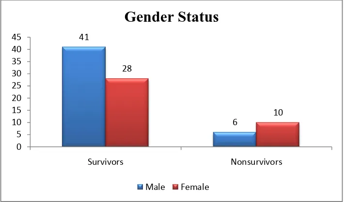
The male-female ratio was 59.42:40.58. Females showed a high rate of mortality.
The occurrence of comorbid conditions like Diabetic mellitus, Hypertension and
Chronic kidney disease showed higher risk of death outcome. Mean RDW was
16.22±0.89 in the case of survivors and 19.08±1.04 in the case of non-survivors which
was statistically significant (p<0.0001) with respect to duration of stay and need for
inotropic support. The mean NLCR was 8.95±1.54 in survivors and 13.24±1.37 in
non-survivors, the results are statistically significant (p<0.0001). A highly significant and
positive correlation of RDW and NLCR with SOFA score was observed.
CONCLUSION
RDW and NLCR measured on admission can be used as prognostic markers in
patients with sepsis, severe sepsis and septic shock.
KEYWORDS:
1
INTRODUCTION
Sepsis is a clinical syndrome characterized by systemic inflammation due to
infection, the severity ranging from sepsis to septic shock. Data from the centre for
Disease Control and Prevention reveals that sepsis is the leading cause of death in
non-coronary intensive care unit patients and the tenth most common cause of death
worldwide, the first being heart disease.1 Despite advances in intensive care and
antimicrobial therapy, the incidence of sepsis and related mortality rate has increased
over the last thirty years.2 The mortality rate is estimated at 30% in sepsis and 80% in
septic shock in the USA 3 and at 12.8% in sepsis and 45.7% in septic shock in Europe.4
Reduced rates of reporting may affect estimations in developing countries.
Sepsis or severe sepsis is defined as the harmful systemic response (including
some degree of hypofunction) with the proven or suspected microbial etiology. Septic
shock is sepsis with hypotension (arterial blood pressure < 90 mmHg below patient’s
normal blood pressure for at least one hour despite fluid resuscitation) or need for
vasopressors to maintain the systolic blood pressure ≥ 90 mmHg or mean arterial blood
pressure ≥70 mmHg. The incidence of sepsis and septic shock continues to increase
worldwide. The mortality increase has been attributable to patients’ advanced age,
preexisting comorbidity, immunosuppressive diseases and therapies or infections with
multi-drug resistant bacteria, patients with chronic diseases for a long period, and those
2
mechanical devices.5 Invasive bacterial infections are a prominent cause of death around
the world-especially among children.
Without consistent and reproducible criteria the extensive pathophysiology
associated with sepsis is difficult to diagnose and treat. A delay in the diagnosis and
treatment of sepsis will result in the rapid progression of circulatory failure, multiple
organ dysfunction and eventually death.6
Treatment guidelines are ambiguous. It involves a prolonged hospital stay for
patients, while receiving complex therapy. The in-hospital mortality risk of 10% in
patients diagnosed with sepsis is widespread and those who develop septic shock
increase their mortality risk greater than 40%.7
Early diagnosis of severity of sepsis and appropriate treatment is essential for the
survival of the patients. There are many biochemical markers, clinical parameters and
scoring systems used to assess the severity and in predicting the mortality in patients with
sepsis- some of which include- estimating serum procalcitonin levels, clinical scoring
systems like Sequential Organ Failure Assessment (SOFA), quick SOFA (qSOFA),
Acute Physiology and Chronic Health Evaluation (APACHE II) scoring systems. The
degree of severity is most often quantified by the Sequential Organ Failure Assessment
(SOFA) score, which can predict the severity and outcome of multiple organ failure.
However, calculating SOFA score is cumbersome. Moreover, assessment of the septic
patient outcome during treatment needs to be focused on, as currently used clinical and
3
The need for simple, cost effective and easily available, yet reliable markers has
pushed researchers in identifying such markers for assessing the severity and predicting
the prognosis of sepsis.
Several inflammatory biomarkers have been evaluated in recent years with the
high sensitivity, specificity, positive and negative predictive values for the early
diagnosis of sepsis as available in literature. The Red Cell Distribution width (RDW) is
one of the various biomarkers which have been shown to predict the mortality and
morbidity of sepsis.
The Red Cell Distribution Width (RDW) is the coefficient of variation of Red
Blood Cell (RBC) volume and is a representation of the RBC size heterogeneity of an
individual patient.8 Recent studies have reported that Red Cell Distribution Width (RDW)
is associated with prognosis in Critical Illness, Heart Failure, Acute Myocardial
Infarction, Pulmonary Embolism, Pneumonia and Cardiac Arrest.9-14
Research has shown that Neutrophil-lymphocyte count ratio (NLCR) may be
considered a novel marker of subclinical inflammation.15 It represents a combination of
two markers; neutrophils, which represent the active nonspecific mediator initiating the
first line of defence and lymphocytes, representing the regulatory or protective
component of inflammation. Neutrophil-lymphocyte ratio (NLR) is calculated by
dividing the number of neutrophil count by number of lymphocyte count, usually from
4
In this work, the haemogram parameters RDW and NLCR which are part of a
complete blood count, easy to evaluate and which do not incur additional costs to routine
analysis are studied; (i) to assess the efficiency of these parameters as prognostic markers
in sepsis and in predicting the clinical outcome after 28 days as assessed by SOFA score
in patients with sepsis, severe sepsis and septic shock and (ii) to investigate whether
changes in RDW and NLCR during the first week correlates with the severity of sepsis
5
REVIEW OF LITERATURE
PREAMBLE
Sepsis, a syndrome of physiologic, pathologic, and biochemical abnormalities induced by infection, is a major public health concern. Multiple definitions and terminologies are currently in use for sepsis, severe sepsis, septic shock, and organ dysfunction. With Considerable advances made into the pathobiology, the definition for sepsis and septic shock is being revised periodically. A 1991 consensus conference developed initial definitions for sepsis wherein sepsis refers to the presence of an infectious systemic inflammatory response
syndrome (SIRS). 16
SystemicInfiammatory Response syndrom (SIRS) in adults requires two or more of the
following. ( Table 1)
Table 1. SystemicInfiammatory Response syndrom (SIRS)
1. Temperature >38 C or< 36 C 2. Pulse>90/min
3. RR>20/min or PaCO2<32 mm Hg
4. WBC count >12000/cmm or <4000/cmm or >10% immature band forms
The definition was not adequate, lacked specificity and deceptive. Even patients with
6
criteria. SIRS may simply reflect an appropriate host response that is frequently adaptive.
Sepsis involves organ dysfunction, indicating a pathobiology more complex than infection in
addition to an accompanying inflammatory response alone
The definition was updated in 2001 and the international consensus defined severe
sepsis as sepsis that leads to dysfunction of any or more organ systems, the organ dysfunction
variables listed as, 17
Arterial hypoxaemia (PaO2/Fi O2 ratio <300) with new pulmonary infiltrates
A new or increased Oxygen requirement to maintain SpO2 more than 90%
Acute oliguria (Urine output < 0.5 mL/kg/hour for at least 2 hours
Serum creatinine more than 176.8 µ mol/L (2.0 mg/dL)
Coagulation abnormality (INR >1.5 or aPT more than 60 secs
Thrombocytopoenia (platelet <100x 109/L ( 100000/ µL))
Hyperbilurubinaemia ( total bilurubin >68.2 µmol/L(4mg/dL))
Arterial hypotension ( systolic BP <90 mmHg, mean BP <65mmHg) or reduction
in systolic BP > 40 mm Hg from baseline)
7
The international consensus defines Septic shock as to satisfying the following criteria:
Arterial hypotension ( systolic BP <90 mmHg, mean BP <65mmHg, or
reduction in systolic BP > 40 mm Hg from baseline) persisting for at least one
hour despite adequate fluid resuscitation (or)
Serum lactate >4 mmol/L(>36mg/dL) after adequate fluid resuscitation .
(The use of vasopressor agents to correct hypotension does not exclude shock).
As per the Surviving Sepsis Campaign (SSC) guidelines updated in 2012, severe sepsis
is defined as sepsis plus sepsis induced organ dysfunction or tissue hypoperfusion.
Septic shock is defined as sepsis induced hypotension persisting despite adequate fluid
resuscitation.
The definition was reanalyzed and modified in 2016 and it recommended that sepsis
should be defined as life-threatening organ dysfunction caused by a dysregulated host response to infection. Septic shock should be defined as a subset of sepsis in which particularly profound circulatory, cellular, and metabolic abnormalities are associated with a greater risk of mortality than with sepsis alone.7,18 Patients with septic shock can be clinically identified with a presentation of
Persistent hypotension requiring vasopressors to maintain Mean Arterial pressure
MAP≥65mmHg and
Serum lactate >2 mmol/L(>18mg/dL) in the absence of hypovolemia
8
Table 2. Definition used to describe the condition of septic shock
Bacteremia Presence of bacteria in blood, as evidenced by positive
blood cultures
Signs of possibly harmful systemic response
Two or more of the following conditions:
(1) fever (oral temperature >38°C [>100.4°F]) or hypothermia (<36°C [<96.8°F]);
(2) tachypnea (>24 breaths/min);
(3) tachycardia (heart rate >90 beats/min); (4) leukocytosis (>12,000/μL), leukopenia (<4000/μL), or >10% bands
Sepsis (or severe sepsis) The harmful host response to infection; systemic response to proven or suspected infection plus some degree of organ hypofunction, i.e.:
1. Cardiovascular: Arterial systolic blood pressure ≤90 mmHg or mean arterial pressure ≤70 mmHg that responds to administration of IV fluid
2. Renal: Urine output <0.5 mL/kg per hour for 1 h despite adequate fluid resuscitation
3. Respiratory: PaO2/FiO2 ≤250 or, if the lung is the only dysfunctional organ,≤200
4. Hematologic: Platelet count <80,000/μL or 50% decrease in platelet count from highest value recorded over previous 3 days
5. Unexplained metabolic acidosis: A pH ≤7.30 or a base deficit ≥5.0 mEq/L and a plasma lactate level >1.5 times upper limit of normal for reporting lab
Septic shock Sepsis with hypotension (arterial blood pressure <90 mmHg systolic,or 40 mmHg less than patient’s normal blood pressure) for at least 1 h despite adequate fluid resuscitationa
or
Need for vasopressors to maintain systolic blood pressure ≥90 mmHg or mean arterial pressure ≥70 mmHg
Refractory septic shock Septic shock that lasts for >1 h and does not respond to fluid or pressor administration
a Fluid resuscitation is considered adequate when the pulmonary artery wedge pressure is ≥12 mmHg or the central venous
pressure is ≥8 mmHg.
9
Fig 1. Sepsis - Disease continuum
10
EPIDEMIOLOGY
Recent estimates indicate that the incidence of sepsis has increased in the past 3
decades.19 About 1.5 million people in the United States are diagnosed with sepsis every year.
The incidence of severe sepsis and septic shock in the United States has increased to 750000
every year (about 3/1000 population).20 It is reported that, one out of every 4 people admitted
with severe sepsis die during hospital stay. There is a similar trend of increase in the incidence
of sepsis worldwide. The reason for this increased incidence may be attributed to increase in
the lifespan of general population, leading to increase number of ageing population, and the
association of chronic diseases like diabetes, cardiac diseases, chronic obstructive pulmonary
disease and malignancy. The frequent use of in-dwelling catheter and immunosuppressants has
also contributed to the rise in the number of patients with sepsis and severe sepsis globally
every year. Sepsis has been more commonly associated with African and Afro- American
males and also HIV infected individuals. In spite of improvement in health care and treatment
modalities, the mortality of patients with severe sepsis and septic shock continues to be high.
AETIOLOGY:
In 2007, a prevalence study was conducted in over 14000 patients in Intensive Care
units across more than 70 countries to identify the etiological pattern of sepsis. In the study,
51 % of the patients were considered to be infected, of which respiratory tract infection was
found to be most common. In another study conducted by Mayr et.al., respiratory tract
infection was found to be the most common cause in adults , followed by bloodstream
11
and catheter related infections.21 Microbial invasion of bloodstream is not essential in all cases
of sepsis, as local inflammation can lead to distant organ dysfunction. Studies have shown that
only 20 to 40 percentage of cases of severe sepsis and 40 to 70 percentage cases of septic
shock yield positive blood culture for bacteria or fungus.
More frequent use of broad-spectrum antibiotics in critically ill patients who remain in
the ICU for longer periods of time has resulted in a high degree of bacterial resistance over
time.22,23 Antibiotic resistance is challenging, resulting in longer duration of hospital stay and
ventilator dependence, although the effect on mortality is uncertain.24-26 Among causative
agents, Gram Negative organisms were found to be the most common cause of sepsis (62%),
but, of late, incidence of Gram positive organisms as a cause of sepsis is on the rise and are
now becoming almost as common as Gram negative organisms (46%). 19, 27-29.This trend may
be due to the greater use of invasive procedures and increasing magnitude of hospital acquired
infections26. Recent European prevalence of infection in Intensive Care (EPIC-2) study
reported that the incidence of Gram negative organisms to be around 62 % compared to Gram
positive organisms at 42%.The study also showed that among individual organisms,
Staphylococcus aureus was found to be the most common pathogen (20.5%) followed by
Pseudomonas (19.9 %) and Enterobacteriae, particularly E.Coli. A large meta-analysis of
more than 500 studies demonstrated that Gram negative bacteraemia leads to more mortality
than Gram positive bacteraemia30. Coagulase negative staphylococci and E coli were the
commonest cause of bloodstream infections. Pseudomonas aeruginosa had the highest
12
was the next common cause (41%). Candida and Acenobacter species also accounted to high
mortality rates (40%).
Recent studies have shown that there has been an increased incidence of sepsis among
paediatric population over the past decade. This has been largely due to the disproportionate
increase in the incidence of sepsis among neonates, particularly among the low birth weight
category31. Again, respiratory tract infections were found to be the most common cause of
sepsis even among paediatric population (49%) followed by primary bacteremia (18.1%).
RISK FACTORS
Risk factors for severe sepsis can broadly be divided into risk factors for infection,
contingent upon developing infection, and risk factors for organ dysfunction.
Bacteraemia
Patients with bacteraemia often developed systemic consequences of infection. In a
study conducted in 2015 in South Korea in patients with sepsis, whose blood cultures were
positive for either bacteria or fungi, about 95% of positive blood cultures were associated with
sepsis severe sepsis or septic shock.
Sex and race
Sepsis has been found to be more common among male gender and black people
(African and Afro- American).
Environmental factors
There exists an inverse relationship between socioeconomic status and the risk for
13
Age
Elderly patients, particularly above 65 years, are at increased risk of developing sepsis.
The incidence of sepsis increases with age, and older adults above 65 years of age have an
increased risk of mortality from sepsis compared to their younger counterparts. Age is also
an independent prognostic marker in sepsis. Elderly individuals need greater nursing care and
close monitoring. Studies have shown that in-hospital mortality rates are higher among elderly
individuals affected with sepsis.
Immunosuppression
Immunosuppression is an important risk factor for the development of sepsis.
Conditions that affect the host defence mechanisms and those drugs that lead to
immunosuppression make the patient more vulnerable for developing frequent infections and
their poor immunity make them candidates for the development of sepsis. Some of these
conditions include renal failure, diabetes, neoplasms, hepatic failure, AIDS, patients on
corticosteroid therapy or immunosuppressant medications. The risk of progression to severe
sepsis, septic shock and Multi Organ Dysfunction Syndrome (MODS) is more common among
immunosuppressed individuals which may lead to high mortality.
Malignancy and diabetes
Diabetes and malignancies alter the immune system resulting in an increase in the risk
for developing sepsis. Studies have shown that there is high incidence of nosocomial
infections among patients with diabetes, particularly among patients with poor glycemic
14
Community acquired pneumonia
Patients who developed community-acquired pneumonia were found to have an
increased risk of developing sepsis and an increased risk of progression to severe sepsis and
septic shock.
Genetic factors
There is confirmatory evidence from experimental and clinical studies that genetic
factors can increase the risk of infection in few cases. Monogenic defects underlie
vulnerability to specific infection, but genetic factors are typically genetic polymorphisms.
Genetic studies of susceptibility to infection have initially focused on defects of antibody
production or a lack of T- cells, phagocytes, natural killer cells or complement, but recently
genetic defects that impair the recognition of pathogens by the innate immune system have
been recognised. Study by Sorensen et al., suggests that genetic factors may play a crucial role
in outcomes of infectious diseases compared with cardiovascular disease. In this study,
children whose biological parents died due to infectious causes had an 8 fold increased risk of
death due to infections33. In comparison, the increased risk of death due to cardiovascular
causes was four fold if their biological parents died of cardiovascular causes.
PATHOPHYSIOLOGY
The normal host responds to any infection in a peculiar fashion aiming towards
localising and restricting the bacterial invasion. The host responds by triggering the activation
of circulating macrophages resulting in the production of pro-inflammatory and anti-
15
the innate immune cells, particularly macrophages, to identify and bind to the microbial
components. This process may occur as follows.
When the microorganisms enter the host, the host immune cells have certain
pattern recognition receptors on their cell surface that identify and attach to the Pathogen
Associated Molecular Patterns (PAMP) 1of the microorganism. There are three major groups
of Pattern Recognition Receptors:
1) Toll -like receptors (TLR)
2) Nucleotideoligomerization domain (NOD) leucine Rich repeat proteins and
3) Retinoic acid inducible gene I (RIG--i) like helicases.
The host immune cells also contain triggering receptors expressed on myeloid cell (TREM-1)
and Myeloid DAP -12 associating Lectin (MDL-1) on their surface which identify and bind to
the microorganisms2.The binding of Toll like receptors triggers a signalling cascade by
activating the cytosolic nuclear factor -kb (NF-kb). The activated NF-kb migrate from the
cytoplasm to the nucleus, attaches itself to the transcription sites, and trigger the activation of a
large set of genes resulting in the release of pro-inflammatory cytokines like Tumor necrosis
factor-Alpha,Interleukin-1, chemokines like Intercellular Adhesion Molecule-1(ICAM -1) and
Vascular Cell Adhesion Molecule-1(VCAM-1) and nitric oxide.
The polymorphonuclear leukocytes also get activated and express adhesion molecules
on their surface that leads to their aggregation and margination to the vascular endothelium.
The polymorphonuclear leukocytes then undergo a series of steps such as rolling, adhesion,
diapedesis, and chemotaxis to move to the site of injury.34 The poly morphonuclear leukocytes
16
local inflammation. This process is regulated by the release of proinflammatory and anti
-inflammatory mediators by the macrophages at the site of injury that is triggered by the
invasion of tissue by the microorganisms.35-37
The balance in the pro-inflammatory and anti- inflammatory mediators control and
regulate the inflammatory process, which leads to homeostasis, overcoming of the infectious
insult and ultimately results in tissue repair and healing38. When this inflammatory response to
infection exceeds the boundary of local environment and becomes a more generalized
response, it leads to widespread tissue damage and is called sepsis. In the absence of any
infectious etiology, this process is called Systemic Inflammatory Response Syndrome (SIRS).
The reason why the localised immune response sometimes crosses the local environment
leading to sepsis is still not clear.
It may be due to the direct effect of microorganisms or their toxic products.
Bacterial cell wall components like endotoxin, peptidoglycan, muramyl dipeptide and
lipotechoic acid and bacterial products like Staphylococcal enterotoxin-B, toxic shock
syndrome toxin-1, pseudomonas exotoxin A, and M-protein of hemolytic group A
streptococci may contribute to the progression of a local infection to sepsis.39-41
Large quantities of pro-inflammatory cytokines released during sepsis may
spill into the bloodstream leading to the progression of local infection to sepsis. These include
tumor necrosis factor-alpha and interleukin-1 whose plasma levels peak early and eventually
decrease to undetectable levels. There is strong evidence that TNF - α has an important role to
17
The complement system is a protein cascade which helps in clearing
pathogens.44, 45 There is proven evidence in animal studies that activation of complement
system plays a vital role in sepsis. 46, 47
Genetic susceptibility
Single nucleotide polymorphism (SNP) is the commonest type of genetic variation.
Various SNPs are associated with high degree of susceptibility to infection. Some of these
include SNPs of genes that encode cytokines like TNF-α, IL-10, IL-18,IL-1 receptor
antagonist, IL-6 and Interferon gamma; cell surface receptors like CD14, MD2 , toll-like
receptors 2 and 4; lipopolysaccharide ligands like lipopolysaccharide binding protein,
bactericidal permeability increasing protein, mannose-binding lectin, heat shock protein 70,
angiotensin I-converting enzyme, and caspase-12. 48
SYSTEMIC EFFECTS OF SEPSIS
When the immune response to bacterial invasion becomes generalised, it leads to
widespread cellular injury. Organ dysfunction eventually follows this cellular injury. The
precise mechanism of cellular injury is not understood, but various mechanisms have been
proposed explaining the process of cellular injury. These include tissue ischemia, cytopathic
injury and an altered rate of apoptosis.
A) TISSUE ISCHEMIA
During sepsis there occurs a significant derangement in metabolic auto- regulation
which is the process that matches oxygen availability to changing tissue oxygen demand.
18
lesions decrease the cross sectional area available for tissue oxygen exchange reading to tissue
ischemia and cellular injury49.
B) CYTOPATHIC INJURY
The pro- inflammatory mediators released during sepsis may lead to mitochondrial
dysfunction through a variety of mechanisms, like direct inhibition of respiratory enzyme
complexes, oxidative stress damage and mitochondrial DNA breakdown.50 Such mitochondrial
injury leads to cytotoxicity. .
C) APOPTOSIS
Apoptosis is the primary mechanism by which senescent or dysfunctional cells normally
get eliminated. It is the principal process by which inflammation gets terminated once an
infection has subsided. The pro-inflammatory cytokines that are released during sepsis delay
the process of apoptosis in the activated macrophages and neutrophils, disrupting the normal
inflammatory response mechanism and leading to the development of multiple organ failure.
In addition to this, there is also an extensive apoptosis of lymphocytes and dendritic cells
which alters the immune response that occurs during sepsis. The extent of lymphocytic
apoptosis correlates with the severity of sepsis and the level of immunosuppression.
Apoptosis has also been found to occur in epithelial cells, endothelial cells and parenchymal
cells. Several animal studies have shown that inhibiting the process of apoptosis has been
19
20
21
ORGAN SPECIFIC EFFECTS OF SEPSIS
The cellular injury accompanied by the release of proinflammatory and anti
-inflammatory mediators leads to the development of organ failure. Multi organ dysfunction is
not uncommon.
Circulation:
Circulatory dysfunction leads to diffuse vasodilatation resulting in hypotension.
Inflammatory mediators like nitric oxide and prostacyclin are released by the endothelial cells
in sepsis to induce appropriate vasodilatation for the purpose of improving metabolic auto
-regulation. But diffuse vasodilation occurs as unintended consequences of these inflammatory
mediators, which results in hypotension. Nitric oxide, particularly is believed to play an
important role in the process of vasodilatation during septic shock.53,54 When nitric oxide
reaches the systemic circulation, it depresses the auto regulation at all the- central, regional
and micro regional levels of circulation .Vasodilatation is not the only cause of hypotension in
sepsis. It may also be due to the redistribution of intravascular fluid which occurs as a
consequence of increased endothelial permeability and reduced arterial vascular tone. In
addition to these diffuse effects on circulation, there are also localised effects.
During sepsis myocardial depressant substances that are released into the
circulation impair the systolic and diastolic ventricular performance of the heart. The
myocardium may still be able to use the Frank- Starling law and maintain the cardiac output in
normal individuals. But patients with pre-existing cardiac disease are often unable to increase
22
Sepsis is associated with a reduction in the number of functional capillaries
as a result of which there is subnormal oxygen extraction through the micro circulation.
Sepsis induces phenotypic changes to endothelial cells. This occurs through
direct and indirect interactions between the endothelial cells and components of the bacterial
wall. These phenotypic changes lead to endothelial dysfunction, which is associated with
coagulation abnormalities, reduced leukocytes, decreased red blood cell deformability,
upregulation of adhesion molecules, adherence of platelets and leukocytes and degradation of
the glycocalyx structure.55 Diffuseendothelial activation results in widespread tissue edema.
Micro particles from circulating and vascular cells also participate in the
deleterious effects of sepsis-induced intravascular inflammation 56.
Lung
Endothelial injury that occurs in the pulmonary vasculature during sepsis impairs
capillary blood flow and leads to increased micro vascular permeability.This leads to
interstitial and alveolar edema. The neutrophil entrapment in the pulmonary vasculature
further amplifies the injury, the end result being pulmonary edema and hypoxemia. This type
of pulmonary edema that occurs in sepsis is called as acute respiratory distress syndrome.
Gastrointestinal tract
The circulatory abnormalities that occur in sepsis disrupt the GIT‘s normal barrier
function, leading to translocation of bacteria and endotoxin from the gut into the systemic
23
Liver
Liver dysfunction can contribute to the initiation and progression of sepsis. Normally,
the reticuloendothelial system of the liver is the first line of defence in clearing bacteria and
bacteria-derived products that enter the portal system from the gut. Liver dysfunction can
prevent the elimination of enteric-derived endotoxin and bacteria-derived products, which
results in direct spillover of these harmful products into the systemic circulation. 57, 58
Kidney
Sepsis is often associated with acute kidney injury. The renal hypoperfusion that occurs
in sepsis leads to organ dysfunction. Systemic hypotension, direct renal vasoconstriction,
release of cytokines such as Tumor Necrosis Factor, may also contribute to renal injury.
Nervous system
Central nervous system complications occur very commonly in septic patients.
Encephalopathy and peripheral neuropathy are the commonest complications.
Encephalopathy: The pathogenesis of the encephalopathy is poorly defined.
A high incidence of brain microabscesses was noted in one study, but the significance of
hematogenous infection as the principal mechanism remains uncertain.
Peripheral neuropathy: Studies suggest that at least 25 percent of patients
admitted to medical or surgical intensive care units have some degree of acquired paresis59,60.
Affected patients manifest a sensorimotor polyneuropathy characterized by limb muscle
weakness and atrophy, reduced or absent deep tendon reflexes, sensory loss to light touch and
24
in this condition is unknown. CNS dysfunction may be attributed to changes in metabolism
and alterations in cell signalling due to the release of inflammatory mediators.
CLINICAL MANIFESTATIONS AND COMPLICATIONS
The clinical manifestation of patients with sepsis includes signs and symptoms that are
common to any septic response and those manifestations that occur as a complication of the
septic response. Moreover, some of the clinical manifestations can give a clue to the primary
etiology contributing to the sepsis. Most patients are hyperthermic, whereas some patients
maybe normothermic or hypothermic. Hypothermia is commonly seen in elderly, neonates and
alcoholics. Hyperventilation producing respiratory alkalosis is a very common early
manifestation in sepsis. Disorientation, confusion and other manifestations of encephalopathy
can develop early. Blood glucose levels increase in septic patients, particularly in diabetics.
Protein catabolism is accelerated. Serum albumin levels decrease due to impaired hepatic
synthesis and due to the movement of albumin into the interstitial spaces.
COMPLICATIONS
Cardio pulmonary complications
Alveolar epithelial injury and increased alveolar capillary permeability leads to
increased pulmonary water content. This interferes with gas exchange and produces arterial
hypoxemia. Ultimately acute lung injury and acute respiratory distress syndrome will result.
.Hypotension occurs as a result of hypovolemia and generalised redistribution of blood flow in
sepsis. Dehydration may further contribute to hypotension. Also, as a part of the septic
response, the myocardial function becomes depressed within the first 24 hours in most patients
25
maintained in spite of the myocardial depression. The myocardial function restores to normal
over several days. So the primary cause of hypotension is usually a decreased systemic
vascular resistance.
Renal complications
Oliguria, azotaemia, proteinuria are the commonest renal complications. Most renal
failure occurs due to acute tubular necrosis as a result of hypovolemia and hypotension.
Coagulopathy
Thrombocytopenia is common in patients with sepsis. In patients with DIC, the platelet
counts are very low, usually less than 50000. The exact mechanism for thrombocytopenia is
unclear.
Complications of Sepsis
26
Neurological complications
Confusion, altered sensorium (acute encephalopathy) occurs as early manifestation of
sepsis. Studies show that encephalopathy occurs in around 20 to 60 percentage of septic
patients at some point during the course of illness.
Immunosuppression
Patients with severe sepsis often become immune suppressed. This leads to increased
risk for secondary infection and difficulty in controlling the primary infection.
LABORATORY INVESTIGATIONS AND DIAGNOSIS
The most common laboratory findings that are seen in sepsis include neutrophilic
leukocytosis, thrombocytopenia and hyperbilirubinemia. Urine analysis may show
mild-to-moderate proteinuria. Leukopenia may be seen in some individuals. Peripheral smear may
reveal neutrophilia with neutrophils containing toxic granules. In severe sepsis,
thrombocytopenia usually worsens and may be associated with prolonged thrombin time,
decreased fibrinogen levels and elevated D -dimer levels suggesting DIC. Serum creatinine
and blood urea nitrogen levels are commonly elevated in patients with severe sepsis and septic
shock, indicating renal hypoperfusion and acute kidney injury. Arterial blood gas analysis
usually reveals high anionic gap metabolic acidosis though in many patients early arterial
blood gas analysis may show respiratory alkalosis as a result of hyperventilation. The most
common ECG abnormality seen is sinus tachycardia. Chest X-ray may reveal underlying
27
common and blood cultures may reveal the causative organism. In case of suspected urinary
tract infection, urine cultures are helpful. In patients with in-dwelling catheter or soft tissue
infections, swabs taken from the catheter or wound should be sent for culture.
DIAGNOSTIC AND PROGNOSTIC MARKERS
There is no specific diagnostic test for sepsis. Several diagnostic and prognostic
markers are being studied by researchers in patients with sepsis. Some of these biomarkers
which are proved to be helpful in the diagnosis and predicting the severity and outcome to
some extent are discussed below.
C - reactive protein:
CRP levels are being used extensively as a non-specific marker of inflammation.
Several studies have demonstrated the increase in CRP levels in patients with sepsis62, 63.As
CRP levels are also increased in many other inflammatory conditions like rheumatoid arthritis,
crohns disease, myocardial infarction, CRP is a nonspecific marker of sepsis. Because baseline
CRP levels are often raised in these comorbid chronic inflammatory conditions, changes in
concentrations over time and serial monitoring of CRP levels in patients with sepsis are more
useful than single values64, 65.
Procalcitonin
Many studies have shown that procalcitonin levels are elevated in patients with sepsis66.
Several studies have validated Procalcitonin to be a more reliable marker of sepsis than CRP,
and that, it can be used to distinguish between sepsis caused by bacteria and other
organisms67, 68.There is also evidence based data that higher procalcitonin levels are associated
28
Cytokine Levels
Various inflammatory cytokines, that are key mediators of the sepsis response, have
been studied for their role as biomarkers of sepsis, some of which include IL-6, TNFα and
IL-8. The serum levels of these inflammatory cytokines have been shown to be increased in
patients with sepsis.70
Soluble Triggering Receptor Expressed on Myeloid Cells-1
Serum levels of sTREM-1 are found to be elevated in patients with sepsis. 71-74 Several
studies have suggested that they are more sensitive and specific for infection than other
markers such as CRP and procalcitonin.71,72 A decrease in sTREM-1 levels over time was
associated with a good prognosis in patients with sepsis.75
CD 64
Neutrophilic CD 64 expression have been studied in patients with sepsis and were
found to have moderate sensitivity and specificity in the diagnosis of sepsis.76,77
Clot Waveform Analysis
Toh et al have established in their studies on patients diagnosed with sepsis and severe
sepsis, the optical transmission waveform obtained during measurement of the activated partial
thromboplastin time showed a biphasic pattern. The presence of this abnormal waveform has
29
PREDICTION OF OUTCOME OF SEPSIS: SCORING SYSTEMS
Severity of organ dysfunction has been assessed with various scoring systems that quantify abnormalities according to clinical findings, laboratory data, or therapeutic interventions.
APACHE II (Acute Physiology and Critical Health Evaluation score)
The APACHE II score is one of the many scoring systems available such as SOFA,
SAPS III, RIFLE to assess the prognosis of individual patients based on the set of laboratory
values and the patient’s sign of illness both acute and chronic illness. For APACHE II
scoring, the input data should be that of the initial 24 hours of the patient’s entry into the ICU.
The scores are assigned according to the criteria given in table (Table 3)
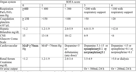
SOFA SCORE
In 1994, The European Society of Critical Care Medicine (ESCCM), developed a
simple and objective score, the Sequential Organ Failure Assessment (SOFA) Score as a
system for measuring the status of the patient in the ICU.79,80 The organ dysfunction in sepsis
is recommended to be identified by an acute change in total SOFA score ≥ 2 points consequent
to infection. The Sequential Organ Failure Assessment (SOFA) score allows for calculation of
both the number and the severity of organ dysfunction in six organ systems, ie, respiratory,
coagulatory, liver, cardiovascular, renal, and neurologic .The score can measure individual or
aggregate organ dysfunction. Different variables and parameters are included in each of the
organ system (Table 4). The function of each is scored from 0 (the Normal function-unless the
patient has a previously known co-morbidity like cirrhosis, chronic kidney disease etc) to 4
30
sum of the highest score per individual during the entire ICU stay is considered the Maximal
SOFA score. A higher SOFA score is associated with an increased probability of mortality81.
The value of SOFA score> 15 is supposed to predict a mortality of 90%. Mean SOFA score is
the average of all total SOFA scores in the entire period of ICU stay. Studies reveal that mean
SOFA score for the first 10 days is significantly higher in non-survivors. Aditi Jain et al
made prospective study on patients of age between 15 and 80 years admitted to ICU over 8
weeks period and concluded that there is a high correlation of ΔSOFA score (significance
p<0. 0001) and the mortality82. The maximum score in survivors (3.92 ±2.17) was
significantly lower than that of non-survivors (8.9 ± 3.45), while duration of the stay did not
correlate with the survival (p = 0.461).
qSOFA score
SOFA score computation is a tedious procedure and uses a treatment related variable
(doses of vasopressor agents). Laboratory variables, namely, PaO2, platelet count, creatinine
level and bilirubin level are needed for full computation. The qSOFA score (Table 5) focuses on only three clinical variables- hypotension (Systolic blood pressure ≤ 100mm Hg ), altered
mental status and tachypnea ( respiratory rate > 22/min), the presence of at least two of these
criteria strongly predicts the likelihood of poor outcome in patients with clinical suspicion of
sepsis in the non-ICU environment.
This definition, nevertheless, does not offer clear criteria for sepsis diagnosis. In this
context, sepsis raises multiple problems of diagnosis and prognosis, and further studies are
necessary to identify useful criteria for establishing a rapid and correct diagnosis and quick
31
32
Table 4 SOFA Score
Organ system SOFA score
0 1 2 3 4
Respiration PaO2/FiO2
mm Hg
≥400 < 400 < 300 <200 with
respiratory support
<100 with
respiratory support
Coagulation platelets x103/µL
≥ 150 >150 >100 >50 >20
Hepatic
Bilurubin mg/dL
<1.2 1.2-1.9 2.0-5.9 6.0-11.9 >12.0
CNS
Glascow Coma Scale
15 13-14 10-12 6-9 <6
Cardiovascular MAP ≥70mm Hg
MAP <70mm Hg Dopamine<5 or
dobutamine (any dose) *
Dopamine 5.1-15 or epinephrine≤0.1 or norpinephrine≤0.1*
Dopamine >15 or epinephrine>0.1 or norpinephrine>0.1*
Renal Serum Creatinine mg/dL
<1.2 1.2-1.9 2.0-3.4 3.5-4.9 >5.0 or dialysis
Or urine output Or < 500mL/24 h Or < 200mL/24 h
*Catecholamine doses are given as µg/kg/min for at least 1 hour PaO2 - Partial pressure of Oxygen; FiO2 –Fraction of inspired Oxygen
PaO2/FiO2 ratio is calculated without reference to the use or mode of mechanical ventilation and without reference to the use or level of PEEP.
Glascow Coma Score- For the patient receiving sedation or muscle relaxants, normal function is assumed unless there is evidence of intrinsically altered mentation.
[image:40.612.72.547.101.353.2]
Table 5 qSOFA score
2 or more of
1. Hypotension: SBP less than or equal to 100mm Hg
2. Altered mental status ( any CGS < 15
3. Tachypnoea: Respiratory rate ≥ 22
33
OTHER BIOMARKERS
Extensive studies have been done on identifying specific markers for rapid diagnosis in
order to improve the clinical management of sepsis. The literature focuses on RDW and NLCR
i.e., the haemogram parameters which are easy to evaluate, and which do not incur additional
costs to routine analysis. Normally red cell size variation is known as anisocytosis. The red
blood cell distribution width (RDW) represents an index of the heterogeneity of the
erythrocytes (anisocytosis) which is calculated by dividing the standard deviation of
erythrocyte volume by the mean corpuscular volume (MCV) and multiplying by 100 to
express the result as a percentage.
Structure of the Red cell
Among the cells of human tissues mature RBCs are unique, in the sense that they
normally lack nuclei and cytoplasmic structures such as Lysosomes, endoplasmic reticulum
and mitochondria. Hence they cannot carry out protein synthesis, and are unable to undergo
mitosis and mitochondrial oxidative reactions. RBCs are biconcave discs of 7-8µm in
diameter, but their shape changes to a parachute- like configuration in the capillaries whose
diameter, is less than that of RBCs in the biconcave disc form. As the membrane of red cell is
elastic they resume biconcave shape. Once they re-enter the large blood vessels, loss of
flexibility or elasticity leads to membrane damage and change in shape .This leads to
34
RED CELL DISTRIBUTION WIDTH (RDW)
Red cell distribution width is an index of variation in RBC size or RBC volume. Most
automated instruments produce a quantitative assessment of the variation in red cell volume
indicated by RDW which corresponds to the microscopic analysis of the degree of
anisocytosis. The RDW derived from pulse height analysis can be expressed either as (SD)
standard deviation in fl or as the percent of coefficient of variation (CV) of the measurements
of red cell volume.
RDW-SD is a measurement of width of RBC size distribution histogram and it is
measured by calculating the width at the 20% height level of the RBC size distribution
histogram. Hence RDW-SD is not influenced by the average RBC size, that is, mean
corpuscular volume.
RDW-CV is calculated from standard deviation and MCV by the formula.
RDW-CV (%) = 1 SD of RBC volume / MCV x 100%
Since RDW- CV is obtained mathematically from MCV it is affected by changes in average
size of RBCs.
Normal reference ranges of RDW in adults:
RDW - SD 39-46 fl
RDW - CV 12-14%
Significance of elevated RDW
Early diagnosis of nutritional deficiency (d/t) iron, B12 and folic acid.
Differentiation of iron deficiency anaemia from thalassemia.
35
Identification of Red cell fragmentation, agglutination and dimorphic red cells in
peripheral smear examination.
Red cell distribution width in sepsis
Recent studies have been focusing on evaluating RDW’s prognostic value and use for
the diagnostic role in sepsis. Literature reveals that as RDW is a means of evaluating the
variability in size of erythrocytes it has been used widely in the differential diagnosis of
anemia 83.Since RDW is a marker of unspecific inflammation, it can show high value in many
other diseases such as heart failure, stroke, peripheral arterial disease or chronic pulmonary
diseases 84.Red cell distribution width (RDW) represents an indicator which can vary in sepsis,
under the influence of pro-inflammatory cytokines (TNFα, IFNδ, IL-1β, IL-6)85,released
during the inflammatory process. These cytokines cause inefficient erythropoiesis resulting in
structural and functional changes of erythrocytes, with volume variations and increased RDW.
Elevated value of RDW can also appear in nutritional deficiencies such as iron deficiency
anaemia, vitamin B12 or folate deficiency anaemia, or in blood transfusions86.
Kim et al. evaluated the predictive role of RDW regarding the short and medium-term
mortality in elderly patients with severe sepsis and septic shock and concluded that every one
percent (1%) increase in RDW is equivalent to a 15% increase in the mortality rate in the first
30 days 87. Lorente et al. showed that RDW is a low-cost procedure which should be routinely
performed to identify the risk of death in sepsis. In this study, the prognostic value of RDW
was compared with other biomarkers such as serum malondialdehyde and TNFα, 88
In a study on 349 patients, the function of RDW in predicting septic outcome in sepsis
36
mortality rate. (p<0.0001) was presented. This study demonstrated that an RDW value over
16% is concurrent with a higher APACHE value and risk of death84. RDW was also
considered a good predictor of the length of hospitalisation for septic patients admitted to
intensive care units 89.
The biomarker RDW proved its usefulness as an outcome measure because an
ascending trend of RDW in the first 72 hours of admission is associated with an unfavourable
prognosis, even though the initial values were normal90. Chen et al. analysed almost 7000 adult
patients with sepsis, with a mortality rate of 6.8%. In this study, patients who died had higher
initial values of RDW than the survivors (15.7% versus 13.8%). There were also established
threshold values; for RDW over than 15.6%, the risk of death was 16.7%, for RDW between
14 – 15.6% it was 7.3%, and for RDW under 13.1% the mortality rate was 1.6%. Thus, RDW
was considered to be a superior mortality predictive factor to SIRS criteria, the MEDS
(Mor-tality in Emergency Department Sepsis) or CURB65 scores 91.
SIGNIFICANCE OF NLCR
Neutrophil/lymphocytes count ratio (NLCR) has been the focus of several recent
studies as it is accessible, cheap and readily determined. The importance of this parameter is
related to the pathophysiological mechanism of SIRS, which is characterised by an increased
number of circulating leucocytes, due to the increased number of neutrophils, the first line of
antimicrobial defence. On the other hand, lymphocytopenia appears as a consequence of
37
apoptosis 92. Normally there are 4000-11000 WBCS/ micro litre in the human blood. Of these
granulocytes are the most numerous. Young granulocytes have horse shoe shaped nuclei that
become multilobed as the cells grow older. Most of them contain neutrophilic granules.
Neutrophils
Neutrophils have cytoplasmic granules that contain biologically active substances
involved in inflammatory reactions. The average half- life of a neutrophil in the circulation is 6
hours. They are attracted to the endothelial surface by selectins and they roll along it.
Neutrophil adhesion molecules of the integrin family helps them to get bound to selectins.
They insinuate themselves through the walls of the capillaries by a process known as
diapedesis. Many of those that leave the circulation enter the GI tract and are lost from the
body.Neutrophilic granules contain various proteases and in addition they also contain
enzymes such as NADPH oxidase, catalase and myeloperoxidases. NADPH oxidase is
associated with a sharp increase in oxygen intake and metabolism in the neutrophil,
(Respiratory burst) and this reaction generates plenty of free O-radicals. The myeloperoxidase
catalyses the conversion of Halides and cyanides to their corresponding acid forms. These
acids in turn are potent oxidants by themselves.In addition to myeloperoxidase and NADPH
oxidase neutrophil granules also contain an elastase and two metalloproteinases.
The total body neutrophils can be divided into circulating pool (CGP) and marginating
granulocyte pool. In these two pools, the cells are equal size and they are in constant
equilibrium. MGP represents the neutrophils involved in adhesion and rolling along the
38
venepuncture. So the neutrophil content actually represents about half of the total no of
neutrophils in the vascular compartment.
Lymphocytes
Lymphocytes are motile non phagocytic cells. There are many subpopulations of
lymphocytes which interact with each other and with cells of the monocyte macrophage
system. They help in maintaining both humoral and cell mediated immunity. Proliferating
lymphocytes are enriched with enhanced levels of enzyme n-terminal deoxyribonucleic acid
transferase. It is found in immature lymphoid cells in the bone marrow and thymocytes, but
not in mature lymphocytes. Adenosine de aminase is present in large amounts in
T-lymphocytes and it is necessary for their immune function.
Inflammation
Inflammation is naturally a protective mechanism against invasion of microbes and
toxins. The inflammatory response consists of 2 main components- a vascular reaction and a
cellular reaction. Both the reactions are mediated by chemical factors that are derived from
plasma proteins or cells produced as a result of inflammatory response.
Mechanisms
Endothelial dysfunction secondary to cellular response of blood components heralds the
onset of inflammation. Endothelial dysfunction leads to impaired production of nitric oxide
and prostacylins. This leads to the depletion of anti-atherogenic, antithrombotic and
39
Zahorec first proposed the use of the ratio of neutrophil and lymphocyte counts
[neutrophil lymphocyte count ratio (NLCR)] as an marker of infection in clinical
Applications. 93 Loonen et al., demonstrated that NLCR is a rapidly available biomarker, and
appears most promising in differentiating patients with BSI from those without BSI for
subsequent pathogen identification.94 NLCR is considered to be superior to other biomarkers
such as CRP, leukocytes count or neutrophils count, as a predictor for bacteraemia in patients
admitted to emergency or intensive care units.95 In a study of 40 patients with severe sepsis,
Okashah et al., highlighted NLCR superiority regarding sensitivity, specificity, positive and
negative predictive values to other parameters like lactate, CRP, neutrophils count,
lymphocytes count, or leucocytes count.96 The same study showed the usefulness of NLCR in
prognostic evaluation attributing to the statistically significant correlation with two severity
scores, APACHE II (p=0.01) and SOFA (p=0.01).
Recently, attempts have been made to establish threshold values for NLCR for
predicting the severity and outcome of the disease. A more recent study showed that an initial
value of NLCR over 10 could be correlated with an unfavourable prognosis, as assessed by the
number of SIRS criteria, the presence of organ failures or septic metastasis at admission.
Despite the low number of investigated patients, this study intended to substantiate the
prognostic role of NLCR in sepsis through the statistically significant correlations with
APACHE IV (p=0.01) and APS (p=0.01) scores and with the estimated rate of mortality
(p=0.01)97. A retrospective study, which included 2311 patients with bacteraemia concluded
that a value of NLCR over seven on admission, represented an independent increased
40
The predictive role of NLCR has been evaluated not only in septic patients but also in
patients with tumours, cardiovascular diseases or intestinal inflammatory diseases. The initial
value of this indicator was correlated with the outcome and with the survival rate in patients
with different types of cancers: pulmonary, breast, prostatic, pancreatic, oesophageal 99,
colorectal 100 or hepatocellular carcinoma followed by liver transplant 101.Several studies,
published in cardiovascular medicine domain, showed the prognostic role of NLCR in patients
with an acute coronary syndrome, aortocoronary bypass or congestive heart failure 102,103. Ertas
et al. indicated that NLCR was useful in the prognosis of patients with thromboembolic
Stroke.104 On the other hand, a high correlation between NLCR and the outcome of patients
with gangrenous appendicitis was demonstrated, and was shown to be superior to other
parameters like fever, CRP, Leucocytes number or the Glasgow scale.105
MANAGEMENT OF SEPSIS
Early Management
For patients with severe sepsis or septic shock, (i) stabilization of the airway and
breathing and (ii) restoration of perfusion to the peripheral tissues should be should be done
immediately, by supplemental oxygenation and monitored continuously with pulse oximetry.
Intubation and mechanical ventilation if needed should not be delayed.106, 107
Following initial stabilization, chest radiographs and arterial blood analysis should be
obtained in combination with other clinical parameters to diagnose acute lung injury (ALI) or
41
Central venous Catheter(CVC)
After the assessment of airway and perfusion, inserting a central venous catheter will be
beneficial with most patients with severe sepsis and septic shock for infusion of intravenous
fluids, infusion of blood product and infuse medications. In addition, CVC can be used to
monitor the central venous pressure (CVP) and the central venous oxyhaemoglobin saturation
(ScvO2). Few studies have shown that, managementt of septic shock guided by ScvO2 reduce
the mortality. 108
Restoration of Perfusion
Once the presence of hypoperfusion has been established, early restoration of perfusion
is vital to prevent or reduce multi organ dysfunction. The goals for resuscitation of circulation
include maintaining central venous pressure (CVP) 8 to 12 mmHg, a mean arterial pressure
(MAP) ≥65 mmHg and a urine output ≥0.5 mL/kg per hour.Intravenous fluid administration
should be started as early as possible.
Intravenous fluids
Large volume infusions of IV fluids are the main stay of initial therapy in patients with
severe sepsis or septic shock. Fluid should be administered as rapidly infused boluses 109,110.
The volume status, blood pressure, peripheral perfusion and the presence of lung signs (to rule
out pulmonary edema) must be monitored before and after each bolus. Intravenous fluids
should be administered carefully and a more restrictive approach should be followed with
respect to fluid resuscitation in patients at risk and who have developed acute lung injury or
ARDS.111 The ideal fluid of choice can be either a colloid like 0.9 % normal saline or Ringer’s
42
most commonly used and preferred fluid of choice for many clinicians, the usefulness of
crystalloids have been studied extensively.
In a SAFE (Saline versus Albumin Fluid Evaluation) trial, nearly 7000 critically ill
patients were studied after being randomly assigned to receive 4% albumin to one group and
normal saline to another group. It was observed that, after 28 days, there was no difference in
the outcome between the two groups. In another trial called VISEP trial, a similar comparison
was done between two groups. One group received penta- starch (colloid) and another group
received Ringer’s lactate (crystalloid). The trial was stopped due to an increased trend in 90
day mortality among patients who received pentastarch112.Generally, crystalloids like normal
saline/ Ringer’s lactate are preferred due to the higher cost of colloid.
Vasopressors and Inotropes
In the treatment of severe sepsis and septic shock, though vasopressors are second line
agents, intraveno