0022-538X/05/$08.00⫹0 doi:10.1128/JVI.79.17.11194–11204.2005
Copyright © 2005, American Society for Microbiology. All Rights Reserved.
Endothelial Cells Promote Human Immunodeficiency Virus
Replication in Nondividing Memory T Cells via Nef-, Vpr-,
and T-Cell Receptor-Dependent Activation of NFAT
Jaehyuk Choi,
1Jason Walker,
2Kristina Talbert-Slagle,
3Paulette Wright,
4Jordan S. Pober,
1,5,6,7and Louis Alexander
3*
Section of Immunobiology,1Department of Genetics,2Department of Epidemiology and Public Health,3Department of
Pharmacology,4Department of Pathology,5Department of Dermatology,6and Interdepartmental Program in
Vascular Biology and Transplantation,7Yale University School of Medicine, New Haven, Connecticut
Received 25 March 2005/Accepted 1 June 2005
Human endothelial cells (ECs) enhance human immunodeficiency virus (HIV) replication within CD4ⴙ
memory T cells by 50,000-fold in a Nef-dependent manner. Here, we report that EC-mediated HIV type 1
replication is also dependent on an intactvprgene. Moreover, we demonstrate that despite a requirement for
engaging major histocompatibility complex (MHC) class II molecules and costimulators, EC-stimulated
virus-producing cells (p24high
T cells) do not proliferate, nor are they arrested in the cell cycle. Rather, they are minimally activated, sometimes expressing CD69 but not CD25, HLA-DR, VLA-1, or effector cytokines. Blocking antibodies to interleukin 2 (IL-2), IL-6, IL-7, or tumor necrosis factor do not inhibit viral replication. Cyclosporine effectively inhibits viral replication, as does disruption of the NFAT binding site in the viral long terminal repeat. Furthermore, in the presence of ECs, suboptimal T-cell receptor (TCR) stimulation with phytohemagglutinin L supports efficient viral replication, and suboptimal stimulation with toxic shock
syn-drome toxin 1 leads to viral replication selectively in the TCR-stimulated, V2-expressing T cells. Collectively,
these data indicate that ECs provide signals that promote Nef- and Vpr-dependent HIV replication in memory T cells that have been minimally activated through their TCRs. Our studies suggest a mechanism for HIV
replication in vivo within the reservoir of circulating memory CD4ⴙT cells that persist despite antiretroviral
therapy and further suggest that maintenance of immunological memory by MHC class II-expressing ECs via TCR signaling may contribute to HIV rebound following cessation of antiretroviral therapy.
We have recently reported that human endothelial cells (ECs) markedly enhance human immunodeficiency virus (HIV) replication up to 50,000-fold within previously nonacti-vated memory CD4⫹ T cells that are allogeneic to the ECs (i.e., from a different donor) both in vitro and in vivo in a manner that requires an intactnefgene (9). In contrast, Nef does not greatly influence viral replication in T cells cocultured with allogeneic B cells, macrophages, fibroblasts, or dendritic cells. Fluorescence-activated cell sorter (FACS) analysis per-formed on CD4⫹T cells cocultured with ECs for 6 or 9 days demonstrated that a discrete subset of T cells (between 5 and 20% of the population) expressed very high levels of intracel-lular viral proteins and was therefore likely to be responsible for the production of cell-free virus. In EC–T-cell cultures, viral replication depended upon endothelial expression of ma-jor histocompatibility complex (MHC) class II molecules (prin-cipally HLA-DR), the target molecules recognized by the T-cell receptor (TCR) for antigen of CD4⫹T cells, as well as on CD2, the target of the EC costimulatory molecule LFA-3 (CD58). Furthermore, only CD45RO⫹ memory T cells sup-ported viral replication, consistent with the capacity of ECs to selectively present antigen to memory cells (reference 14 and unpublished data). Moreover, since ECs lacking MHC class II
molecules could exert this Nef-dependent effect in three cell cultures (involving B cells and ECs allogeneic to the T cells), our data suggested that ECs also provide a unique signal that augments Nef-dependent viral replication. In other words, Nef-dependent enhanced HIV replication required signals for T-cell activation, as well as additional (and as yet undefined) EC-specific signals.
Since activated CD4⫹T cells are a primary source of HIV production in vitro (4, 44) and in vivo (21, 46, 65), it is rea-sonable to hypothesize that ECs support viral replication in TCR-activated T cells. The productively infected cells ob-served on days 6 and 9 of culture, i.e., the p24highT cells, could represent the progeny of the cells that had been fully activated and proliferated in response to high-affinity TCR binding to nonself (allogeneic) MHC molecules. The response of mature CD4⫹ T cells to allogeneic cells is predominantly a cross-reaction of TCRs that were selected during thymic develop-ment to recognize peptides derived from foreign proteins bound to self allelic forms of MHC class II molecules with complexes consisting of peptides bound to nonself allelic forms of MHC class II molecules displayed on the surface of the allogeneic cell (MHC alleles are the most diverse genes in the human population). The response to nonself MHC molecules is otherwise identical to activation by foreign proteins (pre-sented as peptides bound to self MHC molecules), depending upon the same intracellular signaling pathways and showing the same requirements for costimulation (41). By limiting di-lution analysis, it has been observed that such direct
recogni-* Corresponding author. Mailing address: Department of Epidemi-ology and Public Health, Yale University School of Medicine, 60 Col-lege St., New Haven, CT 06520. Phone: (203) 785-6917. Fax: (203) 785-7552. E-mail: [email protected].
11194
on November 8, 2019 by guest
http://jvi.asm.org/
tion of nonself MHC class II molecules on ECs can lead to the proliferation of between 0.1 and 1% of the input peripheral blood T cells, the precise frequency depending on the MHC alleles of the EC and T-cell donors (43). However, these acti-vated cells expand to 10% or more of the total by 6 days, as measured by carboxy-fluorescein diacetate succinimidyl ester (CFSE) dilution assays (which measure cellular proliferation). It is possible that it is these cells that represent the high virus producers (p24high), which is underscored by the ability of HIV proteins to augment T-cell activation (3, 16). Alternatively, the p24highT cells may represent a population of input cells, larger than the predicted fully activated population (0.1 to 1%), whose TCRs bind to EC MHC class II molecules with moder-ate to low affinity. While insufficient to support T-cell prolif-eration, these lower-affinity signals may still be sufficient to activate some specific T-cell functions, including the expression of T-cell activation markers, the elaboration of effector cyto-kines, and perhaps HIV replication.
Although these are appealing explanations, viral replication may instead, or in addition, be taking place in unactivated or minimally activated T cells. There is growing evidence for circumstances in which T-cell activation and productive viral infection may be uncoupled. In the early and late stages of HIV infection in vivo, most of the cells that express viral RNA do not express markers of cellular activation, and these cells persist despite antiretroviral therapy (ART) (70). Further-more, resting memory T cells in the gut-associated lymphoid tissue (GALT) are specifically depleted in the initial weeks of simian immunodeficiency virus (SIV) infection, which is thought to establish conditions that eventually lead to progres-sion to AIDS (31, 40). In vitro, infection of thymic stromal histocultures by HIV also leads to replication in cells lacking activation markers (13). High levels of cytokines from highly or moderately activated T cells or ECs could also cause produc-tive infection with minimal activation, thereby enhancing HIV replication in cells that are not receiving a TCR signal (54, 61). Alternatively, low-affinity binding of the TCR with the EC MHC class II molecules may approximate homeostatic signals provided by engagement of the TCR with self MHC class II-peptide complexes. Such signals are not strong enough to induce effector T-cell functions, but they are thought to be necessary for maintenance of CD4⫹T-cell memory function (39, 46).
Here, we report that HIV production in this in vitro model requires Vpr, as well as Nef, viral gene products that modulate T-cell signaling pathways (28). Furthermore, despite the re-quirement for endothelial MHC class II molecules and co-stimulation, viral replication is largely confined to T-cell pop-ulations that do not proliferate and that show minimal evidence of activation. Finally, we present evidence to indicate that viral replication within these cells depends upon subopti-mal TCR stimulation and TCR-dependent activation of the transcription factor NFAT, but not cytokine signals.
MATERIALS AND METHODS
Isolation and culture of human cells.All human cells and tissues were ob-tained under protocols approved by the Yale Human Investigations Committee. Peripheral blood mononuclear cells (PBMCs) were isolated from HIV-seroneg-ative donors by density gradient centrifugation of leukapheresis products by
using lymphocyte separation medium (Gibco BRL, Grand Island, N.Y.). CD4⫹
T cells were isolated from PBMCs by positive selection using Dynabeads (Dynal, Lake Success, N.Y.). The selected population obtained by this procedure was
routinely⬎97% CD4⫹CD3⫹by flow cytometry (data not shown). The cells were
subsequently purified by using further negative selection as described previously
(12). Activated CD4⫹T cells were removed by incubating them with an
anti-HLA-DR antibody at a concentration of 5g/ml (LB3.1; a gift of J. Strominger,
Harvard University, Cambridge, MA) for 20 min, washing them twice, and depleting them by using magnetic beads conjugated to goat anti-mouse antibody (Dynal).
Human umbilical vein endothelial cells were isolated from umbilical cords by collagenase digestion and maintained in culture by using conditions and reagents
as previously described (48). Such cultures are uniformly CD31⫹and von
Wil-lebrand factor⫹and lack CD45. When indicated, the cells were treated with 50
ng/ml of gamma interferon (IFN-␥) (Biosource, Camarillo, CA) per ml for 3 days
prior to coculture with allogeneic CD4⫹T cells. After pretreatment with IFN-␥,
the ECs were uniformly HLA-DR positive (data not shown).
Disruption of the NFAT binding sites in the HIV long terminal repeats (LTRs).The NFAT⫺clones were created by site-directed mutagenesis using the Quick Change Site Directed Mutagenesis kit (Stratagene, La Jolla, CA) with
p83-2 and p83-10 plasmid DNAs (containing 5⬘and 3⬘NL4-3 sequences,
respec-tively) as templates. Individual clones were subjected to fluorescence sequencing (http://info.med.yale.edu/wmkeck) to ensure that the exact desired sequence was used in our experiments.
Generation of virus stocks. To generate infectious pNL4-3 (wild type) or
derivative HIV (Nef⫺ or NFAT⫺), wild-type or mutated p83-2 and p83-10
plasmid DNAs were digested with EcoRI (New England Biolabs, Beverly, MA), column purified (QIAGEN, Valencia, CA), and combined with an equal amount of similarly prepared EcoRI-digested complementary plasmid DNA. The two plasmids were fused by using T4 DNA ligase (New England Biolabs), and the DNA was introduced into the permissive cell line CEMx174 by DEAE-dextran transfection. The transfected cells were cultured in complete medium consisting of RPMI 1640 (Gibco BRL) supplemented with 10% fetal bovine serum (Gibco
BRL), penicillin-streptomycin, andL-glutamine (Gibco BRL). HIV lacking Vpr
(Vpr⫺) was described previously (22) and was obtained from the AIDS Research
and Reference Reagent Program (Rockville, MD). For the generation of HIV stocks, culture supernatants containing virus were harvested daily beginning 48 h
after the first observation of cytopathic effect and stored at⫺70°C until
exper-imental use.
Viral infection.Approximately 1.0⫻106primary quiescent CD4⫹T cells were
cocultured with 1.5⫻105
ECs in C24 well microculture plates (BD Falcon,
Bedford, MA). Alternatively, CD4⫹T cells were pretreated with PHA-L (5
g/ml; Fisher Chemical, Fairlawn, NJ) and interleukin 2 (IL-2) (25 IU;
Biolog-ical Resources Branch, National Cancer Institute, Frederick, MD) for 3 days prior to infection. The cultures were infected with HIV at a multiplicity of infection of 0.05, and cultures containing ECs and T cells were infected within 1 h of coculture. In each case, the infections were carried out in triplicate. After overnight incubation with HIV, nonadherent cells and the culture supernatant were subjected to centrifugation. The supernatant was then removed, the cells were washed with phosphate-buffered saline (PBS) to remove residual virus, and the cells were resuspended with ECs in fresh complete medium. At 3-day
inter-vals, the media were changed; supernatants were aliquoted and stored at⫺70°C.
Where indicated, the cultures were treated with cyclosporine or ethanol vehicle or with rapamycin or dimethyl sulfoxide vehicle (Sigma-Aldrich, St. Louis, MO). These agents were added at the onset of coculture and replaced with every medium exchange.
The growth kinetics of HIV under various experimental conditions were as-sayed by the concentration of HIV p24 in the culture supernatants as assessed by enzyme-linked immunosorbent assay (ELISA; Coulter, Hialeah, FL).
Immunostaining and flow cytometric analyses.T cells were harvested from the EC–T-cell cocultures as described previously (9). The production of intracellular p24 was assessed by using the fluorescein isothiocyanate (FITC)-conjugated or phycoerythrin (PE)-conjugated anti-p24 monoclonal antibody KC-57 (Coulter). The expression of T-cell surface markers was assessed by using PE-conjugated anti-CD4, anti-CD25, anti-CD69, anti-CD49a (VLA-1), anti-HLA-DR, or im-munoglobulin G (IgG) isotype control (Beckman Coulter, Fullerton, CA). For
FACS analysis of the V2⫹population, the cells were stained first with a
monoclonal antibody recognizing V2 (MBP2D5) (Beckman Coulter) for 1 h.
The cells were then washed and stained with a PE-conjugated goat anti-mouse IgG antibody (Beckman Coulter). For intracellular cytokine staining of T cells,
monensin (2M) was added for the last 8 h of coculture. For pharmacologically
stimulated T cells, 10 ng/ml phorbol myristate acetate (PMA) plus 1M
iono-mycin in the presence of 2M monensin were added to replicate cultures for the
last 6 h. The cells were then washed, fixed with 4% paraformaldehyde,
on November 8, 2019 by guest
http://jvi.asm.org/
abilized with 0.1% saponin, and labeled with anti-cytokine antibody as described previously (34). Intracellular cytokines were assessed by using PE-conjugated
anti-IFN-␥, FITC-conjugated anti-IL-4, and FITC-conjugated anti-IL-2 (BD
Pharmingen). Extensive fixation was necessary to abolish infectivity of the cells prior to FACS analyses. The absolute intensity of fluorescent staining of fixed cells for various antigens varied among experiments, but the relative intensities among different subpopulations were preserved. We attribute this to effects of the fixative on masking antigenic epitopes.
For proliferation assays, the cells were labeled with 250 nM CFSE (Molecular Probes, Eugene, OR) for 15 min prior to coculture with ECs as described previously (9). The data were acquired with a FACSCalibur flow cytometer and analyzed with CellQuest software (BD Biosciences).
For analysis of DNA content, the cells were fixed with 70% ethanol overnight.
The cells were incubated with FITC-conjugated anti-p24gag
monoclonal antibody KC-57 for 30 min in the dark. After several washes in PBS, the cells were
incubated in PBS containing RNase A (100g/ml; Sigma-Aldrich) for a
mini-mum of 1 h. Propidium iodide (PI) (50g/ml) was added 20 min before FACS
analyses.
Superarray RNA analyses of T-cell mRNA. CD4lowcells were isolated by
magnetic-bead depletion 9 days postinfection in the Nef⫹virus-infected cultures.
In brief, the cells were incubated with 5 to 15g/ml of␣-CD4 antibody (R&D
Systems, Minneapolis, MN) for 20 min with continuous agitation at 4°C. The cells were then washed two times to remove unconjugated antibody, and the
CD4⫹cells were depleted using magnetic beads conjugated to goat anti-mouse
antibody (Dynal). T cells from the same donor were labeled with CFSE prior to
EC coculture in the uninfected cultures. After 9 days of coculture, CD4⫹T cells
were harvested, and the CFSEhighcells were isolated by FACS using the
FACS-Vantage SE (BD Biosciences).
RNAs from these samples were isolated using QiaShredder and an RNEasy MiniPrep kit (QIAGEN). RNA was reverse transcribed using the Ampho-La-beling kit (Superarray, Frederick, MD), and the cDNA was labeled with biotin-16-dUTP (Roche). Cytokine expression was assessed using a cDNA GEArray Human Common Cytokine Microarray (Superarray, Frederick, MD).
Blocking antibodies.Mouse monoclonal antibodies reactive with human cy-tokines included anti-bone morphogenic protein 6 (BMP6), anti-IL-2, anti-IL-6, anti-tumor necrosis factor (TNF), IgG1 isotype control, and IgG2b isotype con-trol (R&D Systems). The antibodies against IL-7 and its isotype concon-trol were rat monoclonal antibodies (BD Pharmingen). The antibodies were used at 10 times the 50% neutralizing dose; the final concentration of the antibodies ranged from
5 to 10g/ml. The antibodies were added at the time of infection, and these
antibodies were included in every medium change (every 3 days) and were therefore present throughout the duration of the cocultures.
Polyclonal stimulation of EC–T-cell cocultures.For polyclonal stimulation, phytohemagglutinin L (PHA-L; Sigma), toxic shock syndrome toxin 1 (TSST-1; Toxin Technologies, Sarasota, FL), or vehicle was added to EC–T-cell cocultures prior to infection. After 24 h, the supernatant was harvested so that IL-2 expres-sion could be assayed by ELISA (eBioscience, San Diego, CA). At the time of medium exchange (24 h postinfection), PHA-L and TSST-1 were again added so that they were present throughout the duration of the coculture.
RESULTS
ECs enhance Nefⴙvirus replication in minimally activated
CD4ⴙT cells.To determine whether the p24highT cells
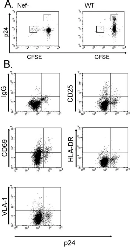
gen-erated in EC–T-cell cocultures represented the population that had proliferated, we labeled CD4⫹T cells with CFSE prior to placing them in coculture with allogeneic MHC class II⫹ECs and infecting them with HIV. In EC–T-cell cocultures infected with anef-deficient strain of virus (Nef⫺) (Fig. 1A), a CFSElow population accumulates, comprising the cells that have prolif-erated repetitively. This response to IFN-␥-pretreated alloge-neic ECs is indistinguishable from that of non-CD4⫹-infected cells (data not shown). However, in EC–T-cell cocultures in-fected with wild-type virus, we observed a novel population of p24highcells that remained CFSEhigh, indicating that they had not proliferated (Fig. 1A). The frequency of the p24high CFSEhighpopulation ranged between 5 and 20% of the starting population, which is substantially higher than the number of input T cells that can proliferate in response to allogeneic ECs
[image:3.585.307.533.72.502.2](0.1 to 1.0%) prior to expansion. Therefore, even if this entire population became p24high, without expansion via prolifera-tion, they could not account for the majority of the total p24high population. An unexpected finding was that in cultures in-fected by wild-type virus, but not by Nef⫺virus, there was a conspicuous absence of CFSElowcells. We suspect that wild-type virus either inhibited T-cell proliferation or, more likely, mediated the death of infected T cells. We did not investigate this issue further in the present study. In the remainder of
FIG. 1. Characterization of HIV type 1-infected CD4⫹ T cells. (A) FACS analyses of CFSE-labeled CD4⫹ T cells cocultured with ECs, infected with either Nef⫺or wild-type (WT) HIV, and harvested on day 9 postinfection. The thin-walled box identifies the CFSEhigh p24highcells, and the thick-walled box identifies the CFSElowp24low cells. Note the expansion of CFSEhighp24highcells in the wild-type-infected cultures. (B) FACS analyses of wild-type HIV-wild-type-infected T cells after 9 days of coculture with ECs stained with antibodies against IgG isotype control, CD25, CD69, HLA-DR, and VLA-1. Note that p24high cells express CD69 but not other markers of activation. The data in panels A and B are from one of three experiments with similar results.
on November 8, 2019 by guest
http://jvi.asm.org/
this study, we focused on the characterization of the p24high CFSEhighvirus producers that emerge when wild-type infected T cells are cocultured with ECs.
We initially examined the activation status of the high-virus-producing T cells. We analyzed the p24highcells for surface expression of early, intermediate, and late activation markers. A few hours after TCR engagement, T cells express the early activation markers CD25, a component of the high-affinity IL-2 receptor, and CD69, a molecule with unknown function. After 24 h of activation, human T cells will stably upregulate MHC class II molecules (e.g., HLA-DR) (15), and after a week will stably upregulate the1integrin, VLA-1 (20). FACS analysis revealed that on day 9 postinfection, the p24highcells did not express CD25, HLA-DR, or VLA-1, although some did ex-press the activation marker CD69 (Fig. 1B). These data are consistent with minimal (and perhaps a very early stage of) activation.
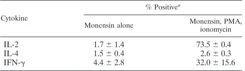
Cytokine synthesis, which can be measured by intracellular staining, is another hallmark of CD4⫹T-cell activation (29). We therefore proceeded to examine the cytokines expressed by the high virus producers. We harvested the cells 9 days after infection and performed intracellular cytokine staining for ef-fector cytokines, IL-2, IL-4, and IFN-␥, using monensin to enhance the signal by blocking secretion. FACS analysis re-vealed that the virally infected cells did not appear to be producing cytokines in the presence of MHC class II-express-ing ECs (Table 1). Intracellular cytokine stainII-express-ing of replicate cultures showed that these same cells are capable of producing IL-2 and IFN-␥in response to pharmacological stimulation by PMA plus ionomycin (Table 1). EC-stimulated cells after 9 days of culture do not produce IL-4 with or without PMA and ionomycin. The absence of intracellular cytokine staining is thus also suggestive of minimal T-cell activation.
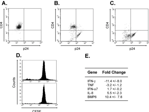
We also characterized the profile of cytokines produced by these cells at the mRNA level. To do so, we took advantage of the well-described abilities of Nef, Vpu, and Env to downregu-late surface CD4 in order to isodownregu-late a population enriched in virus producers (2, 7, 62, 66). Uninfected T cells are uniformly CD4high p24low (Fig. 2A). In contrast, in the HIV-infected cocultures, we found three populations, a CD4highp24low pop-ulation, a CD4lowp24lowpopulation, and a CD4lowp24highcell population (Fig. 2B). In other words, the p24high cells are almost exclusively CD4low, presumably due to high levels of expression of HIV protein. By selecting the cells based on CD4 expression, we enriched for a population of the high virus producers (Fig. 2C). In parallel, we isolated the nonproliferat-ing CFSEhighcells from an uninfected replicate culture by cell
sorting (Fig. 2D). We thus collected RNAs from CD4low in-fected cells and CFSEhighuninfected cells from replicate EC– T-cell cocultures and analyzed them by using Human Common Cytokine Superarrays. In general, the virally infected cells closely resembled the unactivated T-cell population (data not shown). After normalization to GAPDH (glyceraldehyde-3-phosphate dehydrogenase), these cells actually expressed lower levels of IFN-␥RNA than unactivated T cells isolated from the same donor (Fig. 2E). However, consistent with being HIV infected, the p24highT cells produced more IFN-␣7 and IL-8 RNA, the latter presumably due to the effects of Tat (45). The p24high cells also consistently expressed more BMP6 RNA, the function of which is unknown. Despite these few differences, the results from the Superarray analyses generally confirm the lack of T-cell activation.
High virus producers are not arrested in G2by Vpr.Since
virus-producing cells were not dividing, we tested whether an antiproliferative HIV protein could be responsible for causing cell cycle arrest of the host cell. Specifically, it has been re-ported that CD4⫹T cells that have been productively infected with wild-type virus are nonproliferating despite strong stimu-lation (4), whereas CD4⫹T cells infected with avpr-deficient strain (Vpr⫺) that were similarly stimulated underwent multi-ple cell divisions (44). Vpr can arrest infected cells in G2(19, 23, 49), which is advantageous to the virus because G2is the phase of the cell cycle that is optimal for viral replication (18). Strikingly, Vpr contributes strongly to viral replication in EC-stimulated T cells, since its absence results in a 10- to 20-fold reduction in cell-free virus (Fig. 3A). The number of p24high CFSEhighT cells is also drastically reduced in EC–T-cell co-cultures infected with Vpr⫺ virus (Fig. 3B), consistent with reduced cell-free virus levels. Because the p24highpopulation is essentially absent in the Vpr⫺-infected cultures, it was not possible to determine if Vpr actually caused growth arrest. To determine whether the cells infected with wild-type virus were, in fact, arrested in a particular stage of the cell cycle, we stained the T cells with propidium iodide. FACS analysis re-vealed that the p24highcells are not arrested in G
2or in any other specific stage of the cell cycle but instead represent a heterogeneous population with most of the p24high cells in G0/G1(Fig. 3C). Rapamycin inhibits mTOR, which is required for the progression of IL-2-stimulated cells from G1to S (1). To further test if progression beyond G1is required for en-hanced HIV production, we added rapamycin at concentra-tions that were sufficient to inhibit or completely block prolif-eration of polyclonally activated T cells (Fig. 3D). We observed that addition of rapamycin reduced viral replication (though not nearly to the extent of deletion ofvpr) (Fig. 3E) in our in vitro model using previously resting T cells. Our cumulative observations suggest that the necessity for Vpr for high levels of viral replication in p24high CFSEhigh cells cannot be ex-plained by its ability to induce cell cycle arrest.
TCRs and not cytokine signals provide minimal activation
for viral replication.In our previous study, we showed that the
[image:4.585.43.286.90.161.2]ability of ECs to enhance viral replication in cocultured CD4⫹ T cells requires endothelial expression of MHC class II and costimulators, yet our new data revealed that the p24high T cells showed little evidence of T-cell activation (9). Two mod-els can explain viral replication in minimally activated T cells. In the bystander model, the T cells that are p24high do not
TABLE 1. The p24highcells in EC–T-cell cocutures do not spontaneously produce cytokines
Cytokine
% Positivea
Monensin alone Monensin, PMA,
ionomycin
IL-2 1.7⫾1.4 73.5⫾0.4
IL-4 1.5⫾0.4 2.6⫾0.3
IFN-␥ 4.4⫾2.8 32.0⫾15.6
a
Percentage of the p24high
cells that were positive for each intracellular cytokine. These data are from one of three experiments with similar results.
on November 8, 2019 by guest
http://jvi.asm.org/
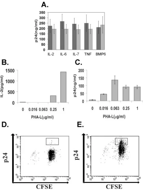
directly recognize MHC class II on the EC surface. Instead, the signals that promote viral replication in these nonproliferating cells, i.e., cytokines, are provided intransby other T cells which are highly or moderately activated. In the suboptimal-signaling model, the T cells that are p24high directly recognize MHC class II on the EC surface but receive signals that are insuffi-cient to fully activate the cells to proliferate and/or elaborate cytokines. Such signals may be provided by EC MHC class II molecules to cells bearing TCRs that interact with low affinity. Although activated T cells appeared to have died off by day 9 postinfection, we did detect cytokines, such as IL-2, in the medium on day 1 postinfection at levels that were similar to those found in uninfected EC–T-cell cocultures (9). Therefore, to test whether the bystander model was operative in our in vitro system, we added blocking antibodies to IL-2, IL-6, IL-7, TNF, or BMP-6, which have been identified as cytokines that can support TCR-independent viral replication (54, 61, 63), to EC–T-cell cocultures. These treatments did not inhibit viral
replication (Fig. 4A), results that are inconsistent with the bystander model.
[image:5.585.54.529.69.428.2]To test whether the suboptimal-signaling model was opera-tive, we cocultured CD4⫹T cells with untreated ECs, added suboptimal doses of PHA-L, and infected them with wild-type virus. Under such conditions, ECs continue to provide co-stimulation, but the TCR-mediated signals are mediated pri-marily through PHA-L rather than MHC recognition. PHA-L provides a dose-dependent increase in the IL-2 produced (Fig. 4B) and dose-dependent proliferation (data not shown). Strik-ingly, suboptimal doses of PHA-L that are insufficient to in-duce detectable cytokine production still support efficient viral replication (Fig. 4C). We next assessed the ability of subopti-mal stimulation with PHA-L to induce a CFSEhigh p24high population. To do so, we labeled the T cells with CFSE prior to adding them to EC cultures and infecting them. On day 7 postinfection, the T cells were harvested and counterstained for p24. We found that cultures activated with suboptimal
FIG. 2. Changes in gene expression induced by HIV infection. FACS analyses after 9 days of EC–T-cell coculture of (A) uninfected CD4⫹cells, (B) wild-type HIV-infected T cells without selection, and (C) wild-type HIV-infected T cells following depletion of CD4highcells. (D) FACS histogram of replicate uninfected cultures without sorting (top) and after sorting (bottom) for the CFSEhighpopulation. (E) Human Common Cytokine Superarray Analyses of RNAs isolated from CD4lowcells from wild-type virus-infected EC–T-cell cocultures compared with unactivated CD4⫹T cells from the same donor. Cytokine expression levels were normalized to GAPDH. Depicted are genes that displayed a⬎1.5-fold change in expression in CD4lowcells (presumably infected) versus CFSEhighcells (from uninfected cultures). The data in panels A to E are from one of three experiments with similar results.
on November 8, 2019 by guest
http://jvi.asm.org/
stimulation, i.e., 0.0625g/ml of PHA-L, displayed negligible T-cell proliferation, but compared to no PHA-L, they show a significant increase in the number of p24highCFSEhigh cells (Fig. 4D and E). These data support the idea that a suboptimal TCR signal promotes HIV replication in T cells cocultured with ECs.
To determine more directly whether the p24highcells were receiving MHC class II signals from ECs, we used a different type of polyclonal stimulator, namely, the superantigen TSST-1. TSST-1 binds to a conserved site on MHC class II molecules and to a site on the V2 chain of the TCR (17). Therefore, like PHA-L, TSST-1 can activate T cells primarily by simulating TCR-MHC interactions. However, unlike PHA-L, TSST-1 requires expression of endothelial MHC class II, and more importantly, TSST-1-mediated activation, unlike PHA-L-mediated activation, is restricted to those T cells that express TCRs containing the V2 receptor chain. We found that TSST-1, like PHA-L, provided a dose-dependent increase in cytokine production (Fig. 5A). If the suboptimal-signaling model were true, TSST-1 should selectively increase viral rep-lication among the cells that express V2. To test this, we cocultured MHC class II-expressing ECs with unselected, rest-ing CD4⫹ T cells in the presence of increasing doses of TSST-1. On day 7 postinfection, we harvested the T cells and stained them for extracellular V2 and intracellular p24. In the absence of TSST-1 (Fig. 5B), the p24highcells, like the input cells, were predominantly V2⫺. In the presence of suboptimal TSST-1, we found that the p24highcells were enriched
selec-tively in the V2⫹ population (Fig. 5C). TSST-1 did not change the proportion of V2⫺ cells that were p24high. In contrast, suboptimal TSST-1 increased the proportion of V2⫹ cells that were p24highby sevenfold (Fig. 5C). These observa-tions directly confirm a role for suboptimal TCR signaling in promoting HIV replication.
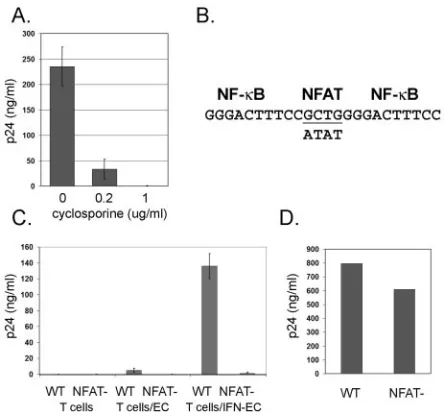
[image:6.585.56.533.70.313.2]Finally, we investigated signals downstream of the TCR that could underlie the EC effect. Cyclosporine inhibits the TCR-dependent activation of calcineurin. Therefore, to assess a role for calcineurin in our in vitro model, we added cyclosporine to EC–T-cell cocultures. Since previous studies had shown that ECs confer cyclosporine resistance on CD4⫹T cells (24), we chose relatively high doses of cyclosporine, namely, 0.2g/ml and 1.0g/ml. Such concentrations are consistent with doses previously used to examine the impact of calcineurin on HIV replication with minimal effect on cyclophilin A incorporation into virions (11, 26). The addition of cyclosporine significantly inhibited viral replication (Fig. 6A). The major target of cyclo-sporine is thought to be calcineurin-dependent activation of NFAT. To directly test whether an NFAT-dependent mecha-nism underlies the EC effect, we disrupted the NFAT binding site in the HIV LTR (26, 27) (Fig. 6B) and tested if this affected the capacity of ECs to enhance HIV production. The recombinant HIV (NFAT⫺) replicated to only very low levels in comparison to the wild-type derivative strain in previously resting CD4⫹T cells cocultured with IFN-␥-treated ECs (Fig. 6C). To ensure that this phenotype was not due to inherently poor NFAT⫺replicative capacity, it was also grown in CD4⫹T
FIG. 3. Characterization of the role of Vpr in enhanced HIV production. (A) Replication of wild-type versus Vpr⫺ HIV in EC–T-cell cocultures 9 days postinfection as assessed by p24 measured in the supernatant by ELISA. (B) CFSE staining of infected cells harvested on day 9 postinfection. The box identifies the CFSEhighp24highcells, which are fewer in cultures infected by Vpr⫺virus (right). (C) PI staining of infected
cells harvested on day 9 postinfection. The lower box identifies cells in the G0/G1state, and the upper box identifies the cells in the G2state of the cell cycle. Note that the majority of cells are in G0/G1. (D) FACS analyses of CFSE-labeled CD4⫹T cells treated with PHA-L and rapamycin. Note that the cells remain CFSEhigh. (E) Replication of wild-type HIV in EC–T-cell cocultures treated with rapamycin or vehicle (dimethyl sulfoxide, 0 nM rapamycin) 9 days postinfection as assessed by p24 measured in the supernatant by ELISA. The data in panels A to E are from one of three experiments with similar results. The error bars indicate standard deviations.
on November 8, 2019 by guest
http://jvi.asm.org/
cells pretreated with PHA and IL-2. Under these conditions, NFAT⫺virus replicated similarly to wild-type virus (Fig. 6D).
DISCUSSION
In the present study, we made the unexpected finding that HIV replication in EC–T-cell cocultures actually occurs exclu-sively in memory CD4⫹T cells that are only minimally acti-vated. We cannot rule out the possibility that highly or mod-erately activated cells contributed to viral replication but died before they could be detected. However, on day 9 postinfec-tion, i.e., the peak of viral replicapostinfec-tion, the cells that are pro-ducing the virus (the p24highT cells) are neither proliferating nor arrested at a specific stage of the cell cycle. Furthermore, other than CD69 expression, the p24highT cells do not express activation markers or effector cytokines. In our system, the
[image:7.585.48.278.69.375.2]virally infected T cells are identified by FACS analysis, which can occur only after they express sufficient intracellular viral proteins. Therefore, we cannot rule out the possibility that the cells had previously demonstrated markers of activation, i.e., surface markers and effector cytokines, but had subsequently downregulated them. This seems unlikely, since we examined the cells for early, intermediate, and late activation markers, and we found that the p24highT cells did not express CD25, HLA-DR, or VLA-1 and only small amounts of CD69. Our results using T cells activated by ECs stand in striking contrast to previous studies, which used similar FACS-based techniques
[image:7.585.305.534.71.452.2]FIG. 4. TCRs and not cytokine signals provide minimal activation necessary for HIV replication. (A) Replication of wild-type HIV in EC–T-cell cocultures 9 days postinfection treated with the indicated blocking antibodies (dark bars) or isotype controls (light bars) as assessed by p24 measured in the supernatant by ELISA. (B) IL-2 measured in the supernatant of infected T cells after 1 day of coculture with ECs in the presence of various doses of PHA-L or vehicle alone. (C) Cell-free virus measured in the supernatant of infected EC–T-cell cocultures on day 7 postinfection as assessed by p24 measured in the supernatant by ELISA. (D and E) FACS analyses of CD4⫹ T cells cocultured with untreated ECs in the presence of (D) 0g/ml and (E) 0.063g/ml of PHA-L. The boxes identify the CFSEhighp24high cells. Note the expansion of CFSEhighp24highcells in the wild-type-infected cultures in the presence of 0.063g/ml of PHA-L. The data in panels A to E are from one of three experiments with similar results. The error bars indicate standard deviations.
FIG. 5. TSST-1 selectively leads to viral replication in V2⫹T cells. (A) IL-2 measured in the supernatant of infected T cells after 1 day of coculture with ECs in the presence of increasing doses of TSST-1 or vehicle alone. (B) FACS analyses of CD4⫹ T cells cocultured with IFN-␥-pretreated ECs in the presence of 0 ng/ml or 1 ng/ml of TSST-1. The upper boxes identify the V2⫹p24highcells, and the lower boxes identify the V2⫺ p24high cells. Note the expansion on the V2⫹
p24highcells in the presence of 1 ng/ml TSST-1. The data in panels A and B are from one of three experiments with similar results. (C) Pro-portions of p24highcells among the V2⫹and the V2⫺populations in
infected EC-T cell cocultures stimulated with 0 or 1 ng/ml of TSST-1. Shown are the means from three independent experiments⫾standard error.
on November 8, 2019 by guest
http://jvi.asm.org/
to show that viral replication under other conditions is re-stricted to highly activated T cells (4, 44).
In a fully quiescent cell, HIV exists as incomplete or com-plete cDNA, neither of which is stable long term, and both are essentially transcriptionally silent (58, 68, 71). Therefore, even in cells that demonstrate limited markers of activation, viral replication appears to require the activation of specific signal-ing pathways. In the current study, we tested two possible means by which MHC molecule- and costimulation-dependent signals could support viral replication in EC-stimulated T cells but not proliferation or cytokine production from these same T cells. In the bystander model, the high virus producers receive selective activation via a factor, i.e., a cytokine produced by other CD4⫹T cells that are highly or moderately activated. This possibility is supported by a previous study, which showed that in autologous dendritic-cell–T-cell cocultures and supe-rantigen, productive HIV replication occurs not only in the superantigen-activated T cells but also in the bystander cells, which did not receive direct TCR activation and were therefore stimulated via cytokines (51). However, in contrast to the pre-vious report on dendritic-cell–T-cell cocultures, we found no direct evidence of highly activated T cells in the EC–T-cell cocultures at the time of peak virus production. In fact, there
was a striking absence of the expected CFSElow population, i.e., the population that proliferated in wild-type-infected cul-tures. In addition, rapamycin, an inhibitor of cytokine-depen-dent mTOR activation, only minimally inhibits viral replica-tion, and the addition of blocking antibodies to IL-2, IL-6, IL-7, and TNF, which have been identified as cytokines that can support TCR-independent viral replication (54, 61, 63), had no effect on viral replication in EC–T-cell cocultures. Our data suggest that the bystander model does not explain why the p24highT cells in cocultures with ECs appear minimally acti-vated.
An alternative to the bystander model is the suboptimal-signaling model. There is a hierarchical activation of T-cell functions, and different functions are activated sequentially with increasing TCR stimulus. For example, T-cell prolifera-tion requires higher stimulaprolifera-tion than does cytokine producprolifera-tion (5). In the suboptimal-signaling model, the high virus produc-ers directly recognize endothelial MHC class II molecules but do not receive a signal sufficiently strong to drive the cell to proliferation or to cytokine production. Our results have led us to favor this model. The experiments with PHA-L demonstrate that suboptimal TCR-mediated stimulation is sufficient for ef-ficient virus replication; furthermore, the experiments with TSST-1 demonstrate that viral replication is restricted to T cells whose TCRs are engaged by endothelial MHC class II molecules. We have also examined the effects of cyclosporine, an inhibitor of the TCR-activated enzyme calcineurin. Al-though this drug has several nonspecific effects (52, 60), its major action is blocking the calcineurin-dependent activation of the transcription factor NFAT. We have demonstrated that cyclosporine, in contrast to rapamycin, effectively inhibits viral replication. More directly, we also showed that the alteration of the NFAT binding site in the HIV LTR blocks the EC effect on HIV replication.
Studies with altered peptide ligands (APLs) have shown that suboptimal T-cell activation preserves some aspects of full T-cell activation but not others (56). Suboptimal T-cell activa-tion with APLs induces only partial TCR- phosphorylation, which in turn leads to the inefficient recruitment of Src family kinases. The biochemical consequence of this is the selective activation of downstream targets. For example, APL-induced calcium signaling, which is reduced and shorter in duration than agonist peptide-induced calcium signaling, can contribute to the selective activation of NF-B or of NFAT (30, 57). In contrast to the IL-2 promoter, where NFAT binds as a het-erodimer with AP-1, NFAT binds in the LTR enhancer as a homodimer (47). Therefore, while IL-2 expression requires both NFAT activation and AP-1 activation (35), HIV replica-tion can occur with the selective activareplica-tion of NFAT (26). Independent studies have shown that selective NFAT activa-tion produces a phenotype similar to that of EC stimulaactiva-tion in HIV-infected T cells. NFAT-driven HIV production leads to preferential infection of memory T cells (50), and viral repli-cation occurs without T-cell proliferation, activation marker expression, or cytokine expression (26). Lastly, selective NFAT activation, like HIV replication in our system, can be aug-mented by EC-specific factors. Wnt, produced by ECs, can inhibit glycogen synthase kinase-3(GSK-3), which in turn inhibits the nuclear export of NFAT (42).
[image:8.585.52.275.68.274.2]A primary role for NFAT in EC-stimulated memory T cells
FIG. 6. Characterization of the role of NFAT in HIV production. (A) HIV release on day 9 postinfection (representing the peak) from EC–T-cell cocultures treated with cyclosporine or vehicle (ethanol). The data are from one of three experiments with similar results. (B) Schematic diagram of the region of the HIV LTR that contains the NF-B and NFAT binding sites. The underlined nucleotides are re-quired for NFAT-LTR interactions. The nucleotides on the lower line indicate the loci that differ between the NFAT⫺strain and the wild-type derivative strain (upper line). (C) HIV released from T cells on day 9 postinfection (representing the peak). T cells represents T cells alone, T cells/EC represents T cells cocultured with untreated ECs, and T cells/IFN-EC represents T cells cocultured with ECs pretreated with IFN-␥. The data are from one of two experiments with similar results. (D) HIV released from CD4⫹T cells isolated from PBMCs and pretreated with PHA and IL-2 on day 8 postinfection (represent-ing the peak). Similar results were obtained us(represent-ing the cell line CEMx174 (not shown). HIV release was assessed by p24 measured in the supernatant by ELISA. The error bars indicate standard devia-tions.
on November 8, 2019 by guest
http://jvi.asm.org/
may explain the important roles of Nef and Vpr in our model system. Nef is expressed even before the virus is integrated (67). In addition, Nef can interact directly with T-cell signaling molecules, including Lck, Vav, Pak2, and PKC-(37), and in response to TCR-derived signals, Nef has been shown to po-tentiate specific TCR-dependent signaling pathways, including those involving ERK (53), MAPK (37), PI-3-kinase (32), and Ca2⫹(38). The rise in intracellular Ca2⫹is the mechanism that leads to calcineurin and NFAT activation. The dependence of the EC effect on an intactnefgene is reminiscent of the effect that HIV-infected macrophages have on the infectivity of rest-ing CD4⫹T cells (59). However, the effect of Nef is operative in the macrophages and not in the resting T cells, whereas in the EC system, the effect of Nef is operative in the resting T cells and not in the ECs. We have shown that ECs, but not other antigen-presenting cells, greatly enhance Nef-dependent viral replication in resting T cells (9). Combined with the find-ings from this study, we suggest that the primary role of Nef is to serve as a signaling molecule that specifically modulates EC-specific, TCR-dependent signals, notably NFAT. Thus, Nef may play multiple roles in the establishment of an HIV-favorable microenvironment in peripheral tissue.
Vpr, a viral protein that is necessary for viral pathogenesis in vivo (33), contributes to viral replication in EC–T-cell cocul-tures in a manner that is independent of its ability to induce G2 arrest. Although we have not determined if the requirement for Vpr occurs prior to or subsequent to integration, we hy-pothesize that Vpr’s ability to promote HIV transcription may underlie its contribution to EC-stimulated viral replication. In support of this, Vpr, a nuclear protein, has been shown to alter signal transduction pathways via direct modulation of tran-scription factor activity in the nucleus (25, 28), and Vpr-medi-ated enhancement of HIV transcription is most pronounced under conditions of suboptimal activation. Vpr’s ability to ac-tivate cyclic AMP responsive element binding factor (CREB) is evident only under conditions that lead to the serine phos-phorylation of CREB but not to CREB-dependent gene ex-pression (28). In addition, Vpr’s ability to potentiate LTR transcription driven by Sp1, a transcription factor that is active in resting CD4⫹ T cells (64), is attenuated both by NF-B activation and by progression of the cell into G2. Moreover, Vpr has been shown to synergize with Nef in augmenting calcineurin-dependent NFAT activation (36–38), which we postulate contributes to the EC effect.
The role of minimal TCR signaling by ECs fits into a general pattern in which HIV subverts normal immune function. CD4⫹T cells require TCR-dependent signals for maintenance of immunological memory (39, 55). In humans, MHC class II molecules are displayed on vascular ECs (8). Therefore, as human memory cells circulate through the blood, ECs are well situated to provide the suboptimal TCR signals required for maintenance of immunological memory. We propose that HIV exploits these signals for virus production. Specifically, we hy-pothesize that EC-expressed MHC class II molecules, perhaps containing self peptides, provide nonredundant signals that enhance virus replication in circulating, latently infected mem-ory T cells.
Our findings have implications for the course of HIV infec-tion in patients. During HIV and SIV infecinfec-tion, cells with limited markers of T-cell activation are productively infected
(70). Furthermore, in the initial weeks of SIV infection, mem-ory T cells are greatly depleted in multiple tissues (which suggests that high-affinity MHC class II-TCR interactions are not required for efficient HIV production in human tissue) and resting memory T cells are specifically depleted in the GALT (31, 40). The loss of these cells (which results directly from viral infection and not via a bystander mechanism) is thought to establish conditions that eventually result in progression to AIDS. Ex vivo analysis of infected patients has shown that resting memory cells represent the T-cell compartment that harbors the highest copy number of viral DNA (6). Further-more, latently infected memory cells are thought to contribute to the viremic “blips” in patients on ART, as well as to break-through virus in patients whose ART regimens are changed and rebound virus in patients who discontinue ART (10, 69). All of these phenomena may involve EC effects on infected memory T cells of the kind described in the present report.
In summary, we have described a novel manner in which vascular ECs can boost HIV replication in minimally activated CD4⫹ T cells. This response depends upon recognition of molecules on the ECs and involves the viral genesnefandvpr
and TCR-dependent activation of NFAT. These findings illus-trate yet another way in which HIV exploits the human im-mune system and may contribute to rebound following discon-tinuance of ART in HIV-infected patients and the depletion of resting memory T cells in the GALT in the initial weeks of infection.
ACKNOWLEDGMENTS
This work was supported by NIH grant R01 HL51014 and American Cancer Society institutional research grant IRG 58-012-42. J.C. and P.W. were supported by NIH training grants GM07205 and T32CA09085, respectively; J.W. was supported by the National Sci-ence Foundation Graduate Research Fellowship Program.
REFERENCES
1.Abraham, R. T., and G. J. Wiederrecht.1996. Immunopharmacology of
rapamycin. Annu. Rev. Immunol.14:483–510.
2.Aiken, C., J. Konner, N. R. Landau, M. E. Lenburg, and D. Trono.1994. Nef induces CD4 endocytosis: requirement for a critical dileucine motif in the
membrane-proximal CD4 cytoplasmic domain. Cell76:853–864.
3.Alexander, L., Z. Du, M. Rosenzweig, J. U. Jung, and R. C. Desrosiers.1997. A role for natural simian immunodeficiency virus and human
immunodefi-ciency virus type 1nefalleles in lymphocyte activation. J. Virol.71:6094–
6099.
4.Bahbouhi, B., A. Landay, and L. Al-Harthi.2004. Dynamics of cytokine
expression in HIV productively infected primary CD4⫹T cells. Blood103:
4581–4587.
5.Bielekova, B., and R. Martin.2001. Antigen-specific immunomodulation via
altered peptide ligands. J. Mol. Med.79:552–565.
6.Brenchley, J. M., B. J. Hill, D. R. Ambrozak, D. A. Price, F. J. Guenaga, J. P. Casazza, J. Kuruppu, J. Yazdani, S. A. Migueles, M. Connors, M. Roederer, D. C. Douek, and R. A. Koup.2004. T-cell subsets that harbor human immunodeficiency virus (HIV) in vivo: implications for HIV pathogenesis.
J. Virol.78:1160–1168.
7.Buonocore, L., and J. K. Rose.1990. Prevention of HIV-1 glycoprotein transport by soluble CD4 retained in the endoplasmic reticulum. Nature
345:625–628.
8.Choi, J., D. R. Enis, K. P. Koh, S. L. Shiao, and J. S. Pober.2004. T
lymphocyte-endothelial cell interactions. Annu. Rev. Immunol.22:683–709.
9.Choi, J., J. Walker, S. Boichuk, N. Kirkiles-Smith, N. Torpey, J. S. Pober, and L. Alexander.2005. Human endothelial cells enhance human
immuno-deficiency virus type 1 replication in CD4⫹T cells in a Nef-dependent
manner in vitro and in vivo. J. Virol.79:264–276.
10.Chun, T. W., R. T. Davey, Jr., M. Ostrowski, J. Shawn Justement, D. Engel, J. I. Mullins, and A. S. Fauci.2000. Relationship between pre-existing viral reservoirs and the re-emergence of plasma viremia after discontinuation of
highly active anti-retroviral therapy. Nat. Med.6:757–761.
11.Cron, R. Q., S. R. Bartz, A. Clausell, S. J. Bort, S. J. Klebanoff, and D. B.
on November 8, 2019 by guest
http://jvi.asm.org/
Lewis.2000. NFAT1 enhances HIV-1 gene expression in primary human
CD4 T cells. Clin. Immunol.94:179–191.
12.Dengler, T. J., and J. S. Pober.2000. Human vascular endothelial cells
stimulate memory but not naive CD8⫹T cells to differentiate into CTL
retaining an early activation phenotype. J. Immunol.164:5146–5155.
13.Eckstein, D. A., M. L. Penn, Y. D. Korin, D. D. Scripture-Adams, J. A. Zack, J. F. Kreisberg, M. Roederer, M. P. Sherman, P. S. Chin, and M. A. Gold-smith.2001. HIV-1 actively replicates in naive CD4⫹T cells residing within
human lymphoid tissues. Immunity15:671–682.
14.Epperson, D. E., and J. S. Pober. 1994. Antigen-presenting function of human endothelial cells. Direct activation of resting CD8 T cells. J.
Immu-nol.153:5402–5412.
15.Evans, R. L., T. J. Faldetta, R. E. Humphreys, D. M. Pratt, E. J. Yunis, and S. F. Schlossman.1978. Peripheral human T cells sensitized in mixed
leu-kocyte culture synthesize and express Ia-like antigens. J. Exp. Med.148:
1440–1445.
16.Fortin, J. F., C. Barat, Y. Beausejour, B. Barbeau, and M. J. Tremblay.2004. Hyper-responsiveness to stimulation of human immunodeficiency
virus-in-fected CD4⫹T cells requires Nef and Tat virus gene products and results
from higher NFAT, NF-B, and AP-1 induction. J. Biol. Chem.279:39520–
39531.
17.Fraser, J., V. Arcus, P. Kong, E. Baker, and T. Proft.2000. Superantigens—
powerful modifiers of the immune system. Mol. Med. Today6:125–132.
18.Goh, W. C., M. E. Rogel, C. M. Kinsey, S. F. Michael, P. N. Fultz, M. A. Nowak, B. H. Hahn, and M. Emerman.1998. HIV-1 Vpr increases viral expression by manipulation of the cell cycle: a mechanism for selection of
Vpr in vivo. Nat. Med.4:65–71.
19.He, J., S. Choe, R. Walker, P. Di Marzio, D. O. Morgan, and N. R. Landau.
1995. Human immunodeficiency virus type 1 viral protein R (Vpr) arrests
cells in the G2phase of the cell cycle by inhibiting p34cdc2 activity. J. Virol.
69:6705–6711.
20.Hemler, M. E., J. G. Jacobson, M. B. Brenner, D. Mann, and J. L. Strominger.1985. VLA-1: a T cell surface antigen which defines a novel late
stage of human T cell activation. Eur. J. Immunol.15:502–508.
21.Ho, D. D., A. U. Neumann, A. S. Perelson, W. Chen, J. M. Leonard, and M. Markowitz.1995. Rapid turnover of plasma virions and CD4 lymphocytes in
HIV-1 infection. Nature373:123–126.
22.Jamieson, B. D., and J. A. Zack.1998. In vivo pathogenesis of a human
immunodeficiency virus type 1 reporter virus. J. Virol.72:6520–6526.
23.Jowett, J. B., V. Planelles, B. Poon, N. P. Shah, M. L. Chen, and I. S. Chen.
1995. The human immunodeficiency virus type 1vprgene arrests infected T
cells in the G2⫹M phase of the cell cycle. J. Virol.69:6304–6313.
24.Karmann, K., J. S. Pober, and C. C. Hughes.1994. Endothelial cell-induced resistance to cyclosporin A in human peripheral blood T cells requires contact-dependent interactions involving CD2 but not CD28. J. Immunol.
153:3929–3937.
25.Kino, T., A. Gragerov, O. Slobodskaya, M. Tsopanomichalou, G. P. Chrou-sos, and G. N. Pavlakis. 2002. Human immunodeficiency virus type 1 (HIV-1) accessory protein Vpr induces transcription of the HIV-1 and glu-cocorticoid-responsive promoters by binding directly to p300/CBP
coactiva-tors. J. Virol.76:9724–9734.
26.Kinoshita, S., B. K. Chen, H. Kaneshima, and G. P. Nolan.1998. Host control of HIV-1 parasitism in T cells by the nuclear factor of activated T
cells. Cell95:595–604.
27.Kinoshita, S., L. Su, M. Amano, L. A. Timmerman, H. Kaneshima, and G. P. Nolan.1997. The T cell activation factor NF-ATc positively regulates HIV-1
replication and gene expression in T cells. Immunity6:235–244.
28.Lahti, A. L., A. Manninen, and K. Saksela.2003. Regulation of T cell activation by HIV-1 accessory proteins: Vpr acts via distinct mechanisms to cooperate with Nef in NFAT-directed gene expression and to promote
transactivation by CREB. Virology310:190–196.
29.Laskay, T., U. Andersson, J. Andersson, R. Kiessling, and M. DeLey.1986. An immunofluorescent method for identifying individual
IFN-gamma-pro-ducing lymphocytes. J. Immunol. Methods95:1–7.
30.Lewis, R. S.2003. Calcium oscillations in T-cells: mechanisms and
conse-quences for gene expression. Biochem. Soc. Trans.31:925–929.
31.Li, Q., L. Duan, J. D. Estes, Z. M. Ma, T. Rourke, Y. Wang, C. Reilly, J. Carlis, C. J. Miller, and A. T. Haase.2005. Peak SIV replication in resting
memory CD4⫹T cells depletes gut lamina propria CD4⫹T cells. Nature
434:1148–1152.
32.Linnemann, T., Y. H. Zheng, R. Mandic, and B. M. Peterlin.2002. Interac-tion between Nef and phosphatidylinositol-3-kinase leads to activaInterac-tion of
p21-activated kinase and increased production of HIV. Virology294:246–
255.
33.Lum, J. J., O. J. Cohen, Z. Nie, J. G. Weaver, T. S. Gomez, X. J. Yao, D. Lynch, A. A. Pilon, N. Hawley, J. E. Kim, Z. Chen, M. Montpetit, J. Sanchez-Dardon, E. A. Cohen, and A. D. Badley.2003. Vpr R77Q is associated with long-term nonprogressive HIV infection and impaired induction of
apopto-sis. J. Clin. Investig.111:1547–1554.
34.Ma, W., and J. S. Pober.1998. Human endothelial cells effectively
costimu-late cytokine production by, but not differentiation of, naive CD4⫹T cells.
J. Immunol.161:2158–2167.
35.Macian, F., C. Garcia-Rodriguez, and A. Rao.2000. Gene expression elicited by NFAT in the presence or absence of cooperative recruitment of Fos and
Jun. EMBO J.19:4783–4795.
36.Manninen, A., P. Huotari, M. Hiipakka, G. H. Renkema, and K. Saksela.
2001. Activation of NFAT-dependent gene expression by Nef: conservation among divergent Nef alleles, dependence on SH3 binding and membrane
association, and cooperation with protein kinase C-theta. J. Virol.75:3034–
3037.
37.Manninen, A., G. H. Renkema, and K. Saksela.2000. Synergistic activation
of NFAT by HIV-1 nef and the Ras/MAPK pathway. J. Biol. Chem.275:
16513–16517.
38.Manninen, A., and K. Saksela.2002. HIV-1 Nef interacts with inositol trisphosphate receptor to activate calcium signaling in T cells. J. Exp. Med.
195:1023–1032.
39.Marrack, P., and J. Kappler.2004. Control of T cell viability. Annu. Rev.
Immunol.22:765–787.
40.Mattapallil, J. J., D. C. Douek, B. Hill, Y. Nishimura, M. Martin, and M. Roederer.2005. Massive infection and loss of memory CD4⫹T cells in
multiple tissues during acute SIV infection. Nature434:1093–1097.
41.Mestas, J., and C. C. Hughes.2001. Endothelial cell costimulation of T cell activation through CD58-CD2 interactions involves lipid raft aggregation.
J. Immunol.167:4378–4385.
42.Murphy, L. L., and C. C. Hughes.2002. Endothelial cells stimulate T cell NFAT nuclear translocation in the presence of cyclosporin A: involvement
of the wnt/glycogen synthase kinase-3 beta pathway. J. Immunol.169:3717–
3725.
43.Murray, A. G., P. Libby, and J. S. Pober.1995. Human vascular smooth
muscle cells poorly co-stimulate and actively inhibit allogeneic CD4⫹T cell
proliferation in vitro. J. Immunol.154:151–161.
44.Oswald-Richter, K., S. M. Grill, M. Leelawong, and D. Unutmaz.2004. HIV infection of primary human T cells is determined by tunable thresholds of T
cell activation. Eur. J. Immunol.34:1705–1714.
45.Ott, M., J. L. Lovett, L. Mueller, and E. Verdin.1998. Superinduction of IL-8
in T cells by HIV-1 Tat protein is mediated through NF-B factors. J.
Im-munol.160:2872–2880.
46.Perelson, A. S.2002. Modelling viral and immune system dynamics. Nat.
Rev. Immunol.2:28–36.
47.Pessler, F., and R. Q. Cron.2004. Reciprocal regulation of the nuclear factor
of activated T cells and HIV-1. Genes Immun.5:158–167.
48.Pober, J. S., M. P. Bevilacqua, D. L. Mendrick, L. A. Lapierre, W. Fiers, and M. A. Gimbrone, Jr.1986. Two distinct monokines, interleukin 1 and tumor necrosis factor, each independently induce biosynthesis and transient expres-sion of the same antigen on the surface of cultured human vascular
endo-thelial cells. J. Immunol.136:1680–1687.
49.Re, F., D. Braaten, E. K. Franke, and J. Luban.1995. Human
immunode-ficiency virus type 1 Vpr arrests the cell cycle in G2by inhibiting the
activa-tion of p34cdc2-cyclin B. J. Virol.69:6859–6864.
50.Robichaud, G. A., B. Barbeau, J. F. Fortin, D. M. Rothstein, and M. J. Tremblay.2002. Nuclear factor of activated T cells is a driving force for
preferential productive HIV-1 infection of CD45RO-expressing CD4⫹T
cells. J. Biol. Chem.277:23733–23741.
51.Scales, D., H. Ni, F. Shaheen, J. Capodici, G. Cannon, and D. Weissman.
2001. Nonproliferating bystander CD4⫹T cells lacking activation markers
support HIV replication during immune activation. J. Immunol.166:6437–
6443.
52.Schmidt, A., L. Hennighausen, and U. Siebenlist.1990. Inducible nuclear factor binding to the kappa B elements of the human immunodeficiency virus enhancer in T cells can be blocked by cyclosporin A in a signal-dependent
manner. J. Virol.64:4037–4041.
53.Schrager, J. A., V. Der Minassian, and J. W. Marsh.2002. HIV Nef increases
T cell ERK MAP kinase activity. J. Biol. Chem.277:6137–6142.
54.Scripture-Adams, D. D., D. G. Brooks, Y. D. Korin, and J. A. Zack.2002. Interleukin-7 induces expression of latent human immunodeficiency virus
type 1 with minimal effects on T-cell phenotype. J. Virol.76:13077–13082.
55.Seddon, B., and R. Zamoyska.2003. Regulation of peripheral T-cell
ho-meostasis by receptor signalling. Curr. Opin. Immunol.15:321–324.
56.Sloan-Lancaster, J., and P. M. Allen.1996. Altered peptide ligand-induced partial T cell activation: molecular mechanisms and role in T cell biology.
Annu. Rev. Immunol.14:1–27.
57.Sloan-Lancaster, J., T. H. Steinberg, and P. M. Allen.1997. Selective loss of the calcium ion signaling pathway in T cells maturing toward a T helper 2
phenotype. J. Immunol.159:1160–1168.
58.Stevenson, M., T. L. Stanwick, M. P. Dempsey, and C. A. Lamonica.1990. HIV-1 replication is controlled at the level of T cell activation and proviral
integration. EMBO J.9:1551–1560.
59.Swingler, S., B. Brichacek, J. M. Jacque, C. Ulich, J. Zhou, and M.
Steven-son.2003. HIV-1 Nef intersects the macrophage CD40L signalling pathway
to promote resting-cell infection. Nature424:213–219.
60.Thali, M., A. Bukovsky, E. Kondo, B. Rosenwirth, C. T. Walsh, J. Sodroski, and H. G. Gottlinger.1994. Functional association of cyclophilin A with
HIV-1 virions. Nature372:363–365.
61.Unutmaz, D., V. N. KewalRamani, S. Marmon, and D. R. Littman.1999.
on November 8, 2019 by guest
http://jvi.asm.org/
Cytokine signals are sufficient for HIV-1 infection of resting human T
lym-phocytes. J. Exp. Med.189:1735–1746.
62.Vincent, M. J., N. U. Raja, and M. A. Jabbar.1993. Human immunodefi-ciency virus type 1 Vpu protein induces degradation of chimeric envelope glycoproteins bearing the cytoplasmic and anchor domains of CD4: role of the cytoplasmic domain in Vpu-induced degradation in the endoplasmic
reticulum. J. Virol.67:5538–5549.
63.Wang, F. X., Y. Xu, J. Sullivan, E. Souder, E. G. Argyris, E. A. Acheampong, J. Fisher, M. Sierra, M. M. Thomson, R. Najera, I. Frank, J. Kulkosky, R. J. Pomerantz, and G. Nunnari.2005. IL-7 is a potent and proviral strain-specific inducer of latent HIV-1 cellular reservoirs of infected individuals on
virally suppressive HAART. J. Clin. Investig.115:128–137.
64.Wang, L., S. Mukherjee, F. Jia, O. Narayan, and L. J. Zhao.1995. Interac-tion of virion protein Vpr of human immunodeficiency virus type 1 with cellular transcription factor Sp1 and trans-activation of viral long terminal
repeat. J. Biol. Chem.270:25564–25569.
65.Wei, X., S. K. Ghosh, M. E. Taylor, V. A. Johnson, E. A. Emini, P. Deutsch, J. D. Lifson, S. Bonhoeffer, M. A. Nowak, B. H. Hahn, et al.1995. Viral
dynamics in human immunodeficiency virus type 1 infection. Nature373:
117–122.
66.Willey, R. L., F. Maldarelli, M. A. Martin, and K. Strebel.1992. Human
immunodeficiency virus type 1 Vpu protein induces rapid degradation of
CD4. J. Virol.66:7193–7200.
67.Wu, Y., and J. W. Marsh.2001. Selective transcription and modulation of
resting T cell activity by preintegrated HIV DNA. Science293:1503–1506.
68.Zack, J. A., S. J. Arrigo, S. R. Weitsman, A. S. Go, A. Haislip, and I. S. Chen.
1990. HIV-1 entry into quiescent primary lymphocytes: molecular analysis
reveals a labile, latent viral structure. Cell61:213–222.
69.Zhang, L., C. Chung, B. S. Hu, T. He, Y. Guo, A. J. Kim, E. Skulsky, X. Jin, A. Hurley, B. Ramratnam, M. Markowitz, and D. D. Ho.2000. Genetic characterization of rebounding HIV-1 after cessation of highly active
anti-retroviral therapy. J. Clin. Investig.106:839–845.
70.Zhang, Z., T. Schuler, M. Zupancic, S. Wietgrefe, K. A. Staskus, K. A. Reimann, T. A. Reinhart, M. Rogan, W. Cavert, C. J. Miller, R. S. Veazey, D. Notermans, S. Little, S. A. Danner, D. D. Richman, D. Havlir, J. Wong, H. L. Jordan, T. W. Schacker, P. Racz, K. Tenner-Racz, N. L. Letvin, S. Wolinsky, and A. T. Haase.1999. Sexual transmission and propagation of SIV and HIV
in resting and activated CD4⫹T cells. Science286:1353–1357.
71.Zhou, Y., H. Zhang, J. D. Siliciano, and R. F. Siliciano.2005. Kinetics of human immunodeficiency virus type 1 decay following entry into resting
CD4⫹T cells. J. Virol.79:2199–2210.