Effects of a long term exercise program on lower limb mobility in peripheral arterial disease patients
276
0
0
Full text
(2) Effects of a long term exercise program on lower limb mobility in peripheral arterial disease patients. Thesis submitted by Robert George Crowther BSpExSc (Hons) in January 2008. For the degree of Doctor of Philosophy in the Institute of Sport and Exercise Science James Cook University.
(3) Statement of access. I, the undersigned, author of this work, understand that James Cook University will make this thesis available for use within the University Library and, via the Australian Digital Theses network, for use elsewhere.. I understand that, as an unpublished work, a thesis has significant protection under the Copyright Act and I do not wish to place any further restriction on access to this work.. _____________________ Signature. _______________ Date. ii.
(4) Statement of sources. Declaration. I declare that this thesis is my own work and has not been submitted in any form for another degree or diploma at any university or other institution of tertiary education. Information derived from the published or unpublished work of others has been acknowledged in the text and a list of references is given.. _____________________ Signature. _______________ Date. iii.
(5) Declaration of ethics. The research presented and reported in this thesis was conducted within the guidelines for research ethics outlined in the National Statement on Ethics Conduct in Research Involving Human (1999), the Joint NHMRC/AVCC Statement and Guidelines on Research Practice (1997), the James Cook University Statement and Guidelines on Research Practice (2001). The proposed research methodology received clearance from the James Cook University Experimentation Ethics Review Committee (approval number H2395).. _____________________ Signature. _______________ Date. iv.
(6) Acknowledgments. I would like to pay acknowledgment and special recognition to the following people:. To my supervisor Associate Professor Warwick Spinks, thank you for being my PhD supervisor and for believing in my capacity to complete the PhD, as well as your support for my continued education in all matters including wine, the English language, university politics and general life. I cannot thank you enough for your time and effort that you have spent on this PhD candidature and I hope we can continue to produce quality research together in the future. I will always remember: Consistency & detail!. To my co-supervisor Dr. Anthony Leicht, I thank you for your time in assisting with testing and organization of the overall project. Thank you for your comments, advice, being a colleague and the countless debates about sports teams, players and rules.. To vascular surgeons Professor Jonathan Golledge and Dr. Frank Quigley thank you for your recruitment of participants and assistance throughout the project.. Thank you to the Faculty of Medicine, Health & Molecular Sciences, School of Public Health, Tropical Medicine and Rehabilitation Sciences and the Institute of Sport and Exercise Science for the stipend scholarship.. To all the staff at ISES JCU especially Rebecca Kerr, thank you for your time and support.. v.
(7) Thank you to Dr. Simon Wills for your effort in program development and expertise, and to Dr. Ambarish Gosswani for providing Matlab program codes for parameterization calculations.. To Jane I will always remember those special years.. To all my friends especially Annemarie, Marshall, Matt, Corey and Dan thank you for keeping me sane on this journey.. To my mother, Edith, I cannot thank you enough for your continuing support, encouragement and belief in my abilities throughout my life.. Finally a special mention must be made of the participants without whom this study would not have been possible, your gifts and support were greatly appreciated.. vi.
(8) Abstract Peripheral arterial disease (PAD) is a chronic arterial occlusive disease of the lower extremities caused by atherosclerosis. The most common presenting symptom of PAD is intermittent claudication (IC) with exercise induced pain experienced in the calves, thighs or buttocks that is relieved with rest. Research investigating the effects of PADIC on lower limb mobility is limited to five studies on the temporal-spatial gait parameters (e.g. stride length, cadence, support times, speed) in PAD-IC populations that produced conflicting results. Gardner et al. (2001) speculated that the temporalspatial gait parameters of individuals with PAD-IC could be improved by participation in exercise programs. To date there has been no attempt to determine the validity of this proposition. There has also been no research on the underlying mechanism of these temporal-spatial gait parameters namely gait kinematics (angular joint displacement, velocity and acceleration). Observed limitations in temporal-spatial gait parameters may be explained by the effects of musculoskeletal abnormalities on lower limb joint kinematics during the gait cycle. Understanding of the relationships between temporalspatial gait parameters and gait kinematics in PAD-IC allows more precise identification of gait abnormality and its effects on lower limb mobility in this population. Analysis of variability in gait kinematics is becoming more commonly used as a clinical tool for evaluation of lower limb mobility in the elderly, lower limb disease populations and individual responses to exercise programs. Increased movement variability in lower limb kinematics has been traditionally associated with decreased movement performance due to disease and aging. However, more recent research from a dynamical systems perspective has indicated that movement variability may be of functional importance in motor control and may provide flexibility when adjusting to movement constraints imposed by disease.. vii.
(9) Therefore, for the purposes of this thesis, a series of studies were undertaken to investigate 1) the temporal-spatial gait parameters, gait kinematics, lower limb movement variability, walking performance, physiological responses to exercise and physical activity levels of individuals with and without PAD-IC and 2) the effects of a long term exercise program on these same variables in individuals with PAD-IC compared to individuals with and without PAD-IC.. Study 1 (Chapter 3) examined the lower limb mobility characteristics (temporal-spatial gait parameters and gait kinematics) of individuals with PAD-IC and the relationships between lower limb mobility, walking performance, physiological responses to exercise and physical activity levels in this population. Study 2 (Chapters 4 & 5) assessed intralimb joint coordination and single joint movement variability in patients with PADIC and without PAD-IC (CON). Lower limb mobility characteristics were determined via 2D motion analysis. A graded treadmill test was used to assess walking performance (pain free walking distance/time (PFWD/T) and maximal walking distance/time (MWD/T) and peak physiological responses to exercise (VO2peak, HRpeak, RERpeak and VEpeak). Physical activity levels were measured via a 7 d pedometer recording following motion. analysis.. Intralimb. coordination. variability. was. measured. using. parameterization, vector coding and normalized root mean square techniques applied to relative motion plots of various joint couplings. Single joint movement variability was measured using spanning set and coefficient of variation. Study 3 (Chapter 6) examined the effects of a 12 mth exercise program on the lower limb mobility of individuals with PAD-IC. A further aim was to examine the extent to which lower limb mobility contributes to long term exercise induced changes in walking performance, peak physiological responses to exercise and physical activity levels in PAD-IC patients.. viii.
(10) Finally study 4 (Chapter 7) investigated the effects of a 12 mth exercise program on walking performance and lower limb movement variability using intralimb joint coordination and single joint assessment techniques in individuals with and without PAD-IC.. Compared to CON, PAD-IC temporal-spatial gait parameters were significantly lower (P < .05), except for single support ipsilateral limb time. PAD-IC participants spent a greater percentage of time in gait support phases, took longer to complete a stride and had reduced stride length and walking speeds during the gait cycle. Participants with PAD-IC joint angular kinematics showed significantly reduced displacement of ankle plantar flexion (P = .017), knee ROM (P = .021) and hip extension (P = .016) compared to the CON participants during the gait cycle. All joint minimum and maximum angular velocities and accelerations, physiological responses to exercise (walking) and physical activity levels were significantly lower for PAD-IC compared to the CON participants. The PAD-IC participants displayed significantly higher levels of lower limb movement variability in all joints when assessed using the intralimb joint coordination and single joint movement variability techniques.. The 12 mth exercise program had no significant effect on lower limb mobility, peak physiological responses to exercise or physical activity levels in PAD-IC patients who received normal medical therapy treatment and a 12 mth exercise program (TPAD-IC) compared to PAD-IC patients who received normal medical therapy (CPAD-IC) . However, the TPAD-IC participants demonstrated significantly greater walking performance (171% improvement in PFWT and 120% improvement in MWT) compared with baseline. The 12 mth supervised exercise program made no significant. ix.
(11) impact on the lower limb movement variability of the TPAD-IC group as determined by either intralimb joint coordination or single joint analysis techniques.. The results of these studies show that patients with PAD-IC have reduced lower limb mobility (temporal-spatial gait parameters and gait kinematics) and increased lower limb movement variability. The derived gait kinematics highlighted that the push-off (or toe-off) of the gait cycle in PAD-IC patients is significantly reduced compared to healthy age matched controls. The increased level of lower limb movement variability may be an adaptation to the gradual onset of claudication pain in this population. Patients with PAD-IC also demonstrated reduced walking performance, peak physiological responses to exercise and physical activity levels compared to healthy age matched controls. PAD-IC patients involved in a 12 mth supervised exercise program exhibited no change in lower limb mobility characteristics, physiological responses to exercise or physical activity levels. Gardner et al.’s (2001) speculation that the reduced temporal-spatial gait parameters of PAD-IC patients could be modified to resemble that of age matched controls through the use of an exercise program was not supported by the data. However, a 12 mth supervised exercise program did cause a significant improvement in walking performance in this population sample. It is suggested that the improvement in walking performance may be due at least in part, to adaptation of peripheral physiological mechanisms.. x.
(12) Table of contents Page Title page……………………………………………………………………….... i. Statement of access……………………………………………………………..... ii. Statement of sources……………………………………………………………... iii. Declaration of ethics……………………………………………………………... iv. Acknowledgements………………………………………………………………. v. Abstract………………………………………………………………………....... vii. Table of contents…………………………………………………………………. xi. List of tables……………………………………………………………………... xvii. List of figures……………………………………………………………………. xix. List of appendices………………………………………………………………... xvi. Chapter 1 Introduction……………………………………………………………………... 1. Aging population………………………………………………………................ 1. Cardiovascular disease in Australia……………………………………………… 1 Peripheral arterial disease and mortality rates…………………………………… 2 Cause and symptoms of PAD……………………………………………………. 3. Diagnosis of PAD-IC…………………………………………………………….. 5. Consequences of PAD-IC………………………………………………………... 7. Treatment of PAD-IC……………………………………………………………. 7 PAD-IC walking performance improvement…………………………………….. 11 PAD-IC and lower limb mobility…………………………………..……………. 12 PAD-IC and lower limb movement variability…………………...……................ 13. xi.
(13) Statement of the problem…………………………………………........................ 17. Hypotheses………...……………………………………………………..………. 18. Format for investigation………………………………………………………….. 19. Limitations and delimitations – Studies 1 & 2…………………………………... 20. Limitations and delimitations – Studies 3 & 4…………………………………... 20. Definition of terms……………………………………………………………….. 21. List of abbreviations and nomenclature………………………………................. 22. Significance of the study…………………………………………………………. 24. Chapter 2 Literature review……………………………………………………………….. 26. Characteristics of the Australian population……………………………………... 26. Peripheral arterial disease………………………………………………………... 28 Diagnosis of PAD-IC…………………………………………………………….. 30 Physical findings…………………………………………………………. 31. Ankle-brachial pressure index…………………………………………… 31 Vascular imaging………………………………………………………… 33 Questionnaire assessment………………………………………............... 33. Walking assessment……………………………………………………… 34 Prediction of PFWD and MWD………………………………………….. 37. Treatment of PAD-IC……………………………………………………………. 39 Risk factors………………………………………………………………. 40. Pharmacological treatment of PAD-IC…………………………………... 41. Surgical treatment of PAD-IC…………………………………………… 43 Exercise programs………………………………………………………... 46. xii.
(14) Exercise program modes…………………………………………. 47. Supervised exercise programs……………………………………. 47. Home exercise programs…………………………………………. 53. Intensity………………………………………………………….. 54. Mechanisms for improved walking performance in PAD-IC…………..……….. 55. Oxygen uptake…………………………………………………………… 55 Leg musculature………………………………………………………….. 56. Peripheral blood flow…………………………………………………….. 57. Angiogenesis……………………………………………………………... 58. Lower limb mobility characteristics……………………………………............... 59. Gait cycle………………………………………………………………… 59 Temporal-spatial gait parameters……………………………………….... 60. Cadence…………………………………………………………... 60. Speed/velocity……………………………………………………. 60. Stride length……………………………………………………… 61 Double support…………………………………………................ 61. Kinematics of the gait cycle……………………………………………… 64 Kinematics of the hip…………………………………………….. 65 Kinematics of the knee………………………………………….... 65. Kinematics of the ankle………………………………………….. 66 Gait across the lifespan…………………………………………………... 67. Pathologies affecting gait………………………………………................ 69. Gait and PAD-IC…………………………………………………. 69. Theories of motor control………………………………………………............... 73. Variability………………………………………………………............... 75. xiii.
(15) Movement variability with aging, injury and disease.………………….... 76. Methods of determining movement variability………………………….. 79. Intralimb joint coordination……………………………………………… 79 Angle-angle diagrams…………………………………………….. 80 Parameterization…………………………………………. 81. NoRMS…………………………………………………... 82. Vector coding…………………………………………….. 83. Single joint movement variability...……………………………… 85 Coefficient of variation (CV)…………………………….. 85 Spanning set……………………………………................ 86. Summary of the literature review………………………………………............... 88. Experimental studies. Chapter 3 Relationship between temporal-spatial parameters of gait, gait kinematics, walking performance, exercise capacity and physical activity level in peripheral arterial disease……………………………………………………... 90. Abstract…………………………………………………………………............... 91. Introduction………………………………………………………………………. 93. Methods………………………………………………………………………….. 94. Results……………………………………………………………………………. 101. Discussion………………………………………………………………………... 108. xiv.
(16) Chapter 4 Intralimb coordination variability in individuals with and without peripheral arterial disease……………………………………………………... 112. Abstract…………………………………………………………………………... 113. Introduction………………………………………………………………………. 115. Methods…………………………………………………………………………... 118. Results……………………………………………………………………………. 124. Discussion………………………………………………………………………... 128. Chapter 5 Lower limb movement variability in patients with peripheral arterial disease…………………………………………………………………………… 131 Abstract…………………………………………………………………………... 132. Introduction………………………………………………………………………. 133. Methods………………………………………………………………………….. 134. Results……………………………………………………………………………. 138. Discussion………………………………………………………………………... 141. Chapter 6 Effects of a long term program on lower limb mobility, physiological responses, walking performance and physical activity levels in patients with peripheral arterial disease………………………………………. 144. Abstract…………………………………………………………………………... 145. Introduction………………………………………………………………………. 147. Methods………………………………………………………………………….. 149. xv.
(17) Results……………………………………………………………………………. 156. Discussion………………………………………………………………............... 168. Chapter 7 Effects of a long term exercise program on walking performance and lower limb movement variability in peripheral arterial disease patients…… 171 Abstract…………………………………………………………………………... 172. Introduction………………………………………………………………………. 174. Methods………………………………………………………………………….. 176. Results……………………………………………………………………………. 183. Discussion………………………………………………………………………... 192. Chapter 8 Summary, conclusions and recommendations………………………………... 195. Summary…………………………………………………………………………. 195. Conclusions………………………………………………………………………. 200. Recommendations for further study……………………………………………... 201. References………………………………………………………………………... 203. Appendix A: Disclosure and informed consent form and the subject consent form used in the experimental studies……………………. 250. Appendix B: Abstracts from conference presentations presented from studies in this thesis during the course of this doctoral candidature…….... 252. xvi.
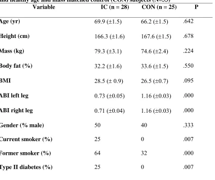
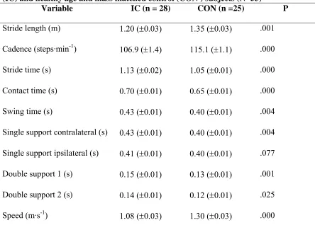
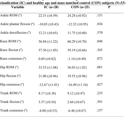
(18) List of tables. Chapter 2 Literature review Table. Page. 1. Rutherford clinical categories of acute limb ischemia (Rutherford, 1991)........ 38 2. Rutherford clinical categories of chronic limb ischemia resulting from PAD-IC (Rutherford, 1991)…………………………………………………… 38 3. Fontaine stages of classification of PAD-IC (Lampman & Wolk, 2003)……... 39. 4. PAD-IC exercise programs research protocol review ………………………... 49 5. PAD-IC exercise program research results review …………………………… 51 6. Female gait range characteristics (Whittle, 2003)…………………………….. 68 7. Male gait range characteristics (Whittle, 2003)……………………………….. 68. Experimental studies Chapter 3 1. Mean (±SEM) descriptive characteristics of intermittent claudication (IC) and healthy age and mass matched control (CON) subjects (N=53)…….. 101 2. Mean (±SEM) temporal-spatial gait parameters of intermittent claudication (IC) and healthy age and mass matched control (CON) subjects (N=53)…….. 103 3. Mean (±SEM) peak and ROM angular kinematics of intermittent claudication (IC) and healthy age and mass matched control (CON) subjects (N=53)………………………………………………………………... 104. 4. Mean (±SEM) peak angular velocity and acceleration values for intermittent claudication (IC) and healthy age and mass matched control (CON) subjects (N=53)………………………………………………………... 106. xvii.
(19) 5. Mean (±SEM) walking performance and physiological responses of intermittent claudication (IC) and healthy age and mass matched control (CON) subjects (N=53)………………………………………………………... 107. 6. Mean (±SEM) seven day physical activity levels of intermittent claudication (IC) and healthy age and mass matched control (CON) subjects (N=53)………………………………………………………………... 108. Chapter 4 1. Descriptive characteristics of peripheral arterial disease (PAD-IC) and healthy age and mass matched control (CON) participants (N=53)…………... 119. 2. Mean (±SD) NoRMS values for peripheral arterial disease (PAD-IC) and control (CON) participants (N=53)……………………………………………. 127 3. Mean (±SD) vector coding values for peripheral arterial disease (PAD-IC) and control (CON) participants (N=53)……………………………………….. 127. Chapter 5 1. Descriptive characteristics of peripheral arterial disease-intermittent claudication (PAD-IC) and healthy age and mass matched control (CON) participants (N=53)……………………………………………………. 139 2. Coefficient of variation (CV) and spanning set values for peripheral arterial disease-intermittent claudication-intermittent claudication (PAD-IC) and control (CON) participants (N=53)……………………………. 141. Chapter 6 1. Descriptive characteristics of participants…………………………………….. 156. xviii.
(20) 2. Mean (±SD) temporal-spatial gait parameter values of participants………….. 158 3. Mean (±SD) angular kinematics values of participants……………………….. 158. 4. Mean (±SD) peak angular velocity and acceleration values of participants…... 164. 5. Mean (±SD) walking performance and physiological responses values of participants…………………………………………………………………….. 166 6. Mean (±SD) seven day physical activity levels values of participants………... 167. Chapter 7 1 Descriptive characteristics of participants……………………………………... 184 2 Mean (±SD) normalized root mean square values……………………………... 189. 3 Mean (±SD) vector coding values……………………………………………... 189. 4 Mean (±SD) coefficient of variation values……………………………………. 190. 5 Mean (±SD) walking performance values……………………………………... 191. List of figures. Chapter 1 Introduction Figure. Page. 1. General approaches to the management of patients with PAD-IC (adapted from Brook et al., 2002)……………………………………………………….. 9. Chapter 2 Literature review 1. Age structure of Australia (Australian Bureau of Statistics, 2006)…………… 26 2. Australian PAD death rates (adapted from National Cardiovascular Diseases and Diabetes Database, AIHW, 2006)……………………………………….... 28 xix.
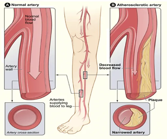
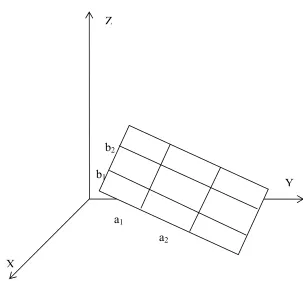
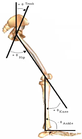
(21) 3. Site of peripheral arterial disease (Nucleus Communications, 2003)…………. 29. 4. Atherosclerotic blockage with diseased artery (National Heart Lung and Blood Institute, 2006)…………………………………………………………. 29. 5. Trophic skin in severe PAD-IC (adapted from MyFoot Shop, 2007)………... 31. 6. A) Palpation of lower limb arteries B) Ankle Brachial Index (ABI) test (Khan, Rahim, Anand, Simel, & Panju, 2006)………………………………... 32. 7. Balloon angioplasty (Texas Heart Institute, 2006).…………………………... 44. 8. A peripheral artery being treated with stenting. A) Balloon angioplasty of the blockage. B) Positioning of a balloon-expandable stent & deployment of the stent. C) Final result with the stent in place (Texas Heart Institute, 2006)... 45. 9. A, B & C SilverHawk event (Mayo Clinic, 2006)…………………………….. 46. 10. Events of the gait cycle (adapted from Neumann, 2002)……………………. 63. 11. Angle conventions used to describe the angular displacements of the trunk, hip, knee and ankle in the sagittal plane during the walking cycle……………. 64. 12. Mean (____) ± SD (·····) of hip ROM during normal walking (adapted from Perry, 1992)……………………………………………………………… 65 13. Mean (____) ± SD (·····) of knee ROM during normal walking (adapted from Perry, 1992)……………………………………………………………… 66 14. Mean (____) ± SD (·····) of ankle ROM during normal walking (adapted from Perry, 1992)……………………………………………………………… 67 15. Angle-angle plot of the sagittal plane hip and knee during a gait cycle……... 80 16. Phase-plane plot of the knee angle and knee velocity during a gait cycle….... 81. 17. Vector coding technique……………………………………………………... 83 18. Rn represents the span of the two vectors, where a1, a2, b1 and b2 represent the components of the respective vectors (Kurz & Stergiou, 2004)… 87. xx.
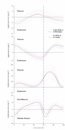
(22) Experimental studies Chapter 3 1. Joint angle conventions………………………………………………………... 98. 2. Trunk, hip, knee and ankle kinematics in the sagittal plane for IC and CON subjects; the dashed line indicates toe-off at ~60% gait cycle (with 0-60% representing stance; 60-100% representing swing)……………………………. 105. Chapter 4 1. Sagittal plane angle convention……………………………………………….. 121 2. Vector coding technique………………………………………………………. 123 3. Joint-joint angular kinematics plot of the hip-knee during the gait cycle in the sagittal plane for PAD-IC and CON participants……………………….. 125. 4. Joint-joint angular kinematics plot of the knee-ankle during the gait cycle in the sagittal plane for PAD-IC and CON participants……………………….. 126. 5. Joint-joint angular kinematics plot of the hip-ankle during the gait cycle in the sagittal plane for PAD-IC and CON participants……………………….. 126. Chapter 5 1. Sagittal plane angle convention……………………………………………….. 136 2. Hip, knee and ankle kinematics in the sagittal plane for PAD-IC and CON subjects; the dashed line indicates toe-off at ~60% gait cycle (with 0-60% representing stance; 60-100% representing swing)……………… 140. Chapter 6 1. Sagittal joint angle conventions……………………………………………….. 153. xxi.
(23) 2. Participant trunk kinematics in the sagittal plane (CON = healthy age and mass matched controls, CPAD-IC = control peripheral arterial disease-intermittent claudication patients, TPAD-IC = treatment peripheral arterial disease-intermittent claudication patients)……………………………. 160. 3. Participant hip kinematics in the sagittal plane (CON = healthy age and mass matched controls, CPAD-IC = control peripheral arterial disease-intermittent claudication patients, TPAD-IC = treatment peripheral arterial disease-intermittent claudication patients)………………… 161 4. Participant knee kinematics in the sagittal plane (CON = healthy age and mass matched controls, CPAD-IC = control peripheral arterial disease-intermittent claudication patients, TPAD-IC = treatment peripheral arterial disease-intermittent claudication patients)…………………………….. 162. 5. Participant ankle kinematics in the sagittal plane (CON = healthy age and mass matched controls, CPAD-IC = control peripheral arterial disease-intermittent claudication patients, TPAD-IC = treatment peripheral arterial disease-intermittent claudication patients)…………………………….. 163. Chapter 7 1. Sagittal plane angle convention……………………………………………….. 178. 2. Vector coding technique………………………………………………………. 181. 3. Hip-knee plot in the sagittal plane (CON, healthy age and mass matched controls; CPAD-IC, control peripheral arterial disease-intermittent claudication patients; TPAD-IC, treatment peripheral arterial disease-intermittent claudication patients)…………………………………….. 186. 4. Knee-ankle plot in the sagittal plane (CON, healthy age and mass matched. xxii.
(24) controls; CPAD-IC, control peripheral arterial disease-intermittent claudication patients; TPAD-IC, treatment peripheral arterial disease-intermittent claudication patients)……………………….……………. 187 5. Hip-ankle plot in the sagittal plane (CON, healthy age and mass matched controls; CPAD-IC, control peripheral arterial disease-intermittent claudication patients; TPAD-IC, treatment peripheral arterial disease-intermittent claudication patients)…………………………………….. 198. xxiii.
(25) Chapter 1. Introduction. Ageing population. Mean life expectancy in Australia increased dramatically during the 20th century, and is expected to continue to increase. The average life expectancy of a new-born male in Australia increased from 55.2 yr in 1901-10 to 76.2 yr in 1997-99, while the life expectancy of a new-born female increased from 58.8 to 81.8 yr during the same period representing an increase of 21 yr and 23 yr for males and females respectively. This increase in life expectancy is due to lower mortality rates at all ages which is caused by increased medical treatment and education. The proportion of the Australian population aged 65 yr – 84 yr is expected to increase substantially, from 12% in 1999 to between 24% and 27% in 2051 and to 25-28% in 2101. The proportion aged 85 yr and over is expected to almost quadruple, from 1.3% in 1999 to approximately 5% in 2051 and approximately 6% in 2101 (Australian Bureau of Statistics, 2002). With this increase in the elderly population of Australian the prevalence of cardiovascular disease will increase.. Cardiovascular disease in Australia. Cardiovascular disease (CVD) is a national health problem in Australia. More than three million adult Australians had a recent and/or long-term cardiovascular condition in 2001. Heart, stroke and vascular disease accounted for 50,290 deaths (37.6% of all. 1.
(26) deaths) in 2002 in Australia. In 2002, coronary heart disease was the major cause of death, accounting for 51.8% of deaths followed by stroke (24.9%), heart failure (5.4%), peripheral vascular diseases (5.1%) and rheumatic fever and rheumatic heart disease (0.5%) (Australian Institute of Health and Welfare, 2004).. From 1991-2002 the CVD mortality rate declined at a rate of 4.3%·yr-1 for males and 4.0%·yr-1 for females. This decline is thought to be partly due to improved survival following cardiovascular events and partly due to a fall in the rate of morbidity of CVD, owing to improvement in and better management of the risk factors (Australian Institute of Health and Welfare, 2004). However, if the current trends in older population numbers continue there may be an increase in CVD mortality rates.. Peripheral arterial disease and mortality rates. Peripheral arterial disease (PAD) is a chronic arterial occlusive disease of the lower extremities caused by atherosclerosis (Aronow, 2004b; Curci & Sanchez, 2003; Gardner, 2001; Hirsch et al., 2001). Atherosclerosis is a condition whereby poor circulation in the blood vessels occurs due to blockages (Olin, 2002; Savage et al., 2001). More commonly known as “hardening of the arteries” it is a condition involving a gradual thickening, hardening, and loss of elasticity in the walls of the arteries, which can be caused by fatty deposits, platelets and calcium. For PAD affected individuals the most common affected arteries include those that supply blood from the heart, to the feet and legs via the aorta, iliac, femoral, popliteal, and tibial arteries (Gardner, 2001; Hiatt, Hirsch, Regensteiner, & Brass, 1995; Olin, 2002; Savage et al., 2001). PAD is estimated to have caused 2,581 (1.9%) of all deaths in Australia in 2002 and was. 2.
(27) responsible for 24,288 hospitalisations (0.4% of all hospitalisations) in 2001-2002 with males being twice as likely to die from PAD as females. The mortality rate for PAD declined at a rate of 5.1%·yr-1 for males and 4.7%·yr-1 for females between 1991-2002. However, the condition is still a health concern particularly for older Australians (Australian Institute of Health and Welfare, 2004).. Cause and symptoms of PAD. In the early stages of PAD a common symptom is ischemic pain (cramping) or fatigue in one or both legs and buttocks during physical activity (Brook, Weder, Grossman, & Rajagopalan, 2002; Comerota, 2003; Gardner, 2001; Hiatt, Regensteiner, Hargarten, Wolfel, & Brass, 1990; Olin, 2002; Schmieder & Comerota, 2001). The symptoms usually subside within minutes of cessation of walking/exercise (Brook et al., 2002; Comerota, 2003). This condition is called “intermittent claudication” (IC) and it limits participation in daily physical activities which can impact on an individual quality of life (QOL) (Aronow, 2004b; Breek, Hamming, De Vries, Aquarius, & van Berge Henegouwen, 2001; Brook et al., 2002; Burns, Gough, & Bradbury, 2003; Johnstone, 2003; Schmieder & Comerota, 2001).. The main risk factors for PAD-IC are smoking, high blood pressure (hypertension), high cholesterol, diabetes, lack of physical exercise and obesity (Aronow, 2004b; Burns et al., 2003; Olin, 2002). Smoking is the most important risk factor for developing PAD-IC (Burns et al., 2003; Christman, Ahijevych, & Buckworth, 2001; Gardner, Killewich, Montgomery, & Katzel, 2004; Hiatt, 2001). Cessation of smoking has been shown to decrease PAD-IC symptoms improving health and consequently QOL. 3.
(28) (Gardner, 2001; Hiatt et al., 1990; Olin, 2002; Savage et al., 2001; Schmieder & Comerota, 2001). Hypertension is another risk factor for PAD-IC (Burns et al., 2003; Olin, 2002) with individuals twice as likely to develop PAD-IC compared to those in comparable health who do not have high blood pressure (Armen & Smith, 2003; Burns et al., 2003; Olin, 2002). As well as high blood pressure, individuals who have diabetes have a greater risk of developing PAD-IC, but the condition also increases the possibility of several other risk factors occurring such as hypertension, poor cholesterol profile (low levels of high density lipoprotein (HDL) cholesterol and/or high levels of low density lipoprotein (LDL) cholesterol) and obesity (Aronow, 2004b; Burns et al., 2003; Olin, 2002). Research has shown that when cholesterol levels are improved, PAD-IC symptoms can remain stable or decrease (Feringa et al., 2007). In addition, controlling the risk factors for PAD-IC can prevent or ameliorate an existing condition (Burns et al., 2003; Olin, 2002). Other factors that may increase the risk of PAD-IC include family history and damaged arteries. Genetic factors are associated with specific lipid and cholesterol abnormalities, which in turn may increase the risk of PAD-IC (Valentine et al., 2004). Elevated levels of C-reactive protein are indicators of persistent inflammation in the arteries, which is known to cause significant damage in blood vessels and to be highly associated with PAD-IC (Narins et al., 2004).. As described earlier if an individual has PAD-IC, the arteries in the legs are obstructed with fatty deposits that can restrict blood flow (Feinglass, McCarthy, Slavensky, Manheim, & Martin, 1996). During any activity involving the lower limbs there is an increased demand for blood and oxygen in the legs that cannot be met when the arteries are blocked. This results in the lower limbs receiving an insufficient supply of blood and oxygen, resulting in pain/cramping in the lower limbs (Olin, 2002). Upon cessation. 4.
(29) of activity the demand for oxygen and blood in the legs decreases and pain/cramps are eased. Symptoms of advanced PAD-IC involve obstruction of the arteries in the legs to the degree that even rest offers no relief and pain continues even when lying, sitting or standing. This condition is referred to as “ischemic rest pain” (Aronow, 2004b; Brook et al., 2002; Hiatt et al., 1990). In this severe situation, the arteries can become so obstructed that ulcers, withered calf muscles, hair loss over the toes and feet, thick toenails, shiny tight skin and gangrene can develop, which can lead to amputation of the limb(s) (Aronow, 2004b; Schmieder & Comerota, 2001).. Diagnosis of PAD-IC. Many individuals with PAD-IC either do not report symptoms and/or may not experience leg pain in the early stages (Aronow, 2004b; Olin, 2002). It is recommended that individuals should be evaluated for PAD-IC if they have risk factors such as heart disease, high cholesterol, obesity, smoking, leg pain during walking, or ulcers on their legs (Brook et al., 2002; Comerota, 2003). PAD-IC diagnosis is relatively simple, by comparing blood pressures taken in the arm and ankle arteries known as Ankle-Brachial Index (ABI) using Doppler ultrasonography, an assessment of PAD-IC is produced (Aronow, 2004b; Brook et al., 2002; Comerota, 2003). The ankle area will have a much lower or undetectable blood pressure reading compared to the arm. Also examination of the skin of the legs and feet for colour changes, ulcers, infection, or soft tissue injuries can be used to diagnose PAD-IC (Comerota, 2003; Olin, 2002). Angiography can also be used to diagnose and define this disease which involves taking x-ray pictures to visualise the inner internal diameter of arteries (Burns et al., 2003; Hirsch et al., 2001; Valentine et al., 2004). A treadmill test (either constant load or graded) can be used to. 5.
(30) test an ABI if further assessment is needed (Gardner, 2001). Patients with PAD-IC demonstrate a 50-60% reduction in walking performance compared to healthy age matched controls, which is also comparable to the walking performance of patients with congestive heart failure. A treadmill test is also useful for determining the severity of the pain while walking and for assessing the effectiveness of treatment therapies (Gardner, 2001).. Questionnaires are also used in the assessment, diagnosis and analysis of treatment progression of PAD-IC as the patient is able to report functional impairments without having to undergo physical testing sessions (Breek et al., 2001; Carlon, Morlino, & Maiolino, 2003; Gardner, 2001; Menard et al., 2004; Stewart et al., 2003). One of the most important questionnaire measures of PAD-IC impact and treatment outcomes is perceived QOL (Breek et al., 2001; Carlon et al., 2003). Questionnaires have been used in PAD-IC studies to assess changes in QOL and include the Short Form-36 (SF-36®) (Ware, 1993), the Nottingham Health Profile, and the EuroQol (The EuroQoL Group, 1990) questionnaires (Chong, Garratt, Golledge, Greenhalgh, & Davies, 2002; Cook & Galland, 1997). However, these questionnaires are generic and are designed for application to other conditions and it has been suggested that they may fail to focus on PAD-IC and thus fail to assess changes in perceived QOL (Chong et al., 2002). The Walking Impairment Questionnaire (WIQ) (Regensteiner, Steiner, Panzer, & Hiatt, 1990) and the Intermittent Claudication Questionnaire (ICQ) (Chong et al., 2002) involve function-specific measures of outcome and have been shown to be practical, reliable, valid, and responsive measures of perceived QOL in PAD-IC affected individuals (Chong et al., 2002). It has become standard practice for researchers to use a combination of PAD-IC and non-PAD-IC questionnaires to quantify the impact of. 6.
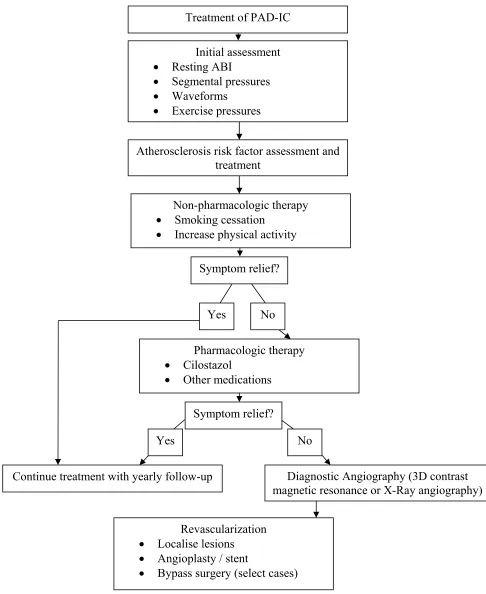
(31) PAD-IC on performing activities of daily living (ADL) and perceived QOL (Gardner, 2001).. Consequences of PAD-IC. The consequences of PAD-IC are a decrease in QOL, an increase in CVD risk and therefore an increase in CVD mortality rate. An individual diagnosed with PAD-IC, has the same risk of death from cardiac events or stroke as a person with evident heart disease (Burns et al., 2003). This risk increases with the degree of PAD-IC. In rare cases, blood clots can develop suddenly in the major arteries in the leg, a condition known as acute occlusion (Aronow, 2004b). Symptoms of acute occlusion include numbness, pain, coolness, pale skin colour, lack of pulse in the artery, and leg weakness (Aronow, 2004b). This is a very serious event, which can lead to amputation or even loss of life. Individuals that have severe PAD-IC demonstrate deterioration in several physiological aspects (e.g. peak oxygen uptake and heart rate) that may contribute to their inability to exercise and perform ADL which can then lead to diminished QOL.. Treatment of PAD-IC. PAD-IC treatment strategies aim to relieve the symptoms of IC, increase walking performance and physiological responses, modify the cardiovascular risk factors, and improve perceived QOL (Hiatt, 2001; Regensteiner & Hiatt, 1995). Treatment strategies used to manage leg pain and improve function can include vascular surgery, angioplasty, pharmacotherapy and exercise therapy (Figure 1)(Aronow, 2004b; Burns et al., 2003; Chong, Golledge, Greenhalgh, & Davies, 2000; Creasy, McMillan, Fletcher,. 7.
(32) Collin, & Morris, 1990; Curci & Sanchez, 2003; de Vries et al., 2002; Gardner, 2001; Leng, Fowler, & Ernst, 2000; Olin, 2002; Ouriel, 2001; Wang, 2004).. Pharmacotherapy treatments such as anti-platelet agents are used to reduce blood clots and fatty deposits thus reducing the risk of heart disease and stroke. Aspirin acts as a non-specific anti-platelet agent to reduce cardiovascular risk in patients with PAD-IC (Olin, 2002). Clopidogrel (Plavix) is a more potent platelet inhibitor recommended for patients with PAD-IC (Aronow, 2004b; Federman, Bravata, & Kirsner, 2004; Olin, 2002). Cilostazol is an agent that improves blood flow and is proving to be useful for disabling PAD-IC (Aronow, 2004b; Dawson, 2001; Dawson et al., 2000; Dawson, Cutler, Meissner, & Strandness, 1998). Other drugs such as anti-hypertensive and cholesterol-lowering agents known as statins may also be beneficial for individuals with PAD-IC (Aronow, 2004b; Mohler, Hiatt, & Creager, 2003; Young-Xu, Chan, Liao, Ravid, & Blatt, 2003). Most patients take combinations of these medications to reduce further development of PAD-IC. Other treatments involve the use of anticoagulants such as aspirin, warfarin, or heparin, to thin the blood and prevent blood clots that occur during surgery (Burns et al., 2003; Curci & Sanchez, 2003). All of these agents however, increase the risk of bleeding. Alteplase also called “Activase” or “t-PA’ and reteplase also called “Retavase” are thrombolytic agents commonly known as a "clotbusters" which are used to diminish existing clots and may be used before, during, or after angioplasty if a blood clot is present (Burns et al., 2003; Curci & Sanchez, 2003).. 8.
(33) Treatment of PAD-IC Initial assessment Resting ABI Segmental pressures Waveforms Exercise pressures. • • • •. Atherosclerosis risk factor assessment and treatment Non-pharmacologic therapy Smoking cessation Increase physical activity. • •. Symptom relief?. Yes. • •. No. Pharmacologic therapy Cilostazol Other medications Symptom relief?. Yes Continue treatment with yearly follow-up. • • •. No Diagnostic Angiography (3D contrast magnetic resonance or X-Ray angiography). Revascularization Localise lesions Angioplasty / stent Bypass surgery (select cases). Figure 1 General approaches to the management of patients with PAD-IC (adapted from Brook et al., 2002).. 9.
(34) In severe cases of PAD-IC where pharmacotherapy treatment is not beneficial other treatments such as opening the obstructed blood vessels (angioplasty) or bypass the obstructed blood vessels (graft surgery) may be undertaken (Chong et al., 2002; Chong et al., 2000; Creasy et al., 1990; de Vries et al., 2002; Gardner, 2001; Wang, 2004). Percutaneous transluminal angioplasty (PTA) is an a procedure used to widen the narrowed or totally-obstructed blood vessels causing IC (Creasy et al., 1990; de Vries et al., 2002; Gardner, 2001; Wang, 2004). This procedure is being increasingly used, especially in patients who have other medical conditions. Some authors believe that it is not only much less expensive, but is also more effective than surgical bypass (Chong et al., 2002; Chong et al., 2000; Creasy et al., 1990; de Vries et al., 2002; Gardner, 2001; Ouriel, 2001; Wang, 2004). The procedure is minor, requiring a local anaesthetic and the patient can return to normal activity in 24-48 h. The advantage of this procedure is that complication rates are low and the effects are permanent. However, if necessary the procedure can be repeated without any greater risk than with the original procedure (Burns et al., 2003).. Regardless of the initial treatment, PAD-IC patients are encouraged to increase their physical activity levels through the use of exercise/walking programs, thereby reducing the risk factors associated with PAD-IC (Hankey, Norman, & Eikelboom, 2006). These programs have been found to be very effective in reducing PAD-IC risk factors and improving QOL. The walking programs typically involve the patient walking until maximal leg pain occurs, followed by rest until the patient is able to walk again (Aronow, 2004b; Gardner, 2001; Regensteiner & Hiatt, 1995; Sorlie & Myhre, 1978; Tan, De Cossart, & Edwards, 2000b). The major benefit of exercise programs is their non-invasive nature and the ease with which patients can engage in the activity.. 10.
(35) Over the past 40 yr there has been considerable research on the effects of exercise training on PAD-IC affected individuals. A meta-analysis of the effects of exercise rehabilitation studies (Gardner & Poehlman, 1995) reported that the average walking distance to the onset of claudication increased by 179% from 126 to 351 m and average walking distance to maximal claudication pain increased by 122% from 326 to 723 m following a 3 mth exercise rehabilitation program. Due to the dramatic improvement in walking performance of individuals with PAD-IC following an exercise program and the non-invasive nature of the program, exercise training is a good course of action for treatment of PAD-IC. However, it is recommended that a holistic treatment approach be adopted whereby an exercise program is undertaken in conjunction with pharmacology prescription. This holistic approach helps to reduce the risk of CVD and improves walking performance, ADL and perceived QOL in individuals with PAD-IC (Antignani, 2003; Bendermacher, Willigendael, Teijink, & Prins, 2005; Meru, Mittra, Thyagarajan, & Chugh, 2005).. PAD-IC and improved walking performance. It is not clear which mechanism(s) underlie improved walking performance in PAD-IC affected individuals following exercise program participation. Increased blood flow to the peripheral arteries, increased capillary growth in the muscles, increased oxygen perfusion and changes in lower limb mobility (Lumsden & Rice, 2006; Womack, Sieminski, Katzel, Yataco, & Gardner, 1997) have all been suggested as contributing to improved walking performance, yet there is no consensus as to the major contributing factor. Understanding the mechanisms of improved walking performance in this population may assist in the structure and management of PAD-IC treatment programs.. 11.
(36) PAD-IC and lower limb mobility. In order to have freedom of movement and personal fulfillment, a level of functional independence is required (Daley & Spinks, 2000). However, as an individual becomes older, the risk of disease and possible injury increases with ongoing implications for health. The increased incidence of disease with age impacts on the physical activity level and general mobility of individuals and leads to further increases in disease states, risk of injury and reduced QOL (Gardner, Forrester, & Smith, 2001a; Maki, 1997). Research has shown that lower limb mobility characteristics (temporal-spatial gait parameters and gait kinematics) are predictors of functional decline (Brach, VanSwearingen, Newman, & Kriska, 2002; Gill, Williams, & Tinetti, 1995; Guralnik, Ferrucci, Simonsick, Salive, & Wallace, 1995; Spirduso & Cronin, 2001) and have been related to increased risk of falling (Daley & Spinks, 2000; Prince, Corriveau, Hebert, & Winter, 1997; Rubenstein, Powers, & MacLean, 2001; Sadeghi, Prince, Zabjek, Sadeghi, & Labelle, 2002). Therefore understanding how chronic disease influences lower limb mobility characteristics is very important.. PAD-IC is one such disease that can impact dramatically on the lower limb mobility of an individual. Research has indicated that PAD-IC significantly influences temporalspatial gait parameters (Gardner et al., 2001a; Scherer, Bainbridge, Hiatt, & Regensteiner, 1998) however, other studies contradict these findings stating that PADIC has no effect on temporal-spatial gait parameters (McCully, Leiper, Sanders, & Griffin, 1999; Scherer, Hiatt, & Regensteiner, 2006). Gardner et al., (2001) speculated that exercise programs could be used to improve the temporal-spatial gait parameters of. 12.
(37) PAD-IC affected individuals to the extent that they would more closely resemble the gait characteristics of healthy age matched controls.. The measurement of temporal-spatial gait parameters usually involves capturing video footage of a full gait cycle (ipsilateral heel contact to ipsilateral heel contact) and analysis of certain aspects (e.g. stride length, knee range of motion) of the gait cycle (Whittle, 2003). Typically, temporal-spatial gait parameters of interest in the gait cycle include stride length, cadence, stride time, contact time, swing time, support time and speed. Determination of temporal-spatial gait parameters and angular gait kinematics (joint angular displacement, velocity, acceleration) allows conclusions to be made about general mobility, health and therefore perceived QOL (Whittle, 2003).. PAD-IC and lower limb movement variability. Temporal-spatial gait parameters and joint angular kinematics may not always indicate alterations in the biomechanical patterns of the lower limbs. Variability of the lower limb angular kinematics (movement variability) was once thought to be “noise” and not beneficial to human walking. However, the lower limb joint movement variability has been shown to reveal more information about human locomotion. From a Dynamical Systems theory perspective variability is defined as the level of variation between multiple attractors (which represent states in which movement components are brought into relation with each other) that permits flexible and adaptive motor system behaviour, thereby encouraging free exploration of performance contexts by each individual (Newell & Corcos, 1993). Variability is said to share a relationship with stability, whereby an increase in movement variability will produce an increase in. 13.
(38) instability. This relationship has been used to demonstrate the process of learning a motor skill. In the initial stages of learning a skill there is a high level of variability, as the person performing the skill has multiple attractors and so there is a high level of instability. As the skill is learnt, movement variability is reduced and therefore the performance becomes stable (Newell & Vaillancourt, 2001). This relationship exists in the processes of learning to walk. When an infant begins to walk there is a high level of movement variability and instability. However, as the child learns to move the legs, gain balance and control the number of possible independent dimensions of movement in a system (i.e. degrees of freedom), the movement variability is reduced and walking becomes more stable (Newell & Vaillancourt, 2001).. Lower limb movement variability research has determined that temporal-spatial gait parameter variability provides information about gait stability where measures of temporal-spatial gait parameters do not. High stride length and stride width variability has been shown to reveal underlining gait instability which may cause poor mobility leading to increased falls and poor obstacle avoidance (Hausdorff, 2005; Hausdorff, Edelberg, Mitchell, Goldberger, & Wei, 1997). However, due to temporal-spatial gait parameter variability being an outcome measure of locomotion and not the mechanism of walking, lower limb movement variability characteristics can be lost. Therefore, assessment of movement variability using joint angular kinematics (i.e. range of movement (ROM) of the knee) is required to provide an in-depth measurement of stability and/or gait changes.. Joint angular kinematics research has determined that both older and injured individuals exhibit reduced movement variability in the lower limbs (Heiderscheit, Hamill, & van. 14.
(39) Emmerik, 2002; Van Emmerik, Hamill, & McDermott, 2005; Van Emmerik, Wagenaar, Winogrodzka, & Wolters, 1999). Therefore, it would appear that the assessment of movement variability using either temporal-spatial gait parameters or the joint angular kinematics would provide distinctly contrasting information about lower limb mobility and motor control characteristics during locomotion (Heiderscheit, 2000). However, given existing research findings it is not possible to conclude that higher movement variability in joint angular kinematics of the lower limbs is either beneficial or detrimental. Researchers continue to investigate this question in order to determine if movement variability can supply information regarding lower limb mobility in relation to disease, obstacle avoidance (risk of falls) and perceived QOL.. There has been only limited research into ways of quantifying variability of joint angular kinematics. Analysis techniques may include linear variable to variable relationships, which may be described as a variable-variable plot or intralimb coordination. The variable-variable plot can involve the angular kinematic ROM of a segment (thigh, shank and foot) and/or angular kinematic ROM of a joint (ankle, knee and hip) and/or angular kinematic velocity of a joint (e.g. hip angle vs. thigh angle, hip velocity vs. knee angle and knee velocity vs. knee angle). Variable-variable plots have been used to examine movement coordination in a variety of activities including slope walking and the effects of patellofemoral injury during walking (Goswami, 1998; Heiderscheit et al., 2002). The variable-variable plot was initially used as a qualitative tool and therefore did not provide any quantitative data concerning individual coordination patterns. However, in recent times these plots have been used to generate quantitative data for gait analysis. Parameterization, vector coding and normalized root mean square are three techniques that have been applied to variable-variable plots to. 15.
Figure


+7
Related documents