Dissertation on
Detection of Autoimmune Thyroiditis in Children with Goitre
attending the Paediatric Department of a tertiary care
Hospital in Chennai
Submitted to
THE TAMIL NADU
DR. M.G.R. MEDICAL UNIVERSITY
in partial fulfilment of the requirement
for the award of degree of
M.D., BRANCH - VII
PAEDIATRIC MEDICINE
ESIC MEDICAL COLLEGE & PGIMSR
K.K. NAGAR, CHENNAI.
THE TAMILNADU DR. M.G.R. MEDICAL UNIVERSITY CHENNAI, TAMILNADU
CERTIFICATE
Certified that this dissertation titled “Detection of Autoimmune
Thyroiditis in Children with Goitre attending the Paediatric Department
of a tertiary care Hospital in Chennai”, is a bonafide work done by
Dr. Saranya P, Post-graduate, ESIC Medical College & PGIMSR, K.K.
Nagar, Chennai, during the academic year 2014-2017.
Prof. Dr. Sowmya Sampath, MD, DNB Dr. S. Shobhana, MD, DCH,
Professor & Head, Principal Guide, Department of Paediatrics, Associate Professor, ESIC Medical College & PGIMSR, Department of Paediatrics,
K.K. Nagar, ESIC Medical College & PGIMSR, Chennai. K.K. Nagar, Chennai.
Prof. Dr. Srikumari Damodaram, MS, MCh
The Dean
DECLARATION
I solemnly declare that this dissertation titled “Detection of
Autoimmune Thyroiditis in Children with Goitre attending the Paediatric
Department of a tertiary care Hospital in Chennai” has been conducted by
me at ESIC Medical College & PGIMSR, Chennai, under the guidance and supervision of Dr. S. Shobhana, MD, DCH, Associate Professor, Department of Paediatrics, ESIC Medical College & PGIMSR, Chennai. This dissertation is submitted to The Tamil Nadu Dr. M.G.R. Medical University, Chennai in partial fulfilment of the University regulations for the award of the degree of
M.D. Branch VII (Paediatrics).
Date:
ACKNOWLEDGEMENTS
At the very outset, I would like to extend my sincere and heartfelt obligation towards all those who have helped me in this endeavour.
Our Dean, Prof. Dr. Srikumari Damodaram MS. Mch (Gastro) for her enormous support given to the post graduates in conducting the study in a smooth manner.
First I would like to express my profound gratitude to my thesis guide
Dr. S. Shobhana MD, DCH, Associate Professor, Department of Paediatrics
for providing her unfailing support, continuous encouragement and motivation throughout the process of my dissertation. The door to her office is always open whenever I ran into a trouble spot or had questions about my research. She consistently allowed this dissertation to be my own work but steered me in the right direction whenever she thought I needed it.
I gratefully acknowledge and express my deep sense of gratitude to
Prof. Dr. Sowmya Sampath MD. DNB, Professor and Head, Department of
Paediatrics, for her constant guidance and encouragement throughout this dissertation work.
I am very much thankful to my co-guide Dr. Sathish Kumar.S MD, Assistant Professor, Dept. of Paediatrics, for his valuable guidance and encouragement at every stage of the study.
I also owe my depth of gratitude to Prof. Dr. Rajalakshmi. V. MD, DCP Professor & Head, Dept. of Pathology and Associate Professors
Dr.Meenakshi Sundaram. K. MD and Dr. Shanmugapriya. S. MD for their
I am thankful to Prof. Dr. Bhuvaneshwari DNB, Professor & Head, Dept.of Radiology and Assistant Professor Dr. Kanika Gupta. MD, for their great contribution towards reporting and ultrasound guided biopsies.
I owe my sincere thanks to Prof. Dr. Malliga. S MD, Professor & Head, Dept. of Biochemistry and Assistant Professor Dr. Thuthi Mohan.MD
for their constant efforts in providing laboratory support, throughout my dissertation work.
I extend my sincere gratitude to Dr. Aruna Patil, our Statistician for being ever helpful in analysis of statistics and clearing all our statistical queries. I also acknowledge with a deep sense of reverence, my gratitude towards my parents who gave me an eternal feeling and moral support to do my thesis work.
Thanks also goes to Dr. J. H. Suresh David, senior consultant, all the Assistant Professors, Senior Residents and my fellow post graduate students of the Paediatric Dept. who have directly or indirectly helped me in the completion of my thesis.
LIST OF ABBREVIATIONS USED
Autoimmune Thyroid Disease
ATD
Autoimmune Thyroiditis
AIT
Free Triiodothyronine
FT3
Free Thyroxine
FT4
Fine Needle Aspirat ion Cytology
FNAC
Hashimoto’s Thyroiditis
HT
Heterogeneity Index
HI
Lymphocytic Thyroid Infiltration
LTI
Thyroglobulin Antibody
Tg-Ab
Thyroid Function Test
TFT
Thyroid peroxidase antibody
TPO
Thyroid Hormone
TH
Thyroid Stimulating Hormone
TSH
Urine Iodine
UI
CONTENTS
CHAPTER TITLE PAGE NO.
Introduction
Thyroid hormone in optimal quantities is essential for neurodevelopment and growth in children1. The development of thyroid dysfunction is insidious and may not be accompanied by typical symptoms or clinical signs. Diseases result from both under- and over activity of thyroid gland. Goitre has many causative factors such as defect in synthesis of thyroid hormone, deficiency of iodine & autoimmune disease. Chronic autoimmune thyroiditis, a major cause of goitre in iodine repleted areas2 which results in thyroid dysfunction that is rather difficult to identify clinically in children.
In India, the burden of thyroid disease is not well understood because of the lack of documented statistics. The only studies which were available are on the prevalence of hypo or hyperthyroidism centred on the success of iodisation program. Though many of the studies are done in the adult population, exclusive Paediatric studies are very few.
AIT results in a spectrum of diseases varying from euthyroidism to frank hypothyroidism or occasional hyperthyroidism3. So children with goitrous autoimmune thyroiditis need periodic monitoring of thyroid function.
There are genetic and non genetic factors that increase the risk of autoimmune thyroiditis in childhood. Positive family history is noted in 50% of ATD4. Environmental factors like geographic variation, seasonality, smoking, stress, infections, iodine & medications like amiodarone have been postulated to precipitate autoimmunity
Many genetic as well as environmental factors interact with the host, which leads to the appearance of auto antigens within the thyroid gland and accumulation of antigen-presenting cells in them. Auto-reactive immune cells (T lymphocytes) get activated by the antigen-presenting cells, due to this break in the immune tolerance. These cells then interact with the thyroid cells by invading the thyroid gland and results in auto immunity.
Review of Literature
Ever since Dr. Hakaru Hashimoto first described Hashimoto thyroiditis in 1912, the condition was widely diagnosed from different parts of world by different diagnostic tools. Being an autoimmune disease it has got varied presentation ranging from a euthyroid goitrous patient to hypothyroid and even hyperthyroid presentation.
In recent years, old entities such as multi nodular goitre in adults and puberty goitre seen in adolescence were now considered to be occuring due to autoimmune thyroiditis5. But the clinical suspicion for the disease is very less and the disease goes under diagnosed in our scenario not only because of lacunae in our understanding but also due to logistical reasons.
Historical aspects:
thyroiditis is considered to be the most frequent autoimmune thyroid disease in paediatric patients.
Need for early detection of autoimmunity
AIT is no longer considered an infrequent form of hypothyroidism in children. Early diagnosis and treatment of autoimmune thyroiditis prevents severe neurological disorders during growth spurts in children7. The most frequently encountered difficulties are in school performance, hyperactivity and easy fatigability, intolerance to cold and difficulty in concentration. Appropriate treatment is essential before the onset of puberty often to reach the adult final height consistent with their genetic potential.
Autoimmune thyroiditis is one of the important causes of acquired hypothyroidism with familial as well as genetic implications. In the study by
Meena Desai et al 4, it was noted that diagnosing autoimmune thyroiditis in children may lead to the detection of the disease in the adult members of the family who may be benefited with early treatment.
Thyroid hormone role
is essential for normal growth of bone as well as general development. T3 through IGF and IGFBPs stimulate differentiation and proliferation of osteoblast by regulating synthesis and action of growth factors9. TH also plays important role in brown and white adipose tissue development and function. T3 regulates both lipolysis and lipogenesis. Studies done in hypothyroid rats have shown that lack of thyroid hormone not only causes diminished axonal growth but also results in reduced dendritic arborisation in the cortex, hippocampus, and cerebellum, visual & auditory cortex.
Pathogenesis of autoimmune thyroiditis
Major pathogenetic mechanism that has been implicated in the occurence of autoimmune thyroiditis is break down in self-tolerance to thyroid autoantigens. The possible inciting events included were environmental triggers resulting in either abnormal proliferation of regulatory T cells or exposure of the antigens within the thyroid which were sequestered normally. A strong genetic component also associated with autoimmune thyroid disease similar to other autoimmune disorders particularly with polymorphisms in protein tyrosine phosphatase-22(PTPN22) and cytotoxic T lymphocyte-associated antigen-4(CTLA4) which are immune regulation lymphocyte-associated genes10. Induction of autoimmunity in the thyroid gland leads to apoptosis of thyroid epithelial cells thereby progressive depletion of the thyroid parenchyma and replacement by mononuclear cell infiltration and fibrosis.
Hypothyroidism and its sequelae
Hypothyroidism leads to mental retardation only if it occurs in the newborn period, but when it is acquired in a later stage it usually leads to delay in bone maturation or bone age then ultimately affecting their growth. Hypothyroidism in adolescent children will have clinical features of tiredness, decline in school performance, constipation dry hair, coarse skin, hair loss, decreased appetite, brittle nails and cold intolerance. Girl children usually have menstrual irregularity rather than amenorrhea.
Chronic untreated hypothyroidism leads to more significant physical alterations. Carotene impregnation makes the skin appear cereous, pale and yellowish in colour. Myxedema occurs due to the increased muco polysaccharide concentration in the subcutaneous tissue. Severe myxedema usually results in slow muscle action, slow tendon reflexes and pseudo muscular hypertrophy.
TPO antibodies
B-cell-reactive epitopes which may be genetically determined and are believed to be stable within each patient. It is thought to represent the surface antigen which leads to cell-mediated cytotoxicity.
Many TPO assays have now been standardized against the World Health Standard National Institute of Biological Standards and Control. WHO provides a reference preparation for detecting anti-TPO antibody6.Calibrators use international standards given by WHO for establishing the reference concentration of antibody values.
Hypothyroidism and autoimmune thyroidtis
cytostatic, thyro suppressive and substitution therapy, antilipemic, bisphosphonates and other drugs considered significant for autoimmune thyroiditis. These valuable insight into this condition from his article not only motivates us to conduct this cross sectional study but also to be instrumental in bringing about policy change in using iodised salt usage.
Fortification of salt with iodine was questioned by many structured studies as iodine itself was found to break immune tolerance and brings about autoimmune disorders.(12,13)
Usha Menon et al 14, in her study on “thyroid status and auto-immune
median urine iodine excretion was 211.4 mcg/l (mean 220.3 +/- 99.5 mcg/l) indicating iodine sufficiency in the study population. She found thyroid function abnormalities in 19.6% of subjects while subclinical hypothyroidism was present in 9.4% of subjects. Among the euthyroid patients, 9.5% and 8.5% respectively had positive anti-TPO and anti-TG antibodies. Among those with abnormal thyroid function, 46.3% had positive TPO and 26.8% were anti-TG positive. Remarkable finding in her study was significant proportion of this iodine-sufficient adult population had thyroid disorders. However she emphasised the requirement of further studies to characterise the reasons for such high prevalence. She concluded that the focus of public health strategies should not only include iodine deficiency but equal focus should be given to the recent hazards of thyroid dysfunction secondary to thyroiditis.
Bagchi et al 16,in his study using animal model further enlightened our knowledge regarding this entity. In his study, different amounts of potassium iodide were added to the diets of chicken strains which were genetically susceptible to autoimmune thyroiditis. Administration of iodine in the first 10 weeks of life increased the incidence of the disease, as diagnosed by histology, the measurement of auto antibodies to triiodothyronine, thyroxine, and thyroglobulin. Further support for the relation between iodine and autoimmune thyroiditis was provided by an experiment in which iodine-deficient regimens decreased the incidence of thyroid auto antibodies in a highly susceptible strain. With these results he suggested that excessive consumption of iodine in the United States may be responsible for the raised incidence of autoimmune thyroiditis.
Hoogendoorn et al17, studied the prevalence of thyroid dysfunction and
abnormally high and low TSH were associated with the presence of anti-TPO antibodies.
Johannes ott et al 18 , has conducted the cross sectional study over 31 year period to evaluate the incidence of Lymphocytic Thyroid Infiltration (LTI). In his large scale sample collection of 1050 patients, who had undergone uni or bilateral thyroid surgery for benign goitre he selected 150 patients in each group. He reanalysed cut section of removed thyroid gland and graded LTI in scale of 0 – 4 according to Williams and Doniach43. Positive correlation were observed between LTI grading (r=0.077, p=0.013) and Hashimoto thyroiditis (r= 0.044, p = 0.078). He also observed increasing trend in incidence of HT after introduction of iodine therapy in Austria (2 out of 450, 0.4% in 1979 to 1989 vs 6 out of 600, 1% in 1994 to 2009 period; p<0.0001). He found overall increasing trend in Hashimoto thyroiditis over the past 31 years. So we decided to collect this valuable information from our study to find incidence of goitre and autoimmune thyroiditis even in patients on iodised salt.
Early detection of autoimmunity
efficacy by statistical analysis and other clinical parameters are highlighted in various studies are discussed briefly.
Thyroid function test
Gopalakrishnan et al 3, conducted a study in Delhi which lies in the sub-Himalayan plains where the existence of iodine deficiency is well established. Iodine fortification was implemented two decades ago in these zones so his study was to determine the status of iodine nutrition in school-aged children, the prevalence of autoimmune thyroiditis and its correlation with thyroid function test. His study involved data collection from 4,320 schoolchildren (2,218 [51.3%] boys) out of which goitre was detected in 396 children (prevalence 9.2%). He found 112/396 children (28.3%) had evidence of autoimmune thyroiditis (AIT) with median UI in the group of children with AIT was 16.6 µg/dl (P<0.01) which was significant when compared with UI in children with goitre (13.3 µg /dl).
Filippo De Luca et al19, in his survey regarding recent trend in Hashimoto thyroiditis found out that 52.1% of patients were euthyroid while 41.4% were hypothyroid and 6.5% were hyperthyroid. He found thyroid dysfunction was mainly present in young age group and its expression is related to various variables. In childhood, euthyroidism was found to be the most frequent presentation of Hashimotos thyroiditis and he also found that all those with euthyroidism and subclinical hypothyroidism will have progressive deterioration in their thyroid function.
Fine Needle Aspiration Cytology (FNAC)
Fazal I Wahid et al 20, conducted a descriptive study in the department of Ear, Nose, Throat, Head and Neck Surgery, Postgraduate Medical Institute Lady Reading Hospital Peshawar to study the efficacy of FNAC in comparison with histopathological examination of solitary thyroid nodule. He conducted study on 82 patients with solitary thyroid nodule, fulfilling the inclusion criteria. After taking detailed history, thorough examination, relevant investigation and informed consent fine needle aspiration cytology was performed in all cases by the same cyto pathologist.
comparison with HPE. He concluded that FNAC is safe, minimally invasive and a cost effective diagnostic tool in establishing diagnosis.
FNAC showing autoimmune thyroiditis
Cytopathology: Cytological characteristics of a thyroid nodule with typical diagnostic features of Hashimoto’s thyroiditis (i.e. lymphocytic infiltrate, Hurthle cells, lympho glandular bodies and crushed lymphocytes).
antibodies, hormonal profiles, and radionuclide thyroid scan and thyroid ultrasound were studied along with fine needle aspiration cytology grading. He correlated cytological grades with above parameters and the correlation indices were evaluated by standard statistical methods. He found most of the patients in study were females (70/ 92.11%) who came with a diffuse goitre (68/ 89.47%).
Thyroid antimicrosomal antibody was elevated in (46/70) 65.71% patients, while cytomorphology in FNAC were positive in 75 (98.68%) but Hypothyroid features was noted in only 73.68% (56) of the study population. He clearly highlighted the utility of FNAC over other tests but he could not correlate between grades of cytomorphology and clinical, biochemical, ultrasonographic and radionuclide parameters.
Anca staii et al 22, in his study concluded that cytology will uncover a
Ultrasonography of thyroid gland (USG)
Marwaha RK et al 23, conducted a study on 695 school children (244 boys and 451 girls) aged 5-18 year presenting with goitre to evaluate the role of thyroid ultrasound in children with autoimmune thyroiditis diagnosed either on cytopathology or by the presence of thyroid peroxidase antibodies. The study was conducted for a period of two years during which children were subjected to thyroid ultrasound, cytopathology, anti -thyroid peroxidase antibody estimation and thyroid function tests. He found 16% of children with goitre had hypoechogenicity on ultrasound, abnormal thyroid function tests was seen in 25.6%, 10.6% had positive anti-TPO antibodies and 15.2% had cytopathological evidence of thyroiditis. Those with hypoechogenicity had higher percentage of thyroiditis in terms of thyroid dysfunction (46.8% vs. 21.2%; P<0.01), thyroid peroxidase antibody positivity (30.6% vs. 6.8%; P<0.01) and cytopathology (41.4% vs 10.3%; P<0.01) than those with normal echogenicity. He concluded that USG is considered to have a useful but only limited role that it excluding thyroid disorder in children. The echogenicity is less sensitive for the diagnosis of AIT in children than in adults.
negative predictive values as predictor for autoimmune thyroiditis of 88.3% and 93% respectively clearly demonstrating that a decreased echogenicity indicates a high probability of diagnosis of these diseases.
To be more objective in approach he suggested the echogenecity of the thyroid parenchyma to be rated as follows:
Grade1 – Echogenicity similar to the pre thyroid musculature; Normal
Grade2– Hypoechoic if similar to submandibular gland and hyperechoic in relation to the pre thyroid musculature;
Grade 3 – Iso or hypoechoic in relation to the pre thyroid musculature.
Grade 4 – diffuse and marked parenchyma hypoechogenicity; thyroid gland with more volume.
Since in cases where grade 3 was observed, the sensitivity was 56% and positive predictive value was 95%. The sensitivity value of grades 2 and 3 for the diagnosing the disease was 84%, while sensitivity of anti TPO antibody was 58%. The specificity of grade 1 to rule out chronic autoimmune thyroiditis was 96%. He also suggested that 95% of patients with echogenicity grade 3 at US had high concenterations of TPO Ab as reviewed from previous studies. So it has been clarified that the patients with the highest pattern namely grade4 have shown the lowest T4 levels, highest anti-thyroid antibodies and TSH concentrations. So we decided to include the suggested stratified methodology to collect data regarding heterogenecity rather than collecting binary response.
Sostre et al25,also emphasised the valuable information from ultrasound
distinct sonographic patterns which correlates with the disease severity. The higher the pattern the larger the glands, higher antibody titre level, lower T4, higher TSH and higher the incidence of hypothyroidism. She highlighted that sonogram not only helps in diagnosis but also to predict functional impairment which no other single test can fetch.
correlation was noticed in normal controls. There were no significant correlations of HI with FT3, FT4, Tg-Ab or TSH in both the groups.
Thyroid antibodies
In a cross sectional study performed on 100 consecutive patients with a cytopathological diagnosis of AITD by Ajit S Shinto et al 28, from Amala Institute of Medical Sciences, certain remarkable evidence were accumulated for our study. He evaluated the prevalence of serum autoantibodies in cytopathologically proven AITD and assessed the correlation between thyroid autoantibodies, thyroid functional status ultrasonography of the thyroid, and thyroid gland FNAC. He found Anti thyroid peroxidase antibodies (TPO Ab) were tested to be positive in 89% and negative in 11% of patients while antithyroglobulin antibody (ATG Ab) was estimated to be positive in 64 % and negative in 36% of patients. Of the 89 TPO-positive cases, 60.7% were found to be hypothyroid, 6.7% were hyperthyroid, and 32.6% euthyroid. Among 64 ATG Ab-positive patients he found 53.1% cases to be hypothyroid, 42.2% euthyroid and 4.7% were hyperthyroid. But in the 36 ATG Ab-negative patients, 58.3% were hypothyroid. He concluded that TPO Ab was more sensitive than ATG Ab in predicting hypothyroidism and also in detecting autoimmune thyroiditis which was found to be statistically significant.
Myung Ho Rho et al29 has conducted a study to evaluate the efficacy of
from each patient. The correlation between patient serological data and thyroid parenchyma pathology was analyzed. TPO Ab was found to be higher in lymphocytic thyoiditis patients which was statistically significant (p<0.05). An abnormal TPO Ab level was found in 86 of the 598 patients (14.4%) and specificity of TPO Ab was found to be 96.9%. The study also pointed out correlation between thyroiditis and papillary carcinoma. The results indicate that TPO Ab is associated with degree of thyroid inflammation and its detection is specific in diagnosing autoimmune thyroiditis.
Study with multiple parameters
cent were biochemically mildly hypothyroid. 92.3 per cent cases had elevated antibody levels. She also observed heterogeneous echo texture with increased vascularity in a majority number of cases. In her conclusion she suggested that “in an endemic zone for goitre, all women of the child bearing age should be screened for HT”.
Aim of the study
To detect autoimmune thyroiditis in children under 15 years of age presenting with goitre.
Objectives:
1. To screen all children with goitre for thyroid dysfunction.
2. To detect autoimmunity in these children by anti-thyroid antibodies and FNAC.
Materials and Methods
Materials and Methods
This study was conducted at ESIC Medical College & PGIMSR, Chennai. The study population comprised of 107 children who satisfied the inclusion criteria. After getting written informed consent from parents and caregivers TSH, Free T3, Free T4, TPO antibodies, Lipid profile, CBC, ESR, USG and FNAC were done in all the cases.
The goitrous children were screened for autoimmunity based on clinical parameters, Thyroid function tests, thyroid peroxidase auto antibodies, Ultrasonography and Fine needle aspiration cytology. The efficacy of multiple parameters in detecting autoimmunity was compared with FNAC of thyroid.
Study design: Observational study
Study centre:
Department of Paediatrics, ESIC Medical College and PGIMSR, K.K Nagar, Chennai.
Duration of the study:
Study population:
Children (less than 15 years) with goitre attending paediatric department
Sample size:
107 patients
Sample size Calculation:
Expected Proportion 0.075 Precision 5 Desired confidence interval (1-α) % 95 Required sample size 107
Inclusion criteria
All children with goitre less than 15 years of age attending paediatric department at ESIC Medical College & PGIMSR, Chennai -78.
Exclusion criteria
Sick children
Children on treatment with steroids
Children more than or equal to 15 yrs of age
Methodology:
1. All children less than 15 years of age who presented with goitre in our Out-patient department were recruited in to the study.
2. Written informed consent was obtained from the children and the parents or caregivers.
3. Detailed history taking and thorough clinical examination was done and documented on a predesigned proforma.
4. Goitre was graded according to WHO grading system. 5. Other demographic parameters were noted.
6. Under sterile aseptic precautions blood sampling for the following investigations were done in all cases
TSH
Free T3, Free T4
Thyroid peroxidise antibody (TPO Ab)
Lipid profile
ESR
CBC
7. Ultrasound of the thyroid and Fine-needle aspiration cytology direct/USG guided were done in all the children.
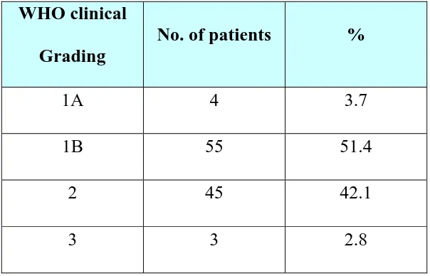
WHO grading of goitre:
Grade 0: Not palpable or visible even when the neck is extended Grade 1: When the goitre is palpable
1A Goitre detected on palpation
1B Goitre palpable and visible when neck extended
Grade 2: Goitre visible when neck is in the normal position Grade 3: Large goitre visible from a distance
Goiter grade 2
Thyroid function test:
used were, T4 = 5 – 12 μg/dl, T3 = 80 – 180 ng/dl and TSH = 0.5 – 5 µIU/L. Depending on these results of thyroid function patients were graded as euthyroid, hypothyroid or hyperthyroid.
Serum sample for TFT
Anti TPO antibody assay:
Anti –TPO antibodies were analysed by electrochemiluminiscence assay. The measurement ranges from 5 - 600 IU/ ml. Reference range vary according to the manufacturer. In our study Anti-TPO antibody values more than 60 IU/ml were considered to be TPO positive.
TPO analyser
USG of thyroid
Ultrasonography of the thyroid:
Sostre and Reyes grading system:
Grade 1 (G1) diffusely enlarged gland with a normoechoic (similar to normal
tissue) pattern
Grade 2 (G2) Multiple hypoechoeic foci or patches scattered throughout an otherwise normoechoic gland; a pattern suggestive of focal rather than diffuse involvement
Grade 3 (G3) Enlarged gland with diffuse but mild hypoechogenicity
Grade 4 (G4) Enlarged gland with diffuse and marked hypoechogenicity
Fine Needle Aspiration Cytology:
Ultrasound guided FNAC
Technique:
FNAC of thyroid was carried out by using non-aspiration/aspiration technique. Nodule is held with left hand & a 25 gauge needle attached to a 10 ml syringe held in the right hand is inserted, gentle suction applied, cellular material aspirated by moving the needle in and out and then the needle is withdrawn.
In case of large swelling multiple point aspiration was carried out. Smears were fixed with 95% ethanol. Alcohol fixed slides were stained with haematoxylin and eosin. The smears were evaluated by a single pathologist.
Outcome measures:
To compare efficacy of auto antibodies against FNAC as a sensitive indicator of autoimmunity
Statistical Methods:
Descriptive and inferential statistical analysis has been carried out in the present study. Results on continuous measurements are presented on Mean SD (Min-Max) and results on categorical measurements are noted as Number (%). Significance is assessed at 5 % level of significance. The following assumptions on data is made, Assumptions: 1.Dependent variables should be normally distributed, 2.Samples should be random drawn from the population, Cases of the samples should be independent,
To study the significance of parameters on continuous scale between two groups (Inter group analysis) on metric parameters, Student t test (independent, two tailed) was used.
Chi-square/ Fisher Exact test was used to find the significance of parameters studied on categorical scale between two or more groups.
Significant figures :
** Strongly significant (P value : P0.01)
* Moderately significant ( P value:0.01<P 0.05) + Suggestive significance (P value: 0.05<P<0.10)
Observation and Results
Among a total of 107 patients recruited into the study who satisfied the inclusion criteria, we made the following observations.
Age distribution:
Fig1. Age distribution (n=107)
Out of the 107 children studied, 43% (n=46) were between 10 to 15 years age group, 56.1% (n=60) of them aged between 5 to 10 years and one child (0.9%) aged less than 5 years. The mean age was 10.13±1.84 years.
Fig 2. Gender distribution
We observed that 68.2% (n=73) of the study population were girls and 31.8% (n=34) were boys.
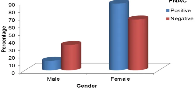
Fig 3 : Gender distribution in relation to FNAC findings (n=8)
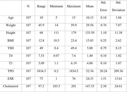
[image:54.595.137.473.459.613.2]Table 1: Descriptive statistics of the study population (n=107)
N Range Minimum Maximum Mean
Std. Error
Std. Deviation Age 107 10 5 15 10.13 0.18 1.84 Weight 107 45.9 14 59.9 29.56 0.76 7.87 Height 107 68 111 179 135.59 1.10 11.38
BMI 107 12.8 10.5 23.4 15.85 0.25 2.62 TSH 107 49 0.4 49.4 5.00 0.79 8.15 T4 107 7.53 0.07 7.6 1.40 0.10 1.02 T3 107 5.09 1.1 6.19 4.00 0.10 1.07 TPO 107 1834.3 0.2 1834.5 52.54 20.24 209.36 ESR 107 75 1 76 24.33 1.51 15.61 Cholesterol 107 97.5 103.5 201 147.53 2.38 24.61
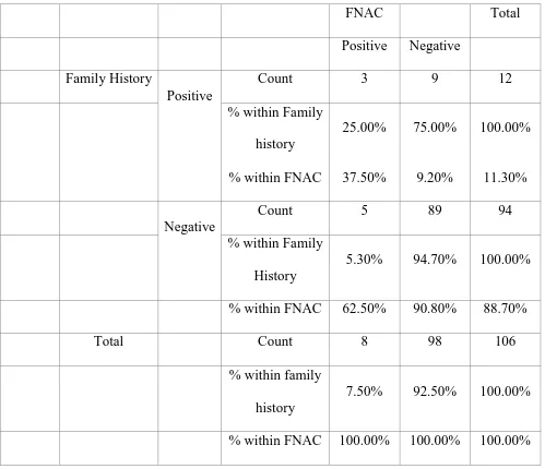
Table 2: Association of Positive Family History with FNAC positivity
FNAC Total Positive Negative
Family History
Positive
Count 3 9 12 % within Family
history
25.00% 75.00% 100.00%
% within FNAC 37.50% 9.20% 11.30%
Negative
Count 5 89 94 % within Family
History
5.30% 94.70% 100.00%
% within FNAC 62.50% 90.80% 88.70% Total Count 8 98 106
% within family history
7.50% 92.50% 100.00%
% within FNAC 100.00% 100.00% 100.00%
Pearson Chi-square Test, P value = 0.015 significant.
Table 3: Distribution of clinical grading of goitre (n=107)
WHO clinical
Grading
No. of patients %
1A 4 3.7 1B 55 51.4
2 45 42.1 3 3 2.8
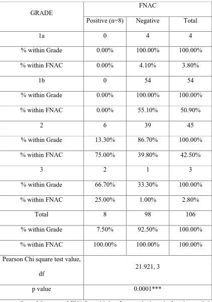
Table 4. Association of WHO Grades of Goitre with FNAC (n=107)
GRADE
FNAC
Positive (n=8) Negative Total
1a 0 4 4
% within Grade 0.00% 100.00% 100.00% % within FNAC 0.00% 4.10% 3.80%
1b 0 54 54 % within Grade 0.00% 100.00% 100.00% % within FNAC 0.00% 55.10% 50.90%
2 6 39 45 % within Grade 13.30% 86.70% 100.00% % within FNAC 75.00% 39.80% 42.50%
3 2 1 3
% within Grade 66.70% 33.30% 100.00% % within FNAC 25.00% 1.00% 2.80%
Total 8 98 106 % within Grade 7.50% 92.50% 100.00% % within FNAC 100.00% 100.00% 100.00% Pearson Chi square test value,
df
21.921, 3
p value 0.0001***
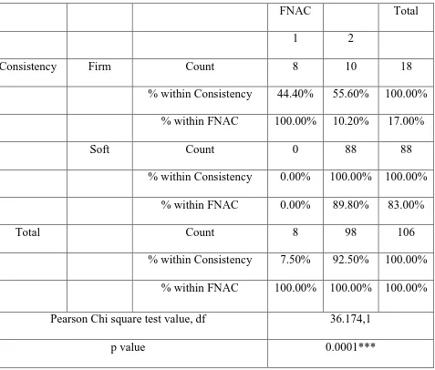
Table 5: Consistency of goiter among study group (n=107)
Consistency No. of patients %
Soft 89 83.2 Firm 18 16.8
In our study, 18 children out of 107 had firm consistency.
Table 6: Association of consistency with FNAC positivity
FNAC Total 1 2
Consistency Firm Count 8 10 18 % within Consistency 44.40% 55.60% 100.00%
% within FNAC 100.00% 10.20% 17.00% Soft Count 0 88 88
% within Consistency 0.00% 100.00% 100.00% % within FNAC 0.00% 89.80% 83.00% Total Count 8 98 106
% within Consistency 7.50% 92.50% 100.00% % within FNAC 100.00% 100.00% 100.00% Pearson Chi square test value, df 36.174,1
p value 0.0001***
[image:59.595.66.543.306.712.2]Fig 5: Association of consistency with TPO positivity
Among 11 TPO Positive cases, 7 (63.6%) had firm goitre.
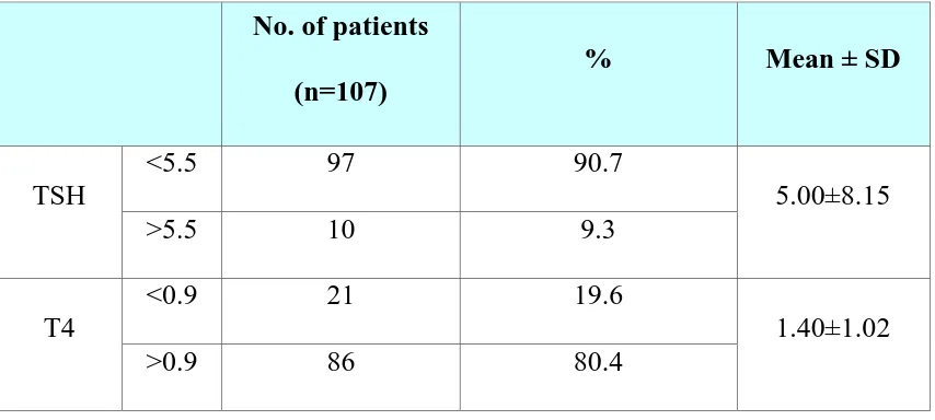
Table 7: Thyroid profile of the study population (n=107)
No. of patients
(n=107)
% Mean ± SD
TSH
<5.5 97 90.7
5.00±8.15 >5.5 10 9.3
T4
<0.9 21 19.6
1.40±1.02 >0.9 86 80.4
Based on the thyroid profile, 10 children (9.3%) were found to be biochemically hypothyroid, 97 children were Euthyroid.
0.00% 10.00% 20.00% 30.00% 40.00% 50.00% 60.00% 70.00%
soft firm
11.50%
63.60%
TPO positive (n=11)
[image:60.595.86.513.414.606.2]Fig 5: Incidence of thyroid dysfunction in children with goitre:
Among the 107 children, 7.5% (n=8) were overt hypothyroid, 1.9% (n=2) were sub clinically hypothyroid and rest of them 90.6% (n=97) were euthyroid.
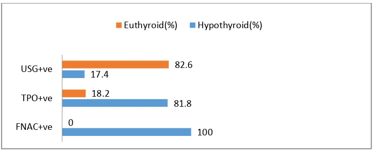
Fig 6: Association of Hypothyroidism with FNAC, TPO and USG
positivity (n=10).
On analysing thyroid profile, 100% of FNAC positive cases and 81.8 % of anti-TPO antibody positive cases were hypothyroid.
7.50% 1.90%
90.60%
Thyroid dysfunction
Hypothyroid
Subclinically hypothyroid Euthyroid
100 81.8 17.4
0
18.2
82.6
FNAC+ve TPO+ve USG+ve
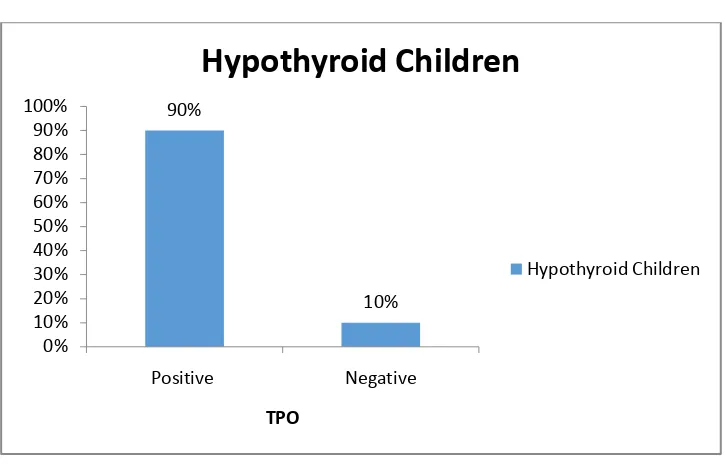
[image:61.595.113.484.491.641.2]Fig 7: TPO Positivity in hypothyroid children with goiter:
P<0.001**, Significant, Chi-Square test
90%
10%
0% 10% 20% 30% 40% 50% 60% 70% 80% 90% 100%
Positive Negative
TPO
Hypothyroid Children
Fig 8: TPO Findings of the patients studied:
Among 107 children with goiter, 11 children (10.3 %) had TPO Positivity.
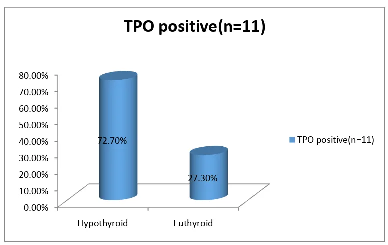
Fig 9: Thyroid dysfunction in anti-TPO antibody positive cases
Among the 11 TPO positive cases, 8 of them had hypothyroidism while 3 had no thyroid dysfunction.
10%
90%
TPO
Positive Negative
0.00% 10.00% 20.00% 30.00% 40.00% 50.00% 60.00% 70.00% 80.00%
Hypothyroid Euthyroid
72.70%
27.30%
TPO positive(n=11)
[image:63.595.103.491.409.654.2]Fig 10: FNAC findings of patients studied:
Overall 8 (7.5%) of the studied population were found to have autoimmune thyroiditis by FNAC.
Fig 11: FNAC findings in hypothyroid children with goiter:
P<0.001**, Significant, Fisher Exact test
Among the FNAC positive cases who showed evidence of lymphocytic thyroiditis, there was 100% biochemical hypothyroidism.
7%
93%
Positive Negative
0 20 40 60 80 100
Positive Negative
FNAC 100
2 0
98
[image:64.595.104.491.413.612.2]Table 8: Comparing anti-TPO antibody positivity with FNAC positivity
TPO
FNAC
Positive Negative Total Positive 7 4 11 Negative 1 95 96 Total 8 99 107
Among the 8 FNAC positive cases, 7 of them were also anti-TPO antibody positive.
Table 9: Evaluation of TPO as a screening tool for AIT
Parameter Estimate Lower - Upper 95% CIs
Sensitivity 87.50% (52.91, 97.76 ) Specificity 95.96% (90.07, 98.42 ) Positive Predictive Value 63.64% (35.38, 84.83 ) Negative Predictive Value 98.96% (94.33, 99.82 ) Diagnostic Accuracy 95.33% (89.52, 97.99 )
[image:65.595.82.515.436.618.2]Table 10: Combination of size, consistency and thyroid dysfunction as a
tool in predicting TPO Positivity
Combination of goiter of grade
≥2,Firmness&Hypothyroidism
TPO
Positive Negative Total Positive 7 2 9 Negative 4 94 98
Total 11 96 107
[image:66.595.61.533.493.679.2]In children with all three parameters of higher grade of goitre, firm consistency and hypothyroidism 7 out of 9 had TPO positivity.
Table 11: Evaluation of the combination as a screening tool to detect
autoimmunity
Parameter Estimate Lower - Upper 95% CIs
Sensitivity 63.64% (35.38 ,84.83 ) Specificity 97.92% (92.72, 99.43 ) Positive Predictive Value 77.78% (45.26, 93.68 ) Negative Predictive Value 95.92% (89.97, 98.4 )
Diagnostic Accuracy 94.39% (88.3, 97.4 )
As a screening tool the diagnostic accuracy of the combination is 94.39%.
Table 12: Combination of size, consistency and thyroid dysfunction as a
tool in predicting FNAC Positivity
Grade >2, Firmness & Hypothyroidism
FNAC
Positive Negative Total Positive 8 1 9 Negative 0 98 98
Total 8 99 107
Table 13: Evaluation of the combination as a screening tool to detect AIT
Parameter Estimate
Lower - Upper
95% CIs
Sensitivity 100% (67.56, 100) Specificity 98.99% (94.5, 99.82 ) Positive Predictive Value 88.89% (56.5, 98.01) Negative Predictive Value 100% (96.23, 100) Diagnostic Accuracy 99.07% (94.89, 99.83)
[image:67.595.116.478.433.653.2]Discussion
Discussion
Incidence of autoimmune thyroiditis has been on the increase in the post iodisation era since iodised salt is known to unmask the thyroid antigens and induce autoantibody production. Studies have quoted the prevalence of AIT in children ranging from 10.2 to 28.6% 4,2. Kabelitz et al 27, in their Berlin study also concluded autoimmune thyroiditis as the major cause of thyroid disorder in children and adolescents in iodine sufficient areas.
We attempted to detect autoimmunity in children presenting with goitre and screened them for thyroid dysfunction and autoimmunity. Anti thyroid peroxidase antibody positivity, Ultrasonological evidence of hypoechogenecity and Fine needle aspiration cytology suggestive of lymphocytic thyroiditis were considered as evidence of autoimmunity. An attempt was also made to compare the relative efficacy of TPO and USG finding to detect autoimmunity as compared to the gold standard namely FNAC. We also looked into the possibility of using a few clinical and laboratory data as a combination of parameters which could be used to predict autoimmunity so that resources are not wasted in future on indiscriminate screening of all goitrous children for AIT.
observed in other studies, our study too had female (68%) preponderance (Fig 2). We also found the presence of autoimmunity was much higher in girls. Our study reveals that 87.5% of FNAC proven cases (Fig 3) and 81.8% of anti TPO antibody positive cases were girls. Similar observation of greater prevalence of goitre in girls compared to boys was made by samid das et al, siriwerera et al and singh et al 13,33,34 . Thyroid dysfunction was also observed more in girl children than boys with F: M ratio of 9:1 in our study. This was similar to studies by siriweera EM et al 33 where there was a female preponderance with F: M ratio of 10.3:1.
Autoimmune thyroid diseases have strong genetic association and in our study too we found 37.5% of the children with AIT having family history of thyroid dysfunction especially in the mother (Table 2). Since this association is statistically significant, we recommend that adolescent girls of mothers with thyroid dysfunction should be screened for autoimmunity.
From our study we found that besides the larger size firmer consistency of goitre could also be used as an indicator of autoimmunity. Out of 18 cases of goitre with firm consistency (Table 5), 8 of them (44.4%) had FNAC evidence of autoimmunity, 7(38.9%) had TPO positivity and 9 (50%) had evidence of hypothyroidism. All the FNAC positive cases had firm goitres which is found to have statistical significance (Table 6). But we could not make such strong association between USG finding and Firmness. Thus we conclude that in contrast to soft goitre, firm goitre would definitely require further investigations for autoimmunity.
In the spectrum of autoimmunity, thyroid dysfunction can range from hypothyroidism to euthyroidism or even hyperthyroidism. In our study 10 out of 107 children (9.3%) had hypothyroidism (Table 7). Interestingly majority of them (8/10) also had clinical evidence of hypothyroidism (80%) in the form of lethargy, poor school performance and constipation. This is in contrast to Gopalakrishnan et al study where, in a series of 112 children with AIT, majority were euthyroid and only 7.2% had hypothyroidism.3
Many studies have been done using ultrasonography of thyroid gland to diagnose autoimmunity. Ultrasonologically autoimmunity is based on presence of hypoechogenecity, heterogeneity index and vascularity 23,24,26. In our study, out of 23 cases of goitre which had USG evidence of hypoechogenicity (grade III and IV), we found that only a small number had hypothyroidism (17.3%).Even TPO antibody positivity(17.3%). and FNAC(17.3%) positivity were seen in very few cases. Grade IV hypoechogenicity is reported as a good predictor of autoimmunity from other studies23,26, but in our study the only case of grade IV goitre was FNAC negative, TPO negative and had no evidence of hypothyroidism. Thus from our study we conclude that ultrasonography is a poor predictor of autoimmunity. This is in contrast to Marwaha study in which the authors have found ultrasound evaluation a sensitive tool to detect AIT.23
In our series, among the 4 cases of antibody positivity with no cytological evidence suggestive of AIT, only one had hypothyroidism and the other 3 were euthyroid. We plan to follow up these 3 children both clinically and by repeated TFT estimation as studies have stated that nearly 20% of children with autoimmune thyroiditis develop hypothyroidism ultimately35. This fact is also supported by T.Bijoro et al study 36, where the authors report that TPO antibodies appear in the circulation long before thyroid dysfunction or histological changes appear. Thus this study has helped us to detect these 3 cases of AIT very early even before the derangement of thyroid function.
Conclusions
1. Prevalence of autoimmunity among goitrous children was found to be 7.5% by FNAC and 10.3% by anti TPO antibodies.
2. Hypothyroidism was detected in 9.34% of goitres.
3. Though majority of cases of AIT have thyroid dysfunction, in a few cases, antibody positivity may be the only evidence of AIT. Such cases would require regular follow up with serial TFT estimation to detect and treat hypothyroidism early.
4. In a resource poor set up, the screening for autoimmunity could be limited to large goitres of grade 2 or more, which are firm in consistency along with accompanying hypothyroidism
5. Anti TPO antibody positivity is an effective, less invasive and more feasible indicator for detecting AIT in children as compared to FNAC which is considered the gold standard.
Study Limitation
1. Sample size is small. 2. It is not a follow up study
Recommendations
1. Wherever logistically feasible all adolescent children, especially girls with goitre should have initial anti TPO antibody testing along with thyroid profile as it determines the follow up. This assumes importance as any delay in recognition or treatment can affect not only physical growth and school performance but can also cause pubertal delay.
2. In a resource poor setting, screening for autoimmunity could be limited to children with large, firm goitres associated with hypothyroidism.
References
1. Cappa M, Bizzarri C, Crea F. Autoimmune Thyroid Diseases in Children. Journal of Thyroid Research. 2011;2011:1–13.
2. Marwaha RK, Garg MK, Nijhavan VS, Dham DN, Dubey R, Amberdar V, et al. Prevalence of chronic lymphocytic thyroiditis in adolescent girls. J Assoc Physicians India. 1998 Jul;46(7):606–8.
3. Gopalakrishnan S, Chugh PK, Chhillar M, Ambardar VK, Sahoo M, Sankar R. Goitrous autoimmune thyroiditis in a pediatric population: a longitudinal study. Pediatrics. 2008 Sep;122(3):e670-674.
4. Desai MP. Disorders of thyroid gland in India. Indian J Pediatr. 1997 Feb;64(1):11–20.
5. Akkara, B; Personal communication, February 2010.
6. Sinclair D. Clinical and laboratory aspects of thyroid autoantibodies. Annals of clinical biochemistry. 2006;43(3):173–183.
7. Carvalho DDT, Rocha DRTW, Arbex AK. Hypothyroidism in Childhood and Adolescence. Open Journal of Endocrine and Metabolic Diseases. 2016;6(1):72–7.
8. Yen PM. Physiological and molecular basis of thyroid hormone action. Physiological reviews. 2001;81(3):1097–1142.
10. Kumar V, Abbas AK, Aster JC. Robbins Basic Pathology. Elsevier Health Sciences; 2012. 925 p.
11. Portmann L, Fitch FW, Havran W, Hamada N, Franklin WA, DeGroot LJ. Characterization of the thyroid microsomal antigen, and its relationship to thyroid peroxidase, using monoclonal antibodies. J Clin Invest. 1988 Apr;81(4):1217–24.
12. Tunbridge WMG, Evered DC, Hall R, Appleton D, Brewis M, Clark F, et al. The Spectrum of Thyroid Disease in a Community: The Whickham Survey. Clinical Endocrinology. 1977 Dec 1;7(6):481–93. 13. Das S, Bhansali A, Dutta P, Aggarwal A, Bansal MP, Garg D, et al.
Persistence of goitre in the post-iodization phase: micronutrient deficiency or thyroid autoimmunity? Indian J Med Res. 2011 Jan;133(1):103–9.
14. Usha Menon V, Sundaram KR, Unnikrishnan AG, Jayakumar RV, Nair V, Kumar H. High prevalence of undetected thyroid disorders in an iodine sufficient adult south Indian population. J Indian Med Assoc. 2009 Feb;107(2):72–7.
16. Bagchi N, Brown TR, Parish RF. Thyroid dysfunction in adults over age 55 years. A study in an urban US community. Arch Intern Med. 1990 Apr;150(4):785–7.
17. Hoogendoorn EH. Thyroid Function and Prevalence of Anti-Thyroperoxidase Antibodies in a Population with Borderline Sufficient Iodine Intake: Influences of Age and Sex. Clinical Chemistry. 2006 Jan 1;52(1):104–11.
18. Ott J, Meusel M, Schultheis A, Promberger R, Pallikunnel SJ, Neuhold N, et al. The incidence of lymphocytic thyroid infiltration and Hashimoto’s thyroiditis increased in patients operated for benign goiter over a 31-year period. Virchows Arch. 2011 Sep;459(3):277–81.
19. De Luca F, Santucci S, Corica D, Pitrolo E, Romeo M, Aversa T. Hashimoto’s thyroiditis in childhood: presentation modes and evolution over time. Italian Journal of Pediatrics. 2013;39:8.
20. I Wahid F, Fawad Khan S, Ur Rehman H, Ahmad Khan I. Role of Fine Needle Aspiration Cytology in Diagnosis of Solitary Thyroid Nodules. Iran J Otorhinolaryngol. 2011;23(65):111–8.
21. Bhatia A, Rajwanshi A, Dash RJ, Mittal BR, saxena AK. Lymphocytic Thyroiditis – is cytological grading significant? A correlation of grades with clinical, biochemical, ltrasonographic and radionuclide parameters. CytoJournal. 2007;4(1):10.
by cytology which uncovers a pre-clinical state. Thyroid Research. 2010;3:11.
23. Marwaha RK, Tandon N, Kanwar R, Ganie MA, Bhattacharya V, Reddy DHK, et al. Evaluation of the role of ultrasonography in diagnosis of autoimmune thyroiditis in goitrous children. Indian Pediatr. 2008 Apr;45(4):279–84.
24. Höfling DB, Cerri GG, Juliano AG, Marui S, Chammas MC. Value of thyroid echogenicity in the diagnosis of chronic autoimmune thyroiditis. Radiologia Brasileira. 2008 Dec;41(6):409–17.
25. Sostre S, Reyes MM. Sonographic diagnosis and grading of Hashimoto’s thyroiditis. J Endocrinol Invest. 1991 Feb;14(2):115–21. 26. Wakita Y, Nagasaki T, Nagata Y, Imanishi Y, Yamada S, Yoda K, et al.
Thyroid heterogeneity, as indicated by the CV of ultrasonographic intensities, correlates with anti-thyroid peroxidase antibodies in euthyroid Hashimoto’s thyroiditis. Thyroid Res. 2013 Mar 23;6:5.
27. Kabelitz M, Liesenkotter KP, Stach B, Willgerodt H, Stablein W, Singendonk W, et al. The prevalence of anti-thyroid peroxidase antibodies and autoimmune thyroiditis in children and adolescents in an iodine replete area. Eur J Endocrinol. 2003 Mar 1;148(3):301–7.
29. Rho MH, Kim DW, Hong HP, Park YM, Kwon MJ, Jung SJ, et al. Diagnostic value of antithyroid peroxidase antibody for incidental autoimmune thyroiditis based on histopathologic results. Endocrine. 2012 Dec;42(3):647–52.
30. Thomas T, Sreedharan S, Khadilkar UN, Deviprasad D, Kamath MP, Bhojwani KM, et al. Clinical, biochemical & cytomorphologic study on Hashimoto’s thyroiditis. The Indian journal of medical research. 2014;140(6):729.
31. Erdogan M, Erdem N, Cetinkalp S, Ozgen AG, Saygılı F, Yilmaz C, et al. Demographic, clinical, laboratory, ultrasonographic, and cytological features of patients with Hashimoto’s thyroiditis: results of a university hospital of 769 patients in Turkey. Endocr. 2009 Oct 24;36(3):486–90. 32. Kimet al.:Hashimoto’s Thyroiditis in children and adolescents: at
presentation and during long-term follow up.International Journal of Pediatric Endocrinology. 2013 2013 (Suppl 1):P143.
33. Siriweera EH, Ratnatunga NVI. Profile of Hashimoto’s Thyroiditis in Sri Lankans: Is There an Increased Risk of Ancillary Pathologies in Hashimoto’s Thyroiditis? Journal of Thyroid Research. 2010;2010:1–5. 34. Singh N, Kumar S, Negi VS, Siddaraju N. Cytomorphologic study of
35. ScottA. Rivkees, MD. Thyroid disorders in children and adolescents. In: Sperling MA. Pediatric Endocrinology. Elsevier Health Sciences; 2014. p. 445-53.
36. Bjoro T, Holmen J, Kruger O, Midthjell K, Hunstad K, Schreiner T, et al. Prevalence of thyroid disease, thyroid dysfunction and thyroid peroxidase antibodies in a large, unselected population. The Health Study of Nord-Trondelag (HUNT). European Journal of Endocrinology. 2000;143(5):639–647.
37. Hacettepe University Medical Faculty Department of Radiology, Ankara, Turkey, Sarikaya B, Demirbilek H, Akata D, Kandemir N. The role of the resistive index in Hashimoto’s thyroiditis: a Sonographic pilot study in children. Clinics. 2012 Nov 6;67(11):1253–7.
38. Karmisholt J, Laurberg P. Serum TSH and serum thyroid peroxidase antibody fluctuate in parallel and high urinary iodine excretion predicts subsequent thyroid failure in a 1-year study of patients with untreated subclinical hypothyroidism. European Journal of Endocrinology. 2008 Feb 1;158(2):209–15.
39. Brown RS. Autoimmune Thyroiditis in Childhood. J Clin Res Pediatr Endocrinol. 2013 Mar;5(Suppl 1):45–9.
41. Marwaha RK, Tandon N, Karak AK, Gupta N, Verma K, Kochupillai N. Hashimoto’s thyroiditis: countrywide screening of goitrous healthy young girls in postiodization phase in India. J Clin Endocrinol Metab. 2000 Oct;85(10):3798–802.
42. Rallison ML, Dobyns BM, Keating FR, Rall JE, Tyler FH. Occurrence and natural history of chronic lymphocytic thyroiditis in childhood. J Pediatr. 1975 May;86(5):675–82.
43. Bottazzo GF, Doniach D. Autoimmune thyroid disease. Annu Rev Med. 1986;37:353–9.
44. Setian N. Hypothyroidism in children: diagnosis and treatment. Jornal de Pediatria [Internet]. 2007 Nov 12 [cited 2016 Sep 18];0(0). Available from:http://www.jped.com.br/conteudo/Ing_resumo.asp?varArtigo=171 8&cod=&idSecao=1
Proforma for Goitre Screening
Name: ESI NO: Age: SID NO: Sex:
Informant: Reliability: Date of birth: Address:
Contact no: SYMPTOMS
H/o Swelling in the neck: Yes No If yes duration
H/o pain in the neck Yes No
H/o difficulty in swallowing / hoarseness of voice Yes No
H/o constipation or diarrhea Yes No
H/o mental retardation /deterioration of school performance or short attention span
Yes No
H/o decreased appetite or hyperphagia without weight gain
Yes No
H/o cold intolerance or heat intolerance Yes No
H/o palpitaion/ tiredness Yes No
H/o weight gain or weight loss
H/o hair loss Yes No
H/o menstrual irregularities Yes No
H/o headache Yes No
H/o goitrogenic foods: cabbage, cauliflower, beans, sorghum, sweet potato, broccoli, turnip, soy, millet, brassica, cassava
Yes No
H/o drug intake: amiodaronne, lithium, iodide containing cough syrup/ asthma medications
Yes No
H/o dry skin or moist hands Yes No
H/o excessive sweating Yes No
H/o thyroid surgery or drug intake Yes No
Family H/o hypothyroidism/ hearing defect/ MR/ thyroid surgery
Yes No
EXAMINATION
Vitals: Anthropometry:
Heart rate
Blood pressure Pulse pressure
Weight Height
Head circumference
SMR staging
Irritability Yes No
Anaemia Yes No
Lid lag/ opthalmopathy Yes No Yellowish discoloration of skin Yes No
Brittle hair/ sparse hair Yes No
Coarse facies Yes No
Periorbital swelling Yes No
Large tongue Yes No
Alopecia Yes No
Acanthosis nigricans Yes No
Polydactyly/ café-au-iait spots Yes No
Vitiligo Yes No
Muco cutaneous candidiasis Yes No
Ectodermal dysplasia features Yes No
1. L/E thyroid WHO grading: 0 1a 1b 2 3 i. Enlargement: diffuse / single lobe
consistency: soft / firm ii. Tender / non tender
iii. Thyroid bruit
2. CVS : murmur:
3. Muscle hypertrophy 4. DTR : delayed relaxation 5. Disturbances in standing / gait 6. Pyramidal signs:
INVESTIGATIONS
Serum TSH
Normal range (0.6-6.3)
FREE T4 Normal range (5-10 years: 6.4-13 11-15 years: 5.5-12) FREE T3
Total count Differential Count
Platelets
ESR
Lipid profile
FNAC
USG of Thyroid
INFORMATION TO PARTICIPANTS
Investigator: Dr. Saranya P
Name of the Participant:
Title: “Detection of autoimmune thyroiditis in children with goitre
attending the Pediatric department of a tertiary care hospital in Chennai”
You are invited to take part in this research study. We have got approval from the IEC. You are asked to participate because you satisfy the eligibility criteria.
What is the Purpose of the Research:
To detect autoimmune thyroiditis in children under 15 years of age presenting with goitre.
The Study Design:
Detailed history taking and thorough clinical examination was done. TSH, Free T3, Free T4, TPO antibody assay, Lipid profile, Complete Blood Count, ESR, Ultrasonography and Fine Needle Aspiration Cytology of thyroid were done in all the cases.
Benefits, Discomforts and risks:
This intervention has been shown to be well tolerated as shown by previous studies. And if you do not want to participate you will have alternative of setting the standard treatment and your safety is our prime concern. Time :
Date : Place :
Signature / Thumb Impression of Patient Patient Name:
PATIENT CONSENT FORM
“Detection of autoimmune thyroiditis in children with goitre attending the Pediatric department of a tertiary care hospital in Chennai”
Study centre: Department of Pediatrics, ESIC Medical college & PGIMSR, K.K.Nagar, Chennai
Participant name: Age: Sex: I.P.No:
I confirm that I have understood the purpose of procedure for the above study. I have had the opportunity to clarify all my queries and doubts and they have been answered to my satisfaction.
Investigator explained very well about the procedure and I am made aware of the safety, advantages and disadvantages of the technique.
I understand that my participation in the study is purely voluntary and that I am free to withdraw at anytime without giving any reason.
I have understood that the investigator, regulatory authorities and the ethics committee will have access to my health records both in respect to current study and any further research that may be conducted in relation to it, even if I decide to withdraw from the study. I have understood that my identity will not be revealed in anyway and information released to third parties or published, unless as required under the law. I agree not to restrict the use of any data or results that arise from the study.
Without any compulsion I am willing to give consent for the participation of my child in this study.
Date: Signature / thumb impression of patient Place:
Patient name: Signature of the investigator:
ஆய்வுபற்மிதகவல்படிவம்
இடம் :
த ொழிலொளர் நல கொப்பீட்டு மருத்துவமனை மற்றும் மருத்துவ பட்ட
மமற்படிப்புஆரொய்ச்சிநிறுவைம்
மக. மக. நகர்
தசன்னை - 78
ஆய்வாரர் : மரு. . பொ. சரண்யொ
ஆய்வு :
“ குழந்ன களுக்கு ன்னுடல் ொங்கு ிறன் ய்ரொய்டு கு னறபொனட
கண்டரி ல்”
றுப்பதற்க்கானஉரிமற்றும்நம்பகதன்ம:
இந் ஆய்வில் ங்கள் குழந்ன பங்மகற்பன மறுக்க முழு உரினம
உள்ளது. அவ்வொறு மறுக்கும் பட்சத் ில் குழந்ன க்கு கினடக்கும் சிகிச்னச
மற்றும்மசனவயில் எவ்வி மொற்றமும் இருக்கொது. இந் ஆய்வுக்கொக நீங்கள்
வழங்கும் அனைத்து கவல்களும் ரகசியமொக பொதுகொக்கப்படும். எந்
இடத் ிலும்உங்கள் தபயர்பயன்படுத் படமொட்டொது.
நமடமுமமகள்:
ங்கள் குழந்ன இந் ஆய்வில் பங்மகற்கும் முன்பு குழந்ன யின்
மநொயின் ன்னம, மமற்தகொள்ளப்படும் சிகிச்னசமுனறகள், சிகிச்னசக்கு
உ விடும் தபொறுத்து மமற்தகொள்ளப்படும் இரத் பரிமசொ னை மற்றும் அ ன்
அவசியம் ஆகியனவ விளக்கி கூறப்படும். ங்கள் குழந்ன இந் ஆய்வில்
பங்மகற்க நீங்கள் விருப்பம் த ரிவித் ொல், குழந்ன யின் மநொய் மற்றும் முழு
உடல் விவரம், உடல் பரிமசொ னை விவரம், ொயின் மபறுக்கொல விவரங்கள்
மசகரிக்கப்படும். அவற்றின் கவல்கள்ரகசியமொகபொதுகொக்கப்படும்
ாமததாடர்புதகாள்வது?
இந் ஆய்வு குறித்து சந்ம கங்கனள எப்தபொழுது மவண்டுமொைொலும்
கீழ்க்கண்டமருத்துவனரத ொடர்புதகொண்டுத ளிவுபடுத் ிதகொள்ளலொம்
ருத்துவர்தபர் : மரு. பொ. சரண்யொ
ஆய்வில்பங்பகற்பதற்க்கானஒப்புதல்படிவம்
நொன் ______________________ கவல் படிவத் ில் இருந் கவல்கனள
படித்து அறிந்ம ன் / படிக்க மகட்டறிந்ம ன். எைக்கு தகொடுத் கவல்கனளயும்
படித்துபுரிந்துதகொண்மடன். என்னுடயசந்ம கங்கனள ீர்த்துக்தகொள்ளஎைக்கு
வொய்ப்பளிக்கப்பட்டது. எைது மகள்விகளுக்கு ிருப் ி அளிக்கும் வனகயில்
ப ில்கள் வழங்கப்பட்டை. எைது குழந்ன "Foe;ijfSf;F jd;Dly;
jhq;Fjpwd; ijuha;L Fiwghil fz;lwpjy;"
என்ற ஆய்வில்கலந்துதகொள்ள எவ்வி ய�