0022-538X/97/$04.0010
Copyright © 1997, American Society for Microbiology
Increased Lymphomagenicity and Restored Disease Specificity of
AML1 Site (Core) Mutant SL3-3 Murine Leukemia Virus by
a Second-Site Enhancer Variant Evolved In Vivo
STEEN ETHELBERG,1JETTE LOVMAND,1JO¨ RG SCHMIDT,2ARNE LUZ,3
ANDFINN SKOU PEDERSEN1,4*
Department of Molecular and Structural Biology1and Department of Medical Microbiology and Immunology,4University of Aarhus, DK-8000 Aarhus C, Denmark, and Institute of Molecular
Virology2and Institute of Pathology,3GSF-National Research Center for Environment and Health, D-85764 Neuherberg, Germany
Received 28 March 1997/Accepted 30 June 1997
SL3-3 is a highly T-lymphomagenic murine retrovirus. The major genetic determinant of disease is the transcriptional enhancer, which consists of a repeated region with densely packed binding sites for several transcription factors, including AML1 (also known as core binding factor and polyoma enhancer-binding protein 2) and nuclear factor 1 (NF1). Previously, we examined the enhancer structure of proviruses from murine tumors induced by SL3-3 with mutated AML1 (core) sites and found a few cases of second-site alterations. These consisted of deletions involving the NF1 sites and alterations in overall number of repeat elements, and they conferred increased enhancer strength in transient transcription assays. We have now tested the pathogenicity of a virus harboring one such second-site variant enhancer in inbred NMRI mice. It induced lymphomas with a 100% incidence and a significantly shorter latency than the AML1 mutant it evolved from. The enhancer structure thus represents the selection for a more tumorigenic virus variant during the pathogenic process. Sequencing of provirus from the induced tumors showed the new enhancer variant to be genetically stable. Also, Southern blotting showed that the tumors induced by the variant were T-cell lympho-mas, as were the wild-type-induced lymphomas. In contrast, tumors induced by the original core/AML1 site I-II mutant appeared to be of non-T-cell origin and several proviral genomes with altered enhancer regions could be found in the tumors. Moreover, reporter constructs with the new tumor-derived variant could not be transactivated by AML1 in cotransfection experiments as could the wild type. These results emphasize the importance of both core/AML1 site I and site II for the pathogenic potential of SL3-3 and at the same time show that second-site alterations can form a viral variant with a substantial pathogenic potential although both AML1 sites I and II are nonfunctional.
Murine leukemia viruses (MLVs) are non-oncogene-encod-ing simple retroviruses that induce malignancies in the hema-topoietic compartment when injected into newborn mice (3, 11, 47). MLV-induced leukemogenesis is a complex process in which several regions in the viral genome participate (47, 48). Of these, the transcriptional enhancer in the U3 region is a major determinant of the latency and specificity of hematopoi-etic disease induction (5, 7, 14, 17, 20, 38, 42).
The SL3-3 MLV induces T-cell lymphomas with a mean latency of 2 to 6 months depending on the mouse strain used (17, 18, 35, 44). SL3-3 is believed to be derived from weakly pathogenic Akv, the endogenous MLV of the AKR mouse strain (18, 37). Most of the enhancer function of SL3-3 is contained in the U3 region within a 72-bp direct repeat fol-lowed by a third repetition of 34 bp (13). This 72-bp region is made up of binding sites for several cellular transcription fac-tors. Each site is thus present two or three times. The inter-acting factors include the AML1 transcription factor family (45, 54), nuclear factor 1 (NF1) (9, 29), members of the Ets transcription factor family (28, 53), basic helix-loop-helix (bHLH) factor transcription factors (26, 27), the glucocorti-coid receptor (GR) (6), and the c-Myb transcription factor (28,
53). Current thinking holds that the transcription factor bind-ing sites in the SL3-3 repeat region, as in similar regions in other MLVs, match the transcription factor profile of the he-matopoietic target cells, enabling the virus to have a high replication rate and to act as a powerful insertional mutagen, thus determining the specificity and strength of the virus as a pathogen.
A central pathogenic determinant in the SL3-3 enhancer as in other MLV enhancers is the AML1 binding site (core site). The AML1–core-binding factorb(CBFb) transcription factor complex, also known as polyoma enhancer-binding protein 2, CBF, and SL3-3 enhancer factor 1 (45, 46), is encoded by several recently cloned genes (2, 19, 21, 24, 32, 33, 51). The humanAml1gene (CBFA2) as well as theCbfbgene is impli-cated in chromosomal rearrangements in several forms of acute myeloid leukemia in humans (21, 23, 30), and the murine homologs of both genes have been shown to be essential for normal hematopoietic development (4, 34, 49, 50).
The AML1 site I, Ets site, NF1 site, and GRE/E-box con-stellation is found in the majority of MLV enhancers and has been termed the enhancer framework (12). Mutation of any of these sites was found to reduce the pathogenic potential of the Moloney MLV (MoMLV). Mutation of the core and Ets sites, however, had the largest effect and also changed the disease specificity from T-cell lymphomas to erythroleukemia (42). SL3-3 viruses mutated in AML1 site I likewise have a reduced pathogenicity but, unlike MoMLV, still induce T-cell lympho-* Corresponding author. Mailing address: Department of Molecular
and Structural Biology, University of Aarhus, Bldg. 130, C. F. Møllers Alle´, DK-8000 Aarhus C, Denmark. Phone: 45 8942 3188. Fax: 45 8619 6500. E-mail: [email protected]
7273
on November 9, 2019 by guest
http://jvi.asm.org/
mas (1, 14, 25). Mutation of both AML1 site I and site II (core I and core II) results in virtually nonpathogenic SL3-3 virus forms (14). The importance of the NF1 site and the Ets site has also been tested in SL3-3, but in contrast to what applies to MoMLV (42), the NF1 site was found to be dispensable (9) and the Ets site was found to be only slightly important for leukemogenesis (28).
Upstream of the enhancer framework, the SL3-3 enhancer diverges from both MoMLV and Akv and contains a Myb binding site and the AML1 site II. Mutation of the Myb site has recently been found to severely reduce the pathogenicity of SL3-3 (28). Also, the AML1 site II has been found to be important for the pathogenic potential of SL3-3, especially in viruses with an impaired site I (14). Constellations of Myb sites, AML1 sites, and Ets factor sites can be found in other T-cell-specific enhancers, and there is evidence for a cooper-ative effect between the interacting factors in some such en-hancers, including the SL3-3 enhancer (15, 31, 42, 43, 52, 53). Previously, we noted that second-site alterations in the en-hancer framework arose in a few cases during the process of pathogenesis by SL3-3 variants carrying mutations in the AML1 sites. These enhancer framework variants acted as stronger enhancers than the forms they arose from when tested in transient transcription assays in a T-cell line (9). We have now compared the pathogenicity of one such SL3-3 enhancer variant with that of the virus it evolved from, a double AML1 site mutant. The original double mutant is weakly pathogenic and has an altered disease tropism whereas the second-site altered double mutant is significantly more potent and induces T-cell lymphomas, as does wild-type SL3-3. Moreover, in tran-sient cotransfection experiments the new enhancer variant can-not be transactivated by AML1 as can the wild type. These variants thus indicate that the direct interaction of AML1 with the proviral enhancer, although essential for the pathogenicity of wild-type SL3-3, can be partly compensated for by other alterations in the enhancer.
MATERIALS AND METHODS
Cell culture.F9 is a mouse embryonic carcinoma (EC) cell line (41). F9 cells
were maintained in gelatin-coated tissue culture bottles in Dulbecco’s modified Eagle’s medium containing Glutamax-1 (Gibco BRL, Life Technologies) sup-plemented with 10% fetal calf serum, 100 U of penicillin, and 100mg of strep-tomycin per ml. NIH 3T3 cells were grown in Dulbecco’s modified Eagle’s medium with Glutamax-1 containing 10% newborn calf serum plus antibiotics as described above. The murine T-lymphoid cell line, L691 (8), was grown in RPMI 1640 medium with Glutamax-1 and supplemented with 10% newborn calf serum and antibiotics as described above.
Generation of viruses. Generation of the wild-type SL3-3 virus and the
pSL3(atc) and pSL3(dm) variants has been previously described (14). To gen-erate pSL3(TUMdm), thePstI-KpnI fragment of construct pSL3(TUMdmL)cat (9) was inserted into a plasmid with the SL3-3 sequence from thePstI site to the
PvuI site located in the 59untranslated region. ThePstI-PvuI fragment from the resulting plasmid was then inserted into a plasmid carrying the molecular clone of SL3-3, and infectious viruses were produced by transfection of concatemerized
PstI-PstI fragments into NIH 3T3 cells as previously described (14). The cells were monitored by reverse transcriptase assays as described elsewhere (14) and sequencing of long terminal repeat (LTR) regions of proviruses amplified by PCR (see below).
Pathogenicity experiments.Inbred NMRI strain mice which lack ecotropic
endogenous proviruses (16) were injected with 107 infectious virus particles
within 36 h after birth. Control mice were mock injected with complete medium. The mice were checked for tumor development 5 days per week. Mice were sacrificed, X-rayed, and autopsied as described elsewhere (14) when they showed illness or tumor development or at the end of the 18-month observation period. Tumors were diagnosed on the basis of gross appearance of lymphoid organs as previously described (39) and according to cytological and anatomic criteria as described by Pattengale (36).
PCR amplification of proviral DNA.DNA was prepared from frozen tumor
material as described elsewhere (9). PCR was performed with a primer set recognizing the 59end of the U3 region and an SL3-3-specific sequence outside the LTR in the 59untranslated region (9). PCR products were purified with
Dynabeads (Dynal M-280), and the U3 regions were sequenced on an automatic DNA Sequenator (Applied Biosystems).
Transfections and reporter assays.F9 cells were transfected by use of the
calcium phosphate method as earlier described (9). A total of 1.5mg of chlor-amphenicol acetyltransferase (CAT) reporter plasmids was used together with the indicated amounts of AML1 expression plasmids. The total amount of plasmid was kept constant by addition of pUC19. A reference plasmid for transfection efficiency standardization was not used, to avoid interfering with the transactivation potential. NIH 3T3 cells were also transfected by use of the calcium phosphate method. L691 cells were transfected by the DEAE-dextran method as previously described (9). CAT assays were done by the original thin-layer chromatography method, except that the chromatograms were quan-titated on a PhosphorImager (Molecular Dynamics), as earlier described (9).
Plasmids. pSL3(TUMdm)-PBScat, pSL(dm)-PBScat, pSL3(atc)-PBScat,
and pSL3(wt)-PBScat were constructed on the basis of the constructs pSL3 (TUMdmL)cat, pSL3(dm)cat, pSL3(atc)cat, and pSL3cat (9), respectively, by deleting a 288-bpSmaI-BglII fragment including the viral primer binding site (PBS). Plasmids pCBFa2-451 and pCBFb-187 (54) were a kind gift of J. Lenz.
Southern blotting and hybridizations.Southern blotting was performed as
previously described (9). Briefly, the genomic tumor DNA was cut withHindIII, which cuts once within the SL3-3 proviral genome, and 10mg of DNA was resolved on a 0.75% agarose gel and transferred to nylon membranes. In order to detect clonal rearrangements of the T-cell receptor (TCR)b chain, two separate probes were used, one at a time. The probes are designed to recognize rearrangements in either the J1 or the J2 region and have been described before (1). To detect the number of viral integrations of each tumor, an ecotrope-specific probe consisting of 330 bp from theenvregion of the Akv retrovirus was used for hybridization (40). All probes were32P labeled by random labeling.
For detection of the size of the variant U3 structures, genomic tumor DNA was digested withPstI andKpnI. Ten micrograms of DNA was resolved on a 1.5% agarose gel in 13Tris-borate-EDTA buffer and transferred onto a nylon membrane as before. A probe specific for the SL3-3 repeat region was generated as previously described (9).
RESULTS
In a previous study, mutations of three consecutive bp were introduced in various positions in the AML1 sites in the SL3-3 enhancer, and the effect hereof on the pathogenic potential of the virus was investigated (14). Subsequently, SL3-3 proviral enhancer variants were found in three of the resulting murine tumors (9). These variant forms all had retained the original AML1 site mutations but contained small deletions in the enhancer repeat area known to be a binding region for NF1 and also carried additional intact repeat elements. When tested in transient transfection assays in a T-cell line, these second-site enhancer variants proved to be stronger enhancers than the mutant viruses originally injected into the mice. Al-though not proven, along with other lines of evidence, this indicated that the different structures could have arisen as a result of a selection for more pathogenic enhancer variants in the course of the pathogenic process (9).
Generation of infectious SL3-3 virus variants.In order to
test if the second-site variant viruses could be directly impli-cated in the process leading to the formation of the tumors in which they were found, we chose to examine the pathogenicity of one of the variants. We therefore constructed infectious SL3-3 viruses carrying the LTR regions found in one of the tumors and compared the pathogenicity of this virus variant with that of the AML1 mutant it evolved from. The original AML1 site double mutant, SL3(dm), carries mutations in both AML1 binding site I and site II (also termed core I and core II) in both 72-bp repeat elements (Fig. 1) and is identical to the GAC/GTT mutant used previously (14). The tumor-derived structure, SL3(TUMdm), contains a deletion of 28 bp in two repeat elements and an additional intact repeat, plus the orig-inal AML1 mutations and a few point mutations (Fig. 1).
In the previous pathogenicity studies, we used random-bred NMRI mice (14). In the current series, however, inbred NMRI mice were used. Mice of this strain have not been used for pathogenicity studies of SL3-3 before, and it was important to establish whether results obtained with these mice were
on November 9, 2019 by guest
http://jvi.asm.org/
parable to the results obtained with the random-bred mice. We therefore also included in the pathogenicity series another SL3-3 AML1 site mutant, SL3(atc), which carries mutations in AML1 site I only (Fig. 1) and is identical to the previously described ATC mutant (14).
Infectious viruses were generated by transfection of the mo-lecular virus clones into NIH 3T3 cells. Virus production was monitored by reverse transcriptase assays, and the integrity of the variants was confirmed by sequencing of PCR-amplified U3 regions from the infected cells. The viral titer was deter-mined by infectious center assays. Each of the variants gave rise to titers within the same range as those of the wild type.
The tumor-derived AML1 site I-II virus variant is more
pathogenic than the virus it evolved from.All three types of
viruses were injected into newborn NMRI mice together with wild-type SL3-3 virus and virus-free medium as controls. As
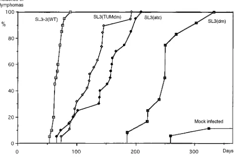
seen in Fig. 2 and Table 1, the tumor-derived variant SL3 (TUMdm), although less pathogenic than the wild type, gives rise to a significantly shorter latency period of disease induc-tion than does SL3(dm), the virus it originally evolved from. SL3(TUMdm) also has a slightly shorter latency period induc-tion than the variant SL3(atc). Thus, this result confirms the hypothesis that SL3(TUMdm) is a more potent viral variant than the parental structure. Furthermore, it formally proves that the tumor-derived SL3-3 variant represents a selection during the tumorigenic process for variants with increased pathogenic potential.
In the previous pathogenicity study using random-bred NMRI mice (14), the wild type had a mean latency of disease induction of 88 days (standard deviation, 25 days), more than 3 weeks longer than that in the present experiment. Likewise, at the end of the 300-day observation period only 50% of the mice infected with the ATC mutant and only 5% infected with the double mutant had developed severe malignancies (14). In this study, a 50% incidence of disease induction was reached after 150 days in the case of the ATC variant, and the last of the double-mutant-infected mice had developed disease after 330 days. Thus, disease induction in the inbred NMRI mouse strain is significantly faster than that in the random-bred strain. The spontaneous lymphoma development in this mouse strain after mock infection, however, was low within the 1-year ob-servation period and can be clearly distinguished from the disease development in the mice infected with the weakly dis-ease-inducing SL3(dm) variant (Fig. 2).
Tumors induced by the selection-derived variant are similar
to wild-type-induced tumors.In order to determine if the SL3
(TUMdm) variant enhancer was genetically stable in the course of the pathogenic process, DNA was prepared from the ma-jority of the tumors and LTR regions from proviruses were PCR amplified and sequenced. As previously noted by us and others (9, 25), several proviral enhancer repeat isoforms are present in the majority of tumors induced by SL3-3. These consist of variants that have lost or gained one or, more rarely, two repeat elements from their enhancers. One or more of these isoforms were found to be present in more than half of the tumors. Generally, the repeat number variants were not otherwise altered and nearly always coexisted in the tumors with the input virus variants. Apart from the SL3(dm) variant (see below), they were found in roughly similar amounts in the different tumors induced by the other three viral forms.
Except for such enhancer repeat variations, only a few oc-casional point mutations, and no major alterations, were de-tected in the proviral genomes of the SL3(TUMdm) variant. Similarly, the integrity of both the wild-type and the ATC mutant proviral genomes was conserved.
[image:3.612.62.292.69.264.2]Histological examination of the tumors induced by the SL3 FIG. 2. Lymphoma incidence of NMRI mice injected with the SL3-3
[image:3.612.316.556.92.173.2]vari-ants, wild type, or virus-free medium. The cumulative incidence of tumor devel-opment is shown versus the number of days after infection of the mice.
TABLE 1. Induction of lymphomas in inbred NMRI mice by SL3-3 wild type and enhancer variants
Virus
No. of mice with malignant lymphomas/
no. of infected mice
Mean latency (days)
SD (days)
SL3-3(wt) 19/19 64 8
SL3-3(TUMdm) 19/19 121 32
SL3-3(dm) 12/12a
248 38
SL3-3(atc) 20/20 145 44
Control 2/21b
aFifteen mice were inoculated with this variant, but three mice died (at days 96, 154, and 208) of causes that did not appear to involve viral induction.
bDetermined 1 year postinoculation.
FIG. 1. The structures of the enhancer repeats of the virus variants used for pathogenicity studies. The organization of the LTR region of SL3-3 is shown at the top with the binding sites for the transcription factors known to interact with one 72-bp enhancer repeat shown below. The sequences of the enhancer repeats of the three enhancer mutants used are shown in panel A, and the organization of the repeat elements is shown in panel B. The tumor-derived variant had a point mutation in one of the AML1 sites I, indicated by an arrow. The position of some restriction sites used is depicted as well.
on November 9, 2019 by guest
http://jvi.asm.org/
[image:3.612.60.297.545.703.2](TUMdm) variant indicated that they were similar to the wild-type-induced tumors. A total of 13 of 19 mice had thymic enlargements. To firmly establish whether the tumors were of T-cell origin, we made Southern blots on the basis of genomic tumor DNA with probes that detect rearrangements in the TCRb-chain gene. Two different probes, designed to detect both J1 segment and J2 segment rearrangements (1), were used independently. As seen in Fig. 3, rearrangements could be seen in 10 of 12 examined cases. This is similar to what we generally observe with wild-type tumors and confirms that the lymphomas were predominantly T-cell lymphomas.
Tumors induced by the double mutant are not T-cell
lym-phomas. The use of the inbred NMRI mice allowed
charac-terization of a pathogenic potential of the AML1 site double mutant. In the previous experiment with this variant (14), only 1 of 19 mice developed malignancies. Moreover, the single mouse that developed a lymphoma was the one in which the altered variant was found. Therefore, it could not be deter-mined whether the double mutant was pathogenic at all.
In the present study, 12 of 15 animals infected with the AML1 site double mutant developed tumors. The three mice that did not develop tumors died of other diseases within 7 months and are therefore not included in Fig. 2. Only 1 of the 12 animals had thymic enlargement and developed a tumor of diagnostic size located in the thymus; the remaining animals had tumors found in mesenterial lymph nodes or in the spleen. Southern blots of genomic tumor DNA prepared from the tumors induced by the double mutant showed that clonal re-arrangements in the TCRb-chain gene had not occurred in the tumor tissue in 9 of 10 cases (Fig. 4). The single tumor that had TCR rearrangements was the only one that was found in the thymus. The fact that TCR rearrangements were generally not detected could theoretically mean that the tumors were highly polyclonal. However, when the filter was reprobed with anenv
gene-specific probe, one or a few distinct bands were seen in most lanes (data not shown), indicating that the tumors were largely clonal or oligoclonal. Thus, the majority of the tumors
induced by the SL3(dm) variant do not appear to be T-cell lymphomas. Consistent with this, histopathological examina-tion of seven of the nine apparent non-T-cell lymphomas showed that they were follicular center cell lymphomas, indi-cating that they may be B-cell lymphomas (36).
PCR amplification of LTR regions from the double-mutant-induced tumors gave rise to a more complex band pattern than that seen with the other variants and the wild type. In some tumors, bands that could not have arisen due to variations in the number of 72-bp repeat elements alone were seen. The presence of such LTR length polymorphic variants in clonal populations of the tumor tissue was verified for some of the tumors by Southern blotting (data not shown). For these,Pst
I-KpnI digestion of the DNA was used and an enhancer repeat element-specific probe was employed as earlier described (9). In a few of the tumors, major deletions were noted within the repeat region when the PCR bands were separated by size on a polyacrylamide gel and sequenced individually. The two most prominent examples are shown in Fig. 5. These deletions are both of 40 bp, involve the NF1 site regions, and to a varying extent affect the flanking sites. It is interesting to note that the NF1 site region was also affected in the previously found al-tered enhancer structures derived from tumors (9). Moreover, the proviral enhancer repeat amplified from the single tumor that appeared to be a T-cell lymphoma carried several point mutations including a mutation in one of the NF1 sites (not shown). Thus, not all the proviral genomes of the double mu-tants were stable throughout the pathogenic process.
SL3-3 enhancer variants with AML1 site I and II mutations
do not respond to activation by AML1. The AML1 site I-II
double mutant is presumably nonresponsive to AML1 trans-activation. However, the same was not necessarily true in the case of the tumor-selected variant. Although it retained the AML1 site mutations, the deletions in the NF1 site region or the additional repeat element could theoretically have made it AML1 responsive. Also, the 1-bp alteration present in one of the three strong AML1 sites I raised the possibility that the AML1 transcription factor played a role in the increased pathogenic effect as well as the enhancer effect (9) seen with this variant.
To investigate if this was the case, we performed cotransfec-tion experiments using the murine EC cell line F9. Previously reported Northern blot analysis has indicated that F9 cells are FIG. 3. Southern blotting of DNA from 12 different tumors induced by SL3
(TUMdm). The DNA was digested withHindIII, resolved on a 0.8% agarose gel, and transferred to a nylon membrane. This was hybridized with two different probes designed to recognize rearrangements in either the J1 or the J2 region of the TCRb-chain gene. The J1 probe was used in the left panel, and the J2 probe was used in the right panel. The majority of T cells will have one (or sometimes two) rearrangedbchains. Rearrangements can involve either the J1 or the J2 region. Rearrangements of the J1 region normally do not involve the J2 region. Rearrangements of the J2 region normally result in the deletion of the J1 region. AHindIII restriction site exists in the area between the J1 and the J2 regions. Arrows indicate the locations of the germ line configurationHindIII fragments of 9.2 kbp in the case of the J1 probe and 5.3 kbp in the case of the J2 probe.
FIG. 4. Southern blots from 10 different tumors induced by the SL3(dm) mutant and hybridized with the TCR b-chain gene rearrangement-specific probes as described in the legend to Fig. 3. The J1 probe was used in the left panel, and the J2 probe was used in the right panel. The 9.2- and 5.3-kbp germ line TCRb-chain geneHindIII fragments are indicated by arrows.
on November 9, 2019 by guest
http://jvi.asm.org/
specifically devoid of expression of the two murine AML1a -subunit-encoding genes Pebp2aB (2) and Pebp2aA (33) (Aml1/ Cbfa2 and Aml3/Cbfa1, respectively). Binding activity for the MoMLV core site has also been reported elsewhere to be nearly absent in this cell line (41). However, the presence of a PBS in MLV constructs has the effect of shutting down tran-scription in EC cells (22). Therefore, the PBS had to be deleted from our CAT reporter constructs before they could be used in the EC cell line. This was done by deleting a 288-bp fragment ranging from the U5 region to the 59untranslated region from the previously used constructs pSL3(wt)cat, pSL3(dm)cat, pSL3(atc)cat, and pSL3(TUMdmL)cat (9), thereby generating constructs pSL3(wt)-PBScat, pSL3(dm)-PBScat, pSL3(atc)-PBScat, and pSL3(TUMdm)-pSL3(atc)-PBScat, respectively (Fig. 6A). To test if these constructs were functional, they were trans-fected into L691 cells which gave rise to CAT activity levels comparable to those of the equivalent PBS-containing con-structs (data not shown).
We then cotransfected each construct into F9 cells along with the AML1 expression vector pCBFa2-451. This construct encodes the 451-amino-acid protein of the murine Aml1 gene (Cbfa2) driven by the Rous sarcoma virus LTR and is identical to the construct previously used for cotransfection studies with the SL3-3 enhancer by Zaiman et al. (53, 54).
As can be seen in Fig. 6, the wild type could be stimulated approximately seven times by cotransfection of the AML1 ex-pression vector whereas the AML1 site I-II mutant could not be transactivated by AML1. This confirms previous results of Zaiman et al. obtained with the EC cell line P19 (53, 54). More importantly, the tumor-derived SL3-3 enhancer variant pSL3 (TUMdm)-PBScat could not be activated by any of the amounts of the AML1 expression vector used. Thus, it does not seem as if the increased pathogenicity of the SL3 (TUMdm) virus variant can be attributed to effects of the AML1 factor. The effect of cotransfection of the pSL3(atc)-PBScat variant with pCBFa2-451 was also tested. The pSL3 (atc)-PBScat construct showed a limited response to AML1 transactivation relative to that of the wild-type construct (Fig.
FIG. 5. The structures of altered proviral enhancers found in two different tumors induced by the AML1 site I-II mutant. The exact repeat nucleotide structure is shown at the top, and the repeat organization of several different individual types recognized in the tumors is shown below. Enhancers with the input virus repeat conformation and enhancer repeat element isoforms found in the tumors are shown together with the forms carrying the 40-nucleotide dele-tions. -D-, deleted area.
FIG. 6. (A) The schematic structure of the CAT reporter constructs used for the cotransfection experiments. (B) Transient cotransfection data for F9 cells. A total of 1.5mg of each CAT reporter construct was cotransfected with the indicated amount of pCBFa2-451. The level of CAT activity without addition of exogenous AML1 expression plasmid was set to 1. Two to five independent transfections were made with each variant. Standard deviations are indicated by the small bars.
on November 9, 2019 by guest
http://jvi.asm.org/
6B). This is in accordance with the ATC mutation conferring a partial disruption of the ability of AML1 to bind the strong AML1 site I. The basic CAT transcription level without addi-tion of the pCBFa2-451 expression construct was the same for all the constructs, except for the wild-type construct, which was approximately twice as active as the other three constructs (data not shown). This is likely to be due to the presence of minute levels of endogenous AML1 activity in the F9 cell line. The AML1 transcription factor complex consists of both the DNA-binding a subunit and an additional b subunit. Theb subunit is not required for DNA binding and transactivation of theasubunit; however, it stabilizes binding of theafactor to DNA and acts to increase the transactivation level as well (32, 51). We therefore also cotransfected the CAT constructs with a-subunit- and b-subunit-encoding plasmids. However, addi-tion of the CBFbexpression plasmid pCBFb-187 (54) encod-ing the 187-amino-acid splice variant of the murineCbfbgene did not lead to an increase in the transactivation level relative to the one seen with pCBFa2-451 alone. This was the case with all four of the CAT constructs (data not shown). This again is in accordance with results obtained by Zaiman et al., who mention the existence of endogenous CBFb expression as a possible explanation (54).
DISCUSSION
Mutation of the core sites in the enhancer repeats of SL3-3 significantly reduces the pathogenicity of the virus. Hereby, a selection pressure is generated for the formation of more pathogenic variants. However, retroviruses are notorious for their mutation rates, and the finding of viruses with altered enhancers in tumors does not necessarily mean that such vari-ants contributed to tumorigenesis. We have therefore tested the pathogenic potential of one such variant found in a murine tumor originating as a result of infection with an SL3-3 core mutant. The variant was found to be more pathogenic than the virus it evolved from. The alterations acted to restore the pathogenic effect of the core mutations in terms of both dis-ease phenotype and latency of disdis-ease induction. This proves that variant enhancers are selected for increased pathogenicity. Since only the U3 region of the virus variant used in this study was taken from a tumor, potential contributions to patho-genicity from other genomic elements can be ruled out. The differences in tumorigenic potential must reside in the variance in the U3 regions between the original and the selected core mutants. The alterations that arose in the new variant are interesting primarily because they are second-site alterations that did not seem to restore the function of the mutated core sites. That they, however, acted to partly restore the patho-genic potential of the virus indicates that this variant may help to elucidate functions of the enhancer other than the core site function.
The in vivo-selected variant contained a reversion of one of the three consecutive base-pair mutations in one of three mu-tated AML1 sites I. It was a possibility that this reversion acted to restore some of the function of this AML1 site. We there-fore tested the ability of AML1 to transactivate the selected variant in a reporter construct by a cotransfection assay. In contrast to a wild-type construct, the selected variant did not respond to ectopic AML1, nor did the original AML1 site I-II mutant. This indicates that the selected variant does not have functional AML1 sites. However, in similar cotransfection as-says previously performed by Zaiman and colleagues (54) the activity of a site I mutant construct could not be distinguished from the activity of a site I-II mutant construct. This probably indicates that the sensitivity of this assay is limited, although we
find it most likely that the assay would detect a leaky mutation of one of the strong AML1 sites I. Furthermore, in a separate investigation the enhancer alterations were found to boost the pathogenic potential in the context of wild-type AML1 sites as well (10). We therefore favor the view that the enhanced pathogenic effect of the selected variant relative to the original mutant is not due to recreation of AML1 responsiveness.
The mechanism by which the alterations increase the patho-genic potential is not known. However, it is interesting to note that alterations involving variations in the number of enhancer repeat elements in combination with deletions of the NF1 areas in the repeat region were also found in tumors induced by the AML1 site I-II mutant in the present study. Presumably, these variants also represent a selection for more pathogenic forms. This indicates that the NF1 site containing part of the 72-bp repeat element lying between the AML1 site I and the bHLH-GR site could be dispensable, or even negatively acting to some extent, for the pathogenicity of SL3-3. It is a well-known effect from studies with synthetic enhancers that mul-tiple transcription factor binding sites placed in close proximity in many instances confer a strong synergistic enhancer effect. Keeping this fact in mind, it can be speculated that the function of the deletions combined with an extended number of repeat units is to create a stronger enhancer by virtue of an increased cooperation between the remaining binding sites. Thus, the two examples of altered repeat structures found in the AML1 site I-II mutant-induced tumors in this study both contain multiple copies of a short repeat segment containing either the Myb and the Ets sites or the Myb and the bHLH sites. In this context, it is interesting to note that the Myb site was recently found to be of major significance for the pathogenic potential of SL3-3 (28).
A special case is presented by the ATC mutant. The ATC mutation is situated in the periphery of the AML1 site I. Band-shift analysis has shown this mutation to moderately impair the binding of a T-cell characteristic nuclear complex assumed to belong to the AML1 protein family, whereas the GTT mutation present in the double mutant completely dis-rupted binding (45). Surprisingly, however, the latency period of disease induction was slightly shorter in GTT-mutant-infect-ed mice than in ATC-mutant-infectGTT-mutant-infect-ed mice (14). This raisGTT-mutant-infect-ed the possibility that the ATC mutation could have effects other than simply reducing the binding of the AML1 complex. The results presented here do not resolve the matter but at least indicate that some of the action of the ATC mutation is to interfere with the ability of AML1 to transactivate.
The AML1 site I-II mutant has been tested before and was found to be virtually nonpathogenic. In the present study, however, a mouse strain more susceptible to disease induction was used. This resulted in tumor development in all mice tested, with a mean latency period of close to 1 year. The tumors induced by this variant were generally phenotypically different from the tumors induced by wild-type SL3-3 as judged by histological observations and the overall organ distribution of the tumors. Moreover, they were not T-cell lymphomas as seen by lack of TCRb-chain gene rearrangements detected by Southern blots. They did not, however, represent a switch to an erythroleukemic disease phenotype as previously found with AML1 site mutants of MoMLV (42). This result further high-lights the importance of the two AML1 binding sites, since an altered tumor phenotype has not been reported with previ-ously tested enhancer mutations in SL3-3. In particular, the tumors previously induced by the GTT mutant (14), i.e., the mutant with the same mutation in the AML1 site I as that carried by the double mutant used in this study but with an intact AML1 site II, have been directly shown to be T-cell
on November 9, 2019 by guest
http://jvi.asm.org/
lymphomas by use of the same Southern blotting technique (1). Therefore, although the effect upon pathogenicity of mutating AML1 site II by itself is quite small, the importance of this site is revealed when site I is mutated as well. The AML1 site II may thus work as a backup site for the stronger AML1 site I. SL3-3 differs from most other MLVs, including MoMLV, by the presence of the AML1 site II and the juxtaposed Myb site in the enhancer repeats. Interestingly, the Myb site was re-cently found to be very important for the pathogenicity of the virus (28), whereas the Ets site and the NF1 site, which are both important for the pathogenic potential of MoMLV, were found to be virtually unimportant (9, 28). The finding that mutation of the AML1 site II in the context of a nonfunctional site I abolishes the T-lymphomagenic potential of SL3-3 fur-ther supports the notion that the additional Myb site and AML1 site II constitute a strong additional T-cell tropic ele-ment for SL3-3, not present in the majority of MLVs.
ACKNOWLEDGMENTS
We thank Annette Balle Sørensen for helpful and encouraging dis-cussions and Jack Lenz for providing us with the AML1 expression plasmids. The technical assistance of Angelika Appold, Anna Nickl, Elenore Samson, and Lone Højgaard is gratefully acknowledged.
This project was supported by the Danish Cancer Society, the Karen Elise Jensen Foundation, the Danish Natural Sciences Research Council, the Danish Biotechnology Program, and the Leo Nielsen Foundation and by European Commission contracts CT-950100 (Bio-technology) and CT-95067 (Biomed-2).
REFERENCES
1.Amtoft, H. W., A. B. Sørensen, C. Bareil, J. Schmidt, A. Luz, and F. S.
Pedersen.1997. Stability of AML1 (core) site enhancer mutations in T
lymphomas induced by attenuated SL3-3 murine leukemia virus mutants. J. Virol.71:5080–5087.
2.Bae, S. C., I. Y. Yamaguchi, E. Ogawa, M. Maruyama, M. Inuzuka, H.
Kagoshima, K. Shigesada, M. Satake, and Y. Ito.1993. Isolation of PEBP2
alpha B cDNA representing the mouse homolog of human acute myeloid leukemia gene, AML1. Oncogene8:809–814.
3.Ben-David, Y., and A. Bernstein.1991. Friend virus-induced
erythroleuke-mia and the multistage nature of cancer. Cell66:831–834.
4.Castilla, L. H., C. Wijmenga, Q. Wang, T. Stacy, N. A. Speck, M. Eckhaus,
P. M. Marin, F. S. Collins, B. A. Wynshaw, and P. P. Liu.1996. Failure of
embryonic hematopoiesis and lethal hemorrhages in mouse embryos het-erozygous for a knocked-in leukemia gene CBFB-MYH11. Cell87:687–696.
5.Celander, D., and W. A. Haseltine.1984. Tissue-specific transcription
pref-erence as a determinant of cell tropism and leukaemogenic potential of murine retroviruses. Nature312:159–162.
6.Celander, D., B. L. Hsu, and W. A. Haseltine.1988. Regulatory elements
within the murine leukemia virus enhancer regions mediate glucocorticoid responsiveness. J. Virol.62:1314–1322.
7.DesGroseillers, L., and P. Jolicoeur.1984. The tandem direct repeats within
the long terminal repeat of murine leukemia viruses are the primary deter-minant of their leukemogenic potential. J. Virol.52:945–952.
8.Duch, M., K. Paludan, P. Jørgensen, and F. S. Pedersen.1994. Lack of
correlation between basal expression levels and susceptibility to transcrip-tional shutdown among single-gene murine leukemia virus vector proviruses. J. Virol.68:5596–5601.
9.Ethelberg, S., B. Hallberg, J. Lovmand, J. Schmidt, A. Luz, T. Grundstro¨m,
and F. S. Pedersen.1997. Second-site proviral enhancer alterations in
lym-phomas induced by enhancer mutants of SL3-3 murine leukemia virus: negative effect of nuclear factor 1 binding site. J. Virol.71:1196–1206.
10. Ethelberg, S., A. B. Sørensen, J. Schmidt, A. Luz, and F. S. Pedersen.An
SL3-3 murine leukemia virus enhancer variant more pathogenic than wild type obtained by assisted molecular evolution in vivo. Submitted for publi-cation.
11. Fan, H.1990. Influences of the long terminal repeats on retrovirus
patho-genicity. Semin. Virol.1:165–174.
12. Golemis, E. A., N. A. Speck, and N. Hopkins.1990. Alignment of U3 region
sequences of mammalian type C viruses: identification of highly conserved motifs and implications for enhancer design. J. Virol.64:534–542.
13. Hallberg, B., and T. Grundstro¨m.1988. Tissue specific sequence motifs in
the enhancer of the leukaemogenic mouse retrovirus SL3-3. Nucleic Acids Res.16:5927–5944.
14. Hallberg, B., J. Schmidt, A. Luz, F. S. Pedersen, and T. Grundstro¨m.1991.
SL3-3 enhancer factor 1 transcriptional activators are required for tumor
formation by SL3-3 murine leukemia virus. J. Virol.65:4177–4181.
15. Hernandez-Munain, C., and M. S. Krangel.1995. c-Myb and core-binding
factor/PEBP2 display functional synergy but bind independently to adjacent sites in the T-cell receptor delta enhancer. Mol. Cell. Biol.15:3090–3099.
16. Leib-Mo¨sch, C., J. Schmidt, M. Etzerodt, F. S. Pedersen, R. Hehlmann, and
V. Erfle.1986. Oncogenic retrovirus from spontaneous murine osteomas. II.
Molecular cloning and genomic characterization. Virology150:96–105.
17. Lenz, J., D. Celander, R. L. Crowther, R. Patarca, D. W. Perkins, and W. A.
Haseltine.1984. Determination of the leukaemogenicity of a murine
retro-virus by sequences within the long terminal repeat. Nature308:467–470.
18. Lenz, J., R. Crowther, S. Klimenko, and W. Haseltine.1982. Molecular
cloning of a highly leukemogenic, ecotropic retrovirus from an AKR mouse. J. Virol.43:943–951.
19. Levanon, D., V. Negreanu, Y. Bernstein, A. I. Bar, L. Avivi, and Y. Groner.
1994. AML1, AML2, and AML3, the human members of the runt domain gene-family: cDNA structure, expression, and chromosomal localization. Genomics23:425–432.
20. Li, Y., E. Golemis, J. W. Hartley, and N. Hopkins.1987. Disease specificity
of nondefective Friend and Moloney murine leukemia viruses is controlled by a small number of nucleotides. J. Virol.61:693–700.
21. Liu, P., S. A. Tarle, A. Hajra, D. F. Claxton, P. Marlton, M. Freedman, M. J.
Siciliano, and F. S. Collins.1993. Fusion between transcription factor CBF
beta/PEBP2 beta and a myosin heavy chain in acute myeloid leukemia. Science261:1041–1044.
22. Lund, A. H., M. Duch, and F. S. Pedersen.1996. Transcriptional silencing of
retroviral vectors. J. Biomed. Sci.3:365–378.
23. Miyoshi, H., T. Kozu, K. Shimizu, K. Enomoto, N. Maseki, Y. Kaneko, N.
Kamada, and M. Ohki.1993. The t(8;21) translocation in acute myeloid
leukemia results in production of an AML1-MTG8 fusion transcript. EMBO J.12:2715–2721.
24. Miyoshi, H., K. Shimizu, T. Kozu, N. Maseki, Y. Kaneko, and M. Ohki.1991.
t(8;21) breakpoints on chromosome 21 in acute myeloid leukemia are clus-tered within a limited region of a single gene, AML1. Proc. Natl. Acad. Sci. USA88:10431–10434.
25. Morrison, H. L., B. Soni, and J. Lenz.1995. Long terminal repeat enhancer
core sequences in proviruses adjacent to c-mycin T-cell lymphomas induced by a murine retrovirus. J. Virol.69:446–455.
26. Nielsen, A. L., P. L. Nørby, F. S. Pedersen, and P. Jørgensen.1996. E-box
sequence and context-dependent TAL1/SCL modulation of basic helix-loop-helix protein mediated transcriptional activation. J. Biol. Chem.271:31463– 31469.
27. Nielsen, A. L., P. L. Nørby, F. S. Pedersen, and P. Jørgensen.1996. Various
modes of basic helix-loop-helix protein-mediated regulation of murine leu-kemia virus transcription in lymphoid cell lines. J. Virol.70:5893–5901.
28. Nieves, A., L. S. Levy, and J. Lenz.1997. Importance of a c-Myb binding site
for lymphomagenesis by the retrovirus SL3-3. J. Virol.71:1213–1219.
29. Nilsson, P., B. Hallberg, A. Thornell, and T. Grundstro¨m.1989. Mutant
analysis of protein interactions with a nuclear factor I binding site in the SL3-3 virus enhancer. Nucleic Acids Res.17:4061–4075.
30. Nucifora, G., and J. D. Rowley.1995. AML1 and the 8;21 and 3;21
translo-cations in acute and chronic myeloid leukemia. Blood86:1–14.
31. Oelgeschlager, M., I. Nuchprayoon, B. Luscher, and A. D. Friedman.1996.
C/EBP, c-Myb, and PU.1 cooperate to regulate the neutrophil elastase pro-moter. Mol. Cell. Biol.16:4717–4725.
32. Ogawa, E., M. Inuzuka, M. Maruyama, M. Satake, F. M. Naito, Y. Ito, and
K. Shigesada.1993. Molecular cloning and characterization of PEBP2 beta,
the heterodimeric partner of a novel Drosophila runt-related DNA binding protein PEBP2 alpha. Virology194:314–331.
33. Ogawa, E., M. Maruyama, H. Kagoshima, M. Inuzuka, J. Lu, M. Satake, K.
Shigesada, and Y. Ito.1993. PEBP2/PEA2 represents a family of
transcrip-tion factors homologous to the products of the Drosophila runt gene and the human AML1 gene. Proc. Natl. Acad. Sci. USA90:6859–6863.
34. Okuda, T., J. van Deursen, S. W. Hiebert, G. Grosveld, and J. R. Downing.
1996. AML1, the target of multiple chromosomal translocations in human leukemia, is essential for normal fetal liver hematopoiesis. Cell84:321–330.
35. Østergaard, M., L. Pedersen, J. Schmidt, A. Luz, J. Lovmand, V. Erfle, F. S.
Pedersen, and P. G. Strauss.1997. Mapping of a major osteomagenic
de-terminant of murine leukemia virus RFB-14 to non-long terminal repeat sequences. J. Virol.71:645–649.
36. Pattengale, P. K.1994. Neoplastic lesions of the mouse lymphoid system, p.
168–176.InP. Bannasch and W. Goessner (ed.), Pathology of neoplasia and preneoplasia in rodents. Schattauer, Stuttgart, Germany.
37. Pedersen, F. S., R. L. Crowther, D. Y. Tenney, A. M. Reimold, and W. A.
Haseltine.1981. Novel leukaemogenic retroviruses isolated from cell line
derived from spontaneous AKR tumour. Nature292:167–170.
38. Rosen, C. A., W. A. Haseltine, J. Lenz, R. Ruprecht, and M. W. Cloyd.1985.
Tissue selectivity of murine leukemia virus infection is determined by long terminal repeat sequences. J. Virol.55:862–866.
39. Schmidt, J., V. Erfle, F. S. Pedersen, H. Rohmer, H. Schetters, K. H.
Mar-quart, and A. Luz.1984. Oncogenic retrovirus from spontaneous murine
osteomas. I. Isolation and biological characterization. J. Gen. Virol.65:2237– 2248.
on November 9, 2019 by guest
http://jvi.asm.org/
40. Sørensen, A. B., M. Duch, H. W. Amtoft, P. Jørgensen, and F. S. Pedersen.
1996. Sequence tags of provirus integration sites in DNAs of tumors induced by the murine retrovirus SL3-3. J. Virol.70:4063–4070.
41. Speck, N. A., and D. Baltimore.1987. Six distinct nuclear factors interact
with the 75-base-pair repeat of the Moloney murine leukemia virus en-hancer. Mol. Cell. Biol.7:1101–1110.
42. Speck, N. A., B. Renjifo, E. Golemis, T. N. Fredrickson, J. W. Hartley, and
N. Hopkins.1990. Mutation of the core or adjacent LVb elements of the
Moloney murine leukemia virus enhancer alters disease specificity. Genes Dev.4:233–242.
43. Sun, W., B. J. Graves, and N. A. Speck.1995. Transactivation of the Moloney
murine leukemia virus and T-cell receptorb-chain enhancers bycbfandets
requires intact binding sites for both proteins. J. Virol.69:4941–4949.
44. Thomas, C. Y.1986. AKR ecotropic murine leukemia virus SL3-3 forms
envelope gene recombinants in vivo. J. Virol.59:23–30.
45. Thornell, A., B. Hallberg, and T. Grundstrom.1991. Binding of SL3-3
enhancer factor 1 transcriptional activators to viral and chromosomal en-hancer sequences. J. Virol.65:42–50.
46. Thornell, A., B. Hallberg, and T. Grundstrom.1988. Differential protein
binding in lymphocytes to a sequence in the enhancer of the mouse retro-virus SL3-3. Mol. Cell. Biol.8:1625–1637.
47. Tsichlis, P. N., and P. A. Lazo.1991. Virus-host interactions and the
patho-genesis of murine and human oncogenic retroviruses. Curr. Top. Microbiol. Immunol.171:95–173.
48. van Lohuizen, M., and A. Berns.1990. Tumorigenesis by slow-transforming
retroviruses—an update. Biochim. Biophys. Acta1032:213–235.
49. Wang, Q., T. Stacy, M. Binder, P. M. Marin, A. H. Sharpe, and N. A. Speck.
1996. Disruption of the Cbfa2 gene causes necrosis and hemorrhaging in the central nervous system and blocks definitive hematopoiesis. Proc. Natl. Acad. Sci. USA93:3444–3449.
50. Wang, Q., T. Stacy, J. D. Miller, A. F. Lewis, T. L. Gu, X. Huang, J. H.
Bushweller, J. C. Bories, F. W. Alt, G. Ryan, P. P. Liu, B. A. Wynshaw, M.
Binder, P. M. Marin, A. H. Sharpe, and N. A. Speck.1996. The CBFbeta
subunit is essential for CBFalpha2 (AML1) function in vivo. Cell87:697–708.
51. Wang, S., Q. Wang, B. E. Crute, I. N. Melnikova, S. R. Keller, and N. A.
Speck.1993. Cloning and characterization of subunits of the T-cell receptor
and murine leukemia virus enhancer core-binding factor. Mol. Cell. Biol.
13:3324–3339.
52. Wotton, D., J. Ghysdael, S. Wang, N. A. Speck, and M. J. Owen.1994.
Cooperative binding of Ets-1 and core binding factor to DNA. Mol. Cell. Biol.14:840–850.
53. Zaiman, A. L., and J. Lenz.1996. Transcriptional activation of a retrovirus
enhancer by CBF (AML1) requires a second factor: evidence for coopera-tivity with c-Myb. J. Virol.70:5618–5629.
54. Zaiman, A. L., A. F. Lewis, B. E. Crute, N. A. Speck, and J. Lenz.1995.
Transcriptional activity of core binding factora(AML1) andbsubunits on murine leukemia virus enhancer cores. J. Virol.69:2898–2906.